A new review published in the American Journal of Respiratory Cell and Molecular Biology discusses the causes of, and potential treatments for, idiopathic pulmonary fibrosis (IPF).
Fibrosis, but not focused on fibroblasts
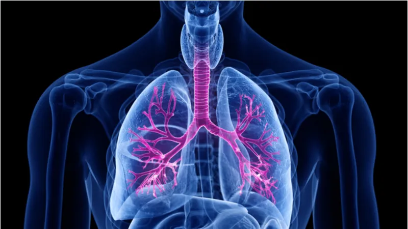
In the lungs, the trachea (airway) breaks out into a tree-like system of nodes. Each branch ends in alveoli, which are responsible for gas exchange between the air and the bloodstream. This is where the distal epithelium resides, and it has two types of cells: AEC1 and AEC2. AEC1 cells are involved in the gas exchange itself, as their thin membranes allow the entry of oxygen and the release of carbon dioxide. AEC2 cells are cubic in shape, and they secrete a surfactant that prevents the alveoli from collapsing. Furthermore, they are able to self-renew and differentiate into AEC1 cells, so they function like stem cells [1].
The direct cause of the fibrosis in IPF is fibroblasts. Previous research has shown that cellular senescence is linked to IPF, and one paper has shown that the senescence-associated secretory phenotype (SASP) is linked to aberrantly activated fibroblasts [2].
However, this review cites research showing that IPF begins with the distal epithelium, not the fibroblasts, and that targeting the disease here may be sufficient to slow or reverse it [3]. The researchers portray this as wound healing gone wrong, and in addition to genetic predispositions for the disease, they cite multiple aspects of aging that may contribute to IPF.
Extracellular matrix dysfunction
The accumulation of cross-links in the extracellular matrix has been associated with multiple aspects of aging, including arterial stiffness. Here, the reviewers cite research showing that the extracellular matrix of IPF patients affects fibroblasts more strongly than where the fibroblasts themselves originated [4]. The lungs of IPF patients are less elastic than the lungs of people without this disease, and one experiment showed that stretching IPF tissue causes the release of TGF-beta 1 [5], a compound that has positive effects in normal concentrations but is known to cause fibrosis in excess.
![]()
Cellular senescence and apoptosis
Research has shown that fibroblasts are not the only cells whose senescence implicates IPF; senescent distal epithelial cells are involved as well. In particular, the senescence marker and transcription factor p53 is directly linked to the progression of the disease, as it drives aberrant cellular behavior in AEC2 cells [6].
The reviewers also suggest that premature or aberrant induction of apoptosis (cell death) pathways might also be partially responsible for the disease. One mouse experiment reduced GSTP, a chemical known to activate apoptosis, and the effects on IPF were positive [7].
Loss of proteostasis and mitochondrial dysfunction
One cause of IPF may be unfolded or misfolded proteins, which is one of the hallmarks of aging: the loss of proteostasis. In the context of IPF, the loss of proteostasis places stress on the endoplasmic reticulum (ER), the part of the cell that is responsible for protein maintenance. In one mouse experiment, the unfolded protein response was triggered in the ER of AEC2 cells, which led directly to fibrosis [8]. The reviewers also cite multiple research papers in which mutations to the genes responsible for the surfactant proteins of AEC2 cells are shown to drive the development of IPF.
Mitochondrial dysfunction, another hallmark of aging, was also cited as a cause of ER stress and, ultimately, IPF. Specifically, the reviewers highlight the lack of autophagy. In autophagy, cells consume their own organelles, eating parts of themselves as part of survival and maintenance. While too much autophagy can lead to apoptosis, not enough can cause dysfunctional mitochondria to proliferate, and these dysfunctional mitochondria are linked to IPF [9].
Potential treatments
The reviewers first point out that there is a strategy that has never been shown to work: directly targeting the fibroblasts themselves. Attempting to treat IPF with anti-inflammatories has also returned generally negative results.

They then go on to show the difficulties inherent in targeting signaling pathways, particularly in the case of IPF, as the number of interlinked pathways makes it difficult to create a targeted drug. There are a great number of clinical trials aimed at these signaling pathways in IPF, although none of them have yet surpassed Phase 2.
The reviewers hold that, on top of a lack of drugs that target the distal epithelium, there is an unmet need for more advanced approaches to IPF, including stem cell therapies, gene therapies, and other treatments that go beyond simple pharmacology, such as nanoparticles and nanotechnology-based antibodies.
Abstract
Idiopathic pulmonary fibrosis is a fatal interstitial lung disease with limited therapeutic options. Current evidence suggests that IPF may be initiated by repeated epithelial injury in the distal lung followed by abnormal wound healing responses which occur due to intrinsic and extrinsic factors. Mechanisms contributing to chronic damage of the alveolar epithelium in IPF include dysregulated cellular processes such as apoptosis, senescence, abnormal activation of developmental pathways, aging, as well as genetic mutations. Therefore, targeting the regenerative capacity of the lung epithelium is an attractive approach in the development of novel therapies for IPF. Endogenous lung regeneration is a complex process involving coordinated cross-talk between multiple cell types and re-establishment of a normal extracellular matrix environment. This review will describe the current knowledge of reparative epithelial progenitor cells in the alveolar region of the lung and discuss potential novel therapeutic approaches for IPF focusing on endogenous alveolar repair. This article is open access and distributed under the terms of the Creative Commons Attribution Non-Commercial No Derivatives License 4.0 (http://creativecommons.org/licenses/by-nc-nd/4.0/).
Conclusion
This review is extremely in-depth, and it cites multiple avenues of research that have discovered many different biological problems that are linked to IPF. This raises the concern that IPF may be fundamentally caused by multiple upstream factors, so there cannot be a single treatment to cure it, just as there is no one cure for all cancers. However, as research progresses and we learn more about how to deal with this frustrating and deadly disease, we may be able to restore AEC2 populations, decrease fibrosis, and, ultimately, allow people to breathe easier.
Literature
[1] Barkauskas CE, Cronce MJ, Rackley CR, Bowie EJ, Keene DR, Stripp BR, Randell SH, Noble PW, Hogan BLM. Type 2 alveolar cells are stem cells in adult lung. The Journal of clinical investigation 2013; 123: 3025-3036.
[2] Álvarez, D., Cárdenes, N., Sellarés, J., Bueno, M., Corey, C., Hanumanthu, V. S., … & Rojas, M. (2017). IPF lung fibroblasts have a senescent phenotype. American Journal of Physiology-Lung Cellular and Molecular Physiology, 313(6), L1164-L1173.

[3] Yuan T, Volckaert T, Redente EF, Hopkins S, Klinkhammer K, Wasnick R, Chao C-M, Yuan J, Zhang J-S, Yao C, Majka S, Stripp BR, Günther A, Riches DWH, Bellusci S, Thannickal VJ, De Langhe SP. FGF10-FGFR2B Signaling Generates Basal Cells and Drives Alveolar Epithelial Regeneration by Bronchial Epithelial Stem Cells after Lung Injury. Stem Cell Reports 2019; 12: 1041-1055.
[4] Parker MW, Rossi D, Peterson M, Smith K, Sikström K, White ES, Connett JE, Henke CA, Larsson O, Bitterman PB. Fibrotic extracellular matrix activates a profibrotic positive feedback loop. The Journal of clinical investigation 2014; 124: 1622-1635.
[5] Froese AR, Shimbori C, Bellaye P-S, Inman M, Obex S, Fatima S, Jenkins G, Gauldie J, Ask K, Kolb M. Stretch-induced Activation of Transforming Growth Factor-ß1 in Pulmonary Fibrosis. American Journal of Respiratory and Critical Care Medicine 2016; 194: 84-96.
[6] Xu Y, Mizuno T, Sridharan A, Du Y, Guo M, Tang J, Wikenheiser-Brokamp KA, Perl AKT, Funari VA, Gokey JJ, Stripp BR, Whitsett JA. Single-cell RNA sequencing identifies diverse roles of epithelial cells in idiopathic pulmonary fibrosis. JCI Insight 2016; 1: e90558-e90558.
[7] Roach KM, Sutcliffe A, Matthews L, Elliott G, Newby C, Amrani Y, Bradding P. A model of human lung fibrogenesis for the assessment of anti-fibrotic strategies in idiopathic pulmonary fibrosis. Sci Rep 2018; 8: 342.
[8] Borok Z, Horie M, Flodby P, Wang H, Liu Y, Ganesh S, Firth AL, Minoo P, Li C, Beers MF, Lee AS, Zhou B. Grp78 Loss in Epithelial Progenitors Reveals an Age-linked Role for Endoplasmic Reticulum Stress in Pulmonary Fibrosis. 2020; 201: 198-211.
[9] Hill C, Li J, Liu D, Conforti F, Brereton CJ, Yao L, Zhou Y, Alzetani A, Chee SJ, Marshall BG, Fletcher SV, Hancock D, Ottensmeier CH, Steele AJ, Downward J, Richeldi L, Lu X, Davies DE, Jones MG, Wang Y. Autophagy inhibition-mediated epithelialmesenchymal transition augments local myofibroblast differentiation in pulmonary fibrosis. Cell Death Dis 2019; 10: 591-591.