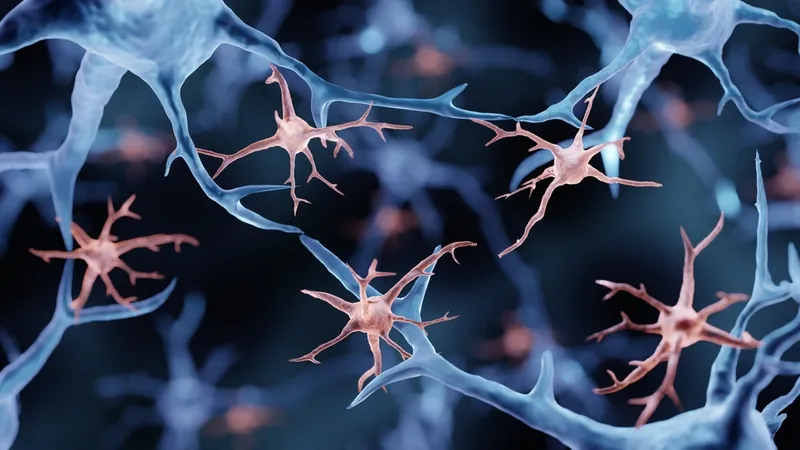
Scientists show that when done right, partial elimination of microglia, the immune cells of the brain, lowers age-related inflammation and cellular senescence [1].
Aging makes microglia bitter
Brain health is of utmost importance for geroscientists, since the brain is the one organ that cannot be replaced, and microglia are the brain’s resident immune cells. Reacting to pathogens and injuries, microglia get activated and drive an inflammatory response. Yet, in the brain, just as elsewhere in the body, this immune response becomes dysregulated with age, leading to the constant low-grade inflammation known as inflammaging, a key feature of many age-related diseases. Brain inflammaging has been linked to neurodegeneration, including Alzheimer’s disease [2]. Some microglia also become senescent and, as a result, toxic.
If you can’t fix them, kill them
To counter these detrimental effects, scientists have been experimenting with microglia depletion or blocking microglia function using genetic and pharmacological tools [3].
In this new paper, the researchers studied partial pharmacological microglia depletion in young and old mice using various doses of the microglia-killing drug PLX5622.
First, they analyzed the amount of microglia in the brain. An interesting early finding was that old mice had more microglia than the young ones. Treatment with PLX5622 eliminated microglia in a dose-dependent manner, with the smaller dose (300 mg/kg) reducing the overall number of microglia by 30-40% and the higher dose (1200 mg/kg) by 70-80%. However, more is not always better. Though there were more microglia in the old mice, a higher percentage of their microglia succumbed to the drug, which partially closed the gap.
Inflammation and senescence
The researchers wanted to know how depletion of microglia affects inflammation. To begin with, they found that several pro-inflammatory markers were substantially upregulated in old, untreated mice compared to young mice, which makes sense in light of the age-related chronic inflammation.
In young mice, the depletion of microglia did not lead to significant changes in the expression of inflammation markers, indicating that young microglia are mostly quiescent and do not contribute to inflammation. Conversely, in aged mice, the treatment substantially reduced the levels of inflammation-related markers.
Interestingly, the levels of the pro-inflammatory cytokine IL-1ß were significantly reduced only by the low dose and not by the high dose of the treatment.
Additional tests showed a five-fold increase in the senescence marker p16Ink4a in the brains of old untreated mice compared to young mice, confirming that old brains contain more senescent cells. In young mice, the treatment did not decrease the marker levels; here, again, the young microglia were found to be benign, with no signs of age-related senescence.
The low-dose treatment led to a significant decrease in p16Ink4a expression compared to the untreated group. Yet, similarly to the IL-1ß case, p16Ink4a levels in the high-dose group were much higher than in the low-dose group, though not as high as in the untreated controls.
Decimate, don’t eliminate
Additional investigation led the researchers to conclude that the high-dose treatment has an ambiguous effect on inflammation and senescence: the mass death of microglia probably leads to the overactivation of the remaining ones and also of astrocytes, another type of brain cell. The authors suggest that the diseased cells leave a lot of cellular debris that the remaining ones have to deal with.
Previous research supports the notion that although killing microglia is a promising therapeutic strategy, it should be exercised with caution: while partial depletion has been shown to improve cognition in a mouse model of Alzheimer’s [4], other studies have found that complete genetic depletion of the microglial population can cause a cytokine storm and worsen cognitive dysfunction [5].
The researchers conclude that low-dose partial pharmacological depletion of microglia is the way to go, as higher doses trigger unacceptable side effects. Another reason to choose the more balanced approach is because of the similarity of microglia to some other immune cells. As a result, the researchers argue, complete depletion of microglia in vivo could compromise immune function.
Conclusion
Age-related microglial activation is a known problem that currently has no solution. Modulating the very number of microglia in vivo is a bold idea that deserves serious consideration. As this research shows, even a modest decrease in microglial numbers can substantially reduce inflammation and senescence in the brain, while complete depletion probably does more harm than good.
Literature
[1] Stojiljkovic, M. R., Schmeer, C., & Witte, O. W. (2022). Pharmacological depletion of microglia leads to a dose-dependent reduction in inflammation and senescence in the aged murine brain. Neuroscience.
[2] Giunta, B., Fernandez, F., Nikolic, W. V., Obregon, D., Rrapo, E., Town, T., & Tan, J. (2008). Inflammaging as a prodrome to Alzheimer’s disease. Journal of neuroinflammation, 5(1), 1-15.
[3] Coleman, L. G., Zou, J., & Crews, F. T. (2020). Microglial depletion and repopulation in brain slice culture normalizes sensitized proinflammatory signaling. Journal of neuroinflammation, 17(1), 1-20.
[4] Spangenberg, E. E., Lee, R. J., Najafi, A. R., Rice, R. A., Elmore, M. R., Blurton-Jones, M., … & Green, K. N. (2016). Eliminating microglia in Alzheimer’s mice prevents neuronal loss without modulating amyloid-ß pathology. Brain, 139(4), 1265-1281.
[5] Parkhurst, C. N., Yang, G., Ninan, I., Savas, J. N., Yates III, J. R., Lafaille, J. J., … & Gan, W. B. (2013). Microglia promote learning-dependent synapse formation through brain-derived neurotrophic factor. Cell, 155(7), 1596-1609.


