Researchers publishing in Nature Aging have investigated one of the core biological reasons behind the decline of stem cells’ ability to proliferate.
Heterochromatin and euchromatin
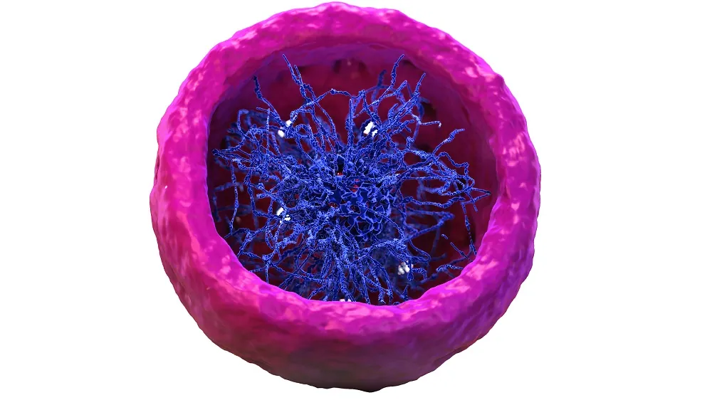
The genetic component of cells is comprised of two main parts: heterochromatin and euchromatin. Heterochromatin is largely silent, as it is a tightly packed mass that consists primarily of non-coding DNA. Euchromatin, on the other hand, is rich in actively transcribed genes and is considerably less packed.
However, heterochromatin can be methylated and demethylated just like euchromatin can. Age-related changes in methylation, the hallmark of aging known as epigenetic alterations, can happen to heterochromatin as well [1]. The regions to which this happens are known as partially methylated domains (PMDs), and they are associated with the lamina, the protective envelopes around nuclei that preserve genomic stability; laminal dysfunction is the key aspect of the rapid aging known as progeria. Changes to PMDs are associated with aging and cancer [2].

Read More
DNA methylation, these researchers explain, is mediated by the DNA methyltransferases (DNMTs), which produce 5mC, which promotes the stability of heterochromatin [3]. On the other hand, the TET family of enzymes oxidize 5mC to produce 5hmC, which is associated with the accessibility of euchromatin. The balance between DMNTs and TETs in regulating both forms of chromatin has not been fully explained [4].
Removing TET appears to help stem cells
These researchers used a mouse model that did not produce TET enzymes. The primary focus of this study was on hematopoietic stem and progenitor cells (HSPCs), which are responsible for creating blood. As expected, in 18- to 24-month-old wild-type mice, HSPCs were less able to divide and proliferate than in their young counterparts. The researchers state that “Tet2 depletion in HSPCs effectively rescued this age-related functional decline”.
Similarly, when cells from TET-knockout and wild-type mice were given to mice that had their natural cells’ proliferation destroyed by radiation, HSPCs from the TET-knockout mice proliferated far more readily. The stem cells of older wild-type mice spend more time in a non-dividing state, but this effect of aging was less pronounced in the TET-knockout mice. While rapid proliferation is often associated with cancer, the researchers found no evidence of these cells becoming cancerous.
There were also considerably fewer changes to gene expression. Wild-type mice have upregulated immune response and downregulated blood creation (hematopoiesis) as they age, but these effects of aging were also substantially blunted in TET-knockout mice.
A closer examination of the chromatin revealed interesting findings. Many epigenetic alterations were the same between the wild-type and TET-knockout groups. However, in the heterochromatin, 5hmC was substantially upregulated in older mice; the TET-knockout group, without the primary enzyme responsible for 5hmC, did not have this level of upregulation with age. The non-coding region has real effects after all.
Old viral DNA is expressed with age
Endogenous retroviruses (ERVs) are fragments of old viral DNA that have remained in the genome for millions of years. Normally, these genes are seldom expressed. However, as age-related changes to the location of heterochromatin occur, these genes become expressed more often, causing inflammation within cells. The HSPCs of TET-knockout mice did not have as many age-related location disturbances nor as much age-related upregulation of ERVs, and therefore they had less upregulation of inflammatory genes as well.
While this paper is very in-depth, and it certainly does not recommend TET knockout as a solution to human aging, it is clear that such complexities of biology need to be addressed if we are to get a handle on why our cells lose their fundamental abilities with age. Further work in this area may lead to better understanding and solutions to the problems of epigenetic alterations and genomic instability.
Literature
[1] Hernando-Herraez, I., Evano, B., Stubbs, T., Commere, P. H., Jan Bonder, M., Clark, S., … & Reik, W. (2019). Ageing affects DNA methylation drift and transcriptional cell-to-cell variability in mouse muscle stem cells. Nature communications, 10(1), 4361.
[2] Brinkman, A. B., Nik-Zainal, S., Simmer, F., Rodríguez-González, F. G., Smid, M., Alexandrov, L. B., … & Stunnenberg, H. G. (2019). Partially methylated domains are hypervariable in breast cancer and fuel widespread CpG island hypermethylation. Nature communications, 10(1), 1749.
[3] Richards, E. J., & Elgin, S. C. (2002). Epigenetic codes for heterochromatin formation and silencing: rounding up the usual suspects. Cell, 108(4), 489-500.
[4] López-Moyado, I. F., Tsagaratou, A., Yuita, H., Seo, H., Delatte, B., Heinz, S., … & Rao, A. (2019). Paradoxical association of TET loss of function with genome-wide DNA hypomethylation. Proceedings of the National Academy of Sciences, 116(34), 16933-16942.


