×

The Blog

Building a Future Free of Age-Related Disease

Rodent on exercise wheel

The Microbiome Might Affect Motivation for Exercise

Scientists publishing in Nature have found that compounds produced by some types of gut bacteria can influence dopamine levels in the brain and, as a result, might influence motivation to go on a morning run [1].

We’ve got company

We tend to think of ourselves as single organisms, but every human body serves as a home to trillions of other living things: bacteria of various species. Until recently, those microscopic squatters were not getting the attention they deserved. During recent years, there has been a boom in microbiome studies, linking the inhabitants of our guts to neurodegenerative [2] and cardiovascular diseases [3], cancer [4], and inflammaging [5]. We now know that microbiome composition differs significantly among individuals, contributing to the variability in health risks and fitness [6].

Exercise is one of the most potent anti-aging interventions known to humans. Most people know they should exercise, and many do, but motivation remains a serious issue. What’s easy for some is daunting for others. The reasons for this variability in motivation are poorly understood and usually catalogued under the vague label of “personality traits”. However, this study suggests that those differences might have a lot to do with the microbiome.

Do you have the guts to go on?

The researchers started with genetically diverse mice and meticulously phenotyped them, accumulating thousands of data points per animal. They learned that genetic differences played only a minor role in the variability in both voluntary and forced exercise capacity. They then used machine learning to identify variables that were strongly predictive of this capacity. Interestingly, the results of 16s rDNA analysis, which is commonly used to identify bacterial strains in the microbiome, were among the most well-correlated with endurance.

The researchers then performed a series of microbiome depletion and transplantation experiments. Sweeping microbiotal ablation with broad-spectrum antibiotics led to a decrease in exercise capacity. When microbiota was transplanted from donor mice to germ-free mice, the performance levels were highly correlated between the donor and the recipient.

The scientists then continued to experiment on genetically identical B6 mice to exclude factors other than microbiome composition. They tried various narrowly acting antibiotics and found that only neomycin had not impaired physical performance. The researchers again turned to 16s rDNA sequencing and identified several members of the Erysipelotrichaceae and Lachnospiraceae families as possible drivers of exercise performance.

Gut-brain dopamine signaling

However, muscle function and oxygen consumption turned out to be largely similar in microbiome-depleted mice and in controls, so the researchers decided to investigate the motivational pathway.

They found that in striatal neurons (the striatum is a part of the brain central to movement control), levels of dopamine, a major regulator of the drive for physical activity, were elevated by exercise in mice with intact microbiota, but not in antibiotic-treated mice – except those treated with neomycin. Those post-exercise spikes in striatal dopamine levels were restored by the same microbiotal transplants that had improved exercise performance, but not by other types of bacteria. Inhibition of dopamine by other means recapitulated the effects of microbiome depletion.

How exactly did the gut communicate with the brain? Experiments ruled out the possibility that this communication was carried out by metabolites in blood, so the researchers turned their attention to nerves. Their results suggest that fatty acid amides (FAAs) produced by some types of bacteria trigger neuronal signaling that ultimately results in increased dopamine levels in the striatum. FAA-fed mice with depleted microbiomes showed improved exercise ability.

Moreover, the researchers were able to engineer an FAA-producing string of E. coli. Mono-colonizing mice’s guts with this strain (but not with regular E. coli) rescued their exercise performance. The bottom line is that when there are few FAA-producing bacteria in the gut, this seems to blunt the exercise-associated dopamine surge, leading to diminished motivation to exercise.

Conclusion

This study increases our understanding of the diversity of microbiota and their importance for human health and fitness. If the researchers are correct and if these results translate to human beings, people might soon be able to increase their motivation to exercise by consuming specific strains of bacteria or FAAs directly.

We would like to ask you a small favor. We are a non-profit foundation, and unlike some other organizations, we have no shareholders and no products to sell you. All our news and educational content is free for everyone to read, but it does mean that we rely on the help of people like you. Every contribution, no matter if it’s big or small, supports independent journalism and sustains our future.

Literature

[1] Dohnalová, L., Lundgren, P., Carty, J. R., Goldstein, N., Wenski, S. L., Nanudorn, P., … & Thaiss, C. A. (2022). A microbiome-dependent gut–brain pathway regulates motivation for exercise. Nature, 612(7941), 739-747.

[2] Chen, C., Liao, J., Xia, Y., Liu, X., Jones, R., Haran, J., … & Ye, K. (2022). Gut microbiota regulate Alzheimer’s disease pathologies and cognitive disorders via PUFA-associated neuroinflammation. Gut.

[3] Witkowski, M., Weeks, T. L., & Hazen, S. L. (2020). Gut microbiota and cardiovascular disease. Circulation research, 127(4), 553-570.Chicago

[4] Helmink, B. A., Khan, M. A., Hermann, A., Gopalakrishnan, V., & Wargo, J. A. (2019). The microbiome, cancer, and cancer therapy. Nature medicine, 25(3), 377-388.

[5] Fransen, F., Van Beek, A. A., Borghuis, T., Aidy, S. E., Hugenholtz, F., van der Gaast–de Jongh, C., … & De Vos, P. (2017). Aged gut microbiota contributes to systemical inflammaging after transfer to germ-free mice. Frontiers in immunology, 8, 1385.

[6] Clauss, M., Gérard, P., Mosca, A., & Leclerc, M. (2021). Interplay between exercise and gut microbiome in the context of human health and performance. Frontiers in Nutrition, 305.

Silica dust

NMN Alleviates Silicate Lung Injury in Mice

A new paper published in Nutrients shows that the well-known NAD+ precursor NMN alleviates lung injury caused by silicate inhalation in wild-type mice.

An antioxidant approach to a common problem

Silicosis is an occupational hazard encountered by people who are regularly exposed to silica dust [1]. It is a common cause of lung injury around the world [2]. Silica dust enters cells easily, but cells find it hard to remove [3]; when they do remove it, it is often through phagocytosis, and the released particles are then often consumed once again [4].

One of the main ways that silicosis harms cells is through cellular stress in the forms of inflammation and oxidation, which are problems that NMN has been reported to alleviate in previous studies related to other conditions [5].

Effective against injury and inflammation

For this study, the researchers used five groups of wild-type mice: a sham group, a silicate inhalation group, groups that received high (one gram per kilogram) and low (half a gram per kilogram) doses of NMN along with silica inhalation, and an NMN control group that only received a gram per kilogram of NMN. These mice were only six to eight weeks old.

After one week of this treatment, the results were already statistically significant. The two NMN treatment groups were found to have significantly less scar-related collagen tissue and significantly fewer lesions than the silica-only group.

NMN Lung Injury 1

Four weeks showed even more significant results. Lung weight, which had significantly increased among the silica-only group, had not increased nearly as much in the NMN groups, which had significantly reduced collagen as well. While both NMN treatment groups benefited, the high-dose group fared better than the low-dose group in lung lesions.

NMN Lung Injury 2

Similar results were reported in reactive oxygen species (ROS), glutathione (GSH), and macrophage count. While low doses of NMN were not shown to be effective against ROS at 7 days, they were effective at 28 days. GSH decreases with silica inhalation but is restored with NMN at low and high doses. Macrophage count increases with silica as well, and this measurement of inflammation is also reduced with NMN, particularly after four weeks.

Gene expression analysis confirmed these findings, showing that GSH-related metabolism, which is mediated by the production of the ROS metabolism factor Nrf2, is a likely cause of these results. In these mice, Nrf2 was found to be decreased with silica inhalation and restored with high-dose NMN. Other genes related to the metabolism of foreign matter and detoxification were found to be decreased with silica and enhanced with NMN at both low and high doses after 28 days.

Conclusion

The researchers note that although they have explored some of the fundamental biology, the lung microenvironment remains largely unexplored. Therefore, despite what has been learned from these experiments, there is not yet a full understanding of how NMN interacts with lung fibroblasts in living tissue.

While this study focuses on environmental exposure rather than intrinsic aging, it is not a leap of logic to suggest that these effects against this particular form of accumulated damage may apply to other forms of accumulated damage as well. However, this is still a mouse study. Further trials are necessary to confirm these results and determine if NMN is effective in alleviating silicosis, or other sources of long-term tissue damage, in human beings.

We would like to ask you a small favor. We are a non-profit foundation, and unlike some other organizations, we have no shareholders and no products to sell you. All our news and educational content is free for everyone to read, but it does mean that we rely on the help of people like you. Every contribution, no matter if it’s big or small, supports independent journalism and sustains our future.

Literature

[1] Barnes, H., Goh, N. S., Leong, T. L., & Hoy, R. (2019). Silica‐associated lung disease: an old‐world exposure in modern industries. Respirology, 24(12), 1165-1175.

[2] Shi, P., Xing, X., Xi, S., Jing, H., Yuan, J., Fu, Z., & Zhao, H. (2020). Trends in global, regional and national incidence of pneumoconiosis caused by different aetiologies: an analysis from the Global Burden of Disease Study 2017. Occupational and Environmental Medicine, 77(6), 407-414.

[3] Rimola, A., Costa, D., Sodupe, M., Lambert, J. F., & Ugliengo, P. (2013). Silica surface features and their role in the adsorption of biomolecules: computational modeling and experiments. Chemical reviews, 113(6), 4216-4313.

[4] Benmerzoug, S., Rose, S., Bounab, B., Gosset, D., Duneau, L., Chenuet, P., … & Quesniaux, V. F. (2018). STING-dependent sensing of self-DNA drives silica-induced lung inflammation. Nature communications, 9(1), 1-19.

[5] Wan, Y., He, B., Zhu, D., Wang, L., Huang, R., Zhu, J., … & Gao, F. (2021). Nicotinamide mononucleotide attenuates doxorubicin-induced cardiotoxicity by reducing oxidative stress, inflammation and apoptosis in rats. Archives of Biochemistry and Biophysics, 712, 109050.

DNA Damage

David Sinclair: Epigenetic Info Loss Is a Cause of Aging

Published in Cell, a new paper by David Sinclair and his team argues that epigenetic dysregulation in the form of information loss is a major driver of aging, but it can be reversed in vivo by partial cellular reprogramming.

Genome or epigenome?

Both genomic instability and epigenetic alterations are included in the Hallmarks of Aging [1], and both have been linked to numerous age-related pathologies. However, some geroscientists, such as the renowned Harvard professor David Sinclair, assign more blame to the latter. Sinclair has developed the “Information Theory of Aging” which postulates that “aging in eukaryotes is due to the loss of transcriptional networks and epigenetic information over time”. The twist is that this epigenetic dysregulation is a byproduct of the cell’s constant attempts to repair DNA damage, so the two hallmarks are interlinked.

According to this “relocalization of chromatin modifiers” (RCM) hypothesis, certain elements of the double strand break (DSB) repair mechanism, such as Sir2, are also in charge of keeping the chromatin structure in order, which is the key to maintaining the cell’s function and identity. When summoned to repair a DSB, those elements leave their usual posts. Sometimes, they fail to return to their initial positions after the repair is completed, especially when there’s a lot of DSBs. This causes gradual dysregulation in chromatin structure and transcription. In addition to several scientific papers [2], the RCM hypothesis is described in detail in Sinclair’s 2019 book “Lifespan: Why We Age, and Why We Don’t Have To”.

DSBs cause aging in the absence of mutations

In this new study, Sinclair’s group tested this hypothesis in a new, ingenious way. The researchers created a model of mild increase in DSB load for use in vitro and in vivo. As opposed to “blunt instruments” such as radiation or chemical damage, which induce too many DSBs and cause serious loss of function, this new model strains the cell’s DSB repair mechanisms without overwhelming them. It does so by conditionally expressing I-PpoI endonuclease (an enzyme that cleaves DNA), which recognizes a specific nucleotide sequence found in some non-essential DNA loci outside of protein-coding genes.

For their in vitro experiments, the researchers used mouse embryonic fibroblasts (MEFs). DSB markers increased four-fold after the system was turned on, but this did not cause changes in cell-cycle profile, apoptosis, or senescence; the cells did not seem to suffer any significant damage. However, the cells became about 1.5-fold epigenetically older than controls, as measured at 89 methylation sites. The cells exhibited several more signs of aging, such as increased sensitivity to DNA-damaging agents and elevated senescence markers. Importantly, no change in mutation frequency was observed, showing that the DSB repair process was faithful and still led to accelerated cellular aging.

The researchers then induced whole-body I-PpoI expression in 4- to 6-month-old mice for 3 weeks. While during the treatment and shortly after, there was no difference in fitness between the study group and controls, subtle differences such as alopecia began appearing one month after the treatment. At the ten-month mark, the treated mice were noticeably less fit, with frailty indices on par with two-year-old mice along with kyphosis and decreased bone density. Various symptoms of brain, muscle, skin, and liver aging were observed as well. The treated mice also showed accelerated epigenetic aging. While no change in mutation frequency was observed one month after the treatment ended, it significantly altered the epigenetic landscape in an aging-like manner.

The researchers then established that intensified DSB repair led to erosion of the epigenetic landscape, disruption of developmental genes, and altered spatial organization of chromatin, which controls gene expression and cellular identity. The paper calls this “the first evidence that faithful DNA repair alters multiple layers of epigenetic information”.

Reprogramming restores the epigenetic landscape

If the treatment indeed weakens cellular identity, it should be easier to nudge the treated cells towards differentiation into other cell types, such as neurons. Using an established cellular reprogramming protocol, the researchers showed that the treated cells were much more susceptible to such redifferentiation than controls. In treated mice, the epigenetic signature of the muscle was shifted towards that of immune cells, which is also what happens during aging.

In a study from 2020 [3], Sinclair’s group showed that partial in vivo reprogramming using Yamanaka factors can reverse epigenetic age and gene expression patterns in old neurons. The researchers interpret this as cells possessing “a back-up copy of youthful epigenetic information that can restore cell identity”. In this study, partial cellular reprogramming did the trick again, rolling back the epigenetic age of cells in vitro and markers of epigenetic aging in vivo.

Conclusion

The authors suggest that the epigenetic dysregulation caused by the normal activity of DSB-repairing machinery drives aging even when the repair is faithful and no mutations occur. Some previous findings seem to support this hypothesis. For instance, epigenetic signatures change significantly with age in most animals but not in long-lived bats.

The researchers also mention successful cloning of animals from old cells (their accumulated mutations get passed to the progeny, but not their aged epigenome). On the other hand, one recent study showed a strong correlation between the rate of somatic mutations and aging across numerous species.

If the researchers are right, this opens the door to reversing aging through partial cellular reprogramming, which is a technique actively pursued by numerous scientists and longevity biotech companies.

We would like to ask you a small favor. We are a non-profit foundation, and unlike some other organizations, we have no shareholders and no products to sell you. All our news and educational content is free for everyone to read, but it does mean that we rely on the help of people like you. Every contribution, no matter if it’s big or small, supports independent journalism and sustains our future.

Literature

[1] López-Otín, C., Blasco, M. A., Partridge, L., Serrano, M., & Kroemer, G. (2013). The hallmarks of aging. Cell, 153(6), 1194-1217.

[2] Oberdoerffer, P., & Sinclair, D. A. (2007). The role of nuclear architecture in genomic instability and ageing. Nature reviews Molecular cell biology, 8(9), 692-702.

[3] Lu, Y., Brommer, B., Tian, X., Krishnan, A., Meer, M., Wang, C., … & Sinclair, D. A. (2020). Reprogramming to recover youthful epigenetic information and restore vision. Nature, 588(7836), 124-129.Chicago

Obese and healthy mouse

Obesity Shown to Affect Brain Aging in Mice

Research published in Immunity & Ageing has shown that obesity has significant, aging-associated effects on behavior and immunity in the brains of mice.

Known effects on humans and mice

The researchers introduce this study by discussing human studies. It is well known that obesity is a risk factor for severe metabolic disorders, such as diabetes and cardiovascular disease [1], and dementia later in life [2].

While such longitudinal studies are highly informative, they do not document any of the fundamental biology involved. Previous research has been conducted in that area, showing that obesity harms the hippocampus [3] and encourages inflammation [4] in mouse models. The effects of aging have also been heavily documented in these models as well, illustrating the well-known phenomenon of inflammaging [5].

The effects of obesity on aged mice have also been documented, showing a broad variety of harms to the brain [6], including gene expressions associated with Alzheimer’s disease [7]. In this paper, the researchers build on that work by analyzing mice of different ages in an effort to determine how obesity and aging interact.

An analysis of four groups

In this study, the researchers used four groups: 16-week-old mice fed a standard diet (SD), 16-week-old fed a high-fat diet (HFD) to induce obesity, 24-week-old mice fed an SD, and 24-week-old mice on the HFD.

This paper began with a hippocampal gene expression analysis. Eight weeks of aging were shown to affect 729 genes, and the change in diet was shown to affect 886 genes. 216 of these genes were the same ones: in these cases, obesity was shown to affect the hippocampus in approximately the same way that aging did. Many of these pathways were related to metabolic dysfunction, and others were related to neurodegenerative diseases such as Parkinson’s, Huntington’s, and Alzheimer’s.

A more involved look showed more counterintuitive data. In many cases, such as in genes related to lymphocyte function and immune cell activation, aging and HFD were shown to independently increase their expression; however, the aged HFD mice had decreased expression instead.

The effects on body size were largely predictable. Both aging and a high-fat diet increase fat mass in mice. However, aged mice fed the high-fat diet were not significantly larger than their younger counterparts fed the same diet, and their individual fat cells were not found to be larger either. They did, however, have significantly worse problems handling glucose; the effects of aging and a high-fat diet were shown to combine in this respect.

Effects on the brain

While neither aging nor a high-fat diet were shown to significantly increase pro-inflammatory cytokines in the hippocampus, aged HFD mice had more immune cells in the brain than their SD counterparts did. The data showed that aging slightly increases this as well, but not to the level of statistical significance. The researchers noted that this obesity-associated increase in microglia is in line with previous research in other animal models [8].

Obesity also caused a more pronounced fear response in these mice. HFD mice in both groups were shown to learn a Pavlovian fear association more quickly; normally, aging slows the development of this learned response. Aging also slows the gradual loss of this response when it is being conditioned out of the mice; however, HFD mice, particularly aged HFD mice, retained their fear responses significantly longer.

Conclusion

While some of the data is notably counterintuitive and bears further investigation, this study largely confirms what is known about obesity and its effects on the brain. Some of this information may be specific to mice; for example, the effects on fear response did not apply to rats [9]. However, this study adds to the body of research showing that obesity has significant, aging-associated, and largely negative effects on the brains of mammals.

We would like to ask you a small favor. We are a non-profit foundation, and unlike some other organizations, we have no shareholders and no products to sell you. All our news and educational content is free for everyone to read, but it does mean that we rely on the help of people like you. Every contribution, no matter if it’s big or small, supports independent journalism and sustains our future.

Literature

[1] Blüher, M. (2019). Obesity: global epidemiology and pathogenesis. Nature Reviews Endocrinology, 15(5), 288-298.

[2] Pedditizi, E., Peters, R., & Beckett, N. (2016). The risk of overweight/obesity in mid-life and late life for the development of dementia: a systematic review and meta-analysis of longitudinal studies. Age and ageing, 45(1), 14-21.

[3] Hao, S., Dey, A., Yu, X., & Stranahan, A. M. (2016). Dietary obesity reversibly induces synaptic stripping by microglia and impairs hippocampal plasticity. Brain, behavior, and immunity, 51, 230-239.

[4] de Heredia, F. P., Gómez-Martínez, S., & Marcos, A. (2012). Obesity, inflammation and the immune system. Proceedings of the Nutrition Society, 71(2), 332-338.

[5] Weyand, C. M., & Goronzy, J. J. (2016). Aging of the immune system. Mechanisms and therapeutic targets. Annals of the American Thoracic Society, 13(Supplement 5), S422-S428.

[6] Valcarcel-Ares, M. N., Tucsek, Z., Kiss, T., Giles, C. B., Tarantini, S., Yabluchanskiy, A., … & Csiszar, A. (2019). Obesity in aging exacerbates neuroinflammation, dysregulating synaptic function-related genes and altering eicosanoid synthesis in the mouse hippocampus: potential role in impaired synaptic plasticity and cognitive decline. The Journals of Gerontology: Series A, 74(3), 290-298.

[7] Tucsek, Z., Toth, P., Sosnowska, D., Gautam, T., Mitschelen, M., Koller, A., … & Csiszar, A. (2014). Obesity in aging exacerbates blood–brain barrier disruption, neuroinflammation, and oxidative stress in the mouse hippocampus: effects on expression of genes involved in beta-amyloid generation and Alzheimer’s disease. Journals of Gerontology Series A: Biomedical Sciences and Medical Sciences, 69(10), 1212-1226.

[8] Gzielo, K., Kielbinski, M., Ploszaj, J., Janeczko, K., Gazdzinski, S. P., & Setkowicz, Z. (2017). Long-term consumption of high-fat diet in rats: effects on microglial and astrocytic morphology and neuronal nitric oxide synthase expression. Cellular and molecular neurobiology, 37(5), 783-789.

[9] Spencer, S. J., D’Angelo, H., Soch, A., Watkins, L. R., Maier, S. F., & Barrientos, R. M. (2017). High-fat diet and aging interact to produce neuroinflammation and impair hippocampal-and amygdalar-dependent memory. Neurobiology of aging, 58, 88-101.

Intestinal molecules

NAD+ Supplement Protects Intestines from Alcohol in Mice

Scientists have shown that the NAD+ precursor nicotinamide riboside (NR) alleviates symptoms of leaky gut caused by ethanol consumption in mice by improving mitochondrial function [1].

Leaky gut drives inflammation

The permeability of the epithelial intestinal barrier is known to increase with age. The resulting condition, also known as “leaky gut,” can be exacerbated by dietary, lifestyle, and environmental factors, such as alcohol consumption [2]. While not lethal, like cancer or cardiovascular diseases, leaky gut is not a trifling matter. Recent studies have shown that intestinal contents, such as the bacterial byproduct lipopolysaccharide, become potent immune system triggers in the bloodstream. This is how a leaky gut causes inflammaging: the persistent systemic inflammation that drives multiple diseases of aging [3].

NR is a precursor to NAD+, a ubiquitous multi-role coenzyme central to energy metabolism, DNA repair, and other important cellular processes. NAD+ levels decrease with aging, while supplementation of NAD+ via its precursors such as NR has been linked to various health benefits in mice and humans [4]. In particular, NR has been shown to rejuvenate aged gut stem cells [5]. NR is a popular supplement that is generally considered safe; however, one recent study found that it might exacerbate metastasizing cancers.

Buttressing the intestinal barrier

In this new study, the researchers investigated the deleterious effects of ethanol on gut permeability in mice and whether they can be alleviated by NR. Over the course of the experiment, mice fed an ethanol-rich diet experienced intestinal barrier deterioration. However, in mice who also received NR, this effect, as measured by lipopolysaccharide (LPS) concentration in serum, was largely abolished.

NR Intestines 1

Tight junctions, the constructions that glue cells together, are an important component of the intestinal barrier and a significant weak spot. Their disintegration is a major cause of leaky gut. The levels of tight junction proteins ZO-1 and occludin were decreased by ethanol, while in the ethanol-NR group, those levels remained on par with controls. Histopathological analysis showed that the structures of intestinal villi and of epithelial cells were deformed in the ethanol group but remained just as healthy in the NR group as in controls.

The researchers also experimented in vitro on Caco-2 cells, which are often used as a model of the intestinal epithelial barrier. When treated with ethanol, the cells showed significantly reduced ZO-1 and occludin levels but not when pre-treated with NR for 48 hours before ethanol exposure.

Recent studies show that energy homeostasis is important for tight junction formation between intestinal epithelial cells [6]. The researchers confirmed that NAD+ levels were greatly depleted in those cells by ethanol, but elevated by NR supplementation – amazingly, above those of healthy controls. Levels of ATP, the molecule considered cellular “energy currency”, showed a similar dynamic.

NR intestines 2

Mitochondrial boost via SIRT1

Since most of the energy production in cells occurs in mitochondria, the researchers also analyzed mitochondrial health. As expected, levels of succinate dehydrogenase (SDH) and citrate synthase (CS), two functional mitochondrial enzymes, were reduced by ethanol but rescued by NR supplementation. Same picture was observed for mitochondrial DNA number.

Mitochondrial biogenesis is promoted by the enzyme SIRT1, a NAD-dependent acetylase of the sirtuin family. Suppressing SIRT1 expression in Caco-2 cells by short interfering RNAs abolished the restorative effect of NR. In particular, NR lost its ability to improve mitochondrial membrane potential. In the absence of SIRT1, NR also failed to increase the levels of both ZO-1 and occludin. This led the researchers to conclude that NR counters the effects of ethanol by promoting intestinal mitochondrial biosynthesis in a SIRT1-dependent manner.

NR intestines 3

Intestinal dysfunction including gut dysbiosis and barrier disruption contributes to the development of diseases in the liver and other organs. The intestinal barrier consists of physical, secretory, immunological and microbic components. TJs are the important components of epithelial barrier, whose integrity is essential in blocking gut microbes and adverse products such as LPS translocated to the circulation. In this study, we focused on the intestinal barrier function affected by ethanol and NR. Our study reports for the first time that ethanol induces intestinal epithelial barrier injury via destroying mitochondrial function. Supplementation of NR, which is a NAD precursor, protects against ethanol-induced intestinal epithelial injuries via maintaining mitochondrial function by promoting mitochondrial biogenesis in a SirT1-dependent manner.

Conclusion

Leaky gut has been implicated in inflammaging, making the integrity of the intestinal barrier an important target for geroscientists. This study shows that NAD+ supplementation through NR dramatically alleviates leaky gut symptoms caused by ethanol (which in humans is usually a result of alcoholic beverage consumption), but these results can probably be extrapolated to other causes. A clear limitation of this study is its modest sample size (5-8 mice per group), so more research is needed.

We would like to ask you a small favor. We are a non-profit foundation, and unlike some other organizations, we have no shareholders and no products to sell you. All our news and educational content is free for everyone to read, but it does mean that we rely on the help of people like you. Every contribution, no matter if it’s big or small, supports independent journalism and sustains our future.

Literature

[1] Li, W., Zhou, Y., Pang, N., Hu, Q., Li, Q., Sun, Y., … & Yang, L. (2023). NAD Supplement Alleviates Intestinal Barrier Injury Induced by Ethanol Via Protecting Epithelial Mitochondrial Function. Nutrients, 15(1), 174.

[2] Bishehsari, F., Magno, E., Swanson, G., Desai, V., Voigt, R. M., Forsyth, C. B., & Keshavarzian, A. (2017). Alcohol and gut-derived inflammation. Alcohol research: current reviews, 38(2), 163.

[3] Kavanagh, K., Hsu, F. C., Davis, A. T., Kritchevsky, S. B., Rejeski, W. J., & Kim, S. (2019). Biomarkers of leaky gut are related to inflammation and reduced physical function in older adults with cardiometabolic disease and mobility limitations. Geroscience, 41(6), 923-933.

[4] Mehmel, M., Jovanović, N., & Spitz, U. (2020). Nicotinamide riboside—the current state of research and therapeutic uses. Nutrients, 12(6), 1616.

[5] Igarashi, M., Miura, M., Williams, E., Jaksch, F., Kadowaki, T., Yamauchi, T., & Guarente, L. (2019). NAD+ supplementation rejuvenates aged gut adult stem cells. Aging cell, 18(3), e12935.

[6] Hall, C. H., Lee, J. S., Murphy, E. M., Gerich, M. E., Dran, R., Glover, L. E., … & Colgan, S. P. (2020). Creatine transporter, reduced in colon tissues from patients with inflammatory bowel diseases, regulates energy balance in intestinal epithelial cells, epithelial integrity, and barrier function. Gastroenterology, 159(3), 984-998.

Updates

The Hallmarks of Aging’s Original Authors Offer a Fresh View

The year 2023 started with the publication of two remarkable review papers in Cell and Cell Metabolism by researchers addressing the hallmarks of aging and their interplay with the hallmarks of cancer [1,2]. These papers were authored by the same team that published the original 2013 Hallmarks of Aging paper [3].

Much-needed update

The original paper on the Hallmarks of Aging systematized the processes underlying aging. As such, it became a very common discussion topic in the rejuvenation world and was frequently cited in research papers related to aging. That paper determined nine hallmarks of aging under three criteria: the process is observed during normal aging, it speeds up aging if worsened, and it reverses some aspects of aging if abolished by therapeutic interventions.

Since then, several research groups have proposed the addition of other hallmarks, including five new ones suggested back in August 2022 and cellular enlargement.

The authors note that although the originally suggested hallmarks have withstood research scrutiny over the last decade, their original paper has required an update that integrates recently obtained knowledge, particularly from mammalian studies.

Therefore, their updated list of twelve hallmarks of aging includes the somewhat reorganized previous hallmarks plus three new ones: chronic inflammation, disabled macroautophagy, and dysbiosis [1]. All of these hallmarks are interconnected and interdependent.

The old and the new

The researchers initially discuss some of the already accepted and established hallmarks of aging. First, they focus on genomic instability, which includes nuclear architecture changes along with nuclear and mitochondrial DNA aberrations that accumulate with age.

The second hallmark, telomere attrition, is closely connected to genomic instability. However, it is a separate hallmark, and it has been shown to modulate aging when manipulated via telomerase activation in mammals.

The third hallmark of aging is known for its utility as a measurement of biological age: epigenetic alternations. There is a number of such aging-associated changes which ultimately lead to gene expression disbalances including histone modifications, non-coding RNAs, and reactivation of retrotransposons.

Loss of proteostasis resulting in protein misfolding and aggregation is a fourth hallmark of aging and was separated out from disabled macroautophagy. The authors consider this to be a hallmark of its own because, outside of its major role in proteostasis, autophagy ensures the proper turnover of organelles.

The authors then discuss deregulated nutrient sensing, with a special focus on diet as a practical target for anti-aging interventions. Mitochondrial dysfunction, cellular senescence, and stem cell exhaustion are all well-established hallmarks of aging which have proven to be promising targets for lifespan extension, at least in model organisms.

Altered intercellular communication is a hallmark of aging that involves several processes involved in cell-to-cell communication. It includes aging-associated damages to the extracellular matrix and pro-aging factors circulating in the blood.

Finally, the authors introduce chronic inflammation and dysbiosis (microbiome alterations) as the new meta-cellular hallmarks of aging. The former stems from all the damages and aberrations that result from the other hallmarks. The latter has been addressed by fecal microbiota transplantation, which proved beneficial in animal models.

Aging and cancer

In their second review publication [2], the researchers explored the overlap between the hallmarks of aging and cancer. They note that both phenomena share some features, which they coined “meta-hallmarks.” These include genomic instability and chronic inflammation. Meanwhile, such hallmarks of aging as telomere attrition favor oncogenesis and therefore represent “antagonistic hallmarks”.

The authors highlight a very complex relationship between aging and cancer. Although aging is the most prominent risk factor for various cancers, it is not necessarily true that targeting molecular drivers of aging is oncoprotective.

It is conceivable that by targeting the meta-hallmarks, it is possible to both delay aging and prevent cancer, while a more intricate tuning of antagonistic hallmarks is required. Regardless, experimental validation is needed to check if these assumptions are correct.

Abstracts

Aging is driven by hallmarks fulfilling the following three premises: (1) their age-associated manifestation, (2) the acceleration of aging by experimentally accentuating them, and (3) the opportunity to decelerate, stop, or reverse aging by therapeutic interventions on them. We propose the following twelve hallmarks of aging: genomic instability, telomere attrition, epigenetic alterations, loss of proteostasis, disabled macroautophagy, deregulated nutrient-sensing, mitochondrial dysfunction, cellular senescence, stem cell exhaustion, altered intercellular communication, chronic inflammation, and dysbiosis.

Both aging and cancer are characterized by a series of partially overlapping “hallmarks” that we subject here to a meta-analysis. Several hallmarks of aging (i.e., genomic instability, epigenetic alterations, chronic inflammation, and dysbiosis) are very similar to specific cancer hallmarks and hence constitute common “meta-hallmarks,” while other features of aging (i.e., telomere attrition and stem cell exhaustion) act likely to suppress oncogenesis and hence can be viewed as preponderantly “antagonistic hallmarks.”

Conclusion

All the hallmarks of aging are intimately related, which means that a truly successful anti-aging intervention would target several hallmarks at once to extend lifespan. Indeed, known substances with reported anti-aging effects, such as metformin and spermidine, are multimodal. Some of these therapeutics also possess anti-cancer properties. At the same time, as highlighted by the authors, there is a certain hierarchy among the hallmarks, which may guide researchers on their path to developing anti-aging interventions.

We would like to ask you a small favor. We are a non-profit foundation, and unlike some other organizations, we have no shareholders and no products to sell you. All our news and educational content is free for everyone to read, but it does mean that we rely on the help of people like you. Every contribution, no matter if it’s big or small, supports independent journalism and sustains our future.

Literature

[1] López-Otín, C., Blasco, M. A., Partridge, L., Serrano, M. & Kroemer, G. Hallmarks of aging: An expanding universe. Cell 0, (2023)

[2] López-Otín, C., Pietrocola, F., Roiz-Valle, D., Galluzzi, L. & Kroemer, G. Meta-hallmarks of aging and cancer. Cell Metab. 35, 12–35 (2023)

[3] López-Otín, C., Blasco, M. A., Partridge, L., Serrano, M. & Kroemer, G. The hallmarks of aging. Cell 153, 1194–1217 (2013)

Aubrey de Grey LEVF

Aubrey de Grey on LEVF and Robust Mouse Rejuvenation

Dr. Aubrey de Grey is a legend in the longevity field who has been steadfastly promoting the idea of life extension since well before it became mainstream. While with SENS Research Foundation, de Grey made significant contributions to geroscience, and at Longevity Summit Dublin last year, he announced the creation of his new brainchild, Longevity Escape Velocity Foundation (LEVF).

Now, the first major and long-awaited LEVF-funded project is being launched: Robust Mouse Rejuvenation (RMR). This is envisioned as a rolling research program aiming to increase both the mean and maximum lifespan of mice by at least 12 months with various combination therapies started late in life. For the first study, four therapies have been chosen: rapamycin, a senolytic, hematopoietic stem cell transplantation (HSCT), and telomerase expression. A groundbreaking experiment by any measure, RMR got us excited, and we reached out to Aubrey to discuss both RMR and LEVF in depth.

The following interview has Arkadi asking questions in bold and Aubrey de Grey answering in normal font.

We are obviously very excited about LEVF’s RMR project. Could you walk our readers through its design and goals?

I believe we’ll have two outcomes. One of them scientific, and the other more, if you like, rhetorical. We want to get mice to live a lot longer than they do now: at least a year longer, starting the treatment or treatments only after middle age. The idea is that this will appeal more directly to people who care, vote, pay taxes, and make donations than if you do early-onset interventions. So, I decided to put numbers on this, to have a milestone that clearly says this is where we want to get to. We believe this will be a sufficiently dramatic result.

What does “sufficiently” mean? The audience that I still care about the most is my prominent colleagues in the community: people who talk to the general public and do a lot of media appearances. We all know each other very well, we’re all good friends. So, I have a very accurate idea of how impressive a result needs to be in order to stimulate these people to say things publicly that they wouldn’t previously have said.

First, we have to take a strain of mice that are inherently healthy and long-lived. Of course, the standard strain that’s normally used are C57Bl/6 (“Black 6”) mice. We start late, at 18 months of age. That’s roughly one year less than the average lifespan of this strain. The idea is to double that one year, i.e. to give an average lifespan of three and a half years rather than two and a half. We also want to have at least a year of increase in maximum lifespan. As I’m sure you know, maximum lifespan is normally defined not as the lifespan of the last surviving mouse but rather the average lifespan of the last 10%.

This is important because many interventions can increase mean lifespan but not maximum. The interpretation that most people, including me, put on that kind of result is that the intervention is hitting some but not all the mechanisms of aging, and those that it’s not hitting are still proceeding unabated.

Meaning, we have compression of mortality instead of an increase in maximum lifespan?

Of course, we must be very careful with what we mean by compression of mortality, because that’s at a population level. We don’t know if any given individual mouse actually had a shorter time when they were going downhill health-wise. So, in this study we absolutely want to increase maximum as well as average lifespan.

There are various other things that we’re throwing into the mix. First, we’re using combination therapies. We take the view that rejuvenation therapies, those that repair damage instead of just slowing down the creation of damage, are inherently more partial in how much they do across the board of different types of damage. We figured out that we might get much more than the sum of the parts by putting multiple of these things in at the same time.

The second thing, again, coming back to rejuvenation, is that we do recognize that there are knock-on effects. In other words, you may be fixing one particular type of damage, but having done that has a beneficial effect on the rest of metabolism and somehow slows down other things as well. We believe that is the basic reason why we see in the literature a small but non-trivial number of reports of increase in maximum lifespan, as well as mean, by rejuvenation-type interventions. So, we believe that those are the most promising ones to start with, and we want to combine them.

In our current, initial, study (this is the first round of what we believe will be a rolling research program) we are taking four interventions, and one of them is not of the type I’ve described: rapamycin. You can think of it as a non-rejuvenation control if you like.

But it’s not just a control, because we want to know how well it synergizes with bona fide rejuvenation therapies that repair damage. Indeed, the entire design of the study is based on my analysis of what will give us the most information, per dollar spent, about the synergies between different treatments. In this study, we have a thousand mice in total, 500 of each sex, and we have split those mice into 10 groups of each sex, so 50 mice per group, per sex.

What are those ten groups? Of course, there’s a control group that gets nothing, and there’s also a group that gets all four of our interventions, but there are also four groups that get exactly one intervention, so that we have some kind of baseline. And then we have four groups that get three out of the four. This is very important. The purpose of this is to determine whether there are antagonistic interactions between things, which is possible.

With this, we believe that we’ll basically get all the possible information about those interactions. We believe that we would not gain appreciably more information if we also did the six more groups where you do exactly two out of the four. We will be able to extrapolate with very high confidence what their results would be. That saves us a lot of money.

What was your rationale for choosing the interventions? You made some intriguing choices here.

As I said, we did want to have rapamycin in the mix as a non-rejuvenation control. It’s a calorie restriction mimetic and probably the most effective one out there. The real question is, why the other three?

There were two main reasons. One was that there were already studies by others showing increase in mean and maximum lifespan in long-lived strains of mice when starting late in life. So, by and large, we are reproducing the protocols that these other groups used over the years.

The other big criterion is that the interventions should, as far as we can say, be targeting different types of damage. As I mentioned, we are drawing on the idea that there will be knock-on effects from one type of damage onto others.

Still, would it be fair to say that among those four interventions, we only have robust life extension data in mice for rapamycin?

Among the calorie restriction mimetics, I would agree with you. But remember, we are restricting ourselves to late onset interventions, and there are not a lot of those. One of the most horrifying things in the history of the field was the fact that a decade ago, when the NIH had this enormous piece of good luck, when they accidentally did this study starting at twenty months of age rather than four months, which was their original intention…

Yes, with rapamycin.

They got this fantastic result that was as good as calorie restriction itself, starting at that same kind of age. It’s so obvious that what they should have done was to go back and revise the guidelines for further studies to focus more on late-onset studies.

Of course, I understand why that didn’t happen – because people in academia are constantly fixated on what are diplomatically called “positive results”, on getting their stuff published in those high-profile journals. That means doing stuff that’s more likely to work, even if it’s less informative, which is insane. But it’s the way of the world. It’s a good example of the reason why I chose the direction I did 20 years ago and decided to lead a series of independent non-profits that were funded by philanthropy rather than by grants.

Not only might late-onset studies be more informative, but using pre-aged mice also allows you to drastically shorten the whole process, right?

Yes. Also, we are planning on putting out interim data all the time, very frequently, once a week or once every two weeks, because we’ve got two things going for us. The first one is, as you say, starting late means that the overall experiment will take two years rather than four years. But the other one, which is just as important, is that because we are mostly doing rejuvenation therapies that remove damage rather than just slowing down the creation of new damage, we have a good chance of seeing they’re working from the divergence of survival curves quite quickly, like after only six months.

Did you consider dietary restriction of any kind as one of the interventions?

We thought about it. At this point, I believe that the data on rapamycin is strong enough that it doesn’t matter which of the two you do. And, honestly, there’s just less labor involved in putting rapamycin in the chow. It was a more straightforward way to go.

With such a lofty goal at hand, would you like to make some predictions about the results? For instance, which interventions or combinations are more likely to succeed?

Definitely not. Let’s be clear: I do not actually have lofty expectations for this first experiment. We’ve been saying from the beginning that this is a rolling research program, and our top priority is, as soon as we get this one kicked off, we’re going to design the next one, and to bring in the money, which is about three million dollars for each round.

So, no, I have no idea what we’re going to get with this one, but I’m hoping that we’ll be able to do subsequent rounds more than once a year – maybe every nine months or so – because we don’t need to wait for the results of the first one to decide how to do the second one. We’re also incorporating masses of information from the community, from literature, and we already have a plenty good list of things that we’d like to try in the next round.

Have you decided on what senolytic will be used?

Yes, we just decided on it, so it’s going to be exclusive for you. We’re going to use conjugated navitoclax. As you probably know, navitoclax has a reputation as a reasonably good senolytic. However, it’s not very specific. But Manuel Serrano had this extraordinarily simple and brilliant idea based on the fact that most senescent cells have a high expression of beta-galactosidase.

You can encapsulate your navitoclax, or any other drug for that matter, in galactose. If it goes into a regular cell, then nothing will happen, while if it ends up in a senescent cell, the galactose will be broken down, the navitoclax will be liberated and will probably kill the cell. It’s just brilliant.

So, he published a bit on that a few years ago, but the manufacturing of this encapsulation is finnicky and hard to reproduce. A few years ago, he and some of his colleagues in Spain decided to try a variation on the theme: conjugated navitoclax. Conjugated means that you actually make it into a pro-drug. You covalently attach galactose to the molecule in a location that makes the molecule not work. But because it’s galactose, if the cell is producing beta-galactosidase, galactose will be cleaved off in senescent cells and only in senescent cells, and, lo and behold, you get the same result. This turned out to be a lot more reproducible.

With mTERT, you will be using a protocol that was only used once in a small proof-of-concept study, including the rather unconventional intranasal method of delivery. How comfortable are you with that?

It was a small study, true, but the results were very impressive, and it was done by very good people. George Church would not have put his name on the paper otherwise. Obviously, you have to weigh all these things.

What will you be measuring?

We’re going to measure all sorts of stuff in addition to lifespan. We will focus heavily on function with tests such as the rotarod, so that we have good information on healthspan. We’ll be doing that in different ways. First, we’ll have a bunch of non-invasive things that measure agility, visual acuity, physical appearance, including alopecia and kyphosis (the bending of the spine). These are well-established measures of biological age.

In addition, we will be sacrificing some mice at various periods during the study and asking what condition they’re in. On top of that, we will be looking at mice that die naturally during the experiment and figuring out what they died of. So, we’re really covering all the bases.

The innovation that I introduced, and I don’t think this has ever been done before (I wonder why because it’s a bit obvious) is that rather than choosing our cull points at particular chronological age intervals, we look at the survival curve of each group. Say, we wait until 20% of them have died, and then we kill a few and ask what state they’re in. This is, to my mind, obviously an improvement on the way things are normally done, because there’s no point in measuring two different things if you know in advance that they are highly correlated.

We’re basically factoring out that correlation with lifespan. If you’re comparing one group with another group, you may be looking at them at different ages, but if it’s the same survival point, you expect them to be roughly equally healthy, and you can find out whether some of them are healthier in one way and less healthy in another way. We feel we’ll get much more information that way. This was my innovation, but it’s been very well received so far. I’m pretty happy with this.

If you do the culling at different chronological points for different groups, could this complicate intergroup analysis?

Not at all. It’s going to reveal things that would normally not be so well-revealed. If you do it at chronological age, and the therapy is working, then the mice will be on average biologically younger. But you knew that already because you saw how many of them have died, right? So, you’re actually learning less because things are already tightly correlated. This method factors out the correlation with lifespan and therefore gets you more information, a better signal to noise ratio, so to speak.

Say you achieve this goal of 30-40% life extension in mice. How do you think that would translate to humans?

Our goal here is twofold, as I said earlier. We would love to identify therapies that will translate to humans, and we certainly would predict that damage repair therapies, meaning rejuvenation therapies, will in general translate more directly, more effectively across species than therapies of the kind that I call “messing with metabolism”. That’s because damage is more similar across species, across mammals anyway, whereas metabolism has a lot of differences.

We hope that will happen, but whether or not it happens, we will certainly achieve the rhetorical goal. We will achieve the goal of people saying that Aubrey de Grey was right all along (albeit maybe not in those words!), and we’re within striking distance of achieving this with humans. That’s very important because this will make Oprah Winfrey start saying that this is coming, and that’s “game over”. The following day, it will become impossible to get elected unless you promise to put proper money into this. Those two goals are of equal importance.

RMR is not a new idea. What happened that finally made it possible?

Two things. First, we’ve got enough money to do the experiment (it’s really expensive). Second, we have the substrate, the interventions that have already shown individual efficacy in other people’s hands. We didn’t have that until a couple of years ago for any damage-repair treatment.

Two years ago, at SENS Research Foundation, Alexandra Stolzing and I took a first step in this direction, combining just two interventions – a senolytic and a stem cell therapy. Since they fired both of us, it’s not gone so well. But the idea was just about that – only just. And now we can throw in a couple of other things, and it’s time to put our pedal to the metal on this.

Tell me about your new foundation, LEV (Longevity Escape Velocity) Foundation. By the way, it’s a bold name that tells me you haven’t abandoned the idea of reaching escape velocity.

Ha! Well, people don’t donate to Aubrey de Grey because they want the work they’re supporting to be timid.

The key thing is that we have moved on enormously over the past decade. Certainly, in the past 20 years since I started talking about rejuvenation. If you remember my journal, Rejuvenation Research, that I started back in 2004, on three separate occasions the publishers asked me to change the name of the journal “because people think it’s about cosmetics, and it’s harming the circulation”. And I had to fight back hard.

It was only about four years ago that I obviously won that battle. I actually wrote an editorial called “Rejuvenation Reclaimed”. That’s when we started getting actual conferences, and companies from the very top labs, using the word properly rather than in the way it had been used.

Although this battle is won, we still have to convince people that this is worth doing. And many people are deep-seated defenders of aging. They believe that we shouldn’t or can’t do anything about aging. That’s why they persist in going on with this completely unscientific nonsense about compression of morbidity, which is never going to happen to a significant degree, because the healthier you are, the longer you are going to live, irrespective of how long ago you were born.

It’s still, “Yes, we might be able to extend lifespan a bit by this kind of approach, but people are still going to get sick and die”. Of course, this is what’s going to happen if people get the first generation of rejuvenation therapies that add 20 to 30 years of extra life, and then we stop developing new therapies – but anyone who thinks we would stop has rather a lot of the history of technology to explain away.

To me, the time frame that I place on reaching longevity escape velocity, on getting those first 20 years or so – that’s the speculative part. But the idea that there will be any chance of failure to maintain longevity escape velocity once we get to it is completely crazy. And I don’t understand why people don’t get that. Of course, I do understand why people pretend not to get it.

So, now I’m doing what I always do, placing myself at the tip of the spear and taking the bullets, opening the doors that other people can walk through afterwards. And, yes, the name of the foundation is very much emblematic of that.

Another part of it is that I have a long-standing and public interest in cryonics. I believe that if we’re going to save so many lives in due course by rejuvenation therapies, we have the duty to save as many lives as we can of people who are not going to make the cut. So, we’ve also put quite substantial money into a couple of startups in the cryonics space.

How is LEV different from what we’ve seen before and how do you see its role in the longevity space?

How is it different from SENS Research Foundation? I’m actually in control. Also, SENS Research Foundation was created in 2009, when I didn’t know anything about how to run an organization or even how to tell whether it was being run.

I’ve learned a few things over the years, some of them the hard way. First of all, governance. The board of directors of SRF was chosen by my co-founder Mike Kope, who was a CEO with me, whereas the board of directors of this foundation were chosen by me with one thing in mind above all – a gold-plated track record of respect for donor intent, which is exactly what was flouted at SRF. Of course, in addition to that, they have a wide spectrum of skills you need on a board of directors. So, I’m very happy about how that’s going.

Also, just being leaner and meaner. Any organization that’s been around for a decade accumulates baggage. It becomes harder to make decisions, more bureaucracy. If I look back at the ghastly stuff that happened in 2021, I’m thinking that I probably haven’t lost much time. I did initially, but by virtue of having my own organization, I’m moving a lot faster than I would have been able to in SRF. I’m steadily resaving all the lives that SRF’s directors caused to be lost.

I understand that for the new foundation, advocacy and outreach are an important part. We at Lifespan.io obviously can relate, so please tell me more about it.

At SRF, it was already important for me to do advocacy and outreach, but what we actually did as an organization was very little. SRF’s outreach consisted mostly of fundraising, and other aspects were basically me going out and doing it on my own.

Here, we’re doing it a bit differently. We’re emphasizing it more – because we can. We’ve moved to the point where the conversation in the wider world, including the corridors of power, is a lot more sophisticated than it was. Lots of people have genuinely got the message that aging is a medical problem, and we might be able to fix it very soon.

At the moment, we’ve been funding two groups. One of them, the Alliance for Longevity Initiatives, is focused on Capitol Hill. They are interfacing very energetically with members of Congress to get legislation changed, and this can work. Just a week ago, there was a change to the rules governing the FDA, which allows them to approve drugs based on animal-free testing, with things such as organoids, and this was partly due to our work.

In parallel with that, because elected representatives care about having their finger on the public pulse, we’re funding a group called the Healthspan Action Coalition, which is led by the most amazing people, starting with Bernie Siegel and Melissa King. Bernie for nearly 20 years has run by far the premier networking event in regenerative medicine. Melissa was the first executive director of CIRM – California Institute for Regenerative Medicine. She also led the campaign to have that institute refunded at 5 billion dollars at the last round of California elections. They’re both amazing, and they’ve recruited more people.

I describe HAC as the antidote to the AARP. They want to speak to the same audience that AARP does, the older generation, but they want to speak in a language of hope rather than the language of fatalism.

Speaking of fatalism: as a veteran in the field, what is your mood overall? Do you feel despair or exhaustion sometimes, or are you mostly optimistic, or maybe both?

I have the great good fortune that, first, I have a lot of intrinsic energy and fight in me. It takes a hell of a lot to slow me down, to make me despondent or anything. Just as importantly, I have people around me with just as much fighting spirit as I have. No, I don’t ever feel significantly despondent. And, of course, looking at the data, you can’t deny that progress has been made.

I don’t like to praise myself too much, but I feel it’s pretty much unarguable that I’ve made a significant contribution over the years, and there are many more contributions that I’m in a position to make. I’m not going to just rest on my laurels and spend my time doing math in a hot tub.

What are the most promising directions in geroscience today? Specifically, but not limited to, I’d like to hear your thoughts about cellular reprogramming.

The fact that damage repair has become the dominant school of thought in terms of doing something about aging changes everything. It means that there’s no longer a kind of running battle between people who favor different theories of aging, whatever the hell that ever meant. At this point, everyone knows that a lot of things are going on at the same time, and they’re only weakly interacting with each other, and that a comprehensive approach is going to involve multiple different interventions applied to the same people at the same time. That’s huge. That means I don’t have to justify that to people anymore. But that also means that one can’t point to any particular direction that people are taking and say: this is the dominant, the most promising one.

As to partial reprogramming: obviously, there’s a huge amount of money in it now. It’s the main thing for Altos, for Retro, for New Limit (or so we think). This means, if it works, we will know that pretty soon. But if you ask me based on what’s known today, what my expectation is, I think that we’re probably going to need different ways to do it. Simply titrating the amount of Yamanaka factors that we express, whether by using mRNA, or by having inducible promoters or whatever… these things will stop the mouse or the human from getting teratomas, but I don’t think they’re going to stop them from getting regular cancers, simply because the body of even people your age or my age is already chock-full of cells that have spontaneously accumulated most of the mutations needed to become cancerous.

With partial reprogramming, you’re taking cells like those, whose “cytostatic cage”, so to say, meaning, their network of mechanisms that stops them from dividing inappropriately, is already impaired, and you’re whacking that cage with a sledgehammer. The occasional cell here and there is not going to survive that, it will be knocked into being cancerous. Of course, you’ll never notice that by looking at cell culture. What’s worse, you also can’t tell this by looking at mice, because they don’t live long enough. The cancers that kill mice start getting going really early on. So, this is my problem with partial reprogramming.

We would like to ask you a small favor. We are a non-profit foundation, and unlike some other organizations, we have no shareholders and no products to sell you. All our news and educational content is free for everyone to read, but it does mean that we rely on the help of people like you. Every contribution, no matter if it’s big or small, supports independent journalism and sustains our future.
Gero logo

Gero Enters Research Collaboration with Pfizer

Gero today announced that it has entered into a research collaboration with Pfizer to apply Gero’s machine learning technology platform to discover potential therapeutic targets for fibrotic diseases using large-scale human-based data.

As part of this research collaboration, the companies will leverage Pfizer’s expertise and Gero’s technology platform with the aim to identify genes and pathways linked to fibrotic diseases. Pfizer may advance the potential therapeutic targets and would be responsible for further preclinical and clinical development.

“Human data-driven drug discovery avoids the “preclinical trap” and has the potential to identify clinically relevant targets against human (not rodent) diseases. However, genetics-based target identification against age-related diseases is hindered by the irreversible component of human aging,” said Peter Fedichev, CEO of Gero. “Our technology platform allows us to separate irreversible effects of aging from reversible disease phenotypes to potentially identify the most actionable therapeutic targets.”

Gero will receive an upfront payment and is eligible to receive discovery milestone payments if the project progresses.

“We are excited to work with Pfizer, one of the world’s leading biopharmaceutical companies, to potentially identify targets against fibrotic diseases, which have a large unmet need.” said Alex Kadet, CBO of Gero. “We believe that combining our platform technology with Pfizer’s extensive disease expertise has the potential to identify high-value targets in this disease area.”

About Gero

Gero is a preclinical-stage physics-enabled biotechnology company creating therapeutics against chronic diseases with a mission to slow down human aging itself.

Gero applies machine learning algorithms originating from the physics of complex systems to build clinically relevant disease progression models (phenotypes) in real-world human data and identify clusters of diseases with shared biology. Then the AI-enhanced genetic study on the phenotypes in conjunction with Whole-Exome sequencing data reveals novel therapeutic targets potentially applicable to several indications simultaneously.

Gero’s research has been published in Nature Communications (123) and Science journals and is covered by Scientific American.

Mouse DNA

Gene Therapy to Induce Epigenetic Reprogramming

A preprint published in bioRxiv by scientists working at Rejuvenate Bio has described how gene therapy that allows for OSKM expression can be used to increase the lifespans of mice.

A new handle on old problems

The researchers begin this study with a discussion of the known problems of aging research. They bring up the difference between lifespan and healthspan, and they note that much research in this field still involves waiting for whole organisms to die. They also bring up partial reprogramming, which involves using the Yamanaka factors, OSKM, to rejuvenate cells back to a more youthful state.

Previous research has shown that transgenic mice are positively affected by OSKM induction in models of progeria [1] and heart disease [2]. Those mice were genetically engineered from birth to express the OSKM factors in the presence of doxycycline, which was administered in carefully timed doses in order to spur partial, rather than total, reprogramming.

From transgenics to gene therapy

These researchers took the logical next step. Instead of using transgenic mice, they used an adeno-associated virus (AAV) to modify wild-type mice so that they express OSK in the presence of doxycycline. (M, which refers to the potentially carcinogenic c-Myc, was omitted in this study.) At 124 weeks, these mice were extremely old by mouse standards.

Both the control group and the treatment group were injected with doxycycline in weekly on/off cycles. By itself, doxycycline was not found to significantly alter the lifespan of mice. However, the genetically modified mice received significantly reduced frailty and increased lifespans.

AAV OSKM Mice

Other benefits were visible at the epigenetic level. The genetically modified mice had significantly less age acceleration according to the Lifespan Uber Clock, an epigenetic clock trained on these tissues. A related experiment on human keratinocytes showed similar benefits, with a substantially reduced predicted age.

AAV OSKM Mice 2

The researchers note that there were no seriously significant side effects in any of the treated animals, such as teratomas, which can occur with uncontrolled OSK expression. They also note that significant RNA analysis will be required to determine the precise pathways by which OSK rejuvenates cells and tissues.

Conclusion

This study was relatively brief and simple compared to most studies of its kind, but the results are clear and easy to interpret. This research shows that OSK induction can be used to increase the lifespan of living animals, even if they are already very old.

It is clear that the authors’ biotechnology company, Rejuvenate Bio, wants to pursue this potentially groundbreaking approach in human beings in a way that satisfies the FDA. Only clinical trials will be able to determine if lifespan, frailty, and epigenetic metrics can truly be improved in people.

We would like to ask you a small favor. We are a non-profit foundation, and unlike some other organizations, we have no shareholders and no products to sell you. All our news and educational content is free for everyone to read, but it does mean that we rely on the help of people like you. Every contribution, no matter if it’s big or small, supports independent journalism and sustains our future.

Literature

[1] Ocampo, A., Reddy, P., Martinez-Redondo, P., Platero-Luengo, A., Hatanaka, F., Hishida, T., … & Belmonte, J. C. I. (2016). In vivo amelioration of age-associated hallmarks by partial reprogramming. Cell, 167(7), 1719-1733.

[2] Chen, Y., Lüttmann, F. F., Schoger, E., Schöler, H. R., Zelarayán, L. C., Kim, K. P., … & Braun, T. (2021). Reversible reprogramming of cardiomyocytes to a fetal state drives heart regeneration in mice. Science, 373(6562), 1537-1540.

Completely different

Silly Walking for Serious Exercise

Researchers have shown that a particular walking style that many people immediately recognize can count as high-intensity exercise [1].

Does exercise have to be boring?

In the fight against aging, anything goes. As we have reported numerous times, exercise is one of the most effective anti-aging interventions known to humans. Physical activity, especially of high intensity, confers many important health benefits, including increased cardiovascular fitness [2]. It also has been linked to lower mortality [3].

Sadly, high levels of physical activity are hard to maintain; otherwise, this wouldn’t be America’s top New Year’s resolution. Hence, there is an urgent need for high-intensity exercise that is affordable, readily available even to a sedentary city-dweller, safe, and, preferably, more fun than the local gym. That’s a lot to ask for, but a group of scientists came up with an ingenious solution. The results of their groundbreaking study were published in the special holiday edition of the prestigious British Medical Journal.

Ridiculously effective

The researchers asked 13 healthy volunteers to recreate the silly walks from “Ministry of Silly Walks”, a memorable sketch by Monty Python. In this sketch, Mr. Teabag, acted by John Cleese, employs a hilarious gait to get to his workplace at the Ministry of Silly Walks, where he is awaited by Mr. Putey (Michael Palin), an inexperienced wannabe asking for a government grant in order to continue the development of his own silly walk.

However, Mr. Putey’s prototype walk turns out to be rather underwhelming, and Mr. Teabag is reluctant to approve the request. He cites the dire financial situation at the ministry, which is funded worse than the Ministry of Defense, despite being at least as important (something that we in the longevity community can relate to).

While imitating the two characters’ movements, the participants in this study had to wear gear that measured their CO2 emissions, which is a common method of assessing exercise intensity. Confirming Mr. Teabag’s skepticism, the “Putey walk” increased energy expenditure only slightly, while attempts to imitate Mr. Teabag’s mastery proved much more energy-consuming. In men, the average energy expenditure during the “Teabag walk” reached 8.7 METs (resting metabolic equivalents), which is on par with cycling and jogging. In women, it was slightly less: 7.1 METs. Anything above 7 METs usually qualifies as high-intensity exercise.

Each minute of “Teabag walking” resulted in burning an average of 8 more kilocalories in men and 5.2 in women compared to participants’ regular gaits. Energy expenditure, as expected, was linearly and positively correlated with body mass.

Silly Walks

Previous research suggests that as few as 75 minutes of vigorous physical activity per week robustly reduce disease and mortality risks [4]. This threshold can be achieved by simply doing your daily walking Teabag-style. Also, a study that we recently covered showed that even short bursts of vigorous activity, a minute or two in duration, can have an effect comparable to much longer bouts.

The researchers theorize that evolution favors the most energy-efficient forms of locomotion, which makes achieving high energy expenditure harder. “Inefficient walks”, as they call them, such as those featured in Monty Python’s sketch, help to solve this problem. The scientists stress that their intention was in no way to mock people with unusual gaits or physical disabilities.

Jolly good fun

An additional important component sets silly walking apart from most other types of exercise, increasing its attractiveness as a potential anti-aging intervention. Research has shown that laughter is associated with lower all-cause and cardiovascular mortality [5]. Not only did the scientists report high levels of laughter from the participants themselves, a “Teabag walker” in an urban setting would reliably elicit laughter from numerous passers-by, thus raising overall life expectancy.

We did not measure minutes spent laughing or number of smiles as secondary outcomes while walking inefficiently. Smiling during the inefficient walking trials could not be observed due to participants’ mouths being obscured by the facemask worn during data collection. However, all participants were noticeably smiling upon removal of the facemask. Moreover, bursts of laughter from the participants were frequently noted by the supervising investigator, almost always when participants were engaging in the Teabag walk.

Conclusion

This study proves yet again that art can be prophetic, but due to its small sample size, more research is needed. Although this clearly has immense potential importance for public health, it is never possible to determine if the necessary funding will be secured, and we cannot speculate as to whether or not the NIH will accept silly walks as part of its remit. Meanwhile, people have been taking matters into their own hands with local Silly Walks events. As International Silly Walk Day is tomorrow, it’s the perfect opportunity to practice.

We would like to ask you a small favor. We are a non-profit foundation, and unlike some other organizations, we have no shareholders and no products to sell you. All our news and educational content is free for everyone to read, but it does mean that we rely on the help of people like you. Every contribution, no matter if it’s big or small, supports independent journalism and sustains our future.

Literature

[1] Gaesser, G. A., Poole, D. C., & Angadi, S. S. (2022). Quantifying the benefits of inefficient walking: Monty Python inspired laboratory based experimental study. bmj, 379.

[2] Nystoriak, M. A., & Bhatnagar, A. (2018). Cardiovascular effects and benefits of exercise. Frontiers in cardiovascular medicine, 5, 135.

[3] Lee, D. H., Rezende, L. F., Joh, H. K., Keum, N., Ferrari, G., Rey-Lopez, J. P., … & Giovannucci, E. L. (2022). Long-term leisure-time physical activity intensity and all-cause and cause-specific mortality: a prospective cohort of US adults. Circulation, 146(7), 523-534.

[4] Kraus, W. E., Powell, K. E., Haskell, W. L., Janz, K. F., Campbell, W. W., Jakicic, J. M., … & 2018 Physical Activity Guidelines Advisory Committee. (2019). Physical activity, all-cause and cardiovascular mortality, and cardiovascular disease. Medicine and science in sports and exercise, 51(6), 1270.

[5] Sakurada, K., Konta, T., Watanabe, M., Ishizawa, K., Ueno, Y., Yamashita, H., & Kayama, T. (2020). Associations of frequency of laughter with risk of all-cause mortality and cardiovascular disease incidence in a general population: findings from the Yamagata study. Journal of Epidemiology, 30(4), 188-193.

targeting aging

Targeting Cdc42 Improves Stem Cells in Old Mice

Researchers publishing in the Nature journal npj Regenerative Medicine have zeroed in on a major reason behind stem cell exhaustion and determined that inhibiting it has significantly rejuvenative effects.

A protein that suppresses stem cells

In both humans and mice, the protein Cdc42 increases with aging. This is because its natural inhibitor, Cdc42GAP, decreases in expression. Mice with Cdc42GAP genetically knocked out experience substantially faster aging than wild-type mice [1], and an increase in Cdc42 is associated with aging in human beings [2].

These researchers have previously reported that inhibiting Cdc42 in aged mice for only four days increased their lifespan [3]. This new research aimed to discover more of the effects of Cdc42 and to determine if they are due to its effects on stem cells.

Inhibiting Cdc42 improves fitness in older mice

For their first experiment, the researchers injected aged mice with the Cdc42 inhibitor CASIN and compared them to young mice and an aged control group. The results were striking. In a rotarod test, almost none of the aged mice could stay on for the full five minutes like the young mice could; after five days of CASIN, almost all of them could. This performance was significantly better than the control group.

CASIN treatment

In cellular studies, Cdc42 was also shown to spur aging phenotypes in skeletal muscle taken from young mice, while CASIN treatment was shown to youthen the phenotypes of the same tissue taken from older mice. The researchers found that tissue taken from CASIN-treated older mice also benefited, with significantly increased myofiber cross-sectional area compared to untreated older mice.

Encouraged by these results, the researchers then challenged aged CASIN-treated mice, along with a control group, with the muscle-damaging drug Notexin. In the CASIN-treated mice, muscle stem cells restored the damaged tissue to a significantly greater degree than in the untreated mice.

Restoration of blood-making cells

Beneficial results were shown in the bone marrow. CASIN did not seem to affect the absolute number of the resident hematopoietic stem cells (HSCs), which are responsible for forming components of blood. However, the relative placement and polarity of HSCs changes with age, and CASIN treatment was shown to restore these metrics to their youthful values. Gene expression was also shown to be more youthful.

These cells were also shown to be more effective in basic function. Normally, aged stem cells do not perform as efficiently as their younger counterparts and differentiate into fewer critical immune cells. This limits the effectiveness of plasma-based rejuvenation treatments [4].

However, in lethally irradiated mice, stem cells taken from CASIN-treated mice behaved significantly more like their younger counterparts in multiple critical ways, including engraftment and B cell differentiation. These effects were not perfect, nor were they statistically significant in every tested metric.

Conclusion

Cdc42 is clearly an important component of stem cell exhaustion, one of the hallmarks of aging, and its increase with aging has visibly deleterious effects. Interventions that reduce Cdc42 might be useful in the treatment of sarcopenia and other aspects of muscle and blood aging, particularly if other stem cell-depleting factors are targeted at the same time. As always, human clinical trials will be necessary to determine if this is truly the case.

We would like to ask you a small favor. We are a non-profit foundation, and unlike some other organizations, we have no shareholders and no products to sell you. All our news and educational content is free for everyone to read, but it does mean that we rely on the help of people like you. Every contribution, no matter if it’s big or small, supports independent journalism and sustains our future.

Literature

[1] Wang, L., Yang, L., Debidda, M., Witte, D., & Zheng, Y. (2007). Cdc42 GTPase-activating protein deficiency promotes genomic instability and premature aging-like phenotypes. Proceedings of the National Academy of Sciences, 104(4), 1248-1253.

[2] Amoah, A., Keller, A., Emini, R., Hoenicka, M., Liebold, A., Vollmer, A., … & Geiger, H. (2022). Aging of human hematopoietic stem cells is linked to changes in Cdc42 activity. haematologica, 107(2), 393.

[3] Florian, M. C., Leins, H., Gobs, M., Han, Y., Marka, G., Soller, K., … & Geiger, H. (2020). Inhibition of Cdc42 activity extends lifespan and decreases circulating inflammatory cytokines in aged female C57BL/6 mice. Aging cell, 19(9), e13208.

[4] Ho, T. T., Dellorusso, P. V., Verovskaya, E. V., Bakker, S. T., Flach, J., Smith, L. K., … & Passegué, E. (2021). Aged hematopoietic stem cells are refractory to bloodborne systemic rejuvenation interventions. Journal of Experimental Medicine, 218(7), e20210223.

Immunis

Immunis Begins Clinical Trial of Stem Cell Secretome

Intending to treat sarcopenia, the private biotechnology company Immunis has begun a human clinical trial of a stem cell secretome product that affects the immune system. The company’s full press release is included here.

Immunis, Inc., a private biotech company developing an innovative treatment for age and disease-related immune decline, announces its first-in-human injection of IMM01-STEM for its STEM-MYO Phase 1/2a clinical trial. The goal is to assess the safety and tolerability of Immunis’ investigational stem cell secretome product, IMM01-STEM, in patients with muscle atrophy related to knee osteoarthritis, an inflammatory disease contributing to age-related disability. Immunis is actively recruiting patients for the trial in collaboration with the California Institute of Regenerative Medicine (CIRM) Alpha Clinic.

As we age, our immune system health progressively declines, resulting in an array of degenerative diseases, including muscle atrophy. Age-related muscle atrophy, or sarcopenia, combined with muscle wasting from disease or disuse, negatively affects quality of life. Currently, no treatment exists to attenuate this muscle loss or enhance muscle regeneration. IMM01-STEM is a novel, investigational secretome comprised of natural immunomodulators that may address these clinical needs.

“IMM01-STEM underwent intense scientific scrutiny to reach this stage of new drug development. This first-in-human injection marks the starting point for observing IMM01-STEM’s immense potential in humans,” said Erin Curry, PA-C, MPH, Director of Medical Affairs at Immunis.

About STEM-MYO

Immunis’ FDA-awarded Phase 1/2a clinical trial is an open-label dose escalation study to assess the safety and tolerability of IMM01-STEM in elderly participants with muscle atrophy associated with knee osteoarthritis. Up to 18 patients will be enrolled in the trial and randomly assigned to an IMM01-STEM dose cohort. Treatment will be administered in the form of intramuscular injections twice a week for four weeks (8 injections total). Male or female patients are eligible for the study if they satisfy the following criteria: (1) 50 to 75 years of age at the time of signing the informed consent, (2) have a grade 2 or 3 knee osteoarthritis as defined by Kellgren-Lawrence analyses, (3) have quadricep weakness (<7.5N/kg/m2), (4) can ambulate >50 feet unassisted, (5) are negative for human immunodeficiency virus, hepatitis B surface antigen (HBsAg), and hepatitis C at the Screening Visit and (6) have a body mass index (BMI) of <40kg/m2. For additional information about Immunis’ trial participant qualifications, please visit: www.immunisbiomedical.com

About Alpha Clinic

Alpha Clinic is a network of top medical centers in California that specializes in stem cell clinical trials. The Network is successful in providing accelerated delivery of treatments to their patients through strong partnerships between patients, medical providers and clinical trial sponsors. These partnerships leverage industry and academic resources to develop high-quality research and innovative stem cell therapies for patients. For additional information about Alpha Clinic please visit: www.cirm.ca.gov

About Immunis Inc.

Immunis is a private biotechnology company developing a novel immunomodulatory secretome product for the various manifestations of age and disease-related immune decline. The STEM product line leverages Immunis’ leading-edge capabilities in stem cell technologies to deliver a product of all natural, all human immune modulators in their natural relative physiological concentrations. For additional information about Immunis’ IMM01-STEM Phase 1/2a trial please visit: www.immunisbiomedical.com

Cautionary Note Regarding Forward-Looking Statements

This communication contains statements that constitute “forward-looking statements” within the meaning of the Private Securities Litigation Reform Act of 1995, as applicable. Forward-looking statements include, but are not limited to, statements regarding our plans, beliefs, expectations and assumptions, as well as other statements that are not necessarily historical facts. You are cautioned that these forward-looking statements are only predictions and involve risks and uncertainties. Further, any forward-looking statement speaks only as of the date as of which it is made, and we do not intend to update or revise any forward-looking statements. This communication also contains market data related to our business and industry which includes projections that are based on several assumptions we believe are reasonable and most significant to the projections as of the date of this communication. If any of our assumptions prove to be incorrect, our actual results may significantly differ from our projections based on these assumptions. This communication is neither an offer to sell nor a solicitation of an offer to buy any of the securities described herein.

We would like to ask you a small favor. We are a non-profit foundation, and unlike some other organizations, we have no shareholders and no products to sell you. All our news and educational content is free for everyone to read, but it does mean that we rely on the help of people like you. Every contribution, no matter if it’s big or small, supports independent journalism and sustains our future.
Injured muscle

Senescent Cells Harm Muscle Regeneration in Mice

New research published in Nature has shown that senescent cells hamper muscle regeneration through inflammation and fibrosis [1].

Cellular senescence is one of the hallmarks of aging. However, there is a growing understanding that, just like aging itself, senescence is a complex and heterogeneous phenomenon [2]. Senescent phenotypes differ considerably depending on what causes them and in what tissue and cell type they occur. This heterogeneity makes it harder to devise therapies against senescence, so numerous attempts are underway to improve our understanding of cellular senescence, including the vast NIH initiative SenNet. This research contributes to those efforts by studying cellular senescence in the context of muscle injury and regeneration.

Both different and alike

One of the problems with studying senescent cells in vivo is that they are scarce. Both in young (3- to 6-month) and very old (28-month) mice, the established senescence markers p16 and beta-galactosidase (β-gal) were undetectable in resting muscles. However, both signals were greatly increased three days after the researchers simulated muscle injury by injecting cardiotoxin. These markers were both more abundant and more persistent in the muscles of old mice, which correlates with their slower recovery. Transient accumulation of senescent cells in both groups was confirmed by additional markers. The researchers also found p16- and β-gal-positive cells in biopsy samples of injured human muscles.

Many of the cell types found in muscles had senescent counterparts. The researchers were able to isolate these cells and perform single-cell RNA analysis, which revealed 16 upregulated and 33 downregulated genes common to most of them. Inflammatory and fibrotic/matrix-remodeling factors were among the most upregulated. However, senescent cells of various types were closer in their proteomes to non-senescent cells of the same type than to each other. On the other hand, senescent cells were also quite distinct from their non-senescent counterparts, with about 2,000 to 5,000 differentially expressed genes, depending on the cell type, with little overlap. This confirms that although senescent cells share some characteristics, senescence is also specific to cell types.

Senescent cells hamper muscle regeneration

The researchers then analyzed the role of senescent cells in muscle regeneration. In muscles treated with ganciclovir, which decreases senescence, they observed better regeneration along with reduced fibrosis and inflammation. Treatment with the popular senolytic duo dasatinib + quercetin (D+Q) produced similar results. On the other hand, transplantation of senescent cells, but not of non-senescent cells of the same type, into a healing muscle hampered regeneration. Specifically, the presence of senescent cells blunted stem cell proliferation required for muscle regeneration.

Senescent cells proved detrimental to muscle regeneration in both young and old mice. The researchers note that this goes against the popular theory that senescent cells play a beneficial role in wound healing, at least in younger bodies [3]. Therefore, this finding warrants further investigation.

Blocking a SASP component mitigates the damage

Senescent cells are characterized by the senescence-associated secretory phenotype (SASP). However, this cocktail of molecules, largely considered detrimental to neighboring cells, can be very diverse. In this study, depending on cell type and conditions, the number of SASP components ranged from 78 to 363. Despite this heterogeneity, in all senescent cells, pathway enrichment identified two major functions of SASP: inflammation, including lipoprotein remodeling and TNF/NF-κB signaling, and fibrosis, including matrix organization, collagen metabolism, and TGFβ signaling.

The researchers also found a striking similarity between inflammation profiles of young injured tissues, old healthy tissues, and senescent cells. Their takeaway is that “the SASP of senescent cells, transiently present in injured young muscles, mimics inflammaging, which is exacerbated in injured aged muscle.” Further analysis of the SASP revealed that it activated downstream signaling pathways in receiving non-senescent cells, inhibiting cell cycle and proliferation.

Among SASP elements common for senescent cells of all types, the protein C36 caught the scientists’ attention, because a computer-generated C36 signaling network predicted that it might be an important SASP regulator. CD36 blockade in injured muscles did not affect the number of senescent cells but reduced the levels of several SASP proteins. It also improved regeneration in both young and old muscles, reduced inflammation and fibrosis, and boosted muscle strength.

After CD36 production was blocked in senescent cells in vitro, transplantation of such cells did not hamper muscle regeneration. CD36 blockade also eliminated the negative effect of senescent cells on the proliferation of co-cultured stem cells in vitro.

Conclusion

This study shows that while senescent cells are indeed quite heterogeneous, there are also some common pathways that can be targeted to counteract those cells’ negative effects on aged tissues. It also highlights the role of senescent cells in inflammation and fibrosis, two major drivers of aging and age-related diseases. Interestingly, the results challenge the existing dogma that at least in young tissues, senescent cells play a positive role in wound healing.

We would like to ask you a small favor. We are a non-profit foundation, and unlike some other organizations, we have no shareholders and no products to sell you. All our news and educational content is free for everyone to read, but it does mean that we rely on the help of people like you. Every contribution, no matter if it’s big or small, supports independent journalism and sustains our future.

Literature

[1] Moiseeva, V., Cisneros, A., Sica, V., Deryagin, O., Lai, Y., Jung, S., … & Muñoz-Cánoves, P. (2022). Senescence atlas reveals an aged-like inflamed niche that blunts muscle regeneration. Nature, 1-10.

[2] Kirschner, K., Rattanavirotkul, N., Quince, M. F., & Chandra, T. (2020). Functional heterogeneity in senescence. Biochemical Society Transactions, 48(3), 765-773.

[3] Moiseeva, V., Cisneros, A., Cobos, A. C., Tarrega, A. B., Oñate, C. S., Perdiguero, E., … & Muñoz‐Cánoves, P. (2022). Context‐dependent roles of cellular senescence in normal, aged, and disease states. The FEBS

100th birthday cake

Novel Longevity Gene Variants Identified in Centenarians

In a study published in International Journal of Molecular Sciences, a team of researchers known for their studies on long-lived individuals has discovered four new genetic loci that partially explain extreme longevity [1].

Genetics can be friend or foe

Centenarians are people who have lived for at least a century. Not only do these genetically fortunate people live longer, they enjoy extended healthspans.

Although centenarians can be found worldwide, there are certain regions famous for their unusually high concentration, the so-called Blue Zones. These include Sardinia (Italy), Okinawa (Japan), Loma Linda (USA), Icaria (Greece), and Nicoya (Costa Rica).

There have been plenty of speculations regarding why Blue Zone inhabitants are exceptionally long-lived, including a combination of genetic predisposition, healthy lifestyles, and other environmental factors.

Nevertheless, as this study confirms, exceptional longevity is a heritable trait. Therefore, no matter how many veggies someone eats or how many marathons someone runs, without a family history of long-lived people, that person is still unlikely to become a centenarian.

However, because centenarians are being actively studied, it is conceivable that once their longevity-promoting genetic factors are determined, they could be manipulated to give us longer and healthier lives.

Up until now, only the APOE locus has been consistently shown to be associated with longevity based on genome-wide association studies (GWASs) in centenarians. This could be partly due to the difficulty of recruiting a large number of long-lived people.

In this study, the researchers aggregated data from four centenarian studies: the New England Centenarian Study (NECS), the Long Life Family Study (LLFS), the Southern Italian Centenarian Study (SICS), and the Longevity Genes Project (LGP). They also supplemented it with more cases compared to their initial analysis back in 2017 [2].

Large datasets

The participants were divided into two groups: 2,304 extremely long-lived individuals (EL) and 5,879 controls. The EL group was composed of those surviving beyond the 99th percentile in their birth year and sex cohort, e.g. 98 y.o. males and 100 y.o. females for 1920. The controls were selected from the general population, oftentimes having parents with usual survival.

In addition, the researchers used replication cohorts to validate the results that they obtained from analyzing this data. These cohorts are extensive datasets of parental survival GWASs for hundreds of thousands of people.

The researchers show that five genetic loci are associated with exceptional longevity. As expected, the APOE locus was among the five, with 30 single-nucleotide polymorphisms (SNPs) that reach significance. These are mutations that change a single letter of the genetic code.

To get a deeper insight into the molecular basis behind the association between the discovered loci and extreme longevity, the researchers performed serum protein expression analysis.

The good four 

The first novel extreme longevity-associated gene is the long non-coding RNA (lncRNA) CDKN2B-AS1, which is located on chromosome 9 and has a cluster of 36 SNPs that reach significance.

Interestingly, one specific SNP was replicated in all the replication cohorts, had a stronger effect in females, and was associated with a lower expression of CCL15, CHGA, and KLK10 proteins. These proteins are known as prognostic markers for different types of cancer.

The second gene, RPLPOP2, is located on Chromosome 11, harbors 25 EL-associated rare SNPs, and encodes a ribosomal protein. Unfortunately, a proteomic analysis for the lead SNP could not be performed. This longevity-associated SNP was more prevalent among Southern Italian and Ashkenazi Jewish peoples.

The third extreme longevity-associated locus was discovered on Chromosome 8 harboring such genes as GATA4, NEIL2, FDFT1, CTSB, and DEFB136. The longevity version of a lead SNP was replicated in two replication cohorts and is associated with a different expression of several proteins, including the pro-inflammatory IL18BP.

Finally, a longevity SNP was detected in the ENPEP gene, which encodes the enzyme glutamyl aminopeptidase. It was associated with a differential expression of 14 proteins, including SENP7, which acts as a stress sensor.

Abstract

We performed a genome-wide association study (GWAS) of human extreme longevity (EL), defined as surviving past the 99th survival percentile, by aggregating data from four centenarian studies. The combined data included 2304 EL cases and 5879 controls. The analysis identified a locus in CDKN2B-AS1 (rs6475609, p = 7.13 × 10⁻⁸) that almost reached genome-wide significance and four additional loci that were suggestively significant. Among these, a novel rare variant (rs145265196) on chromosome 11 had much higher longevity allele frequencies in cases of Ashkenazi Jewish and Southern Italian ancestry compared to cases of other European ancestries. We also correlated EL-associated SNPs with serum proteins to link our findings to potential biological mechanisms that may be related to EL and are under genetic regulation. The findings from the proteomic analyses suggested that longevity-promoting alleles of significant genetic variants either provided EL cases with more youthful molecular profiles compared to controls or provided some form of protection from other illnesses, such as Alzheimer’s disease, and disease progressions.

Conclusion

This study adds another piece to the extreme longevity puzzle. The newly identified genetic variants explain some of the genetics underlying human extreme longevity. The researchers admit that although they tried to aggregate the data from several datasets, their sample size was still limited, which resulted in several shortcomings.

Nevertheless, the obtained genetic data combined with the proteomic analysis allowed the researchers to focus on the alleles that help maintain certain proteins required for extreme longevity. These include low levels of such proteins as PCSK1N, known as proSAAS, and high levels of such proteins as PPBP, which is involved in immune system functioning, and MFF, which is important for mitochondrial fission.

We would like to ask you a small favor. We are a non-profit foundation, and unlike some other organizations, we have no shareholders and no products to sell you. All our news and educational content is free for everyone to read, but it does mean that we rely on the help of people like you. Every contribution, no matter if it’s big or small, supports independent journalism and sustains our future.

Literature

[1] Bae, H. et al. A Genome-Wide Association Study of 2304 Extreme Longevity Cases Identifies Novel Longevity Variants. Int. J. Mol. Sci. 24, 116 (2022)

[2] Sebastiani, P. et al. Four Genome-Wide Association Studies Identify New Extreme Longevity Variants. J. Gerontol. A Biol. Sci. Med. Sci. 72, 1453–1464 (2017)

Emma Teeling Interview Image

Emma Teeling on What We Can Learn from Bats

Emma Teeling is a professor at University College of Dublin, and her research focuses on bats. In this interview, Emma explains why many bats, despite having tiny body sizes and leading very metabolically demanding lifestyles, are so amazingly long-lived. Will we ever be able to learn these animals’ secrets of beating the odds? Emma is optimistic on that.

How did you get into science in general and geroscience in particular?

I did an undergraduate in zoology in University College Dublin in Ireland. I was very interested in trying to understand different processes that would allow species to evolve. I originally was a field biologist and did my master’s degree in animal behavior, but I realized that to understand real evolution and all the different evolutionary histories and environmental changes that led us to have the species that inhabit our planet, I had to embrace genetics.

I decided I’m going to study genetics, but I wanted to study mate-choice behavior in domestic cats. After I got my Master’s, I took a year out, I traveled around Africa, and then I started writing applications to various universities – that it would be a great idea to do a PhD on mate-choice behavior in domestic cats.

I was very happy with this new project idea, but I was looking for somebody who had money to fund it. I saw an advertisement to apply for PhD up in Queens University at Belfast to look at the evolution of echolocation in bats, to try and make evolutionary trees by using genes.

Way back in the “Dark Ages”, when I started my PhD, it took seven years to sequence 400 base pairs of a spider silk gene, so this was a brand new exciting field of biology. However, I thought: OK, but this man has money, so I approached him with my project – why don’t we spend your money on my project?

 So, you pitched your cat idea to him?

I did. I went to Belfast, pitched the cat idea, but they said to me: “Why are you interested in these cats? Looks like you’re more interested in genetics and evolution. Why don’t you apply for this position to study bats?” And so I did, still hoping I could get that cat project funded. Long story short, I got a full scholarship in Belfast to study echolocation in bats by building phylogenetic trees from genes. Within six weeks of starting this project and reading more in depth about bats, I was completely hooked. They are the most fascinating of all mammals. So, I started to study bats by chance.

First, we found that our understanding of how bats evolved was completely wrong, including our understanding of how echolocation evolved. For instance, we found that echolocation either had multiple origins or was lost in the big fruit bats. I was very lucky because we were using new cutting-edge molecular technology, revolutionizing and rewriting the mammalian tree of life. The whole field was moving in this direction – we could now sequence genes, make evolutionary trees, understand how traits evolved.

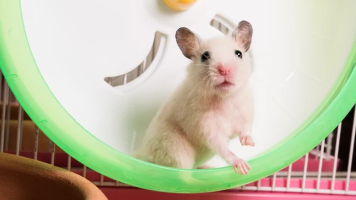
The next thing was to realize that bats do some really unusual things. They live for a ridiculously long time despite their small body size, without showing signs of aging. They tolerate many viruses without getting sick.

I started to read literature about bat lifespan. There’s a pattern in nature that big things live slow and long, while small things live fast and die young. Bats are some of the smallest mammals on earth, but most have very long lifespans for their body sizes.

Now, flight is the most metabolically costly form of locomotion. Bats use up to three times more energy than any other mammal of the same size. Having a very high metabolic rate typically can be very damaging. Cells produce a lot of reactive oxygen species that break up DNA, stimulate the immune system, and generally wreak havoc. Arguably, this is what drives aging. It looks like bats have evolved something to deal with these deleterious effects of flight.

Another thing I was very interested in is what bats do to avoid cancer. They really have the lowest cancer rate of all. They also tolerate all those viruses without getting sick. Why are there so many different types of viruses in these bats? These questions were formed in 2001, before we had this horrible pandemic right in our face.

Three years later, I was able to return to Ireland, got a position in UCD (University College Dublin), something I’d always dreamed of. I managed to get some nice grants to study bumblebee bats in Thailand, I got a presidential young researcher’s award. But what really changed my relationship with aging research was the grant I was able to secure from the European Research Council.

I had a second baby, struggling through all of this. Believe me, life can be hard for an academic mother trying to push science in Ireland. We teach an awful lot. But it was always in the back of my mind: we need to understand how bats do not get cancer. I’ve always said that the key to understanding how we can survive cancer lies in bats.

The European Research Council is one of the most amazing funding bodies in the world. It funds high-risk, high-gain projects. Anybody can apply, but it’s exceedingly competitive, and when you write this grant, you have to imagine what would you do if there were no barriers. I said I wanted to study bats and understand how they can help us slow down aging. How are their slow pace of aging and their unique immune responses connected, and how is it driven by flight?

There are 19 species of mammals that live longer than humans when adjusted for body size, and 18 of those are bats. It’s as if humans lived to about 250 with no signs of aging. But if you want to study bats in aging context, the longest-lived bats, those of the genus Myotis, there is a problem: they don’t do well in captivity.

One of my PhD students had worked in France, and the French care about their wildlife. And there was this grassroots organization called Bretange Vivante. It’s a conservation organization that had been studying this colony of long-lived Myotis bats for about 20 years.

They caught the entire colony in 2010. These were very long-lived bats that were also large enough for us to non-lethally sample them. They were caught and tagged as babies, and then we would recatch them. So, when I started writing the ERC grant, I was very lucky to find this population of long-lived Myotis myotis, or greater mouse-eared bats, in Brittany. It came to an integrative collaboration with this grassroots organization that was interested in ecology, because bats are so important for the ecosystem to function.

The only way you can measure a bat’s age is you catch it as a baby, say, in 2010, you put in a little microchip, like you would in a dog or a cat, and then, when you catch the same individual in 2020, you know it’s 10 years of age. What was really cool about this population is that they were captured every single year by this organization, and they were big enough so that we could take non-lethal samples.

If you want to study an animal in the context of aging, you have to be able to sample it every year as it ages, which means you have to recapture it. What happens with these species is that the females come back to the same roost – in this case, beautiful old Gothic churches. Like me, they give birth in the same place they were born in!

I wanted to be able to take a wing puncture and less than 140 microliters of blood. So, the idea was to take these samples, and look at biomarkers of aging. We wanted to compare young, middle-aged, and older bats and see whether they show the same pace of dysregulation with age as humans do, and what are the mechanisms that regulate this?

We wanted to look at telomeres – do they shorten in bats with age or not? We also wanted to look at their mitochondria – with such a high metabolic rate, do their mitochondria show the level of oxidative damage that would be expected given all the free radicals that are produced? We wanted to sequence the entire blood transcriptome, because blood is an overall proxy for health in multiple tissues. We were also going to look at their microRNA that can potentially regulate many different levels of transcription, at the genomes, at DNA repair mechanisms, at their immune system to see any evidence of adaptive selection going on.

With this, I got the grant. I couldn’t believe my luck. It’s one thing to write all this down on paper and to convince reviewers, but the first year, oh my goodness gracious, it was difficult. However, you have to make it work, and nobody has ever done anything like this before, because it’s bloody well hard.

My Irish group had very bad French, and people from that organization in France had no English. It was a clash of cultures, but we all had to work together, because we have three weeks in the entire year to sample this colony of over 500 individuals. The entire village would come together and help us put up traps in these churches. So, we capture the bats, measure them, weigh them, microchip the babies and flash-freeze the samples.

We had to work out how to do it, and it was so hard. I remember lying in the grass at three in the morning and crying, going: “What on earth have you done, you stupid woman?” But in the end, we made it work.

Yes, it is pretty amazing. I was wondering, considering these animals are so tiny, does taking even non-lethal samples affect their fitness in any way?

It does not. We were obviously worried about it, so we did population predictions to see if there’s a change in terms of animals returning year after year. If taking samples affects fitness, the manipulated bats would die in greater numbers, but that didn’t happen. Also, we catch the same females year after year, and they are all pregnant. So, it looks like our manipulation doesn’t affect them, otherwise we wouldn’t be doing it, because they are protected species.

If bats barely age, what do they die of?

First, bad weather. A very wet spring is hugely problematic for bats. Typically, the recruitment back after year one is about 55%, which means you lose half the babies every year. But once they get to two, you don’t seem to lose them. What correlates with fitness, for instance, when we look at telomeres, is weather, not aging. Think about their biology. They have their babies in July, then they start hibernating in October, and they have to get really fat in August and September to be able to survive hibernation.

Their first year, they have to be born early enough to be able to get fat enough to survive the winter. So, what these females can do if spring is really bad when they wake up from hibernation, and they’re pregnant because they’ve made it in the swarming, is they will slow down their gestation to match it with the food supply. The babies are born later and have less time to get fat. We have found a correlation between stress and telomere dynamics, and it’s been confirmed in many different populations of bats. So, bad weather is a major threat. And they also get eaten by owls.

So, what you’re saying is that stress affects fitness that affects telomere growth?

Yes. Let’s not forget that as the longest-lived bats age, bats of the genus Myotis, their telomeres don’t shorten. On the cohort level, you see this change that can only be attributed to bad environmental conditions. In the US, bats also die of white-nose syndrome, which is a fungal disease of the skin. It grows when the bats are in hibernation. When they drop down their temperature to nearly four degrees, you find fungus growing. So, these are the things bats can die of.

Interesting. In naked mole rats, when we put them in sterile lab conditions, we can’t really tell what their true maximum lifespan is because they just refuse to die, at least the queens. I understand bats don’t like captivity, but theoretically, if we could put them in a sterile environment that were to their liking, what do you think their maximum lifespan would be?

It’s a great question, but I actually don’t know the answer. Obviously, I don’t think they’re immortal. Yes, we haven’t optimized our husbandry yet, but say, if we were to make a perfect environment for these bats, would they show signs of senescence? I honestly don’t know.

Let’s talk about some concrete anti-aging defenses they have. So, they don’t experience telomere attrition – or, as I understand, they regrow their telomeres during hibernation?

All of this is not published yet, so for the record I can only say that potentially, they could be doing it in hibernation. A better question is, how are they maintaining their telomeres? In cell lines, when we looked at RNA, we didn’t find any evidence for telomerase expression, but maybe if we look at the protein level, we will see something. It looks like bats are maintaining their telomeres, either via telomerase or an alternative mechanism. Either way, they are doing this without getting cancer. The big question is, how do they not get cancer?

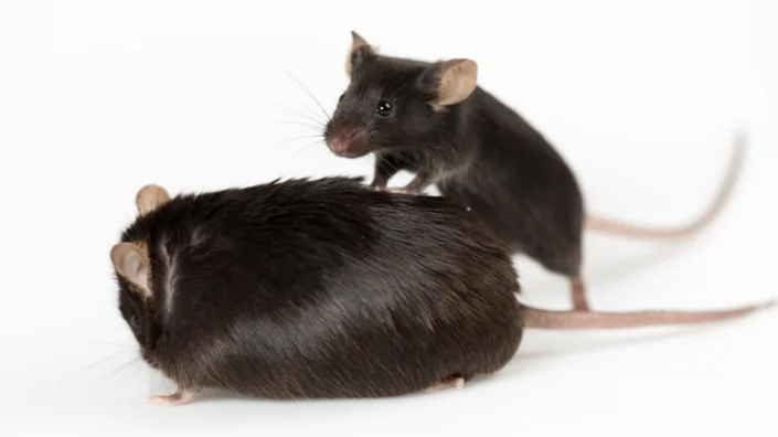
We looked at the whole blood transcriptome in young, middle-aged, and older Myotis bats to see if there were any age-related patterns. When you look at how the blood transcriptome changes in bats, you see a huge difference between babies and one-year-olds – that’s because they’re developing. After that, there’s very little change. In mice, for example, there’s a huge difference.

You also find that as they age, they have this immune balance, like human supercentenarians. They have high inflammation, but also high anti-inflammation to keep the homeostasis. There’s also evidence of maintaining mitochondria.

The big difference is their ability to repair DNA that goes up with age. They maintain a very tight cellular regulation system. I think they’re really good at regulating cell division. Potentially – this is just me thinking out loud, there’s not a lot of evidence for it, but this is what we have to do to move the field forward – potentially, they have a much better immune regulation of cells that go malignant.

We need to ask the question, what are they doing to prevent cancer? They somehow manage to maintain long telomeres without the side effect of getting cancer. We’re looking into that at the moment.

This is where the immune system comes in. We talked about inflammation. As we age, we become highly inflamed, which is now considered as a major driver of aging and disease. Bats have a way of getting that balance right, and this potentially evolved from flight.

Flight is highly metabolically costly and drives inflammation. Bats are the only mammalian order that has achieved true self-powered flight and therefore must have evolved mechanisms to control the constant sterile inflammation they experience from flight.

Interestingly, bats are missing these things called PHYIN genes, the entire family. PHYIN genes make proteins that make an inflammasome which is usually required for the immune system to fight disease by causing inflammation. However, an out-of-control inflammatory response and too much inflammation causes problems. Bats have lost these key genes. The only mammalian order to do this. This means they have this modulated immune response that’s very different from what we see in most other animals.

What you find when you sequence bat genomes is that bats have this expansion of anti-viral genes. They’ve lost these inflammatory genes, they have selection on downstream immune system genes, and they seem to have evolved this perfect Goldilocks response – enough inflammation to control the virus and then enough anti-inflammation to control their immune response.

Because viruses are abundant in their colonies?

The question is, which came first? I think they first had to evolve this anti-inflammatory response to be able to deal with the high levels of inflammation that they experience from all their ROS. They’ve had to evolve this dampened immune response to DNA damage and to ROS damage induced by flight.

Potentially, they lost their PHYIN genes to cope with this, but then they had to evolve a different mechanism to deal with all the pathogens. I think this is how evolution acts: one thing gets broken, one thing gets fixed, or maybe it happens altogether.

My hypothesis is they had to evolve this very aggressive anti-viral response dampened by an equally aggressive anti-inflammatory response, and the two go hand in hand. This means, when they get sick from a virus, they are able to first neutralize the virus, and then neutralize their own immune response.

So, they don’t get cytokine storms?

Exactly. There’s been research recently when they infected live bats and also bat cells with SARS-CoV-2, and the bats didn’t get sick from it. The question is how? There’s a theory about aging that inflammation is really at the heart of it all. Every one of the pillars of aging can be driven by inflammation, and the immune system plays a huge role.

I think bats have done more than just that. I don’t know if you’ve heard of this hypothesis by Claudia Franceschi called “garb-aging”. The idea is that aging is driven by accumulation of damage. Damaged DNA, damaged proteins, not being able to remove them.

The idea is that bats had to evolve mechanisms to limit, repair, and remove damage. They do this by limiting their inflammation. They also have brilliant DNA repair mechanisms. But there’s another thing bats do – as they age, their ability to remove damage via autophagy increases. It decreases in us, humans, and also in mice, but it increases in bats. We saw that in our field and lab studies.

Interestingly, autophagy is also viral regulation. Again, there’s this link between repairing damage and removing damage but also being able to deal with pathogens. I think the two things have to go hand in hand.

This sounds like a case of “what doesn’t kill me makes me stronger”, meaning that bats had to evolve those coping mechanisms that ultimately make them live longer.

Yes, I think that long life is a side effect of evolving protective mechanisms to deal with flight. Being able to carry all those viruses is also a side effect of those mechanisms that protect them from the pathogenic effects of flight.

Here’s another hypothesis that’s quite provocative. When you look at bats’ genomes, they have the smallest genome size of all animals, yet they have the highest diversity of integration of viruses within their genome.

So, a small genome is harboring all those integrated viruses. Why would you do that? Why would evolution allow for this? Potentially, this allows for more genomic plasticity, for evolution, for change, but maybe this is selected for in bats.

We have a paper that was just accepted in Cell, a collaboration with Professor Thomas Zwaka in Mount Sinai hospital in New York. He was very interested, when the pandemic broke out, to study bat lung cells to understand what mechanisms bats have evolved to protect themselves against SARS-CoV-2. The idea was to make induced pluripotent stem cells, but the traditional Yamanaka factors that work in all other animals, didn’t work.

When they tweaked the recipe enough to make those iPSCs, this re-awakened all the endogenized viruses, a plethora of different viruses, and that gives you a huge insight into what viruses bats have survived in the past. Indeed, these fossilized viruses in bat genomes tell a story of a long-evolutionary history of bats and viruses.

When you look at those iPSCs, it looks like a pro-viral environment. We’ve argued in the paper, and it’s Thomas’ hypothesis, that, potentially, it’s adaptive to bats. Maybe this is how they inoculate themselves. It’s just a theory, of course, lots still to do! To understand what’s happening in bats, we need to bring all fields together, we need great minds.

We were able to confirm this in a bat of a different sub-order, and we now can make bat cells in the lab, including all immune cells. We can even make organoids, and this can give us some amazing insights. I firmly believe that the key to extending human healthspan is out there, that nature has already found all the solutions. They are in bats, and we just need to uncover them.

I totally subscribe to this idea that the solution is out there, but even theoretically, how can we benefit from all those mechanisms that have evolved in other species, and how far away are we from doing this?

We’re finding all those things that are happening in bats, but in order to make it work in humans, first you need to put it into a system that humans already know can be compared to their own biology. So, we need to identify the regulators of longer healthspan in bats and knock them into a model system such as C. elegans and see if they extend healthspan.

You can totally do it, and we are currently working on a collaboration with Björn Schumacher. The idea is that once we identify these regulators, which are mostly conserved across species, and if we find out if they have lifespan or healthspan effects on C. elegans, we can then validate it in genetically modified “bat-mice”. Linfa Wang’s group in Singapore has already done this; they’ve shown they can knock bat immune genes into mice and are investigating if they have a healthspan effect.

So, the tools are actually there. But after we have a bunch of targets, which should take us about six years, how do we find the drugs that can help us? As you know, in the longevity field, we have Alex’s (Zhavoronkov) work that can potentially speed this up using AI. You can drug the pathways, but you first have to identify them, that why we need the bats. Luckily, we share the same genes, bats are mammals too.

There seem to be two competing approaches, with some geroscientists betting on drugs, and others on less traditional things like gene engineering and cellular reprogramming.

I think it’s easier to take a drug.

The question is whether it’s going to work.

This is always the question, but drugs work, just look at aspirin. I think it doesn’t have to be one or the other, but my problem with gene engineering is, when do you do it? Do you do it at the embryonic stage, at the egg stage?

For me, the easiest thing with bats will be to emulate their immune response, to find out how we can regulate and stop this pathogenic and inflammatory response that you get from diseases and that ultimately kills you.

When the first patients died of COVID-19 in Ireland, I was devastated because my research was ten years behind. We just showed that bats have a certain inflammatory/anti-inflammatory ratio, they have more IL-10 than TNF as disease continues. The doctors in our hospital were using that proxy to figure out whether or not a patient was going to need to go on a ventilator. If they had a bat-like immune profile, where there’s more IL-10 than Il-6 (similar to inflammatory TNF), they weren’t going on the ventilator. This is something you can potentially change with a drug.

Or take DNA repair mechanisms. What do bats do to repair their DNA? They may have an increased level of expression of some central genes as they age. We now have about 20 genes with expression varying wildly in bats and other mammals. They might not be the main effector that would be targeted in humans, but they are in the pathway, and those are pathways that we know how to drug. I think in the near future, our ability to drug pathways will advance.

What’s hampered aging research in the past? Too much focus on short-lived organisms, but there was no other option. We didn’t have the tools to study long-lived species, but we have them now. I think it all has to go together. Fields have to interact with each other and to respect each other, and that’s how we find novel solutions.

Let’s say, worst comes to worst, and we find that it’s not translatable. No matter what, we’ve found a huge, wonderful insight into how the aging process works. We have uncovered brand new biology. Personally, I believe we can solve it, we can apply it to humans, but we need to find the right direction first.

Private capital is playing an increasingly big role in longevity research. Do you think research of long-lived species can be commercialized? Do you have a pitch for a potential investor?

I do. “Every single penny you spend on aging research today could be better placed, because you’ve been studying short-lived organisms who are great at dying, instead of studying animals who naturally evolved longer healthspans and are great at living. We can do this now and I can show you the way”.

We would like to ask you a small favor. We are a non-profit foundation, and unlike some other organizations, we have no shareholders and no products to sell you. All our news and educational content is free for everyone to read, but it does mean that we rely on the help of people like you. Every contribution, no matter if it’s big or small, supports independent journalism and sustains our future.