×

The Blog

Building a Future Free of Age-Related Disease

Long Life Family Study

Data Released from the NIA Long Life Family Study

Since 2005, a team of researchers in Denmark and the United States have been conducting the National Institute on Aging’s Long Life Family Study (LLFS). Enrolled in the study were 4953 individuals from two generations of family, including siblings, spouses and offspring. Families recruited from the United States and Denmark had to prove a family history of longevity to be eligible.

A broad, longitudinal study

This study involves two in-person visits to assess healthy aging phenotypes in families, which were not limited to cognition, physical, metabolic, heart health, and inflammation indicators. It uses the Family Longevity Selection Score (FLoSS) to recruit participants. The FLoSS can be described as a family score that estimates a living family member’s lifespan. How the researchers determined the score formula can be seen in their methods paper [1]. Since data shows that longevity can run in families [2,3] this database sets itself apart from other population-based studies by being able to interpret research questions on longevity and healthy aging.

The Framingham Heart Study (FFS) study was started in 1948 by the National Heart, Lung and Blood Institute. Over time, it has had 15,000 people, which includes the original participants, their children, and their grandchildren in the Framingham, Massachusetts area of the United States. The original aim of this study was to identify what contributes to heart attack and stroke. This database has enabled research investigators to make discoveries regarding a number of different chronic diseases. For instance, we can thank this database for discovering that high blood pressure and unfavorable blood cholesterol levels are major risk factors for heart disease and stroke. Currently, this study involves various populations, so it is not aimed at recruiting families with a history of longevity like the LLFS study.

The results

In the LLFS and FHS groups, based on sex and age, the LLFS partipants had lower plaque artery build-up as showed by medium intima-media thickness (IMT). IMT is used as a marker of subclinical, non-symptomatic atherosclerosis obtained by carotid ultrasonography [4].

Results also showed that LLFS adult participants have higher levels of HDL cholesterol, higher lung function, lower prevalence of hypertension, lower rates of diabetes, and lower rates of coronary artery disease. The cognition tests showed that the LLFS adult participants scored higher on memory, attention and semantic processing. It is interesting to note that the prevalence of participants with obesity is about the same in both cohorts based on BMI. Other markers of body composition would be useful in the future that measure fat, muscle, and bone to confirm this finding.

The LLFS children had significantly lower rates of chronic lung disease, diabetes, and peripheral artery disease. Additionally, compared to the FHS children, LLFS children showed more healthy traits, such as more favorable blood pressure, physical performance, cognitive performance and lipid profiles.

Though the results from this study suggest that, on average, the LLFS participants appear to be healthier than the FHS participants based on age and sex, it is not consistent across all LLFS participants. In particular, one LLFS study in 2013 referenced 18 families that showed exceptional memory compared to 539 LLFS families [5]. Some of the LLFS families also varied in other health markers, such as grip strength, which also did not tend to be heritable in families.

The study also compared specific phenotypes between the LLFS and the FHS [4] using genome-wide association (GWAS). GWAS uses genomic technology to scan entire genomes of large numbers of people quickly in order to detect genetic variants correlated with a disease or a trait. With the GWAS data, they conducted analyses on heart health, anthropometrics, lipids, blood sugars, lungs, blood-based biomarkers, physical performance measures, brain and psychological traits, and a healthy aging index that can be seen in table 3 of this preprint publication [4]. They calculated heritability at each visit and over time, and they ultimately determined that the healthy aging phenotypes determined by the GWAS analyses are heritable in the LLFS cohort. To learn more, check out our article on multi-omics longevity genes.

On average, the LLFS families showed healthier aging profiles than the FHS familes for all age/sex groups and for many of the healthy aging phenotypes.

Conclusion

The LLFS families have lower rates of disease in the older adults and show healthier aging profiles in its participants as compared to the FHS families. The LLFS and FSH have different eligibility criteria; therefore, trying to compare them directly is like comparing apples to oranges. Despite their cohort differences, it can be useful to compare these different population datasets to further understand the etiology of healthy aging and longevity.

We would like to ask you a small favor. We are a non-profit foundation, and unlike some other organizations, we have no shareholders and no products to sell you. All our news and educational content is free for everyone to read, but it does mean that we rely on the help of people like you. Every contribution, no matter if it’s big or small, supports independent journalism and sustains our future.

Literature

[1] Sebastiani, P., Hadley, E. C., Province, M., Christensen, K., Rossi, W., Perls, T. T., & Ash, A. S. (2009). A family longevity selection score: ranking sibships by their longevity, size, and availability for study. American journal of epidemiology, 170(12), 1555–1562. https://doi.org/10.1093/aje/kwp309

[2] Willcox, B. J., Willcox, D. C., He, Q., Curb, J. D., & Suzuki, M. (2006). Siblings of Okinawan centenarians share lifelong mortality advantages. The journals of gerontology. Series A, Biological sciences and medical sciences, 61(4), 345–354. https://doi.org/10.1093/gerona/61.4.345

[3] Gudmundsson, H., Gudbjartsson, D. F., Frigge, M., Gulcher, J. R., & Stefánsson, K. (2000). Inheritance of human longevity in Iceland. European journal of human genetics : EJHG, 8(10), 743–749. https://doi.org/10.1038/sj.ejhg.5200527

[4] Wojczynski, M. K., Lin, S. J., Sebastiani, P., Perls, T. T., Lee, J., Kulminski, A., Newman, A., Zmuda, J. M., Christensen, K., & Province, M. A. (2021). NIA Long Life Family Study: Objectives, Design, and Heritability of Cross Sectional and Longitudinal Phenotypes. The journals of gerontology. Series A, Biological sciences and medical sciences, glab333. Advance online publication. https://doi.org/10.1093/gerona/glab333

[5] Barral, S., Cosentino, S., Costa, R., Andersen, S. L., Christensen, K., Eckfeldt, J. H., Newman, A. B., Perls, T. T., Province, M. A., Hadley, E. C., Rossi, W. K., Mayeux, R., & Long Life Family Study (2013). Exceptional memory performance in the Long Life Family Study. Neurobiology of aging, 34(11), 2445–2448. https://doi.org/10.1016/j.neurobiolaging.2013.05.002

Stroke

Senolytic Therapy Shows Potential for Stroke Recovery

The newest study published in the International Journal of Molecular Sciences investigates the role senescent cells may play in cerebral ischemia-reperfusion injury and how senolytic therapy may be beneficial for those recovering from stroke.

Stroke and reperfusion injury

Ischemic stroke occurs when an artery in the brain is occluded, preventing an area of brain tissue from receiving fresh oxygen and nutrients from the bloodstream. It is one of the leading causes of death and disability and occurs primarily in elderly individuals [1].

Timely treatments can restore blood flow to the ischemic tissue and reduce the severity of the stroke. While restoring blood flow is critical to the tissue’s survival, reperfusion itself has been shown to set off a cascade of events, contributing to the overall tissue damage [2]. Referred to as ischemia-reperfusion (IR) injury, this damage-causing cascade involves inflammation and reactive oxygen species (ROS) [3].

Cellular senescence is a state that is pro-inflammatory and anti-proliferative. Because of its connection to aging, the cellular stress response, ROS, and inflammation, researchers at Seoul National University hypothesized that it may play an important role in IR injury. They tested this hypothesis in both in vitro and in vivo models. Additionally, they investigated whether the senolytic drug ABT263 (Navitoclax), which selectively kills senescent cells, could alleviate some of the effects of IR injury [4].

IR induces senescence in astrocytes, which can be eliminated by ABT263 in vitro

In cell culture, IR injury was modeled by first depriving rat astrocytes of oxygen and glucose, then reintroducing both into the culture system. This process did not cause cell death, but SA-ß-gal staining (a measure of senescence) increased from 22% to 60% of cells.

These cells were then treated with ABT263. After treatment, cell viability remained unchanged in astrocytes that were not exposed to the IR injury model. The ones that were, however, had their viability decrease. SA-ß-gal positive cells also decreased from 60% to 36% of the population.

ABT263 alleviates IR injury in rats

IR injury in vivo was modeled by occluding and re-opening the middle cerebral artery in rats. This occlusion was associated with an increase in the expression of senescence marker p16 and the inflammatory markers NOS2 (also a SASP factor), MPO, and GFAP. Meanwhile, ABT263 treatment reduced their expression to non-IR injury levels. Treatment also reduced the infarct volume by more than half and improved behavioral measures of neurological function.

In conclusion, this study demonstrated that intravenous ABT263 treatment attenuated inflammation through elimination of senescent cells and improved functional outcomes after IR in the brain. ABT263, a senolytic drug, is a novel therapeutic candidate for cerebral ischemia with IR injury.

Conclusion

Like all studies, this study has several limitations that should be noted when interpreting its results. Only astrocytes were used in the in vitro experiments, but many other cell types in the brain have been shown both to become senescent and to play a role in IR injury.

The authors also only used SA-ß-gal staining in vitro and only p16 in vivo to identify senescent cells. While both are very commonly employed, the senescent cell phenotype is complex and requires more than a single measurement to distinguish senescence from other cell fates. It is difficult to say for certain that these cells were senescent in these experiments.

This study also falls short in another common issue in aging research: their animals weren’t old. The rats were only 8-9 weeks old when they were treated. An aged rat (just like an aged human) might respond very differently both to IR injury and to the senolytic treatment.

Although many questions remain unanswered, this study is the first to demonstrate the potential role of senescent cells and of senolytic therapy in cerebral IR injury. The decreased infarction volume is particularly promising for the prospects of future research.

We would like to ask you a small favor. We are a non-profit foundation, and unlike some other organizations, we have no shareholders and no products to sell you. All our news and educational content is free for everyone to read, but it does mean that we rely on the help of people like you. Every contribution, no matter if it’s big or small, supports independent journalism and sustains our future.

Literature

[1] Gorelick, P.B. The global burden of stroke: Persistent and disabling. Lancet Neurol. (2019). https://doi.org/10.1016/S1474-4422(19)30030-4

[2] Nour, M., Scalzo, F., & Liebeskind, D.S. Ischemia-reperfusion injury in stroke. Interv. Neurol. (2012). https://doi.org/10.1159/000353125

[3] Eltzschig, H.K. & Eckle, T. Ischemia and reperfusion—from mechanism to translation. Nat. Med. (2011). https://doi.org/10.1038/nm.2507

[4] Lim, S. et al. Senolytic therapy for cerebral ischemia-reperfusion injury. A meta-analysis of genome-wide association studies identifies multiple longevity genes. Int. J. Mol. Sci. (2021) https://doi.org/10.3390/ijms222111967

Metformin bottle

What is Metformin? A Summary of N,N-dimethylbiguanide

We take a look at this diabetic drug that some researchers think may slow down aging.

What is metformin?

Metformin is a prescription drug and is available as a tablet or an oral solution. It is a commonly used first-line medication for the treatment of type 2 diabetes and is also used in the treatment of polycystic ovary syndrome.

You can find metformin tablets in two forms: immediate-release and extended-release. The immediate release tablet goes by the brand name Glucophage, and the extended release goes by Glucophage XR, Glumetza, and Fortamet.

Both types of tablet are sold as generic drugs, as the patent on metformin expired back in 2001. Generic drugs typically cost significantly less than the brand name versions, though in some circumstances, they may not be available in all of the strengths  that brand name suppliers offer.

Metformin is in the biguanide class of drugs, a group of drugs that work in a similar way. It was originally discovered in 1922, but it did not receive approval for another fifty years or more [1]. The French physician Jean Sterne initiated the first study in humans in the 1950s; it was registered as a medicine in France in 1957, but it was not approved until 1995 in the United States [2].

The World Health Organization lists metformin on its model list of essential medicines, a list of the most effective and safe medicines that are critical in a health system. To that end, metformin is believed to be one of the most widely used drugs used for the treatment of diabetes.

How does metformin work?

It works by decreasing glucose production in the liver, reducing the amount of glucose your body absorbs, and boosts the effect of insulin on your body. Insulin is a hormone that assists your body in removing excessive sugar from your bloodstream, and this helps to reduce your blood sugar levels, a cause of diabetes.

In this manner, metformin helps people with type 1 diabetes to manage their condition by keeping their high blood sugar under control.

Metformin and anti-aging

There is a constant process of balance happening in our cells between anabolic processes that produce energy from nutrients and catabolic processes that consume that energy. This balancing act becomes critical when there is a scarcity of nutrients to keep our cells powered and functioning.

Famine is an example of when this balance becomes vitally important to keep someone alive. When such conditions are experienced our cells switch to a pro-survival state and prioritize survival and energy conservation instead of growth. By entering this state our cells have a better chance to survive and help us to remain alive. Without a doubt our ancient ancestors who often experienced periods of starvation and famines would have benefited from this.

Metformin seems to trigger the same pro-survival state in our cells by reducing the activity of our mitochondria, the powerhouses of our cells. This causes the mitochondria to slow down the pace at which they convert nutrients into a universal cellular energy known as adenosine triphosphate (ATP).

The reduction in ATP triggers an enzyme known as AMPK which detects low energy levels. This then activates various pro-survival mechanisms including autophagy to conserve energy and keep the cells alive.

Metformin may also support longevity by lowering both insulin resistance and blood sugar levels. By improving the way the body manages insulin it may even help non-diabetics to live a longer and healthier life.

Some researchers, such as Dr. Nir Barzilai, believe that metformin may prove useful as a way to slow down aging and is leading a clinical trial called TAME to find out if it can. We talked to him about his interest in metformin in the “We Can Live Healthier for Longer” interview.

Potential health benefits

There is some evidence that metformin may help to prevent the cardiovascular and cancerous complications of diabetes [3-4]. Metformin is also not associated with weight gain, so it is ideal for diabetics who suffer from weight control issues, which are common in diabetes.

Generally speaking, metformin is well tolerated and appears to have protective effects on the vascular system [5], though some common side effects can include diarrhoea, nausea and abdominal pain. There is risk of high blood lactic acid if taken inappropriately and in overly large doses [6]. It should not be used by people with significant liver damage or kidney problems.

Metformin is of interest to people working in the field of rejuvenation biotechnology, and mouse studies show that it increases lifespan and healthspan [7]. There is also some data that suggests that type 2 diabetics that take metformin may also live longer than non-diabetics [8].

Metformin side effects

Taking metformin can cause mild side effects, including diarrhea, nausea, stomach pain, heartburn, and gas. More serious potential side effects include lactic acidosis and hypoglycemia (low blood sugar). You should call your doctor if you experience any side effects while taking metformin.

However, metformin has a number of known interactions with other drugs, including diabetes drugs such as insulin and glyburide, along with blood pressure management drugs such as furosemide and hydrochlorothiazide.

Cholesterol-modifying drugs, such as NAD+ precursor niacin, are worth noting, as this is a popular supplement in the life extension community and can make metformin less effective in lowering blood sugar levels.

Using metformin with the glaucoma drugs acetazolamide, brinzolamide, dorzolamide, and methazolamide may increase your risk of lactic acidosis. Metformin has known interactions with topiramate, a drug used to treat nerve pain and seizures, and may raise your risk of lactic acidosis. Phenytoin, which is used to treat seizures, can make metformin less effective in lowering your blood sugar. Cimetidine, which is used to treat heartburn and other stomach problems, can interact with metformin and increase your risk of lactic acidosis.

Some hormone drugs may also make metformin less effective in lowering blood sugar. Corticosteroids like budesonide, fluticasone, prednisone, and betamethasone have potential interactions. Estrogens such as estradiol, conjugated estrogens (Premarin), and birth control pills or patches can also interact. Isoniazid, an anti-tuberculosis drug, can also make metformin less effective in lowering blood sugar.

Thyroid drugs may make metformin less effective in lowering your blood sugar too. These include levothyroxine, liothyronine, and liotrix.

Lactic acidosis is an uncommon but very serious side effect of metformin. This is when lactic acid builds up in the bloodstream and is a medical emergency that requires treatment in the hospital. It is fatal in about half of people who develop it.

Disclaimer

This article is only a very brief summary, is not intended as an exhaustive guide, and is based on the interpretation of research data, which is speculative by nature. This article is not a substitute for consulting your physician about which supplements may or may not be right for you. We do not endorse supplement use or any product or supplement vendor, and all discussion here is for scientific interest.

We would like to ask you a small favor. We are a non-profit foundation, and unlike some other organizations, we have no shareholders and no products to sell you. All our news and educational content is free for everyone to read, but it does mean that we rely on the help of people like you. Every contribution, no matter if it’s big or small, supports independent journalism and sustains our future.

Literature

[1] Fischer, J., & Ganellin, C. R. (Eds.). (2010). Analogue-based drug discovery II. John Wiley & Sons.

[2] Stargrove, M. B., Treasure, J., & McKee, D. L. (2008). Herb, nutrient, and drug interactions: clinical implications and therapeutic strategies. Elsevier Health Sciences.

[3] Malek, M., Aghili, R., Emami, Z., & Khamseh, M. E. (2013). Risk of cancer in diabetes: the effect of metformin. ISRN endocrinology, 2013.

[4] Griffin, S. J., Leaver, J. K., & Irving, G. J. (2017). Impact of metformin on cardiovascular disease: a meta-analysis of randomised trials among people with type 2 diabetes. Diabetologia, 60(9), 1620-1629.

[5] Triggle, C. R., & Ding, H. (2017). Metformin is not just an antihyperglycaemic drug but also has protective effects on the vascular endothelium. Acta Physiologica, 219(1), 138-151.

[6] Lipska, K. J., Bailey, C. J., & Inzucchi, S. E. (2011). Use of metformin in the setting of mild-to-moderate renal insufficiency. Diabetes care, 34(6), 1431-1437.

[7] Martin-Montalvo, A., Mercken, E. M., Mitchell, S. J., Palacios, H. H., Mote, P. L., Scheibye-Knudsen, M., … & Schwab, M. (2013). Metformin improves healthspan and lifespan in mice. Nature communications, 4, 2192.

[8] Bannister, C. 1., Holden, S. E., Jenkins‐Jones, S., Morgan, C. L., Halcox, J. P., Schernthaner, G., … & Currie, C. J. (2014). Can people with type 2 diabetes live longer than those without? A comparison of mortality in people initiated with metformin or sulphonylurea monotherapy and matched, non‐diabetic controls. Diabetes, Obesity and Metabolism, 16(11), 1165-1173.

Skin aging

Nicotinamide Alleviates Skin Aging in Cellular Study

A group of researchers successfully alleviated aspects of skin aging by treating skin cells with the NAD precursor nicotinamide [1].

Hard work done by skin leads to its burnout

Skin aging might not be lethal, apart from the increased risk of skin cancer, but it’s surely conspicuous, frustrating, and emblematic of organismal aging. Being our first line of defense against the elements and all the pathogens that are lurking around, skin, especially its outer layer, the epidermis, is one of the first tissues to experience aging [2]. Since it takes a lot of damage, skin also has high levels of cellular senescence, which is a mechanism that prevents damaged cells from proliferating. As one paper puts it, all this makes skin “an ideal organ to observe and analyze the impact of extrinsic and intrinsic drivers of aging” [3].

In addition to being subjected to faster-than-usual aging, our skin is also characterized by fast turnover of cells. Keratinocytes that constitute 90% of skin cells differentiate from skin stem cells in deeper skin layers and then make their journey to the outer layers. Along the way, they undergo several more divisions and stages of differentiation. Eventually, they reach the top through terminal differentiation and are shed away as dead skin in a process called desquamation.

Ultraviolet (UV) light is a major cause of skin aging, which led to the appearance of the term photoaging. Like any radiation, UV light rips through the cells, inducing DNA strand breaks. DNA damage is one of the known causes of cellular senescence and carcinogenic mutations. As the saying in the longevity community goes, the only real anti-aging cream today is sunscreen.

NAD: the electrician and the repair guy

In this new paper, researchers investigated the effect of nicotinamide (NAM) treatment on skin cells exposed to UV. NAM is a precursor to NAD, which is a simple but indispensable molecule that facilitates chemical reactions by taking and donating electrons. It exists in two forms: NAD+, an oxidizing agent that takes electrons from other molecules, and NADH, which is NAD+ after it acquired an electron.

NAD also plays an important role in DNA repair, being a cofactor for the sirtuin family of enzymes. NAD levels decline with age, which is considered one of the mechanisms of aging [4]. In the skin, NAM supplementation is known to delay terminal differentiation and promote the proliferation of keratinocytes.

NAM is also a form of vitamin B3. It is found in a wide variety of foods, both animal- and plant-based, and is well absorbed, so people rarely develop B3 deficiency. However, many longevity enthusiasts take NAD precursors as daily supplements, such as NMN (nicotinamide mononucleotide). We do not endorse this practice due to the lack of solid data.

NAM brings improvement

The researchers used two types of cellular models: cultured keratinocytes and a 3D model of the skin, complete with layers. Irradiating these with UVB light, which falls into the UV frequency range, resulted in accelerated keratinocyte senescence, loss of proliferation, and premature terminal differentiation, which are all hallmarks of skin aging. UV also caused morphological changes in the skin structure, with the epidermis becoming thinner. UV irradiation also decreased energy metabolism levels in the cells.

NAM treatment was able to partially alleviate the damage. It improved energy metabolism, prevented senescence and premature terminal differentiation of keratinocytes, and restored skin morphology. The researchers suggest that NAM treatment achieved all this mainly by promoting DNA repair.

Shortly after the burst of UV irradiation, the levels of a DNA breakage marker called CPD (cyclobutane pyrimidine dimers) shot up. In controls that did not receive NAM, it went down by as much as 74% after two days, indicating that even in the absence of NAM supplementation, DNA repair mechanisms did a decent job. However, in the NAM group, the decline in CPD levels compared to the pre-irradiation state was even more pronounced: 85%. The difference might seem small, but it is not, considering that any unresolved DNA damage is potentially harmful.

Conclusion

The link between NAD and organismal aging has been well-established, but skin aging is a unique phenomenon. This new study shows how NAD replenishment via NAM treatment can partially reverse concrete aspects of skin aging induced by exposure to ultraviolet light. Exciting as it is, this is early-stage research that will probably not get translated into a new skin lotion any time soon, so for now, you are advised to protect your skin from “photoaging” by using sunscreen.

We would like to ask you a small favor. We are a non-profit foundation, and unlike some other organizations, we have no shareholders and no products to sell you. All our news and educational content is free for everyone to read, but it does mean that we rely on the help of people like you. Every contribution, no matter if it’s big or small, supports independent journalism and sustains our future.

Literature

[1] Tan, C. Y. R., Tan, C. L., Chin, T., Morenc, M., Ho, C. Y., Rovito, H. A., … & Bellanger, S. (2021). Nicotinamide Prevents UVB-and Oxidative Stress-Induced Photoaging in Human Primary Keratinocytes. Journal of Investigative Dermatology.

[2] Krutmann, J., Schikowski, T., Morita, A., & Berneburg, M. (2021). Environmentally-induced (extrinsic) skin aging: Exposomal factors and underlying mechanisms. Journal of Investigative Dermatology.

[3] Gruber, F., Kremslehner, C., Eckhart, L., & Tschachler, E. (2020). Cell aging and cellular senescence in skin aging—Recent advances in fibroblast and keratinocyte biology. Experimental gerontology, 130, 110780.

[4] Schultz, M. B., & Sinclair, D. A. (2016). Why NAD+ declines during aging: It’s destroyed. Cell metabolism, 23(6), 965-966.

Bone Marrow

Using Young Stem Cells to Restore Old Stem Cells

In a study published in Aging, researchers have found that younger hematopoietic cells can restore older hematopoietic cells through microvesicles, which are facilitators of intercellular communication.

Placing young and old together

To begin their experiment, the researchers placed equal amounts of young and old cells in a transwell system, in which a membrane allowed the passage of microvessels but not whole cells. This allowed them to directly observe the effects of this communication, observing both the hematopoietic activity of these cells and their gene expression.

The results were substantial. While the cells exposed to these vesicles (referred to in the study as restored aged cells) did not exhibit the same robustness as genuinely young cells, their fundamental abilities were significantly improved.

The restored cells’ levels of “colony forming units-granulocytic monocytic” (CFU-GM), which represents these cells’ ability to form other blood cells, were much higher than those of aged cells. Over five weeks, the aged cells’ CFU-GM had deeply declined almost to nothing, but the restored cells’ CFU-GM had risen and then fallen to where it had began.

Basic biochemistry was affected

The researchers also examined the cells’ gene expressions. Similarly to the CFU-GM measurements, the restored cells’ gene expressions were not entirely like those of young cells, but they were substantially different from those of untreated old cells. Genes related to cellular activation; apoptosis and survival; the formation of reactive oxygen species; and T-helper cell differentiation and movement were the most strongly affected.

A molecular examination of the pathways involved showed similar results. Analyses of inflammatory compounds, apoptosis-related molecules, and other markers revealed the same fundamental concept: communication from young cells placed the restored cells approximately midway between aged and truly youthful cells.

The impact of youth

Cellular senescence was greatly affected by this intervention. The senescence gene expression profile of the aged cells was 85% over that of young cells, but restored cells had only a 43% increase. The protein p53, a key part of senescence, was significantly downregulated in the restored cells. The researchers also note that IGF-1, which is associated with longevity [1], was upregulated, while fibronectin, which is associated with the pathological condition myelofibrosis [2], was downregulated.

Other experiments confirmed that it was these cells’ youth that was responsible for the effects. In order to rule out the idea that allogeneicity (donor/host differences) was the cause, the researchers placed restored aged cells and ordinary aged cells from the same organism together. This caused the same baseline stimulation: restored aged cells were able to promote restoration in other aged cells.

Isolating the cause

In order to test their hypothesis more thoroughly, the researchers created humanized chimeric mice with each category of these cells. Here, they discovered an unexpected snag: While samples from mice given restored and young cells were able to form chimeric colonies in other mice, the samples from mice given aged human cells were unable to accomplish this at all, making some of the proposed testing impossible.

However, the data they did get was in line with the cellular results. Restored aged cells performed similarly in vivo to how they had in vitro, promoting the formation of blood cells and reducing senescence markers as compared to mice given aged cells, and mice given restored cells were also shown to have improved natural killer (NK) activity.

Microvesicles contain RNA, and the researchers identified the specific miRNA 619 as a potential cause. The cells of humanized mice injected with miR-619 were shown to have cellular profiles more like those of younger animals. Another pair of miRNAs, miR-combo, was shown to have more beneficial effects on some molecular markers but fewer on others.

Conclusion

The researchers conclude their very detailed study by suggesting that their microvesicle and miRNA approach might be applicable in fighting infections and in restoring hematopoietic capacity. Further research is needed to determine if this is the case and to determine what precise microvesicles and miRNA are appropriate for human trials.

If it turns out that there is a way to restore the function of HSCs in human beings with a microvesicle-based therapeutic, the ramifications may go beyond these particular applications. If other stem cell types, such as those responsible for neurons and muscle tissue, are also found to be responsive to microvesicles, it may be possible to use this approach to treat them as well.

We would like to ask you a small favor. We are a non-profit foundation, and unlike some other organizations, we have no shareholders and no products to sell you. All our news and educational content is free for everyone to read, but it does mean that we rely on the help of people like you. Every contribution, no matter if it’s big or small, supports independent journalism and sustains our future.

Literature

[1] Junnila, R. K., List, E. O., Berryman, D. E., Murrey, J. W., & Kopchick, J. J. (2013). The GH/IGF-1 axis in ageing and longevity. Nature Reviews Endocrinology, 9(6), 366-376.

[2] Abbonante, V., Gruppi, C., Catarsi, P., Avanzini, M. A., Tira, M. E., Barosi, G., … & Balduini, A. (2016). Altered fibronectin expression and deposition by myeloproliferative neoplasm-derived mesenchymal stromal cells. British journal of haematology, 172(1), 140-144.

Investigation

Investigation of Dr. Aubrey de Grey Concludes

In August of this year, we reported that Dr. Aubrey de Grey, the then-Chief-Science Officer of the SENS Research Foundation (SRF), was the subject of sexual harassment allegations raised by Laura Deming of Longevity Fund and Celine Halioua of Loyal, a dog longevity startup, via their accounts on Twit­ter and personal websites. The two alleged inappropriate behavior by Dr. de Grey and other unnamed members of SRF.

First investigation

An independent investigation was carried out by the Van Dermyden Makus Law Corporation at the behest of the SENS Research Foundation. Dr. de Grey was placed on administrative leave pending the outcome of this investigation.

At the time, Dr. de Grey issued a statement in a post appearing on his Facebook page. In that statement Dr. de Grey admitted to the inappropriate emails from a decade ago and expressed his regret.

During this first investigation, Dr. de Grey, who had been on administrative leave, was dismissed by the SRF Board for attempting to contact Ms. Halioua. The reason they gave for this decision was that Dr. de Grey had “attempted to exert influence over one of the complainants – an action that ultimately led to Dr. de Grey’s separation from SRF”. The investigator also included this in their report.

The report stated that Dr. de Grey may have acted improperly on two occasions and that he had attempted to influence one of the complainants.

No further evidence of unwelcome conduct was reported and it appears he was clear of involvement in revoking Celine’s scholarship funding provided by SRF.

Second investigation

Based on reports that Dr. de Grey may have engaged in inappropriate behavior, Van Dermyden conducted a second investigation. The investigator reports that there may have been some additional, limited boundary-crossing and unprofessional behaviors over a 12-year period.

The SRF has made it clear on several occasions that Dr. de Grey was dismissed not due to the allegations; rather, it was due to breaking company instructions not to contact a witness in an ongoing investigation. Dr. de Grey provided this explanation in response.

What does this mean for the future?

In a further development on November 8th, Kevin Perrott, an SRF Board member, suggested on Facebook openness to Dr. de Grey making a return to the foundation in the future.

It is my intention that we take this negative situation and turn it around. SRFs reputation is not destroyed in the least, at least not according to the influential people I’ve talked to who are surprised at the level of professionalism with which the crisis was and is being handled. Despite intense pressures not a single Board Member abdicated their responsibility to the organization and have put in so many hours doing damage control that many of their own personal lives have suffered. Frankly, every single Board Member (I’m sure you’ve seen the page on the website) is a volunteer who have put countless thankless hours into helping SRF, not the least being donors and funders, all because of Dr. de Grey. We know who is the reason SRF exists, every single one of us are involved with SRF because of him. Things have been EXTREMELY hard on everyone with the collateral damage that just seems to keep coming. Staff and Board Members have been brought to tears (well.. maybe only me) by the difficulty of some of the decisions we’ve had to make. The investigation reports unfortunately are NOT a farce, they are the key to minimizing the effect of the claims, which as we all know now were way off base. I am very optimistic that if we can find a period of relative peace instead of having to deal with continual chaos, we may be able to find a path to wholeness and trust, but that is a two way street.

He continued a little further down the thread:

It is my precisely stated goal that Dr. de Grey come back to SRF. I have ZERO interest in being in an organization that has the stated goal of curing aging that does not have him healthy and whole working within it. I’m sorry, but there just isn’t another brain that has the capacity to handle all the science the way Dr. de Grey’s does.

We should note that these are Kevin’s personal views and not officially sanctioned by the SRF board. That said, it does suggest that at least one member of the SRF board is willing to try to find a solution to this situation.

Dr. de Grey also seems to be open to a way forward as he recently commented on Facebook.

I want to correct the impression expressed by some people here (and elsewhere) that, to paraphrase, I think very poorly of all SRF board members other than Michael Boocher and Frank Schuler. It is my sincere view that a number of other board members have hearts of gold, and have merely been swept along by a tide of events that simply developed too rapidly. Now that all the salient facts are out (other than a still extreme murkiness surrounding the circumstances of Celine’s dropout from her doctoral studies, something I had nothing to do with), I look forward to working with those board members to rectify the current situation without delay.

Our Opinion: Time to let cooler heads prevail

We believe that everyone has value and should be allowed to work and contribute to the field in an atmosphere of mutual respect and safety. Like in many fields of science, there is an under-representation problem in our field, and we actively support efforts to address this issue.

We take allegations of impropriety seriously and believe that an official investigation was the right and appropriate course of action. It was carried out to ascertain the severity of the claims being made and also to protect the foundation from potentially catastrophic damage that could set back progress in the field for many years.

The SRF board appears generally satisfied that the claims that were made against Dr. de Grey were not as extensive as they originally appeared to be on social media. Unfortunately, social media is a place where discussions frequently become heated and facts distorted. Assumptions are made based on incomplete information, and it can be incredibly difficult to understand what is going on, particularly in a complex situation like this.

The goal that our community has set for itself, the end of age-related diseases, is arguably the hardest thing that humankind has ever done. Ours is a relatively small community in which many of us know each other and where infighting and division can be extremely harmful.

Over the last few weeks, we have seen people taking sides based on incomplete information and engaging in battle with those who disagree. We urge the community to avoid doing this, step back and let cooler heads prevail, and help to create an environment where rational and productive discussion can happen.

We also note that SENS Research Foundation has been a long-time ally of LEAF in its mission to fight age-related disease. Aubrey de Grey is also currently a member of LEAF’s scientific advisory board. As such, we disclose potential conflict of interest when reporting on this case.

Alzheimer's

Plasma Exchange Alleviates Alzheimer’s in a Human Trial

New results from the AMBAR trial show that plasma exchange leads to statistically significant functional and cognitive improvements in patients with mild to moderate Alzheimer’s [1].

As our readers surely know, the world is spending much more resources on fighting the diseases of aging than on solving aging itself. In particular, tens of billions of dollars have been poured into looking for a cure for the deadly Alzheimer’s disease (AD), with few to no results. One analysis puts the rate of success of AD clinical trials at 0.4% [2]. So, when a human AD trial yields encouraging results, it’s big news.

The underlying hypothesis

AMBAR (Alzheimer’s Management by Albumin Replacement) was a clinical trial conducted by the Spanish pharmaceutical company Grifols to study the effects of plasma exchange (PE) on AD. With more than 400 participants, it was registered as a Stage IIb trial in the US and a Stage III trial in the EU and ended a while ago. Plasma exchange involves replacing a part of a patient’s blood plasma with donor plasma or a plasma substitute. In AMBAR, various groups received various concentrations (either 5% or 20%) of medicinal albumin in their plasma transfers.

AMBAR was based on the “peripheral sink” hypothesis. Alzheimer’s disease is linked to the accumulation of the misfolded protein amyloid beta (Aß) in the brain. The hypothesis evolved from an experiment where administration of anti-Aß antibodies lowered brain Aß levels in mice even though the antibodies had not penetrated the blood-brain barrier. This suggested that Aß is cleared by moving it from the brain to the cerebrospinal fluid (CSF), then to the peripheral blood, and out of the body. Therefore, if you clear Aß from the blood, this makes room for more Aß to trickle down.

In the blood, more than 90% of Aß is tied to molecules of another protein, albumin. According to this hypothesis, if we replace albumin from the patient’s blood with fresh albumin via PE, this should also remove a lot of Aß and increase the trickle down effect.

The results

AMBAR’s primary endpoint results caused some furor back in the day. They were not clear-cut but also not as disappointing as the results of the vast majority of other AD trials. Statistically significant slowing in AD progression was observed in the range of 42%-70%, depending on the test, the amount of albumin in the fresh plasma, and the severity of the disease.

Now, the results on the secondary endpoints are in, and they are mostly positive as well. Those secondary endpoints addressed various aspects of cognitive function via a battery of tests, while the primary endpoints were less specific. Importantly, in some cases, there was an upward trend – that is, patients’ scores actually improved during the study period. This is unusual in AD research, where even the most successful interventions merely slow the progression of the disease.

Once again, the results were dependent on the dosage of albumin and on the disease stage. For instance, only the mild-AD / high-dosage group, but not patients with moderate AD, showed significant improvement in language fluency tests. Same goes for mental processing speed tests. The results regarding verbal learning and memory were less affected by the severity of AD but also less pronounced.

Lots of caveats

Unfortunately, deciphering the results might not be easy. After the results on primary endpoints were published, a paper interpreting them and analyzing the AMBAR design appeared. As it points out, despite the results being positive, the mechanism of action remains unclear.

Since plasma may contain many ingredients, it is not easy to determine which are responsible for the positive effect. For instance, mechanisms like inflammation and oxidative stress may contribute to AD’s neuropathology, while PE removes inflammatory cytokines from peripheral blood. Albumin itself is also known to have neuroprotective action unrelated to Aß [3]. Unfortunately, tests that could have ruled out or confirm some of those hypotheses were not performed in AMBAR.

Moreover, there are some discrepancies in the primary endpoints’ and. secondary endpoints’ results. For instance, for the primary endpoints, the effect on patients with moderate AD was more significant, while for the secondary it was the opposite: patients with mild AD had more pronounced benefits.

Importantly, most participants, in addition to albumin, also received immunoglobulin (antibodies) in their plasma transfers to correct for a possible immunological deficit, which is a known side effect of PE. In this new paper, the researchers themselves point out that in some tests, immunoglobulin supplementation accounted for a good deal of the change.

Last but not least, the variance was much higher in the results from the treatment groups compared to the control group. This might mean that while the progression of AD is roughly the same in most people, the PE treatment, for unknown reasons, does not benefit everyone equally.

Conclusion

All those caveats notwithstanding, it is hard not to be optimistic about the results, especially considering the abysmal record of previous AD clinical trials. While AMBAR was designed less than perfectly, and some of its results were not clear-cut, they are good enough to provide a strong incentive for a closer look. Plasma exchange remains a hot topic in longevity research, and AMBAR results strengthen the link between it and aging.

We would like to ask you a small favor. We are a non-profit foundation, and unlike some other organizations, we have no shareholders and no products to sell you. All our news and educational content is free for everyone to read, but it does mean that we rely on the help of people like you. Every contribution, no matter if it’s big or small, supports independent journalism and sustains our future.

Literature

[1] Boada, M., López, O. L., Olazarán, J., Núñez, L., Pfeffer, M., Puente, O., … & AMBAR Trial Group. (2021). Neuropsychological, neuropsychiatric, and quality-of-life assessments in Alzheimer’s disease patients treated with plasma exchange with albumin replacement from the randomized AMBAR study. Alzheimer’s & Dementia.

[2] Cummings, J. L., Morstorf, T., & Zhong, K. (2014). Alzheimer’s disease drug-development pipeline: few candidates, frequent failures. Alzheimer’s research & therapy, 6(4), 1-7.

[3] Khatri, R., Afzal, M. R., Rodriguez, G. J., Maud, A., Miran, M. S., Qureshi, M. A., … & Qureshi, A. I. (2018). Albumin-induced neuroprotection in focal cerebral ischemia in the ALIAS trial: does severity, mechanism, and time of infusion matter?. Neurocritical care, 28(1), 60-64.

Broken DNA

DNA Damage, Inflammation, and the Stem Cell Microenvironment

A review published in Frontiers in Cell and Developmental Biology has explained how DNA damage and its accompanying immune response lead to changes in the microenvironment of stem cells, causing a decline in their function.

A direct relationship

After an introduction, the reviewers begin their discussion by talking about direct DNA damage and its accompanying repair response. They explain that DNA damage can be caused by exogenous (outside the body) sources, such as UV rays and X rays. However, this damage can also be caused by endogenous (inside the body) sources, such as telomere attrition, advanced glycation end-products (AGEs), replication stress, and reactive oxygen species caused by mitochondrial dysfunction. These sources are known to increase with age [1].

Because each cell might be exposed to 100,000 DNA lesions every day [2], we have evolved a wide variety of repair mechanisms to offset this damage, which are collectively known as the DNA damage response. Single-strand breaks, nucleotide excisions that distort the helix, and double-strand breaks all have individual repair mechanisms. However, when the damage is too much, cells can die off through apoptosis or become senescent, ceasing to divide [3].

Direct stem cell aging through this genomic instability leads to systemic and potentially deadly problems, such as tissue degeneration and cancer [4]. The reviewers list a very large number of diseases that are caused by this direct relationship, but of interest here is what this does to the microenvironment.

Immune system activation

The researchers note that immune systems can be activated by both pathogen-associated molecular patterns (PAMPs) and damage-associated molecular patterns (DAMPs). The receptors for recognizing these molecules are very similar, and they lead to similar immune responses, the goal of which is to summon other immune cells to the area and ultimately remove the harmful elements [5].

One of the activators of this response is DNA itself. If DNA is released into cells from the mitochondria or nucleus, which can be caused by a ruptured nuclear lamina (as occurs in progeria and aging), this activates specific DNA sensors that turn on the DAMP immune response [6].

Another problem is that latent endogenous retroviruses, which are sequences that had infected our ancestors’ cells millions of years ago, can be activated upon DNA damage. Once activated, these sequences cause accompanying immune activation in response [7].

Inflammatory responses, which are meant to alleviate short-term infections, instead harm the function of stem cells in the long term, including the bone marrow [8]. This counterproductive activity, which also occurs in other age-related contexts, is the fundamental feature of inflammaging, the growing inflammation associated with aging.

This review goes into exacting detail as to the biochemical pathways involved in this response, including the DNA-sensing enzyme cGAS, the interferon gene stimulator STING, and its relationship to various inflammatory factors, including cytokines and interleukins.

Cellular senescence

The reviewers also discuss cellular senescence, particularly the senescence-associated secretory phenotype (SASP). Interestingly, their view of the SASP, which varies between cells, is not entirely negative. While the reviewers do point out that the SASP is strongly linked to inflammation and can cause cancer, depending on context [9], they also note a study stating that SASP factors, when administered for limited amounts of time, can actually promote healthy tissue regeneration [10].

In fact, in one study that is likely to amaze our regular readers, it has been shown that the senescence marker p16INK4a and the SASP component IL-6 promote in vivo cellular reprogramming through OSKM [11]!

Conclusion

While this review goes into deep detail regarding the intertwined nature of stem cells and inflammation, its reviewers hold that the fundamental problem is simple. What we want medicine to do, in this case, is to harness the power of immune cells to fight cancer and tumors while suppressing their negative effects on healthy stem cell growth and proliferation.

Obviously, this is a tall order and will require an enormous amount of work. DNA damage to stem cells and the associated deterioration of the microenvironment are a significant challenge for many reasons. However, if we are truly going to wipe away age-related diseases, it is a challenge that researchers must one day tackle.

We would like to ask you a small favor. We are a non-profit foundation, and unlike some other organizations, we have no shareholders and no products to sell you. All our news and educational content is free for everyone to read, but it does mean that we rely on the help of people like you. Every contribution, no matter if it’s big or small, supports independent journalism and sustains our future.

Literature

[1] Schumacher, B., Pothof, J., Vijg, J., & Hoeijmakers, J. H. (2021). The central role of DNA damage in the ageing process. Nature, 592(7856), 695-703.

[2] Madabhushi, R., Pan, L., & Tsai, L. H. (2014). DNA damage and its links to neurodegeneration. Neuron, 83(2), 266-282.

[3] Jackson, S. P., & Bartek, J. (2009). The DNA-damage response in human biology and disease. Nature, 461(7267), 1071-1078.

[4] Behrens, A., Van Deursen, J. M., Rudolph, K. L., & Schumacher, B. (2014). Impact of genomic damage and ageing on stem cell function. Nature cell biology, 16(3), 201-207.

[5] Gasteiger, G., D’osualdo, A., Schubert, D. A., Weber, A., Bruscia, E. M., & Hartl, D. (2017). Cellular innate immunity: an old game with new players. Journal of innate immunity, 9(2), 111-125.

[6] Mackenzie, K. J., Carroll, P., Martin, C. A., Murina, O., Fluteau, A., Simpson, D. J., … & Jackson, A. P. (2017). cGAS surveillance of micronuclei links genome instability to innate immunity. Nature, 548(7668), 461-465.

[7] Lee, A. K., Pan, D., Bao, X., Hu, M., Li, F., & Li, C. Y. (2020). Endogenous retrovirus activation as a key mechanism of anti-tumor immune response in radiotherapy. Radiation research, 193(4), 305-317.

[8] Hormaechea-Agulla, D., Le, D. T., & King, K. Y. (2020). Common sources of inflammation and their impact on hematopoietic stem cell biology. Current Stem Cell Reports, 1-12.

[9] Faget, D. V., Ren, Q., & Stewart, S. A. (2019). Unmasking senescence: context-dependent effects of SASP in cancer. Nature Reviews Cancer, 19(8), 439-453.

[10] Ritschka, B., Storer, M., Mas, A., Heinzmann, F., Ortells, M. C., Morton, J. P., … & Keyes, W. M. (2017). The senescence-associated secretory phenotype induces cellular plasticity and tissue regeneration. Genes & development, 31(2), 172-183.

[11] Mosteiro, L., Pantoja, C., de Martino, A., & Serrano, M. (2018). Senescence promotes in vivo reprogramming through p16 INK 4a and IL-6. Aging cell, 17(2), e12711.

Lab mouse

New NF-κB Targeting Drug Improves Healthspan in Mice

New mouse data published in the latest issue of Aging Cell highlights the aging benefits of a novel NF-κB-based drug.

NF-κB in aging and disease 

NF-κB is a transcription factor found in nearly all animal cell types. It is activated in response to multiple physiological insults, such as stress, reactive oxygen species (ROS), and viral and bacterial infection. Its dysregulation has been linked to several age-related diseases, including cancer, inflammation, and obesity [1-3].

NF-κB is central to aging as well [4]. It is a key regulator of the senescence-associated secretory phenotype (SASP) [5], and genetically suppressing its activity improves aging phenotypes in mice [6,7]. Human genome studies have also implicated NF-κB in the long-lives of centenarians [8].

NF-κB as a therapeutic target

Scientists at the University of Minnesota have recently developed the drug SR12343 to reduce NF-κB expression. By acting upstream of the NF-κB activation pathway (interfering with the association between IKKß and NEMO), the drug showed positive effects on acute inflammation [9]. In this study, these researchers investigated its potential to reduce cellular senescence and extend healthspan in three different mouse models [10].

Anti-aging benefits in vitro, in premature aging models, and in naturally aged mice

SR12343 reduced multiple senescence markers in human lung fibroblasts, mouse embryonic fibroblasts, and mouse Zmpste24-/- muscle progenitor cells in vitro.

In both male and female Ercc1-/Δ mice (a model of accelerated aging that mimics human XFE progeria), the drug slightly reduced various symptoms of aging and dramatically reduced a composite frailty score that combined multiple symptoms. Various measurements of senescence were reduced in the liver, muscle, fat, and lung. Muscle and metabolic pathologies were also alleviated in these tissues.

In Zmpste24-knockout mice (another accelerated aging model), SR12343 reduced senescence and various pathological features in skeletal muscle and heart tissue.

Lastly, aged (25-month) wild-type mice were treated with SR12343 for 17 weeks and showed reduced senescence in the lung, liver, and muscle. Investigating age-related liver and muscle pathologies also revealed an improvement for SR12343-treated mice.

In summary, we demonstrate the therapeutic activity of chronic treatment with the IKK/NF-κB inhibitor SR12343 in terms of reducing cellular senescence, extending healthspan, attenuating metabolic abnormality and improving tissue pathologies in murine models of premature aging as well as natural aging. Our results suggest that inhibiting the IKK-mediated activation of NF-κB signaling represents a promising target for the development of drug interventions for healthy aging. Moreover, SR12343 is a potential therapeutic compound warranting further development for anti-aging interventions and treating age-related diseases.

Conclusion 

In this study, SR12343 showed a wide range of therapeutic benefits for aging phenotypes and cellular senescence. It also improved tissue-specific pathologies, especially in muscle and liver. Confirming these results in human cells in vitro and three different models of mouse aging (including natural aging) is a promising sign for the drug’s future development.

Of course, whether these benefits hold in human patients without burdensome side effects remains to be seen. Side effects in particular may be a challenge in  future development, as NF-κB is pervasive in many different cellular functions throughout the body. Additionally, the effects of SR12343 on maximum lifespan are still unknown, as the mice in this study were euthanized at specific time points in order to analyze their tissues.

We would like to ask you a small favor. We are a non-profit foundation, and unlike some other organizations, we have no shareholders and no products to sell you. All our news and educational content is free for everyone to read, but it does mean that we rely on the help of people like you. Every contribution, no matter if it’s big or small, supports independent journalism and sustains our future.

Literature

[1] Kauppinen, A. et al. Antagonistic crosstalk between NF-κB and SIRT1 in the regulation of inflammation and metabolic disorders. Cellular Signalling (2013). https://doi.org/10.1016/j.cellsig.2013.06.007

[2] Amiri, K.I. and Richmond, A. Role of nuclear factor-kappa B in melanoma. Cancer and Metastasis Reviews (2005). https://doi.org/10.1007/s10555-005-1579-7

[3] Baker, R.G., Hayden, M. S., and Ghosh, S. NF-kappaB, inflammation, and metabolic disease. Cell Metabolism (2011). https://doi.org/10.1016/j.cmet.2010.12.008

[4] Adler, A.S. et al. Motif module map reveals enforcement of aging by continual NF-kappaB activity. Genes & Development (2007). https://doi.org/10.1101/gad.1588507

[5] Gorgoulis, V. et al. Cellular senescence: Defining a path forward. Cell (2019). https://doi.org/10.1016/j.cell.2019.10.005

[6] Osorio, F.G. et al. Nuclear lamina defects cause ATM-dependent NF-kappaB activation and link accelerated aging to a systemic inflammatory response. Genes & Development (2012). https://doi.org/10.1101/gad.197954.112

[7] Tilstra, J.S. et al. NF-kappaB inhibition delays DNA damage-induced senescence and aging in mice. Journal of Clinical Investigation (2012). https://doi.org/10.1172/jci45785

[8] Ryu, S. et al. Genetic signature of human longevity in PKC and NF-κB signaling. Aging Cell (2021). https://doi.org/10.1111/acel.13362

[9] Zhao, J. et al. Development of novel NEMO-binding domain mimetics for inhibiting IKK/NF-kappaB activation. PLoS Biology (2018). https://doi.org/10.1371/journal.pbio.2004663

[10] Zhang, L. et al. Novel small molecule inhibition of IKK/NF-κB activation reduces markers of senescence and improves healthspan in mouse models of aging. Aging Cell (2021). https://doi.org/10.1111/acel.13486

Ageless Book

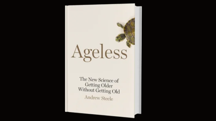
“Ageless” by Andrew Steele: A Perfect Pop-Sci Book on Aging?

People have looked for ways to extend their lives since… well, forever. The oldest known myth on earth, Gilgamesh, is about a Sumerian king embarking on a quest for immortality after his friend’s death. Alchemists were looking for magic death-defying substances, adventurers searched for the legendary Fountain of Youth, and monarchs allegedly took baths filled with young blood.

Yet, even as modern medicine developed into something useful and began conquering one disease after another, research into aging dragged its feet. It was intuitively obvious to scientists that aging was so complex a phenomenon that it was a waste of time and energy trying to cure it.

Not until the 1930s, when experiments with caloric restriction in rats resulted in a considerable lifespan extension, did scientists begin to think of aging as something modifiable, and it took us many more decades to get to the current hope-inspiring situation in the longevity field.

Today, the whole world seems to be talking about lifespan and healthspan. We’ve got articles in the mainstream media, political initiatives, and an increasing supply of money for longevity startups.

With public interest comes related books, but here’s the rub: aging is indeed an extremely complicated phenomenon that we still know appallingly little about. How do you write a popular book on a new and rapidly evolving field of science? How do you explain the basics, and when do you stop when an avalanche of new papers keeps flooding your inbox?

A handful of books on the subject have been written. The most widely known (hence, the most impactful) is, of course, David Sinclair’s “Lifespan: Why We Age and Why We Don’t Have To”, which was published in 2019. Alex Zhavoronkov was one of the pioneers with his “The Ageless Generation“, which saw light back in 2013, and let us not forget “Age Later” by Nir Barzilai and “Finding the Fountain” by Breanna Deutsch, both of which were published last year.

The niche of popular science books on aging is hardly saturated, and Andrew Steele’s “Ageless” is definitely one of the best attempts to date. This is remarkable, considering that Steele is a physicist by training who only recently became interested in longevity.

There is a well-trodden path: discuss the historical attitudes towards aging; explain why combating aging is both possible, important, and morally sound; then list several known mechanisms of aging; and end with a review of possible anti-aging solutions that are currently being researched. Instead of deviating from this simple formula, Steele brilliantly implements it.

Steele is not just a scientist but also, according to his website, a writer and campaigner, so it’s not a surprise that the book is well written. What’s more important is that it does not commit any obvious sins against our current body of knowledge.

As a science journalist covering the longevity field for people who are rather biology-savvy, I sometimes find it hard to gauge what accessible writing for laypersons should look like. Yet, I am pretty sure that Steele manages to strike a good, if not perfect, balance here. I think that at times, the average reader of Steele’s book would be left slightly bewildered, but much more often, Steele gives an explanation that is succinct, fun, and engaging.

The first part of the book, where Steele delves into the historical attitudes and moral aspects of aging, sweeps you away immediately. I particularly admired one of Steele’s arguments, which goes like this: imagine living in a world without aging. In such an ageless society, if a disease that fully recapitulated the symptoms and the prevalence of aging emerged, how would people react? Isn’t it obvious that everything would be done to stop the pandemic? Would the inhabitants of this imaginary world listen to the environmental and economic arguments against fighting aging, or would they be outraged by them? “Aging is not a morally acceptable solution to any problem”, Steele concludes, and I couldn’t agree more.

Having dealt away with the usual anti-longevity nonsense, Steele turns to the evolutionary origins of aging and again does a fabulous job. As fitting for a physicist, Steele deftly tackles the widespread misconception that aging is an increase in entropy, which is inevitable according to the second law of thermodynamics; this is only true for closed systems, while an open system can use external energy to maintain itself in working order.

This part of the book gives a lot of food for thought even for a person familiar with the matter. For instance, Steele discusses the importance of extrinsic mortality for the species’ lifespan (if your environment is perilous, evolution tweaks your body so that you live fast and die young – but only after you had a sporting chance to procreate). The level of extrinsic mortality correlates with lifespan better than many other variables such as body size.

Steele goes on to describe evolutionary strategies that promote a longer lifespan, sometimes to the point of negligible senescence – that is, when chances to die remain constant during the animal’s life. Species that have negligible senescence are not immortal (there are still infectious diseases, predators, natural hazards, etc.), but they can be considered ageless. It seems that we might even have discovered cases of reverse senescence, such as in some fish species, where females grow bigger, stronger, more resilient, and more fertile with time (this gave rise to a term I must share: BOFFFF, big, old, fat, fertile, female fish). All this means that the biological mechanisms that enable negligible or even reverse senescence are out there, and we just need to find them.

Yet, in the next part, which is dedicated to the current state of aging research, Steele never creates an impression that we are on a verge of a breakthrough; he never suggests that we should just wait a bit longer and an anti-aging pill will appear. Instead, he is being extremely honest with his readers, never overpromising nor overselling, and he sometimes maybe even errs on the side of caution. For instance, when reading the part about epigenetic clocks, I felt that Steele didn’t do enough justice to the second generation of clocks, such as PhenoAge. On the other hand, his depiction of the field of cellular senescence might be a bit too bright to my taste, but honestly, these are just minor issues I had to come up with to feel that I’m being even-handed.

When discussing the hallmarks of aging, Steele nicely shows the mind-boggling interconnectedness of aging processes. This is yet another example of honesty that might be slightly discouraging but also builds long-term trust.

Probably every book on aging contains the story of the genetically modified nematodes, but Steele paints the complete picture: how the long-lived mutants were flatly outcompeted by their more agile wild-type peers. The full story is both discouraging because it shows the possible downsides of life extension and encouraging because it explains, yet again, why we do not live much longer: not because it is biologically impossible but because this is the trade-off that evolution decided on, something we might be able to change.

The same goes for the recommendations for the reader that Steele ends his book with. He is meticulously honest on what we know about diet and exercise, to the point that it sometimes feels that you just read one caveat too many.

Steele’s boldness shows, among other things, in him not shying away from the debate about extreme life extension, something that many people in our field try to avoid. Yet even those treacherous waters he manages to navigate expertly.

So, is this the ultimate book on longevity? That, I don’t know, but it is a book that you can recommend (and maybe give – after all, the holiday season is almost upon us) to your friends and relatives. The facts and dilemmas from the book would serve as great conversation starters. Maybe, begin your next social interaction with the parable of an ageless world where aging suddenly emerges – and see what happens.

After all, let’s be honest: we need allies. The nascent longevity field has a lot going on but little to show for. We need hope; we need people to believe that the idea itself is feasible. We need to ignite their curiosity, to make them spread the word, donate, call their representatives in parliaments and demand action.

With his book, Steele joins the ranks of our most skillful and charismatic ambassadors. I wish him luck in this righteous crusade.

We would like to ask you a small favor. We are a non-profit foundation, and unlike some other organizations, we have no shareholders and no products to sell you. All our news and educational content is free for everyone to read, but it does mean that we rely on the help of people like you. Every contribution, no matter if it’s big or small, supports independent journalism and sustains our future.
Blueberries

Blueberry Extract Alleviates Alzheimer’s in Mice

A group of researchers has confirmed the neuroprotective effect of blueberry extract and identified a possible mechanism of action that might facilitate drug development [1].

The delicious cure?

Blueberries have long been on the list of “superfoods” that supposedly protect our health and maybe help us live a bit longer. Flavonoids, a class of plant-derived molecules, are probably responsible for many of blueberries’ protective qualities. We covered one of those flavonoids, pterostilbene, in a previous article.

Anthocyanins, one of the subtypes of flavonoids, are natural pigments that give many plants their purple, red, or black colors. Purple cauliflower, for example, owes its stunning appearance to anthocyanins, and blueberries have even more of these compounds. Various gut enzymes metabolize protocatechuic acid (PCA) from anthocyanins, and it has been credited with antioxidant and anti-inflammatory activity [2].

Can humble blueberries help cure Alzheimer’s disease (AD)? Anthocyanins will never become a magic pill for AD, but they have shown some neuroprotective qualities, including in older adults [3]. However, the problem with anthocyanins is that they are not well absorbed by the body: one study found that only a small fraction of anthocyanins is absorbed into plasma [4]. The researchers behind this new study hoped to demonstrate that the actual neuroprotective work is done by PCA, which is absorbed much more readily.

When the garbage chute stops working

The mechanics of AD aren’t perfectly clear and are hotly debated, but we know that it is closely linked to, if not directly caused by, the accumulation of two proteins: amyloid ß and tau, with the first forming amyloid plaques, and the second forming so-called tau-tangles. Normally, harmful proteins are disposed of by autophagy, the process by which misfolded proteins, dysfunctional organelles, and other cellular junk are enclosed in organelles called autophagosomes, transported to other organelles called lysosomes, and then broken down and recycled.

Recent evidence shows that the dysregulation of this autophagy-lysosome pathway precedes the formation of amyloid plaques and tau-tangles. Researchers have also found an increased presence of autophagosomes in AD-stricken neurons [5], which probably means that the function of lysosomes is impaired and they stop accepting material from autophagosomes, leading to the accumulation of those “cellular garbage trucks”.

Anthocyanins, on the other hand, have been shown to promote autophagy. They also improve cognitive function in aged rats. However, until now, it was not entirely clear whether blueberries owed their neuroprotective effect to anthocyanins. The objectives of the study were to verify yet again the neuroprotective effects of blueberry extract (BBE) and their link to autophagy as well as to confirm that these effects are, at least in large part, mediated by PCA.

PCA is what matters

The researchers experimented in vivo with genetically modified mice that overexpress amyloid precursor protein (APP). These mice are routinely used as a murine model of AD. In these experiments, the scientists confirmed that BBE treatment reduced neuron damage by promoting autophagy. There were fewer dead neurons, neuronal morphology was restored, and autophagy markers were upregulated.

In subsequent in vitro experiments, PCA treatment reduced cytotoxicity caused by amyloid ß in hippocampal neurons. At high concentrations, PCA restored cell viability almost to a normal level after it had dropped some 40% following amyloid ß administration. The researchers also found that amyloid ß inhibits the autophagy-lysosomal pathway in neurons, while PCA treatment alleviated that effect. The researchers concluded that PCA is largely responsible for BBE’s neuroprotective qualities and that PCA can be used as a new foundation for anti-AD drugs.

Conclusion 

Though eating blueberries seems to have some neuroprotective effects, this is probably not enough to cure Alzheimer’s, for various reasons, including the low levels of absorption of anthocyanins by the body. This study identifies PCA, a seemingly better agent, as a molecule that is largely responsible for BBE’s neuroprotective effects.

These findings can lead to the development of new anti-Alzheimer’s drugs, which would be most welcome considering the current lack of effective treatments. The research also highlights the role of autophagy in AD, which means that AD can potentially be targeted by other drugs that increase autophagy, such as rapamycin and its analogs.

We would like to ask you a small favor. We are a non-profit foundation, and unlike some other organizations, we have no shareholders and no products to sell you. All our news and educational content is free for everyone to read, but it does mean that we rely on the help of people like you. Every contribution, no matter if it’s big or small, supports independent journalism and sustains our future.

Literature

[1] Li, H., Zheng, T., Lian, F., Xu, T., Yin, W., & Jiang, Y. (2021). Anthocyanin-rich Blueberry Extracts and Anthocyanin Metabolite Protocatechuic Acid promote Autophagy-lysosomal Pathway and alleviate Neurons Damage in vivo and in vitro Models of Alzheimer’s Disease. Nutrition, 111473.

[2] Choi, J. R., Kim, J. H., Lee, S., Cho, E. J., & Kim, H. Y. (2020). Protective effects of protocatechuic acid against cognitive impairment in an amyloid beta-induced Alzheimer’s disease mouse model. Food and Chemical Toxicology, 144, 111571.

[3] Krikorian, R., Shidler, M. D., Nash, T. A., Kalt, W., Vinqvist-Tymchuk, M. R., Shukitt-Hale, B., & Joseph, J. A. (2010). Blueberry supplementation improves memory in older adults. Journal of agricultural and food chemistry, 58(7), 3996-4000.

[4] Vitaglione, P., Donnarumma, G., Napolitano, A., Galvano, F., Gallo, A., Scalfi, L., & Fogliano, V. (2007). Protocatechuic acid is the major human metabolite of cyanidin-glucosides. The Journal of nutrition, 137(9), 2043-2048.

[5] Nixon, R. A., Wegiel, J., Kumar, A., Yu, W. H., Peterhoff, C., Cataldo, A., & Cuervo, A. M. (2005). Extensive involvement of autophagy in Alzheimer disease: an immuno-electron microscopy study. Journal of Neuropathology & Experimental Neurology, 64(2), 113-122.

Heart Attack

How Telomerase Reverse Transcriptase Protects the Heart

Research published in Circulation has shown that mitochondrial, but not nuclear, telomerase reverse transcriptase (TERT) protects against damage caused by heart attacks in a murine model.

A non-canonical role

While TERT’s name comes from its main function, the protection of telomere length in the cellular nucleus, the researchers make it clear that TERT has other functions as well, even when it is not expressed in the nucleus. For example, prior research has shown that mitochondrial telomerase protects against DNA damage in cancer cells by decreasing the amounts of reactive oxygen species (ROS) [1].

The mouse experiment

In order to determine the contributions of mitochondrial and nuclear TERT to cellular protection, the researchers tested four lines of mice. Wild-type mice were used as the controls, another strain had a complete knockout of TERT, another strain had no TERT in the nucleus but additional TERT in the mitochondria (mitoTERT), and the final strain had no TERT in the mitochondria but additional TERT in the nucleus (nucTERT).

They then subjected these mice to ischemic heart attacks and observed their recovery. One of the first things they found was that nucTERT mice performed slightly worse than TERT-knockout mice. Because their immediate death rate was so high (50%) and the size of the resulting infarctions did not differ from TERT-knockout mice, the researchers chose not to continue experiments with nucTERT mice.

Instead, they focused on the mitoTERT mice, which had substantially lower death rates than even the wild-type mice. Vascularization, a measurement of the blood vessels entering tissue, was substantially reduced in TERT-knockout mice, but less so in wild-type and mitoTERT mice. While the hearts of wild-type and TERT-knockout mice were substantially enlarged 28 days after heart attack, mitoTERT mice had much less enlarged hearts. Wild-type mice had smaller scars than TERT-deficient mice, and mitoTERT mice had even smaller scars than wild-type mice.

In sum, the researchers had found that mitoTERT mice had only slightly reduced heart function after their injuries, while wild-type and TERT-knockout mice were much more injured. These results clearly show that the researchers’ hypothesis is correct: mitochondrial TERT is protective against heart attack.

A closer examination

The researchers performed an in-depth examination of the functions of mitochondrial TERT in the cell. They found that cardiomyocytes (heart muscle cells) that had additional mitochondrial TERT were protected from apoptosis (cellular death), both at baseline and when exposed to hydrogen peroxide. Mitochondrial TERT was also found to increase migration in a wound test.

The researchers pin their results on the actions of Complex I, a critical component of mitochondrial respiration. After performing a proteomic analysis, the researchers found that one of the key differences between wild-type, TERT-knockout, and mitoTERT cells was a protein called Prohibitin. This protein was found to be increased in TERT-knockout mice but decreased in mitoTERT mice, and prior research has found that it is increased with aging [2].

How can this discovery be used?

Armed with this new knowledge, the researchers then investigated two methods of potentially defending against ischemic disease. They considered remote ischemic preconditioning (RIPC), a procedure in which patients are artificially given ischemic attacks in one organ in order to protect them from additional ischemia in other organs. The researchers sampled the mitochondrial TERT levels of patients that had undergone this procedure, finding that their levels were greater than those of a placebo group.

The researchers then examined TA-65, a telomerase activator that has been shown to be generally safe in humans. Their investigation found that TA-65 improved mitochondrial TERT in human endothelial cells and made them more like those of mitoTERT mice.

Conclusion

This research is in its infancy, and the exact biochemical relationships between mitochondrial TERT, respiratory complex I, and Prohibitin have not been elucidated. However, it seems clear that further research into this area might lead to effective interventions for ischemic heart attack, including both preventative measures and post-operative treatments.

We would like to ask you a small favor. We are a non-profit foundation, and unlike some other organizations, we have no shareholders and no products to sell you. All our news and educational content is free for everyone to read, but it does mean that we rely on the help of people like you. Every contribution, no matter if it’s big or small, supports independent journalism and sustains our future.

Literature

[1] Singhapol, C., Pal, D., Czapiewski, R., Porika, M., Nelson, G., & Saretzki, G. C. (2013). Mitochondrial telomerase protects cancer cells from nuclear DNA damage and apoptosis. PloS one, 8(1), e52989.

[2] Miwa, S., Jow, H., Baty, K., Johnson, A., Czapiewski, R., Saretzki, G., … & Von Zglinicki, T. (2014). Low abundance of the matrix arm of complex I in mitochondria predicts longevity in mice. Nature communications, 5(1), 1-12.

Bladder

Twin Drug Combo Convinces Cancer Cells To Eat Themselves

In an important proof-of-concept study, scientists have successfully employed a ferroptosis activator and an mTOR inhibitor against bladder cancer cells [1].

Ferroptosis – “the iron death”

Ferroptosis is a distinct type of cell death that gets less attention and is less well understood than the more ubiquitous and familiar apoptosis. As its name suggests, ferroptosis requires iron to activate complex pathways that eventually lead to the cell’s demise. Since cancer cells generate higher levels of iron, they are more susceptible to ferroptosis, which is why it has been receiving a lot of recent attention from oncologists. Some ferroptosis inducers have already been tested against cancer in early research, even if scientists may not know the exact mechanisms of their action yet. Ferroptosis also definitely plays a role in aging (again, not fully elucidated) – for instance, one recent study found a significant age-dependent increase in ferroptosis in numerous tissues in mice [2].

Autophagy and cancer – a complex relationship

It is becoming increasingly evident that ferroptosis, at least sometimes, uses autophagy pathways. Autophagy is the process of recycling cellular garbage such as misfolded proteins, and it has a complex, context-dependent relationship with cancer. Autophagy is thought to have a protective function during tumorigenesis, but once tumor cells are established, autophagy supports tumor survival and metastasis. Several previous studies showed the contrasting effects of autophagy in bladder cancer [3].

Autophagy can be employed against cancer, such as via inhibitors of the nutrient sensing complex mTOR, of which rapamycin is the most famous. The logic behind this particular facet of autophagy-cancer interplay goes like this: cancer cells require a lot of energy for growth and their unstoppable proliferation. mTOR inhibition, on the other hand, tricks cells into thinking that nutrients are in short supply. This slows down cellular metabolism and activates autophagy so that cells obtain nutrients by recycling those leftover molecules. Neglected when life is good and nutrients are abundant, autophagy results in slower proliferation but also in cleaner and leaner cells. Upregulating autophagy is probably one of the mechanisms behind rapamycin’s geroprotective action [4].

How can autophagy be involved in cell death if it is supposed to make cells younger and healthier? This is probably because biology creatively uses whatever is at hand, so many biological processes have multiple, sometimes mutually exclusive, purposes. Depending on the context, autophagy can accompany cell death, be a part of a signaling pathway that leads to cell death, or even directly cause it [5].

Twin power

In their study, the researchers first experimented with the ferroptosis inducer Fin56. In a series of experiments, they confirmed that Fin56 induced death in many, but not all, bladder cancer cells; that this cellular death was ferroptosis; and that this ferroptosis was autophagy-dependent in at least two lines of bladder cancer cells. Autophagy inhibition, as expected, greatly reduced Fin56-mediated ferroptosis. According to the researchers, not only does ferroptosis correlate with autophagy, it actively uses autophagy mechanisms to kill cells.

Torin 2, an mTOR inhibitor, was added to the equation. After 72 hours of treating two bladder cancer cell lines with the Torin 2 – Fin56 duo, only a tiny fraction of the cells remained viable. Yet, the results were heavily dependent on dosage: this almost complete eradication of the cancer cells was achieved only at the highest concentrations of Torin 2. The safe dosage in living organisms, including humans, remains to be seen. What is clear, though, is that these two autophagy-related processes running simultaneously greatly amplify the overall effect, which can potentially be used in cancer therapies.

Conclusion

Bladder cancer is the 10th most common cancer worldwide, responsible for an estimated 549,000 new cases and 200,000 deaths per year. As recent research shows, current methods of choice in cancer therapy, including chemotherapy, radiotherapy, are harmful and also seem to accelerate aging in survivors [6]. This makes finding new, less toxic methods a necessity. The interplay between autophagy and cancer is intriguing and complex, given that both its promotion and inhibition seem to have anti-cancer effects, sometimes in the same cancer type. This new research expands our understanding of the subject by showing how autophagy can be synergistically amplified by a duo of molecules to effectively quash bladder cancer cells.

We would like to ask you a small favor. We are a non-profit foundation, and unlike some other organizations, we have no shareholders and no products to sell you. All our news and educational content is free for everyone to read, but it does mean that we rely on the help of people like you. Every contribution, no matter if it’s big or small, supports independent journalism and sustains our future.

Literature

[1] Sun, Y., Berleth, N., Wu, W., Schlütermann, D., Deitersen, J., Stuhldreier, F., … & Stork, B. (2021). Fin56-induced ferroptosis is supported by autophagy-mediated GPX4 degradation and functions synergistically with mTOR inhibition to kill bladder cancer cells. Cell Death & Disease, 12(11), 1-14.

[2] Zheng, H., Jiang, L., Tsuduki, T., Conrad, M., & Toyokuni, S. (2021). Embryonal erythropoiesis and aging exploit ferroptosis. Redox Biology, 102175.

[3] Li, F., Guo, H., Yang, Y., Feng, M., Liu, B., Ren, X., & Zhou, H. (2019). Autophagy modulation in bladder cancer development and treatment. Oncology reports, 42(5), 1647-1655.

[4] Sarkar, S., Ravikumar, B., Floto, R. A., & Rubinsztein, D. C. (2009). Rapamycin and mTOR-independent autophagy inducers ameliorate toxicity of polyglutamine-expanded huntingtin and related proteinopathies. Cell Death & Differentiation, 16(1), 46-56.

[5] Denton, D., & Kumar, S. (2019). Autophagy-dependent cell death. Cell Death & Differentiation, 26(4), 605-616.

[6] Wang, S., Prizment, A., Thyagarajan, B., & Blaes, A. (2021). Cancer Treatment-Induced Accelerated Aging in Cancer Survivors: Biology and Assessment. Cancers, 13(3), 427.

Thoughtful old man

How Uncontrolled Diabetes Is Linked to Cognitive Impairment

A team of Spanish researchers has examined how glucose imbalance leads to neurological disorders through the failure of adipocytokines.

What are adipocytokines?

These compounds’ name seems a bit concerning: “adipo-” refers to fat, and a cytokine is a cellular signal most commonly known for being part of the deadly cytokine storm associated with severe immunological problems. However, the two adipocytokines that these researchers studied, adiponectin and leptin, have a beneficial, neuroprotective effect through specific receptors in the brain [1].

The researchers cite a great number of previous studies, some of which are contradictory, in elucidating this relationship. They note another hypothesis that suggests that the ratio between these two compounds (Ad/L) is the most important; however, previous research in this direction has also been contradictory.

However, one thing is clear: metabolic syndrome is dangerous to the brain, as inflammation and hyperglycemia (a characteristic symptom of diabetes) are linked to cognitive impairment through the infliction of oxidative stress [2].

A Bayesian examination

Using Bayesian computer modeling, the researchers set out to examine the precise relationship between metabolic syndrome and brain dysfunction. What they discovered was an extremely complicated relationship between the Ad/L ratio, insulin resistance, and triglycerides or glucose. In the case of elevated triglycerides and high insulin resistance, a higher Ad/L ratio was neuroprotective; however, if glucose was elevated instead, this ratio became associated with reduced cognitive function.

Most critically, and most understandably, the researchers singled out glucose metabolism as playing a key role in the loss of neuroprotection. Even in the presence of insulin resistance, if glucose is controlled, adipocytokines can continue to protect the brain; if it goes out of control, this protective ability is lost.

Furthermore, the researchers single out a particular part of the brain as being particularly vulnerable: the rostral anterior cingulate cortex (rACC). This is responsible for carrying information between key areas of the brain, such as behavior, decision making, learning, and memory.

The authors of this study cite prior research showing that lifestyle interventions, such as diet and exercise, are the best way of preventing this decline [3]. However, they go further by saying that it may be worth investigating treating sufferers of metabolism-related cognitive impairment with the adipocytokines involved, particularly leptin, which has shown beneficial effects in the brains of people with a genetic deficiency.

Conclusion

This study is extremely complicated, merging together several seemingly contradictory explanations for how insulin, glucose, and fats interact within the brain. The researchers actually hold that it may not be detailed enough; they were unable to examine every related hormone, for example, and they state that sex-based differences, preclinical Alzheimer’s disease, and other, undiscovered issues may have played various roles in their results.

While this research clearly warrants further investigation, it may be worth a clinical trial to determine if the administration of adipocytokine-enhancing drugs is beneficial. Furthermore, this research makes it even more clear just how harmful uncontrolled glucose is and how important living a healthy lifestyle is, even for rarely mentioned parts of the brain.

We would like to ask you a small favor. We are a non-profit foundation, and unlike some other organizations, we have no shareholders and no products to sell you. All our news and educational content is free for everyone to read, but it does mean that we rely on the help of people like you. Every contribution, no matter if it’s big or small, supports independent journalism and sustains our future.

Literature

[1] Harvey, J. (2007). Leptin: a diverse regulator of neuronal function. Journal of neurochemistry, 100(2), 307-313.

[2] Etchegoyen, M., Nobile, M. H., Baez, F., Posesorski, B., González, J., Lago, N., … & Otero-Losada, M. (2018). Metabolic syndrome and neuroprotection. Frontiers in neuroscience, 12, 196.

[3] Atienza, M., Ziontz, J., & Cantero, J. L. (2018). Low-grade inflammation in the relationship between sleep disruption, dysfunctional adiposity, and cognitive decline in aging. Sleep medicine reviews, 42, 171-183.

Omics

Multi-omics Identifies Longevity Genes, Therapeutic Targets

The latest research in Aging Cell has brought to light new insights into the biology of aging and associated therapeutic candidates.

What is multi-omics?

“-omics” refers to branches of molecular biology that attempt to characterize and/or quantify their components in totality, often using recently developed big data technologies. For example, genetics is the study of individual genes, while genomics is the study of the entire genome. As there are at least 19,000 human genes (if we are only counting the protein-coding ones), genomics requires entirely different strategies than genetics.

Genes from our genome are read and transcribed to make RNA. This RNA is then transported out of the cell nucleus and used to build proteins. The sum of all RNA being expressed in an organism, tissue, or cell is referred to as the transcriptome, while the entire set of proteins present is the proteome. So, genomics looks at the blueprint that is used to make proteins, transcriptomics looks at the set of proteins being synthesized at a given moment, and proteomics looks at the sum of proteins that are present and carrying out various functions.

Additional -omics also exist, such as epigenomics, microbiomics, and metabolomics. Multi-omics then, is the combination of two or more of these techniques and can be a very powerful tool to study the biology of aging.

Multi-omics and longevity

While our genome, transcriptome, and proteome don’t determine everything about us, they make up a significant portion of who we are, including how we age. Recent technological advancements in how scientists can analyze different -omics (sometimes referred to as the genomic revolution) has created a unique opportunity for studying aging and longevity. By studying long-lived individuals and their children, researchers have already identified a number of genes potentially involved in aging, such as the APOE loci [1,2].

However, as the genomic revolution steams ahead, new technologies, analysis techniques, and human datasets to draw from continue to become available. One such analysis technique, Mendelian randomization, allows for the identification of causal relationships while being less susceptible to many of the biases and errors that are common in other techniques [3]. Using Mendelian randomization, the UK Biobank and LifeGen consortium, and a multi-omics approach, researchers at the Université Laval in Quebec have uncovered previously unknown and potentially causal players in aging [4].

Haptoglobin is associated with parental longevity throughout the protein synthesis process

The multi-omics approach was able to identify many proteins that were significantly associated with parental lifespan in the genome-wide, transcriptome-wide, or proteome-wide analyses. However, only one protein, haptoglobin (Hp), showed significant association in all three.

Hp is one of the most abundant proteins in human plasma, regulates hemoglobin and cholesterol levels, and is expressed primarily in the liver. While this analysis shows several potential mechanisms for how Hp may be linked to lifespan, further investigation is needed to determine if it could be a potential anti-aging therapeutic target.

Novel insights into the impact of smoking and cardiovascular disease-related genes on lifespan 

Various proteins that are well-known to contribute to clogged arteries and cardiovascular disease were associated with parental lifespans (SORT1, APOB, LRP8, and CCDC71L). Additionally, potential new treatment pathways for lowering bad cholesterols were identified, such as targeting PCSK9 and LPA.

Similarly, the study turned up many smoking-associated genes, such as cholinergic receptor nicotinic 2, 3/5, and 4 (CHRNA2/3/4/5), and their search for potential drug targets for aging-associated diseases identified many smoking cessation therapies. These results bolster the already strong evidence for the importance of cholesterol levels and smoking behaviors on lifespan.

Do these findings align with the archetypal longevity pathways?

Comparing their findings to the mechanisms of aging that have been identified through model organisms (i.e. mTOR, autophagy, cellular senescence, etc.), only chronic inflammation was also associated with parental lifespan in this study. In particular, this included the IL6R and ICAM5 loci. Further analysis also suggested that targeting these pathways may provide cardiovascular benefits.

The study also identified a previously unknown interaction between RAD52 and WRN. WRN is the major gene involved in Werner progeria (an accelerated aging disease). This connection may have implications both for aging research and Werner syndrome patients, warranting further study.

The lack of representation of other pathways that are stereotypical parts of geroscience is concerning for the prospects of their translation from model organisms into humans. However, this finding is far from a definitive conclusion that drugs utilizing these pathways are doomed to fail.

In conclusion, our study identified new genetically regulated genes across 43 tissues, as well as genetically regulated circulating proteins and metabolites that could potentially regulate human lifespan. Many of these genetic determinants of parental lifespan represent potential therapeutic targets for aging-associated diseases. Our study also underscores the importance of global population health measures such as adopting stricter tobacco control measures as well as the globalization of interventions targeting all apolipoprotein-B-containing particles to prevent the onset of diseases of the cardiovascular system and possibly promote longevity.

Another page in the genomic revolution

While the depth of this study cannot be captured in a brief summary, several other genes were newly identified. These include SVEP1 (which was previously correlated with longevity in the Framingham Heart Study), NEK10, HTT (in skeletal muscle), NRG1, and BECN1. Additional circulating proteins that may be causally associated with parental lifespan included ASPN, ASIP, IGF2R, PLXNB2, and ENPP7. 

As always, these findings must be considered in the context of several study limitations. Notably, the main outcome investigated was parental lifespan. Looking at the lifespan of the study participants’ parents rather than their own was a necessary limitation for this dataset, as many of the participants are still alive.

However, it also introduced limitations such as selection bias (for example, only individuals who had children could be enrolled), the different environments that parents and children were exposed to, and self-reporting errors (i.e. if a participant misremembered the age of their parents at death). Additionally, more technical issues, such as spurious gene prioritization and horizontal pleiotropy, may also have resulted in erroneous findings, although much care was taken to avoid these issues.

Ultimately, as the authors note, only prospective randomized clinical trials can definitively determine if an intervention extends lifespan in humans. Fortunately, this study provides a number of new candidate genes and circulating proteins as potential therapeutic targets that may someday face that challenge.

We would like to ask you a small favor. We are a non-profit foundation, and unlike some other organizations, we have no shareholders and no products to sell you. All our news and educational content is free for everyone to read, but it does mean that we rely on the help of people like you. Every contribution, no matter if it’s big or small, supports independent journalism and sustains our future.

Literature

[1] Deelen, J. et al. A meta-analysis of genome-wide association studies identifies multiple longevity genes. Nature Communications (2019). https://doi.org/10.1038/s41467-019-11558-2

[2] Timmers, P.R. et al. Genomics of 1 million parent lifespans implicates novel pathways and common diseases and distinguishes survival chances. eLife (2019). https://doi.org/10.7554/eLife.39856

[3] Hemani, G. et al. The MR-Base platform supports systematic causal inference across the human phenome. eLife (2018). https://doi.org/10.7554/eLife.34408

[4] Perrot, N. et al. A trans-omic Mendalian randomization study of parental lifespan uncovers novel aging biology and therapeutic candidates for chronic diseases. Aging Cell (2021). https://doi.org/10.1111/acel.13497