×

The Blog

Building a Future Free of Age-Related Disease

Plant-Based Diet

Plant-Based Diet Linked To Reduced Leaky Gut Syndrome

The MaPLE trial in Italy investigated whether or not increasing fruits and vegetables decreases leaky gut syndrome [1].

Background

This study examines increased intestinal permeability (leaky gut) in adults 60 years and older [1].  Age has been reported to be a risk factor for increased intestinal permeability over the age of 50 [2]. The authors of this study hypothesized that consuming a plant-rich, and therefore polyphenol-rich, diet would induce serum metabolomic changes associated with better intestinal permeability through gut bacteria.

Methods

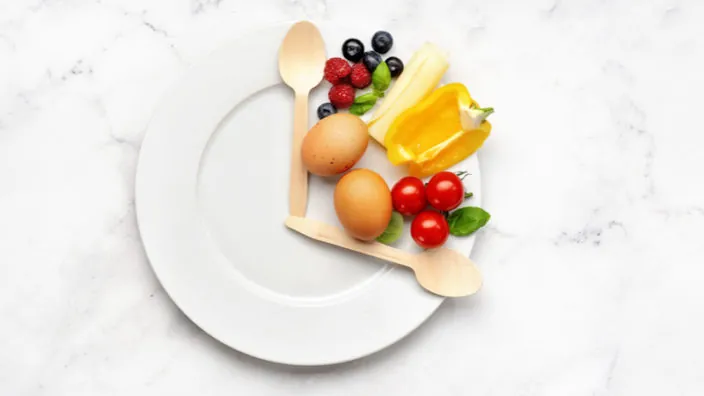
This randomized, controlled trial included eight weeks of eating three portions of polyphenol-rich foods daily, which was followed by eight weeks of being back on a regular diet in a long-term care center. There was an eight-week break between each diet.

The researchers evaluated increased intestinal permeability by measuring serum zonulin levels in 51 participants after an overnight fast. Zonulin is a human protein that is known to be involved in regulating tight junctions in the gut, and higher zonulin levels reflect worse intestinal permeability. A leaky gut may contribute to inflammaging, and having a sustained high zonulin level predisposes people to additional health issues.

Elevated zonulin has been reported in many research studies across different ages and disease conditions, including, but not limited to, celiac disease, type 1 diabetes, pre-diabetes, type 2 diabetes, rheumatoid arthritis, multiple sclerosis, obesity, polycystic ovary syndrome, and irritable bowel syndrome [3,4].

The gut microbiota results were determined by analyzing stool samples. They also collected anthropometric data, blood pressure, and measures of functional, metabolic, inflammation, vascular, and oxidative stress markers, and they used specialized software to analyze three food records per day per person. They estimated the polyphenol content of each food as well using the Phenol Explorer database.

Results

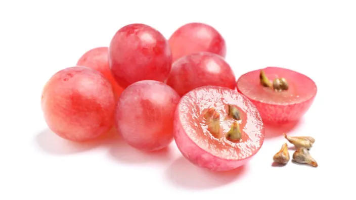
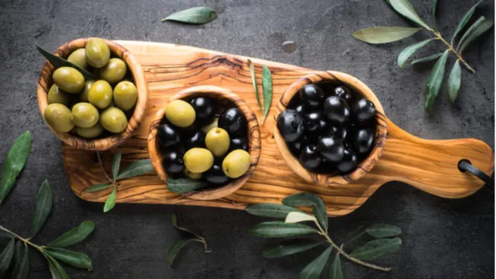
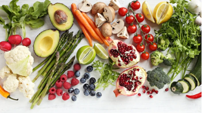
This study showed reduced blood pressure in both men and women following an 8-week polyphenol-rich diet. This diet contained approximately 579 more milligrams of polyphenols in a day than the regular control diet [5]. Polyphenol-rich foods, such as cocoa, green tea and berries, led to significant reductions in zonulin.

This research also explains the secondary outcomes of the initial cohort. The polyphenol-rich diet was significantly lower in animal and plant protein, total fat, monounsaturated fatty acids, polyunsaturated fatty acids, total omega-6 fatty acid, calcium, and iron than the control diet. Stool bacterial communities did not vary in comparison between the diets.

Using a liquid chromatography technique, the researchers determined that seven of the metabolites were significantly increased while three metabolites significantly decreased. The increased metabolites included catechol sulfate, hippuric acid, and 2-methyl pyrogallol sulfate, which are markers of polyphenols being digested in the stomach that then undergo additional metabolism in the liver [6,7,8]. The metabolites 3-methylxanthine, 7-methylxanthine, theobromine, and HPPA-S were also increased after the 8-week polyphenol-rich diet. Lower levels of the metabolic product deoxycarnitine were correlated with a less leaky gut [9], and the researchers reported lower amounts of deoxycarnitine after the polyphenol-rich diet ended.

A heatmap showed the significant correlations. Serum zonulin and serum metabolites were significantly altered by the polyphenol-rich diet interventions and many of the nutrients that the researchers measured.

The researchers also investigated correlations between serum metabolome and gut microbiota. Notably, theobromine, a compound found primarily in cocoa, showed the most statistically significant correlations. In prior research, theobromine has been associated with beneficial gut bacteria that converts plant ligins to the compound enterolactone [10].

In summary, this multi-omics study shows that age, the baseline level of zonulin, and changes in the abundance of the bacteria porphyromonadaceae are primarily responsible for the total level of zolunin.

Conclusion

The major limitation of this study is that it does not prove a causal relationship between a polyphenol-rich diet, zolunin, and leaky gut. There are likely other food components present in this diet that may oppose or promote each other’s effects.

Other polyphenols have been associated with higher expression of tight gut proteins in the stomach, so increasing polyphenol rich-foods in diet may help leaky gut syndrome. Often, diet recommendations may include an anti-inflammatory diet, which includes foods that increase positive gut bacteria and excludes foods that cause bloating, constipation and diarrhea. Leaky gut may also be triggered by lifestyle factors, such as alcohol interacting with tight gut junction proteins [11,12]. There is currently no clear dietary treatment for leaky gut, so evidence-based research is needed to develop a personalized approach.

We would like to ask you a small favor. We are a non-profit foundation, and unlike some other organizations, we have no shareholders and no products to sell you. All our news and educational content is free for everyone to read, but it does mean that we rely on the help of people like you. Every contribution, no matter if it’s big or small, supports independent journalism and sustains our future.

Literature

[1] Peron, G., Gargari, G., Meroño, T., Miñarro, A., Lozano, E. V., Escuder, P. C., González-Domínguez, R., Hidalgo-Liberona, N., Del Bo’, C., Bernardi, S., Kroon, P. A., Carrieri, B., Cherubini, A., Riso, P., Guglielmetti, S., & Andrés-Lacueva, C. (2021). Crosstalk among intestinal barrier, gut microbiota and serum metabolome after a polyphenol-rich diet in older subjects with “leaky gut”: The MaPLE trial. Clinical nutrition (Edinburgh, Scotland), 40(10), 5288–5297. https://doi.org/10.1016/j.clnu.2021.08.027

[2] Nicoletti C. (2015). Age-associated changes of the intestinal epithelial barrier: local and systemic implications. Expert review of gastroenterology & hepatology, 9(12), 1467–1469. https://doi.org/10.1586/17474124.2015.1092872

[3] Sturgeon, C., & Fasano, A. (2016). Zonulin, a regulator of epithelial and endothelial barrier functions, and its involvement in chronic inflammatory diseases. Tissue barriers, 4(4), e1251384. https://doi.org/10.1080/21688370.2016.1251384

[4] Ciccia, F., Guggino, G., Rizzo, A., Alessandro, R., Luchetti, M. M., Milling, S., Saieva, L., Cypers, H., Stampone, T., Di Benedetto, P., Gabrielli, A., Fasano, A., Elewaut, D., & Triolo, G. (2017). Dysbiosis and zonulin upregulation alter gut epithelial and vascular barriers in patients with ankylosing spondylitis. Annals of the rheumatic diseases, 76(6), 1123–1132. https://doi.org/10.1136/annrheumdis-2016-210000

[5] Del Bo’, C., Bernardi, S., Cherubini, A., Porrini, M., Gargari, G., Hidalgo-Liberona, N., González-Domínguez, R., Zamora-Ros, R., Peron, G., Marino, M., Gigliotti, L., Winterbone, M. S., Kirkup, B., Kroon, P. A., Andres-Lacueva, C., Guglielmetti, S., & Riso, P. (2021). A polyphenol-rich dietary pattern improves intestinal permeability, evaluated as serum zonulin levels, in older subjects: The MaPLE randomised controlled trial. Clinical nutrition (Edinburgh, Scotland), 40(5), 3006–3018. https://doi.org/10.1016/j.clnu.2020.12.014

[6] Rechner, A. R., Kuhnle, G., Hu, H., Roedig-Penman, A., van den Braak, M. H., Moore, K. P., & Rice-Evans, C. A. (2002). The metabolism of dietary polyphenols and the relevance to circulating levels of conjugated metabolites. Free radical research, 36(11), 1229–1241. https://doi.org/10.1080/246-1071576021000016472

[7] Pimpão, R. C., Ventura, M. R., Ferreira, R. B., Williamson, G., & Santos, C. N. (2015). Phenolic sulfates as new and highly abundant metabolites in human plasma after ingestion of a mixed berry fruit purée. The British journal of nutrition, 113(3), 454–463. https://doi.org/10.1017/S0007114514003511

[8] Pasinetti, G. M., Singh, R., Westfall, S., Herman, F., Faith, J., & Ho, L. (2018). The Role of the Gut Microbiota in the Metabolism of Polyphenols as Characterized by Gnotobiotic Mice. Journal of Alzheimer’s disease : JAD, 63(2), 409–421. https://doi.org/10.3233/JAD-171151

[9] Semba, R. D., Trehan, I., Li, X., Moaddel, R., Ordiz, M. I., Maleta, K. M., Kraemer, K., Shardell, M., Ferrucci, L., & Manary, M. (2017). Environmental Enteric Dysfunction is Associated with Carnitine Deficiency and Altered Fatty Acid Oxidation. EBioMedicine, 17, 57–66. https://doi.org/10.1016/j.ebiom.2017.01.026

[10] Cardona, F., Andrés-Lacueva, C., Tulipani, S., Tinahones, F. J., & Queipo-Ortuño, M. I. (2013). Benefits of polyphenols on gut microbiota and implications in human health. The Journal of nutritional biochemistry, 24(8), 1415–1422. https://doi.org/10.1016/j.jnutbio.2013.05.001

[11] Wang, Y., Tong, J., Chang, B., Wang, B., Zhang, D., & Wang, B. (2014). Effects of alcohol on intestinal epithelial barrier permeability and expression of tight junction-associated proteins. Molecular medicine reports, 9(6), 2352–2356. https://doi.org/10.3892/mmr.2014.2126

[12] Wang, Y., Tong, J., Chang, B., Wang, B., Zhang, D., & Wang, B. (2014). Effects of alcohol on intestinal epithelial barrier permeability and expression of tight junction-associated proteins. Molecular medicine reports, 9(6), 2352–2356. https://doi.org/10.3892/mmr.2014.2126

Stethoscope Clock

Epigenetic Clock Shows Association With Cardiovascular Aging

A new study published in Mechanisms of Ageing and Development has found correlations between cardiovascular aging and a measure of epigenetic age acceleration [1].

Can epigenetic clocks predict cardiovascular disease?

One way researchers test the validity and usefulness of epigenetic clocks is to compare their outputs to other measures of biological aging, such as the occurrence of age-related diseases [2]. Cardiovascular disease is one of the largest causes of death around the world, and age is its primary risk factor. Despite this, research findings have been inconsistent when studying the relationships between measures of epigenetic age and cardiovascular disease. Many high-profile studies have reported a range of associations between the two, but others have shown no association at all [3].

Measuring epigenetic aging and cardiovascular disease

In this study, scientists from Charité Medical University in Berlin utilized the Berlin Aging Study II [4], which aims to explore factors that impact aging both positively and negatively. Its participants reside in Berlin, and the sub-group used in this study consisted of people between ages 60 and 84 (n=1671).

Participants degree of cardiovascular disease was measured by two common composite scores, the LS7 [5] and FRS [6].

An epigenetic clock that was modified from Vidal-Bralo [7] and uses only 7 CpG methylation sites [6] was used in this study. This clock is much simpler compared to the more popular Horvath (353 CpGs) and Hannum (71 CpGs) clocks.

For this study, the primary variable of interest from the epigenetic clock was epigenetic age acceleration. This is typically defined as the difference between biological age, as estimated by the epigenetic clock, and chronological age. However, the authors also accounted for leukocyte cell distribution (monocytes, lymphocytes, etc.) in addition to chronological age in this analysis. Previous studies have shown that this may be a key variable, as epigenetic measures are often made from leukocytes [8], including the dataset in this study.

LS7 and FRS are associated with epigenetic age acceleration

148 participants showed clinical signs of cardiovascular diseases, but many more showed a variety of risk factors, including high blood pressure, diabetes, high cholesterol, smoking, etc.

Epigenetic age acceleration was correlated with both LS7 and FRS for both men and women, with the absolute value of the correlation coefficient (r) ranging between 0.049 and 0.084. While this is considered a weak correlation, epigenetic age acceleration was more predictive than chronological age, which had a correlation coefficient of r = 0.007. When a linear regression model was applied, the association between epigenetic age acceleration and both LS7 and FRS was statistically significant (p<0.05).

Looking at individual components within both scores, physical activity was the most associated with epigenetic age acceleration for LS7 relative to diet, BMI, HbA1c, total cholesterol, smoking, and blood pressure. Similarly, for the FRS items, HDL cholesterol and total cholesterol were the primary drivers of its association with epigenetic age acceleration, with slightly less impact from chronological age, diabetes, systolic blood pressure, and smoking.

In conclusion, our results provide evidence of a weak association of the epigenetic clock (DNAm age acceleration) with cardiovascular health in the BASE-II cohort. We were able to confirm our initial hypothesis that more favorable results in the Life’s simple 7 (LS7) and the Framingham Risk Score (FRS) would be associated with a lower DNAm age acceleration. The DNAm age acceleration of participants with more favorable scores in the well-established instruments FRS and LS7 resulted to be lower compared to participants with less favorable score results. Moreover, we found our hypothesis that the LS7 may be associated more strongly with DNAm age acceleration than the FRS because it includes physical activity and diet at least partially confirmed. Physical activity resulted to be the driving force in the association between DNAm age acceleration and the Life’s simple 7. As expected, women displayed more favorable results in the CVH scores and had significantly lower epigenetic age acceleration than men. The above findings add to the growing body of evidence supporting the epigenetic clock´s potential as a biomarker of aging in the context of CVH and lifestyle factors.

Conclusion

This study is the first to compare LS7 and FRS with epigenetic age acceleration. Different study population datasets, epigenetic clocks, and measurements of cardiovascular disease have been examined in previous studies. While this study contributes to the muddled findings of previous work to a certain extent, it also suggests that some components of cardiovascular disease (such as physical activity, HDL cholesterol, and total cholesterol) may be more related to measures of epigenetic age acceleration than others. Epigenetic clocks are still relatively new to longevity research and will likely require many more studies such as this one to sort out their optimal parameters, which components of aging they are associated with, and why.

We would like to ask you a small favor. We are a non-profit foundation, and unlike some other organizations, we have no shareholders and no products to sell you. All our news and educational content is free for everyone to read, but it does mean that we rely on the help of people like you. Every contribution, no matter if it’s big or small, supports independent journalism and sustains our future.

Literature

[1] Lemke, E. et al. Cardiovascular health is associated with the epigenetic clock in the Berlin Aging Study II (BASE-II). Mechanisms of Ageing and Development (2021). https://doi.org/10.1016/j.mad.2021.111616

[2] Ferrucci, L. et al. Measuring biological aging in humans: A quest. Aging Cell (2020). https://doi.org/10.1111/acel.13080

[3] Lind, L. et al. Methylation-based estimated biological age and cardiovascular disease. Eur J Clin Invest (2018). https://doi.org/10.1111/eci.12872

[4] Bertram, L. et al. Cohort profile: The Berlin Aging Study II (BASE-II). Int J Epidemiol (2014). https://doi.org/10.1093/ije/dyt018

[5] Konig, M. et al. Historical trends in modifiable indicators of cardiovascular health and self-rated health among older adults: Cohort differences over 20 years between the Berlin Aging Study (BASE) and the Berlin Aging Study II (BASE-II). PLoS One (2018). https://doi.org/10.1371/journal.pone.0191699

[6] D’Agostino, R.B. Sr. et al. General cardiovascular risk profile for use in primary care: the Framingham Heart Study. Circulation (2008). https://doi.org/10.1161/circulationaha.107.699579

[7] Vidal-Bralo, L., Y. Lopez-Golan, and A. Gonzalez. Corrigendum: Simplified Assay for Epigenetic Age Estimation in Whole Blood of Adults. Front Genet (2017). https://doi.org/10.3389/fgene.2017.00051

[8] Quach, A.L. et al. Epigenetic clock analysis of diet, exercise, education, and lifestyle factors. Aging (Albany NY) (2017). https://doi.org/10.18632/aging.101168

Grape seeds

A Grape seed Extract Slows Aging In Mice

Studying grape seed extract, scientists have discovered a new senomorphic compound that enhances chemotherapy and prolongs lifespan and healthspan in mice [1].

Nature’s drugstore

Many drugs have come to us from the world of plants, including promising geroprotective molecules such as resveratrol. However, identifying specific components of plant extracts that exert positive effects can be challenging.

In this new study, the researchers started by screening a library of 46 plant-derived compounds for their senolytic potential – that is, the ability to remove senescent cells.

The researchers treated cells in vitro with a senescence-inducing compound and then applied the chemicals from the library to ascertain their senolytic effects. They also used several known senolytics as controls. The list of compounds that had exhibited a senolytic potential included the well-known curcumin and fisetin, but the researchers decided to focus on grape seed extract (GSE) due to both the magnitude of its effects on senescent cells and its relative paucity of research.

Both senomorphic and senolytic

In a series of experiments, the researchers confirmed that GSE effectively reduces SASP secretion, but in moderate doses, this was not accompanied by senescent cells being wiped out. According to the researchers, this effect is more senomorphic in nature, which means that it preserves senescent cells but alleviates harmful aspects of their phenotype [2]. The senomorphic approach has been gaining popularity recently because senescent cells are not always bad, and their elimination might be ill-advised in certain conditions.

Still, the researchers also tested GSE’s senolytic abilities and found them to be just as impressive. At higher concentrations, GSE eliminated up to 80% of senescent cells without affecting the viability of normal cells.

GSE consists of many different molecules, so which one or ones are responsible for its senolytic action? A component called procyanidin C1 (PCC1) that belongs to the flavonoid family caught the researchers’ attention due to its known ability to induce DNA damage. The scientists repeated the experiments with only PCC1 and found that its effect was very similar to that of GSE. Other GSE components did not exhibit senolytic activity – or at least, not at the same scale as PCC1.

Senolytics and cancer

The researchers then ran a series of experiments in vivo on cancerous tumors in mice. It is known that senescent cells are instrumental in preventing cancer in its early stages but might actually team up with it in later ones [3]. For instance, they can contribute to a tumor’s resistance to chemotherapy [4]. What’s worse, radiotheraphy and chemotherapy themselves induce cellular senescence by inflicting stress on healthy cells.

The researchers induced tumors in mice and then treated them either with a chemotherapeutic known as mitoxantrone, PCC1, or with the mitoxantrone-PCC1 duo. Unsurprisingly, mitoxantrone administration led not just to an anti-tumor effect but also to the appearance of numerous senescent cells in the tumor tissue. However, PCC1 eliminated most of those cells. As a result, the combination treatment remarkably enhanced tumor regression (55.2% reduction in tumor size compared to mitoxantrone alone).

Mice who were receiving the mitoxantrone-PCCC1 combo survived 1.5 times longer than the group treated with mitoxantrone alone. To be sure, PCC1 without mitoxantrone did not have a significant effect on survivability: after all, PCC1 is just a sidekick that clears senescent cells, making the job of the chemotherapeutic agent easier.

Genuine life extension

The researchers then performed additional in vivo experiments to assess PCC1’s ability to attenuate senescence-related physical dysfunction. It is known that even a small number of senescent cells can affect organismal health, including in young organisms. The researchers implanted senescent cells into young mice, which led to a decline in their physical abilities (maximum walking speed, hanging endurance, and grip strength), but this decline was largely reversed by PCC1 treatment.

Finally, to examine the effect of PCC1 on organismal aging, the researchers tested it on a bunch of naturally aging wild type mice. By the age of two years (or around 75 in human years), cellular senescence ran rampant in those mice. Bi-weekly PCC1 treatment that lasted about four months eliminated a large percentage of senescent cells and led to an increase in physical function and longevity. Treated mice lived for almost 10% longer, and their median post-treatment lifespan increased by a whopping 64%.

Lifespan of mice on a senomorphic compound

Conclusion

In this study, the researchers have identified a new powerful plant-derived senolytic compound and conducted extensive experiments to learn how it helps fight cancer and prolongs lifespan and healthspan in mice. By doing so, they also provided additional support to the theory that senolytics can effectively slow aging, even when administered later in life.

We would like to ask you a small favor. We are a non-profit foundation, and unlike some other organizations, we have no shareholders and no products to sell you. All our news and educational content is free for everyone to read, but it does mean that we rely on the help of people like you. Every contribution, no matter if it’s big or small, supports independent journalism and sustains our future.

Literature

[1] Xu, Q., Fu, Q., Li, Z., Liu, H., Wang, Y., Lin, X., … & Sun, Y. (2021). The flavonoid procyanidin C1 has senotherapeutic activity and increases lifespan in mice. Nature Metabolism, 1-21.

[2] Zhu, X. Y., & Lerman, L. O. (2022). Senomorphic, senolytic, and rejuvenation therapies. In Regenerative Nephrology (pp. 405-417). Academic Press.

[3] Wyld, L., Bellantuono, I., Tchkonia, T., Morgan, J., Turner, O., Foss, F., … & Kirkland, J. L. (2020). Senescence and cancer: A review of clinical implications of senescence and senotherapies. Cancers, 12(8), 2134.

[4] Georgilis, A., & Gil, J. (2016). Controlling secretion to limit chemoresistance. Genes & development, 30(16), 1791-1792.

Olives

Olive Derivative Fights Epigenetic Kidney Aging

Researchers publishing in Aging Cell have discovered how and why oleuropein (OLP), a polyphenol derived from olives, ameliorates epigenetic kidney aging.

Upregulation and downregulation

Like nearly everything in biology, this specific part of kidney aging results from a chain of events. Here, the researchers show that DNA methyltransferases (DNMTs), which methylate and thereby epigenetically suppress genes [1], are responsible for suppressing NRF2 and KLOTHO, two beneficial and antioxidant genes known to mitigate multiple aspects of aging [2,3], in the kidneys. This aspect of aging is suppressing the genes that fight other aspects of aging.

As the first step in demonstrating this, the researchers examined the kidneys of both naturally aged mice and mice that were artificially aged through the application of D-galactose (D-gal). In both sets of mice, KLOTHO and NRF2 were significantly downregulated. Fibrosis went up, macrophage infiltration went up, and the senescence marker SA-ß-gal was increased as well.

The researchers also analyzed DNMT levels in both sets of mice. Unsurprisingly, DMNTs and DNA methylation were elevated in both mouse models of aging. At 7 months in wild-type mice, DMNT1 was shown to be elevated; at 16 months, and further at 25 months, DMNT3a and DMNT3b were elevated as well. This was directly correlated with the decline in NRF2 and KLOTHO expression, whose gene sites were found to be heavily methylated.

Suppressing the suppressor

To determine a causal relationship between methylation and gene expression, and hopefully combat this aging, the researchers examined the effects of the synthetic drug SGI-1027 along with the potentially less cytotoxic OLP.

Both treatments worked as intended. Renal (kidney) DNA methylation was significantly decreased with both interventions, more so for SGI-1027 than OLP. Mice given both D-gal and OLP were shown to have only slightly higher amounts of DMNTs than control mice. The suppressor had been, itself, suppressed.

The researchers confirmed their findings by examining the downstream effects. As expected, NRF2 and KLOTHO expression were restored. The associated markers of kidney function were also restored: macrophage infiltration was largely absent; levels of blood urea nitrogen and creatinine, two markers of kidney function, became closer to those of control mice; fibrosis was significantly reduced. As a whole, the intervention was shown to be highly effective in this model.

The researchers then broke this causal chain in multiple places to prove its existence. First, they showed that using dimethyloxallyl glycine to suppress the effects of SGI-1027 and OLP on DMNT (suppressing the suppressor of the suppressor) prevented the positive changes. They also used silencing RNA to render mice deficient in KLOTHO, and these mice did not benefit from this treatment.

Conclusion

As usual, while the results were stark and highly significant, this was a mouse study. While a human study is much more expensive and involved, the significance of these results suggests that a clinical trial of OLP’s effects on kidney function might be the first step in bringing a potentially effective treatment to market, alleviating the suffering of a great many people suffering from renal disease.

We would like to ask you a small favor. We are a non-profit foundation, and unlike some other organizations, we have no shareholders and no products to sell you. All our news and educational content is free for everyone to read, but it does mean that we rely on the help of people like you. Every contribution, no matter if it’s big or small, supports independent journalism and sustains our future.

Literature

[1] Maunakea, A. K., Nagarajan, R. P., Bilenky, M., Ballinger, T. J., D’Souza, C., Fouse, S. D., … & Costello, J. F. (2010). Conserved role of intragenic DNA methylation in regulating alternative promoters. Nature, 466(7303), 253-257.

[2] Silva-Palacios, A., Ostolga-Chavarría, M., Zazueta, C., & Königsberg, M. (2018). Nrf2: Molecular and epigenetic regulation during aging. Ageing research reviews, 47, 31-40.

[3] Kim, S. J., Cheresh, P., Eren, M., Jablonski, R. P., Yeldandi, A., Ridge, K. M., … & Kamp, D. W. (2017). Klotho, an antiaging molecule, attenuates oxidant-induced alveolar epithelial cell mtDNA damage and apoptosis. American Journal of Physiology-Lung Cellular and Molecular Physiology, 313(1), L16-L26.

Brain Blood Vessels

Viagra Identified as a Candidate Drug for Alzheimer’s

After sifting through 1,600 FDA-approved drugs, scientists have shown that Viagra is significantly correlated with lower Alzheimer’s disease risk [1].

Re-repurposing Viagra?

Viagra (sildenafil) is one of the most recognizable drugs in the world. Yet, sildenafil’s hailed effect on erectile dysfunction was discovered serendipitously. Originally, sildenafil was studied as a candidate drug for heart-related chest pain, as it works by relaxing arteries and is prescribed for pulmonary arterial hypertension. This already makes sildenafil a repurposed drug, even before it might be put to yet another purpose.

If Viagra is a celebrity among drugs, Alzheimer’s is a kind of a celebrity among diseases. It probably holds the record for the worst ratio between the amount of money spent on its research and the quality of the results. After dozens of failed clinical trials, the only drug approved by the FDA for treating this disease is donepezil, and its approval caused a major scandal, because its staggering cost of treatment, which threatens to disrupt the whole Medicare system, brings marginal benefits.

Building the gene and protein network

How do you repurpose one of the thousands of approved drugs without waiting for a serendipitous discovery? These researchers approached this question by using state-of-the-art techniques to identify drugs that could potentially treat Alzheimer’s. They began by analyzing the genetic footprint of Alzheimer’s and building the “network” of hundreds of interconnected genes and proteins that are characteristic of the disease. This would have been impossible without the aid of ever-growing computing power alongside AI algorithms.

Knowing the target proteins of existing drugs, the researchers then calculated a proximity score for each target that measures how close it is to Alzheimer’s on the network that they had built. After doing this for more than 1,600 existing FDA-approved drugs, they identified 66 candidates.

Among those candidates, the largest group was of 14 drugs affecting the cardiovascular system, including sildenafil, with 11 nervous system therapeutics being the second-largest group. This is not a coincidence, of course. Brain cardiovascular disorders are a major cause of dementia, and this is known as vascular dementia [2]. For 21 of the 66 drugs, some evidence of their effectiveness against Alzheimer’s disease already exists, and 11 of them are in ongoing clinical trials for it.

A highly significant correlation

The researchers then wanted to see if the actual usage of the selected drugs was correlated with Alzheimer’s prevalence. At this selection stage, two major criteria were used: the drug must be able to penetrate the blood-brain barrier, and it must be widely used enough that an epidemiological analysis can be performed. After the scientists had analyzed data on more than 7 million people on Medicare and commercial insurance plans, sildenafil emerged as a clear winner: its usage was associated with a 69% reduction in Alzheimer’s risk.

The relationship remained highly significant even after controlling for various confounding variables. For instance, individuals with cognitive impairment and dementia may be less likely to be prescribed sildenafil for obvious reasons. Yet, after adjusting for mild cognitive impairment, the association between sildenafil and Alzheimer’s disease remained strong.

Finally, the researchers performed a series of in vitro experiments. Cells derived from patients with Alzheimer’s were turned into induced pluripotent stem cells (iPSCs) and then into neurons. The sildenafil-treated group demonstrated more robust neurite growth compared to the control group. Sildenafil also significantly reduced the accumulation of tau protein, which, like amyloid beta, is associated with Alzheimer’s disease.

Metformin has a chance too

This wasn’t the first time that sildenafil got into the crosshairs of Alzheimer’s researchers. It has been shown to significantly improve cognition and memory in a rat model of vascular dementia and to attenuate tauopathy (the abnormal accumulation of tau protein) in mice that were genetically modified to develop Alzheimer’s.

Sildenafil was also not the only drug significantly associated with Alzheimer’s disease  in this study. One of the others was metformin, which is probably the most promising drug in geroscience today. Metformin scored better than sildenafil on its proximity to the Alzheimer’s network, but its actual correlation with the disease was weaker. The researchers point at a probable reason: metformin is an anti-diabetes drug, and diabetes is a known risk factor for Alzheimer’s disease [3]. This might explain why people on metformin are more prone to developing this disease than people on sildenafil. Metformin is currently in clinical trials for Alzheimer’s disease.

Conclusion

As the researchers themselves note, a proper clinical trial will be needed to prove that there is causation behind the correlation that they have found. Looking beyond the headlines, an even bigger achievement might be the successful use of computational methods in identifying candidate drugs via their target proteins. Repurposing drugs can greatly speed up the discovery of new therapies since we already have safety data for approved drugs.

We would like to ask you a small favor. We are a non-profit foundation, and unlike some other organizations, we have no shareholders and no products to sell you. All our news and educational content is free for everyone to read, but it does mean that we rely on the help of people like you. Every contribution, no matter if it’s big or small, supports independent journalism and sustains our future.

Literature

[1] Fang, J., Zhang, P., Zhou, Y. et al. Endophenotype-based in silico network medicine discovery combined with insurance record data mining identifies sildenafil as a candidate drug for Alzheimer’s disease. Nat Aging (2021).

[2] Iadecola, C. (2013). The pathobiology of vascular dementia. Neuron, 80(4), 844-866.

[3] Lee, H. J., Seo, H. I., Cha, H. Y., Yang, Y. J., Kwon, S. H., & Yang, S. J. (2018). Diabetes and Alzheimer’s disease: mechanisms and nutritional aspects. Clinical nutrition research, 7(4), 229-240.

Chinese Licorice

Chinese Licorice Plant Extract Restores Fat Stem Cells

Researchers publishing in Aging have reported that licochalcone A (LA), a flavonoid that originates from Chinese licorice, restores human adipose-derived stem cells (hADSCs) in cell culture.

Reported positive effects

Previous research on Licochalcone A has discovered that it has anti-inflammatory and anti-tumor effects [1], reduces obesity in mice [2], and protects against liver failure through its antioxidant properties [3], among multiple other benefits. Therefore, the researchers of this paper sought to learn more its fundamental biochemical effects, including how it affects stem cell aging.

Differentiation and glycolysis

For this study, the researchers chose hADSCs that they had aged in their previous work [4], then they tested multiple concentrations of LA in their cell culture in order to determine its cytotoxicity, that is, how dangerous it was to cells.

Instead, at a 25-micromolar concentration, they found that LA was substantially beneficial to the proliferation of hADSCs. SA-beta-galactose, a well-known marker of senescent cells that was upregulated in the control group, was substantially downregulated in the LA culture. The LA-cultured cells had longer telomeres, less of the senescence marker p16Ink4A, and less mRNA relating to the senescence-associated compounds p53 and p21.

Osteogenic (bone-forming) differentiation in hADSCs was also positively affected, according to multiple biomarkers. The three marker genes ALP, OCN, and RUNX2 were all upregulated in the presence of LA. Adipogenic (fat-forming) differentiation was, similarly, downregulated. This is counter to the normal effects of aging, which upregulates adipogenic differentiation while downregulating osteogenic differentiation.

The researchers also examined the cells’ ability to metabolize glucose (glycolysis). Multiple signaling genes related to glycolysis were upregulated, a finding that was confirmed through RNA sequencing analysis. Enzymes relating to glycolysis were also upregulated, as was the relationship between extracellular acidification and oxygen consumption.

Conclusion

The researchers conclude their investigation by stating that the effects of licochalcone A on glycolysis are responsible for the specific differentiation changes shown in this research and what cause its rejuvenative effects.

However, this is only a cellular study, and there are many questions left unanswered. The exact biochemical pathways are not fully explored, and further research should be conducted in order to more deeply explore why this compound has the effects reported here.

If these positive effects of licochalcone A can be confirmed through in vivo studies and human clinical trials, it may be of use in treating multiple age-related diseases, particularly ones related to stem cell exhaustion.

We would like to ask you a small favor. We are a non-profit foundation, and unlike some other organizations, we have no shareholders and no products to sell you. All our news and educational content is free for everyone to read, but it does mean that we rely on the help of people like you. Every contribution, no matter if it’s big or small, supports independent journalism and sustains our future.

Literature

[1] Huang, W. C., Su, H. H., Fang, L. W., Wu, S. J., & Liou, C. J. (2019). Licochalcone A inhibits cellular motility by suppressing E-cadherin and MAPK signaling in breast cancer. Cells, 8(3), 218.

[2] Lee, H. E., Yang, G., Han, S. H., Lee, J. H., An, T. J., Jang, J. K., & Lee, J. Y. (2018). Anti-obesity potential of Glycyrrhiza uralensis and licochalcone A through induction of adipocyte browning. Biochemical and biophysical research communications, 503(3), 2117-2123.

[3] Lv, H., Xiao, Q., Zhou, J., Feng, H., Liu, G., & Ci, X. (2018). Licochalcone A upregulates Nrf2 antioxidant pathway and thereby alleviates acetaminophen-induced hepatotoxicity. Frontiers in pharmacology, 9, 147.

[4] Shen, J., Zhu, X., & Liu, H. (2020). MiR-483 induces senescence of human adipose-derived mesenchymal stem cells through IGF1 inhibition. Aging (Albany NY), 12(15), 15756.

Umbilical Cord

Demonstration That Cord Blood Improves Progeria

A new case report published in the International Journal of Molecular Sciences highlights cardiovascular improvements in a patient with Hutchinson-Gilford progeria treated with umbilical cord blood.

A case study using umbilical cord blood 

Cell therapy has shown potential in treating cardiovascular disease, which is also the most disruptive symptom of progeria. Umbilical cord blood (UCB), in particular, has shown promise in stroke patients and is easily accessible through biobanking [1].

It is difficult to test treatments for progeria in a large number of patients, since it is such a rare condition. Because of this, a case study on UCB was conducted with a single 12-year-, 7-month-old male at CHA University in Korea [2]. He received three doses of UCB four months apart and was followed up for two years.

Due to a lack of a placebo control, progeria studies often compare their results to the typical progression of the disease. Additionally, they can determine the rate of change of specific measurements before treatment, determine the rate of change of those measurements after treatment, and then compare the two rates. In this way, such studies can detect if a treatment slows the progression of progeria, even if a patient continues to decline.

In this study, the patient showed reduced height and weight, which is typical for progeria patients at his age. His growth rate increased from 0.5 cm/year before treatment to greater than 4 cm/year after treatment. This was also increased relative to the growth rates of progeria patients of a similar age. Body weight followed similar trends.

No improvements were found in the patient’s joint range of motion or bone mineral density, although the authors suggest that the patient’s increase in height could have offset benefits by these measures. A slight increase in IQ was also found, although cognitive decline is not typical of progeria.

UCB improves measures of cardiovascular aging

The patient was suffering from cardiovascular decline at a rate that was elevated even for progeria patients prior to the treatment, as measured by pulse wave velocity, intima-media thickness, blood triglyceride levels, and HDL cholesterol levels. Pulse wave velocity slightly improved after treatment, while intima-media thickness remained the same rather than continuing to worsen. Blood triglycerides decreased after treatment, and HDL cholesterol increased, both of which are positive effects in the context of cardiovascular disease.

Anti-inflammatory benefits were also seen with the UCB treatment, reducing mRNA and protein levels of the inflammation factors IL-1ß, TNF-a, MCP-1, CRP, and ICAM-1 after treatment. However, another cytokine, IL-8, was elevated after treatment.

We have administered allogenic CB cell infusions to a patient diagnosed with HGPS, and as far as we know, it is the first case to treat a patient with HGPS via allogenic CB cells. Altogether, the study findings suggest that CB cell therapy exerts therapeutic effects on HGPS pathogenesis by ameliorating inflammation and atherosclerosis, which seemed to progress abruptly immediately before CB cell therapy. Clinical parameters showed favorable outcomes, including stature and body weight gain, arterial elasticity, arterial intima-media thickening rate, and lipid profiles. Furthermore, this case study revealed the possibility of cell therapy to control atherosclerotic conditions, a cause of concern in this aging generation. Further studies, if possible, may provide stronger evidence of the effectiveness and feasibility of this optimistic treatment option for patients with HGPS.

Conclusion

Unfortunately, case studies are not an effective way to make sweeping conclusions about potential treatments. They are necessary for rare diseases such as progeria, but we can only speculate on what role the treatment might have played in the patient’s improvements as opposed to an unrelated factor such as a change in diet or a growth spurt. Nonetheless, the results are interesting and may inspire similar studies in other progeria patients or naturally aged individuals, which could shed more light on the generalizability of these findings.

We would like to ask you a small favor. We are a non-profit foundation, and unlike some other organizations, we have no shareholders and no products to sell you. All our news and educational content is free for everyone to read, but it does mean that we rely on the help of people like you. Every contribution, no matter if it’s big or small, supports independent journalism and sustains our future.

Literature

[1] Laskowitz, D.T. et al. Allogeneic umbilical cord blood infusion for adults with ischemic stroke: Clinical outcomes from a phase I safety study. Stem Cells Transl. Med. (2018). https://doi.org/10.1002/sctm.18-0008

[2] Suh, M.R. et al. Efficacy of cord blood cell therapy for Hutchinson-Gilford Progeria Syndrome – A case report. International Journal of Molecular Sciences (2021). https://doi.org/10.3390/ijms222212316

Gene transcription

Hyperbaric Oxygen Causes Transcriptomic Changes

Transcriptomic results have been published regarding the Shamir Medical Center longitudinal study on hyperbaric oxygen therapy (HBOT), which was conducted between 2016 and 2020 on 35 healthy adults aged 64 and older [1].

Background

Recently, we discussed misconceptions from the media on research developing on HBOT for age reversal. The researchers of this study have also recently published data to support the idea that HBOT may reverse cognitive decline.

In this study, the authors sought to see if HBOT changed the transcriptome, and you can follow the link to learn more about the transcriptome.

Methods

In this study, sixty days of HBOT sessions were conducted at 100% oxygen, at 2 atmospheric pressure, for 90 minutes with five-minute air breaks every 20 minutes. The researchers examined the transcriptome using RNA extracted from whole blood samples, which were taken at a fasting baseline, at the 30th session, at the 60th session, and 1-2 weeks after the final session.

Results

Two weeks after the HBOT sessions began, compared to their baseline, 8 genes were downregulated and 11 genes were upregulated. There were nine differentially expressed genes (DEGs); however, ATP-binding cassette subfamily A member 13 (ABCA13) was the only one with over a 1.5-fold change. In studies that examine DEGs that use fold change analysis, the threshold is typically set at a 1.5-or 2-fold change [2]. The researchers verified their ABCA13 finding with the lab technique RT-qPCR on all available samples, which also yielded significant results.

The researchers continued to compare the differences from baseline at the last HBOT session and at the final extraction. The results show that there was a change in gene expression after 60 sessions of HBOT. 1342 genes were upregulated, and 570 genes were downregulated when compared to the baseline. Five of the genes had a greater than 1.5-fold change.

The authors did discuss two genes related to longevity in prior human studies [3,4,5], although their changes in expression did not meet the 1.5-fold threshold. The gene expression of FOXO3 decreased by 1.22 fold, and RUNX3, which has been shown to decline in aging [6], increased by 1.29 fold. They went on to further explain that the age-related gene expression changes of different groups of people show little overlap with each other.

Conclusion

The authors note that most of the significant changes returned to normal two weeks after the HBOT session were completed, although 19 gene expressions were still measurably different at this time. The main limitation of this study was its lack of a placebo group, so causality cannot be determined.

Due to the transcriptomic results, and the researchers’ prior results on improved cognition, it would be great to see the same research questions studied in larger sample sizes at multiple medical centers, comparing differences between people and geographical locations in order to determine if HBOT may be feasible as a therapy. We will continue to report on this as more research develops, so stay tuned.

We would like to ask you a small favor. We are a non-profit foundation, and unlike some other organizations, we have no shareholders and no products to sell you. All our news and educational content is free for everyone to read, but it does mean that we rely on the help of people like you. Every contribution, no matter if it’s big or small, supports independent journalism and sustains our future.

Literature

[1] Hadanny, A., Forer, R., Volodarsky, D., Daniel-Kotovsky, M., Catalogna, M., Zemel, Y., Bechor, Y., & Efrati, S. (2021). Hyperbaric oxygen therapy induces transcriptome changes in elderly: a prospective trial. Aging, 13(undefined), 10.18632/aging.203709. Advance online publication. https://doi.org/10.18632/aging.203709

[2] Thomas, J. G., Olson, J. M., Tapscott, S. J., & Zhao, L. P. (2001). An efficient and robust statistical modeling approach to discover differentially expressed genes using genomic expression profiles. Genome research, 11(7), 1227–1236. https://doi.org/10.1101/gr.165101

[3] Flachsbart, F., Dose, J., Gentschew, L., Geismann, C., Caliebe, A., Knecht, C., Nygaard, M., Badarinarayan, N., ElSharawy, A., May, S., Luzius, A., Torres, G. G., Jentzsch, M., Forster, M., Häsler, R., Pallauf, K., Lieb, W., Derbois, C., Galan, P., Drichel, D., … Nebel, A. (2017). Identification and characterization of two functional variants in the human longevity gene FOXO3. Nature communications, 8(1), 2063. https://doi.org/10.1038/s41467-017-02183-y

[4] Flachsbart, F., Caliebe, A., Kleindorp, R., Blanché, H., von Eller-Eberstein, H., Nikolaus, S., Schreiber, S., & Nebel, A. (2009). Association of FOXO3A variation with human longevity confirmed in German centenarians. Proceedings of the National Academy of Sciences of the United States of America, 106(8), 2700–2705. https://doi.org/10.1073/pnas.0809594106

[5] Soerensen, M., Dato, S., Christensen, K., McGue, M., Stevnsner, T., Bohr, V. A., & Christiansen, L. (2010). Replication of an association of variation in the FOXO3A gene with human longevity using both case-control and longitudinal data. Aging cell, 9(6), 1010–1017. https://doi.org/10.1111/j.1474-9726.2010.00627.x

[6] Balogh, P., Adelman, E. R., Pluvinage, J. V., Capaldo, B. J., Freeman, K. C., Singh, S., Elagib, K. E., Nakamura, Y., Kurita, R., Sashida, G., Zunder, E. R., Li, H., Gru, A. A., Price, E. A., Schrier, S. L., Weissman, I. L., Figueroa, M. E., Pang, W. W., & Goldfarb, A. N. (2020). RUNX3 levels in human hematopoietic progenitors are regulated by aging and dictate erythroid-myeloid balance. Haematologica, 105(4), 905–913. https://doi.org/10.3324/haematol.2018.208918

Nematode

Late-Life Treatment Extends Nematode Lifespan by Two-Fold

Scientists have managed to extend the lifespan of C. elegans nematode worms by as much as 135% by blocking an insulin-related pathway very late in life [1].

The long-lived mutants

Back in the late 80s and early 90s, experiments with C. elegans, tiny nematode worms, became one of geroscience’s first major successes. Scientists showed that if you knock out a single gene, Daf-2, the worms live up to two times longer [2]. This is not particularly impressive, as C. elegans’ average lifespan is just a couple of weeks. On the other hand, this is what makes C. elegans a popular model organism for studying aging.

The Daf-2 gene belongs to the insulin receptor family. Mammals do not have a single gene that is similar to Daf-2. Instead, Daf-2 corresponds to several pathways in humans, most notably to the insulin/insulin-like growth factor 1 (IGF-1) pathway. Obviously, the two-fold increase in lifespan could not be reproduced in humans, though experiments with blocking IGF-1 via antibodies in mice led to a modest improvement in lifespan and healthspan [3].

Yet, for worms, this longevity comes with a price tag. The modified nematodes develop numerous adverse phenotypes, including decreased fertility, a small body size, and a propensity to enter the so-called “dauer state”, which is similar to hibernation for worms.

Scientists have been trying to work their way around those problems for quite some time. For instance, there were attempts to regulate DAF-2 levels by RNA interference. Although this mostly prevented the development of the unwanted phenotypes, the increase in lifespan was much more modest compared to a Daf-2 knockout, possibly due to the fact that RNAi machinery weakens with age.

Knockout on demand

In this new study, the scientists tried a different approach: instead of a full Daf-2 gene knockout, they created a mechanism that degrades the DAF-2 protein on demand when another protein is introduced.

Using CRISPR, the scientists inserted a new sequence into Daf-2 that added a degron, a part of a protein that promotes its degradation. The degron though could only be triggered by another protein called auxin. Without auxin in the system, the worms would have normal DAF-2 levels. The researchers performed a series of experiments to determine that the hefty 68-amino acid-long degron addition did not interfere with the functions of the DAF-2 protein.

When auxin was introduced, the resulting DAF-2 degradation led to a maximum of a 40% decrease in the protein’s levels rather than to its complete loss. Despite that, when started early in life, the treatment still caused the development of the phenotypes that usually accompany Daf-2 deletion.

A second life for old worms

The breakthrough came when the scientists attempted to start the intervention very late in life, which, for nematodes, is between days 20 and 25; by this time, three-quarters of the worms have already died of old age. The remaining quarter, after receiving auxin that had triggered DAF-2 degradation, continued to live for up to 67 days in total. This corresponds to a 70% to 135% increase in lifespan. The researchers claim that these results surpass even the famous longevity of Daf-2 mutants.

This effect was stronger when auxin was applied on day 25 rather than 20. This might seem counterintuitive, but it is not: the passage of time weeded out less aging-resistant worms. Those that remained were naturally more long-lived, which is probably why the longevity boost they received was more pronounced.

Auxin's effects on nematodes

Image: Dimethyl sulfoxide (DMSO) is a solvent widely used as a negative control for lifespan assays in C. elegans. Interestingly, DMSO itself has recently been shown to mildly extend C. elegans lifespan [4], which means that the longevity-promoting effect of auxin-dependent DAF-2 degradation might be even greater than it seems.

Conclusion

Decades ago, the original experiments with Daf-2 deletion sparked enthusiasm in the longevity community by showing that extreme life extension is possible, at least in simple organisms. This new study provides yet another reason for cautious optimism: it joins the growing cohort of studies demonstrating that late-life interventions can still lead to a major increase in lifespan. This could be especially important in the context of pathways such as insulin/IGF-1, which are essential for healthy development of an organism and should not be targeted earlier in life. The idea of trying life-extending interventions on geriatric animals has been gaining steam lately, and we will continue to cover any new developments.

We would like to ask you a small favor. We are a non-profit foundation, and unlike some other organizations, we have no shareholders and no products to sell you. All our news and educational content is free for everyone to read, but it does mean that we rely on the help of people like you. Every contribution, no matter if it’s big or small, supports independent journalism and sustains our future.

Literature

[1] Venz, R., Pekec, T., Katic, I., Ciosk, R. & Ewald, C. Y. (2021). End-of-life targeted degradation of DAF-2 insulin/IGF-1 receptor promotes longevity free from growth-related pathologies. eLife 10, e71335.

[2] Mao, K., Quipildor, G. F., Tabrizian, T., Novaj, A., Guan, F., Walters, R. O., … & Huffman, D. M. (2018). Late-life targeting of the IGF-1 receptor improves healthspan and lifespan in female mice. Nature communications, 9(1), 1-12.

[3] Kenyon, C., Chang, J., Gensch, E., Rudner, A., & Tabtiang, R. (1993). A C. elegans mutant that lives twice as long as wild type. Nature, 366(6454), 461-464.

[4] Frankowski, H., Alavez, S., Spilman, P., Mark, K. A., Nelson, J. D., Mollahan, P., … & Ellerby, H. M. (2013). Dimethyl sulfoxide and dimethyl formamide increase lifespan of C. elegans in liquid. Mechanisms of ageing and development, 134(3-4), 69-78.

Cancer cells

Building a Universal Immunotherapy for Cancer

Researchers publishing in Cell Reports Medicine have described a method of using off-the-shelf, allogeneic immune cells as cancer therapy.

Overcoming the hurdles of allogeneic cells

Allogeneic cells come from other sources than the individual being treated. As recipients of organ donations know, if the immune system identifies foreign cells, it will attack them. This is known as graft-versus-host disease (GvHD), and it is why many organ recipients take immunosuppressants.

This problem is a major part of why the researchers chose to pursue an approach involving invariant natural killer T cells (iNKTs). They cite extensive prior research showing that these cells are excellent at attacking tumors through multiple methods while not inducing GvHD [1]. On paper, extracting and using iNKTs to fight cancer seems like a straightforward approach.

However, there are problems in extracting and using these cells. There are very few iNKT cells in blood [2], and extracting iNKT cells risks also extracting other immune cells that might cause GvHD. In order to sidestep this problem, the researchers had previously created cells in mice through genetic modification [3], although this approach is not sufficient for creating an off-the-shelf product.

To do that, they had to use a new, in vitro technique, which is the subject of this paper.

A multi-step approach with clear benefits

To develop this product, the researchers started with hemapoietic stem cells (HSCs) derived from human cord blood or peripheral blood stem cells (PBSCs). They used a lentiviral vector to encourage these cells to develop into iNKTs, then put them through a two-step process that stimulates their differentiation.

The researchers claim that half of the cells are successfully transfected by their vector and that the result is high in purity. One single donor of cord blood can result in 500 to 5,000 doses of the product, while a PBSC donor can provide anywhere from 30,000 to 300,000 doses, and the final product contains exceedingly few cells that would produce GvHD.

Through both direct examination and RNA sequence testing, these cells were shown to produce large amounts of cytokines and immune cell markers, which demonstrates their fitness for purpose. However, this wasn’t enough of a test; the researchers needed to test them against tumors themselves, in vitro and in mice.

In both cases, the cells performed well. When compared to PBMC-NK cells, which are taken from an endogenous source, these cells were much more effective in killing cancer cells and reducing the size of tumors. Interestingly, while those endogenous cells suffered significant damage from cryopreservation, the new HSC-iNKT cells did not.

HSC-iNKT cells were also found to be excellent at targeting specific types of cancers after chimeric antigen receptor (CAR) therapy:

The high antitumor efficacy and multiple tumor-targeting mechanisms of CAR-engineered AlloHSC-iNKT cells may provide new opportunities to target hard-to-treat tumors and counteract tumor antigen escape.

Conclusion

This paper outlines an entirely new method of generating T cells to fight cancer in a safe and consistently effective way. If the researchers’ clear optimism is shown to be warranted in human studies, allogeneic HSC-iNKT cells will quickly become the standard for cancer therapy, and cancer patients will expect oncologists to administer off-the-shelf immune therapies in much the same way as less advanced therapies are expected today.

We would like to ask you a small favor. We are a non-profit foundation, and unlike some other organizations, we have no shareholders and no products to sell you. All our news and educational content is free for everyone to read, but it does mean that we rely on the help of people like you. Every contribution, no matter if it’s big or small, supports independent journalism and sustains our future.

Literature

[1] Fujii, S. I., Shimizu, K., Okamoto, Y., Kunii, N., Nakayama, T., Motohashi, S., & Taniguchi, M. (2013). NKT cells as an ideal anti-tumor immunotherapeutic. Frontiers in immunology, 4, 409.

[2] Krijgsman, D., Hokland, M., & Kuppen, P. J. (2018). The role of natural killer T cells in cancer—a phenotypical and functional approach. Frontiers in immunology, 9, 367.

[3] Zhu, Y., Smith, D. J., Zhou, Y., Li, Y. R., Yu, J., Lee, D., … & Yang, L. (2019). Development of hematopoietic stem cell-engineered invariant natural killer T cell therapy for cancer. Cell stem cell, 25(4), 542-557.

Gitcoin

Gitcoin Launches Crypto Funding Round for Longevity

Gitcoin is a platform dedicated to supporting the development of open-source Web3 software – decentralized architecture based upon blockchain technology – and the funding of  “public goods” projects that are intended to benefit everyone.

The longevity literacy project with VitaDAO.

Of course, one such public good is the extension of healthy human lifespan, and accordingly, in its 12th funding round, the Gitcoin community has chosen to raise cryptocurrency donations for projects supporting longevity research – including several projects related to Lifespan.io.

What sets Gitcoin apart is its use of Quadratic Funding, an algorithmic crowdfunding approach championed by Ethereum co-founder Vitalik Buterin, designed to support many projects at once, and allocate larger matching funds to the projects with the most individual contributions. This rewards the projects with the most community support, elevates the power of smaller contributors, and creates a sense of camaraderie between each donor and the larger contributors in the “matching pool”.

You can learn more about Quadratic Funding, and explore a calculator to see how it works, at the website wtfisqf.com.

The Lifespan.io team was happy to work together with the community at VitaDAO to curate the projects in this Gitcoin round, and we are excited to see the growth of Web3 and the potential of decentralized, blockchain-enabled innovations to support longevity research.

We also look forward to continuing to work with the Gitcoin and VitaDAO communities in these areas, so keep an eye out for similar features to make their way into Lifespan.io — you can even help us do so directly by contributing to the related Gitcoin project.

Finally, for people who are unfamiliar with the technology and find it a bit daunting to make cryptocurrency donations, we’re including a step-by-step guide below – using an example of starting with $100 and desiring to support 2 projects:

  1. Get a MetaMask wallet
  2. Acquire $100 (or whatever you like) worth of ETH via Coinbase or other methods. You may need more than you think do to gas fees (see below).
  3. Send this ETH to your MetaMask from Coinbase (or wherever): this may incur a significant gas fee.
  4. After the likely-harsh gas fee you now have ~$70 worth of ETH in your MetaMask wallet.
  5. (Optional but recommended if supporting multiple projects) –  Connect your MetaMask, and transfer your ETH to zkSync
  6. This will trigger another gas fee, so maybe you try to transfer $50 in ETH; leaving the remaining for that gas.
  7. Create a Gitcoin account if you haven’t already.
  8. If you’d like to magnify your matching effect you can do verifications such as verifying your account with SMS, Facebook, Google – the more ways the more your power to pull in matching funds increases.
  9. Finally go to Gitcoin and explore longevity projectssuch as Lifespan.io’s research projects.
  10. Select “Connect Wallet” at the top of Gitcoin once logged in to connect your MetaMask wallet to the site.
  11. Select “Add to Cart” for the projects you want to help.
  12. Let’s assume you select 2 projects to add to cart. Now scroll up to the cart button at the top and click it to go to the checkout
  13. Choose ETH as your currency in the upper left
  14. Put reasonable amounts in for each project maybe .0025 ETH for each (about 10 dollars). Select a % for “Give back to the Gitcoin match pool” if you want to support the Gitcoin platform itself.
  15. Select “I’m ready to check out”, and select zkSync check out if you did step 5 above
  16. Go through the checkout steps and it should ask you to sign a few steps on your Metaask wallet, finally saying “complete payment”. Click that and then just wait for confirmation.
    1. Note: if it gets blocked it likely means you put in more money than you had, so just go back and reduce to .002 ETH for each or some such.
    2. Note: if you did not use zkSync here you’d get charged with the large ETH gas fee for every project, instead of just one fee here for a few bucks. This benefit compounds if you are donating to more projects at once.
    3. Note: The matching amount you pull in diminishes after 10 dollars and 100 dollar thresholds. So if you are not looking to donate a large amount, donating about $10 to each project you care about will still yield a large-size matching effect.
    4. Note: If you are planning on supporting just 1 or 2 projects: the above starting amount is likely sufficient.
    5. Note: Yes; the ETH gas prices are currently crazy!
  17. Thanks for doing this!
We would like to ask you a small favor. We are a non-profit foundation, and unlike some other organizations, we have no shareholders and no products to sell you. All our news and educational content is free for everyone to read, but it does mean that we rely on the help of people like you. Every contribution, no matter if it’s big or small, supports independent journalism and sustains our future.
Backwards Clock

Alpha-Ketoglutarate Decreases Biological Age in Human Study

A commercially available alpha-ketoglutarate-based supplement has been shown to roll back biological age by 7 years on average in a retrospective human study [1].

Live slower, die later

Though metformin and rapamycin are getting most of the attention in the longevity field today, alpha-ketoglutarate (AKG) has emerged as another candidate longevity drug.

These three molecules have something in common: their mechanisms of action, at least partly, mimic caloric restriction. In other words, one of our best anti-aging strategies today is to override the “live fast, die young” paradigm installed in our species by evolution. From what we know, people might be able to live longer if they just live a bit “slower”.

On the technical side, AKG is an intermediary metabolite of the Krebs cycle – a complex chain of reactions that generates energy from nutrients. In addition to regulating the speed of the Krebs cycle, AKG has multiple other functions, including amino acid production and cellular signaling. AKG levels generally decrease with aging. AKG supplementation has been shown to improve lifespan and healthspan in various model animals [2,3], but human studies are scarce.

An epigenetic clock you can buy online

For this trial, the researchers recruited 42 healthy people. The participants were given a multi-month supply of Rejuvant, an AKG-based dietary supplement produced by Ponce De Leon Health (Juan Ponce de León was a Spanish explorer who allegedly searched for the Fountain of Youth).

The median self-reported length of use was 7 months, with individual values varying from 4 to 10 months. At the baseline (beginning of the study) and at the end, the biological age of the participants was measured using another consumer product – TruAge, a commercially available epigenetic test produced by TruMe Labs.

Epigenetic clocks have been around for about a decade and are now into their second generation. They work by measuring DNA methylation patterns that regulate transcription and tend to change with age. Although methylation-based clocks are considered the gold standard for measuring biological age, we do not have much information about TruAge and how it stacks up against well-known clocks such as GrimAge and PhenoAge.

The researchers tried to control for as many confounding variables as possible, such as diet, alcohol intake, smoking, overall health, weight, sleep duration, and physical activity, though the sample size was small: for instance, only one participant was an active smoker, and just six reported a history of smoking.

Seven months of treatment, seven years of results

Based on the questionnaires, the researchers identified a group of 13 participants who did not report any changes over the study period in diet type, drinking frequency, additional dietary supplement intake, sleep duration, and exercise frequency. The scientists used this homogenous subset to initially assess the independent effect of AKG. At baseline, people in this group were, on average, 2.06 years biologically younger than their chronological age, but by the end of the treatment, this gap grew to 9.74 years. This means, epigenetically, these 13 participants became 7.69 years younger over a period of several months.

The results in the entire cohort of 42 patients were even more impressive. At baseline, the participants were on average 0.35 years biologically younger than their chronological age. By the end of the study, this increased to 8.31 years, with a mean difference of 7.96 years. Importantly, the effect was highly uniform, with just two of the 42 participants having their biological age slightly increased. The effect in males and females was also largely the same, with females receiving a somewhat smaller boost (6.98 years compared to 8.44 in males). This might be due to the fact that at baseline, the female participants had, on average, better biological and chronological age comparisons.

In general, people with higher biological age relative to their chronological age, and who were chronologically older, experienced the strongest effects. In other words, AKG seems to work best for the people who need it more: the older and the sicker. Interestingly, the duration of the treatment hardly correlated with the magnitude of the effect. According to the researchers, this indicates that the median treatment duration in this study, or approximately 7 months, may be sufficient to get the maximum out of AKG supplementation.

Conclusion

This exciting study had quite a few limitations. First, the sample size was relatively small. Second, it was built around a commercially available supplement and a commercially available epigenetic age clock that we know little about. Third, it was not placebo-controlled, though the results are probably too definitive to be explained by a placebo effect. Finally, the researchers note that AKG is a known substrate for DNA demethylases, which is why a methylation clock might not be the best way to gauge the effect of AKG on longevity. Despite all those caveats, the results are too spectacular to be ignored. Surely, more data on AKG and aging will be coming in soon, and we will keep you informed.

We would like to ask you a small favor. We are a non-profit foundation, and unlike some other organizations, we have no shareholders and no products to sell you. All our news and educational content is free for everyone to read, but it does mean that we rely on the help of people like you. Every contribution, no matter if it’s big or small, supports independent journalism and sustains our future.

Literature

[1] Demidenko O, Barardo D, Budovskii V, Finnemore R, Palmer FR, Kennedy BK, Budovskaya YV. (2021). Rejuvant®, a potential life-extending compound formulation with alpha-ketoglutarate and vitamins, conferred an average 8 year reduction in biological aging, after an average of 7 months of use, in the TruAge DNA methylation test. Aging (Albany NY), 13:24485-24499.

[2] Chin, R. M., Fu, X., Pai, M. Y., Vergnes, L., Hwang, H., Deng, G., … & Huang, J. (2014). The metabolite a-ketoglutarate extends lifespan by inhibiting ATP synthase and TOR. Nature, 510(7505), 397-401.

[3] Shahmirzadi, A. A., Edgar, D., Liao, C. Y., Hsu, Y. M., Lucanic, M., Shahmirzadi, A. A., … & Kuehnemann, C. (2020). Alpha-ketoglutarate, an endogenous metabolite, extends lifespan and compresses morbidity in aging mice. Cell Metabolism, 32(3), 447-456.

Blood Vessels Eye

Narrowed Neck Arteries Lead to Reduced Blood Flow in Eyes

Researchers publishing in GeroScience have found that carotid artery stenosis, a consequence of hypertension, is linked to a lack of blood flow in the eyes.

A disease that restricts blood flow to the brain

Carotid artery stenosis (CAS) is a dangerous condition that is strongly associated with stroke [1]. It occurs when plaque build-up occludes the carotid artery, which carries blood from the aorta to the head. Its risk factors include smoking, related blood vessel diseases, family history, excessive fats in the bloodstream, and, of course, aging [2].

This disease can result in eye problems ranging from transient vision loss to permanent blindness caused by a retinal stroke [3], which has the same fundamental cause as a cerebral stroke and a heart attack: an occlusion of a blood vessel that leads to tissue death.

Prior research shows that this plaque, when it builds up to an extent that more conservative approaches are no longer recommended, is best directly removed through a procedure known as endarterectomy [4].

A before-and-after study

For their study cohort, the researchers recruited 56 people who were slated to receive endarterectomy procedures. Both before and after these procedures, the researchers examined these patients’ eyes with optical coherence tomography angiography, which analyzes the retinal blood vessels in order to determine vascularization and blood flow. This non-invasive procedure allowed the researchers to closely examine cerebral vascularization without needing to conduct surgery or use any kind of chemical or radiological tracer.

The researchers made several interesting discoveries. Many variables that they assumed would be directly and strongly correlated with vascular density were not. Age, BMI, cholesterol, and aspirin use were among these non-significant variables.

Instead, the researchers found that hypertension was among one of the strongest correlations, as was the use of statins, which are drugs used to treat cholesterol buildup. While carotid artery stenosis and the surgery used to treat it were both significant, they were, surprisingly, less significant than hypertension and statins.

The study also found that kidney failure is very strongly associated with a lack of blood vessel density; however, prior research is conflicting, and the researchers could not determine if a lack of general vascularization is a cause or consequence of kidney failure. Estimated glomerular filtration rate, a measurement of kidney function, was also correlated with vascular density.

Conclusion

Hypertension and occluded arteries are normally associated with cardiac events, and for good reason; heart attack remains the world’s leading cause of death. However, these findings serve as a strong reminder that hypertension is a whole-body problem that causes restriction of blood flow to multiple organs, including the brain and the eyes.

While this surgery was shown to be at least somewhat effective in improving blood flow to the carotid’s downstream organs, it is still a significant procedure. No matter how old you are, if you want to live long enough to enjoy a world where there are effective treatments for age-related diseases, it is a good idea to eat a healthy diet and follow your doctor’s advice in order to minimize your risk of hypertension.

Literature

[1] Flaherty, M. L., Kissela, B., Khoury, J. C., Alwell, K., Moomaw, C. J., Woo, D., … & Kleindorfer, D. (2013). Carotid artery stenosis as a cause of stroke. Neuroepidemiology, 40(1), 36-41.

[2] Mathiesen, E. B., Joakimsen, O., & Bønaa, K. H. (2001). Prevalence of and risk factors associated with carotid artery stenosis: the Tromsø Study. Cerebrovascular diseases, 12(1), 44-51.

[3] Lawrence, P. F., & Oderich, G. S. (2002). Ophthalmologic findings as predictors of carotid artery disease. Vascular and endovascular surgery, 36(6), 415-424.

[4] Hill, M. D., Brooks, W., Mackey, A., Clark, W. M., Meschia, J. F., Morrish, W. F., … & CREST Investigators. (2012). Stroke after carotid stenting and endarterectomy in the Carotid Revascularization Endarterectomy versus Stenting Trial (CREST). Circulation, 126(25), 3054-3061.

Apollo logo

Apollo Health Ventures Closes $180 Million Fund

Apollo Health Ventures, a venture capitalist firm focusing on rejuvenation biotechnology, has raised $180 million in order to fund companies developing interventions that target age-related diseases. We have included the full press release below.

Apollo Health Ventures Closes $180 Million Fund to Drive Breakthrough Innovations Combating Age-related Diseases

BOSTON and BERLIN, December 1, 2021Apollo Health Ventures announces the final closing of its second venture fund to build a portfolio of data-driven biotechnology and healthtech ventures aimed at extending human healthspan. The oversubscribed Apollo Health Ventures Fund II successfully raised $180 million (€157.5 million) to invest in both venture creation as well as externally sourced deals. The new fund is supported by Apollo’s existing international investor base as well as new notable and experienced investors.

“The healthcare sector is experiencing a paradigm shift from treating age-related diseases long after first symptoms have developed to targeting the root causes at earlier stages in the disease pathology,” said Nils Regge, Co-founder and Partner of Apollo Health Ventures. “The successful closing of this fund is not only a recognition of the importance of age-related diseases and their burden to healthcare systems globally, but also our systematic approach in catalyzing and building transformational companies with breakthrough technologies in an exciting therapeutic landscape with tremendous upside potential.”

Advances in understanding the biology of aging coupled with emerging technologies have significantly increased pre-symptomatic detection of age-related damage and dysfunction, paving the way for novel interventions. Apollo Health Ventures’ investments focus on companies targeting well-validated aging pathways with the aim of, for example, maintaining overall cellular health and fitness, reducing tissue damage caused by chronic inflammation, or restoring a healthy immune system to provide more resilience and protection against diseases. COVID-19 is a current prominent example of the impact aging can have on mortality and morbidity risk caused by a less effective and more vulnerable immune system.

Apollo Health’s predecessor fund has successfully built and invested in companies developing differentiated therapeutics against age-related disorders demonstrating the firm’s leadership and understanding of the therapeutic area. Portfolio companies from the fund include Aeovian Pharmaceuticals, a company developing a safer version of rapamycin, a drug which has proven to extend healthspan as well as lifespan in several animal models. Apollo has also co-founded Samsara Therapeutics, the world‘s largest discovery platform developing autophagy-enhancing molecules covering a broad range of therapeutic indications.

Strong corporate and personal track record

The Apollo Health Ventures investment team brings together industry leaders, serial entrepreneurs, and investment professionals with a track record of successfully guiding and advancing the expected next wave of biotech companies.

The company was co-founded by Nils Regge who ranks among the most successful European serial entrepreneurs. Companies he founded or co-founded have raised capital in over 30 fundraising rounds with a total valuation exceeding $1 billion. Since 2017, Nils has been focusing on healthtech and biotech companies. During his career he has been involved in several company exits leading to lucrative investor returns.

Dr. Jens Eckstein joined Apollo Health Ventures in 2019. With more than 25 years of experience in biotech venture capital andleading roles at TVM Capital and SR One (GSK’s corporate VC), he brings a long track record of successful transactions to the firm, including investments in groundbreaking companies like CRISPR Therapeutics.

To broaden Apollo’s investment footprint, Dr. Marianne Mertens has joined Apollo Health Ventures as Partner. She will be responsible for Apollo’s investment- and company building activities. Marianne has over 13 years of experience in the life science industry, including roles in research, management consulting and venture investing. She was formerly with Wellington Partners, a pan-European venture capital firm, and High-Tech Gruenderfonds, Europe’s largest seed stage investor.

In addition, Dr. Jan Adams has been appointed as a Venture Partner and will focus on company creation at Apollo Health Ventures. Prior to joining Apollo, Jan was heading commercial activities as Head of Business Builder / Senior Director at the Merck Innovation Center in Germany and was a Managing Director with EMBL Ventures.

About Apollo Health Ventures

Apollo Health Ventures is a transatlantic venture capital firm specialized in developing and investing in data-driven biotechnology and health tech ventures. Apollo Health Ventures invests in game-changing companies at the seed or early stage and builds companies within the aging sector. Apollo’s team consists of entrepreneurs, seasoned biotech investors and scientists with remarkable track records in life science investments and venture creation.

Contacts

Dr. Steven Leunert

Chief Financial Officer

steven@apollo.vc

Trophic Communications

Stephanie May & Eva Mulder

+49 89 238 877 30

apollo@trophic.eu

We would like to ask you a small favor. We are a non-profit foundation, and unlike some other organizations, we have no shareholders and no products to sell you. All our news and educational content is free for everyone to read, but it does mean that we rely on the help of people like you. Every contribution, no matter if it’s big or small, supports independent journalism and sustains our future.
Fasting diet

Separating Fact from Fiction in Anti-Aging Diets

Dr. Matt Kaeberlein and colleagues recently published a review summarizing anti-aging diets as well as their misconceptions [1].

Caloric restriction, fasting-mimicking diets, and intermittent fasting

While fasting diets have been practiced for centuries, a recent re-emergence of this research has come to the anti-aging field. The general idea behind fasting diets is to cut back on calories, such as simple carbohydrates and protein. The author’s review of these studies showed that they have multiple health benefits in mice. Some of the health benefits of the fasting-mimicking diet shown by Dr. Valter Longo and collaborators are reduced levels of blood glucose, insulin-like growth factor 1, and insulin-like growth factor binding protein 1 in mice and humans [2,3].

Notably, in the randomized clinical trial, human participants with obesity, prediabetes and hypertension showed that a fasting-mimicking diet for five days every three months reduced fasting blood sugar levels, BMI, and blood pressure [2]. Additional clinical trials show that fasting-mimicking diets may have benefits against cancer, multiple sclerosis, and autoimmune disease [4,5,6]. However, in a recent study with cancer patients undergoing chemotherapy, a benefit was not seen between the regular diet and fasting-mimicking diet group in the DIRECT study [7]. A Nature Communications publication discussed this trial and how the fasting-mimicking diet in addition to chemotherapy had low compliance rates [8].

Intermittent fasting studies in mice thus far look promising in inducing health benefits. However, these same results have not been seen in humans. A study protocol in which participants had 1 day of fasting and 1 day of feeding for 3 weeks (often called 1:1 intermittent fasting and alternate-day fasting) in lean healthy people revealed that intermittent fasting did not improve metabolic or cardiovascular biomarkers [9].

The reviewers urged that there needs to be additional research in people and rodents due to the limited timeline and scope of these studies. The reviewers mentioned a similar statement when reviewing time-restricted feeding due to the mixed research results thus far in rodents and humans.

Protein restriction

Numerous studies since the 1920s show lifespan extension in rodents fed protein-restricted diets. These results have been proposed to be due to reduced mTOR signaling, insulin-like growth factor 1, and growth hormone [10,11] along with increased hormone fibroblast growth factor 21 [12]. The reviewers discuss a meta-analysis and an additional study in mice and rats that showed that protein restriction had a greater impact on life extension than caloric restriction [13,14].

Another meta-analysis that reviewed 25 studies showed increased lifespan of mice when protein is replaced with carbohydrates [15]. However, to make things more complex, the absolute lifespan of these mice was lower than that of other studies of the same mouse breed, and the lowest-calorie diets did not yield the longest lifespans. Interestingly, they found that out of all the diets, the results in the longest median lifespan, 139 weeks, consisted of approximately 42% protein, 29% carbohydrates, and 29% fat. This illustrates the need for additional research on protein restriction and its influence from other macronutrients and other variables on longevity.

Ketogenic diets

In human studies, the most commonly used ketogenic diet has approximately 75% of daily calories from fat and only roughly 30 to 50 grams per day carbohydrates [16].

The authors discussed one mouse study, a daily ketogenic diet that had no calories from carbohydrate and failed to increase lifespan. However, a cyclic ketogenic diet that alternates with a regular diet on a weekly basis increased mean lifespan [17]. A different study had the mice eating a low-carbohydrate diet or a ketogenic diet [18]. Median lifespan was increased in both diet groups.

In both of these studies, mTOR activity was decreased in the longer-lived mice that were on the ketogenic diets [17,18]. You can read more about mTOR in our articles on a mouse study, the Dog Aging Project, and a human trial.

Do anti-aging diets from animal studies work in humans?

One area of the paper that the reviewers pointed out very early on is that intermittent fasting, fasting-mimicking diets, and ketogenic diets generally can be considered caloric restriction diets due to typically having 20-40% less calorie intake than the regular diet groups. In some studies, the diets are not matched for calorie intake between the regular diet group and calorie-restricted groups. In these cases, it is obvious that there may be greater weight loss and improved health parameters in the caloric restriction groups. Additionally, it can be hard to determine whether it is the calorie restriction or the dietary composition that induced the health changes. This, along with low adherence rates to these diets, are among the reasons why they are highly debated in the nutrition field.

The final part of the paper goes into weighing the evidence to determine if anti-aging diets work in people. It should be no surprise that when people eat a diet pattern that differs from the typical Western diet and is lower in calories, it will likely result in losing weight. However, there is not yet strong evidence that calorie restriction diets slow aging in people.

Additionally, the reviewers mention that there have been multiple reports of lifespan and healthspan extension from calorie restriction diets as well as multiple reports that calorie restriction diets do not extend lifespan or healthspan. In both human and animal studies, calorie restriction and other diet interventions’ successful outcomes are likely to be dependent on a number of factors that are not fully discovered yet, such as genetic background, gender, stress, and environment.

In one human study, documented side effects included poor sleep, poor temperature fluctuation tolerance, loss of sexual dysfunction and libido, increased risk of infections, psychological problems, muscle weakness, chronic fatigue, social isolation and poor wound healing [19].

Conclusion

On a personal note, I have been a Registered Dietitian for over a decade. I have often heard clients discuss diets or supplements that they are trying because they were given recommendations by a friend, social media influencer, celebrity, or scientist on diets and/or supplements that are based solely on research studies not yet conducted in humans. I believe that this review is a great example of why pursuing any diet should be met with caution, and to avoid joining the hype before it is established with human data.

I agree with the authors, and I have seen while working with clients, that few people will be able to maintain a caloric restriction, a protein restriction, or a ketogenic diet continuously over their entire adulthood. The eating pattern for longevity will certainly be different in different people. Dr. Matt Kaeberlein and colleagues conclude with a call to action by saying:

Future research should focus on better understanding the cellular and molecular mediators of anti aging diets under highly controlled laboratory conditions as well as the impact of genetic and environmental variation on health outcomes associated with these diets.

We would like to ask you a small favor. We are a non-profit foundation, and unlike some other organizations, we have no shareholders and no products to sell you. All our news and educational content is free for everyone to read, but it does mean that we rely on the help of people like you. Every contribution, no matter if it’s big or small, supports independent journalism and sustains our future.

Literature

[1] Lee, M. B., Hill, C. M., Bitto, A., & Kaeberlein, M. (2021). Antiaging diets: Separating fact from fiction. Science (New York, N.Y.), 374(6570), eabe7365. https://doi.org/10.1126/science.abe7365

[2] Brandhorst, S., Choi, I. Y., Wei, M., Cheng, C. W., Sedrakyan, S., Navarrete, G., Dubeau, L., Yap, L. P., Park, R., Vinciguerra, M., Di Biase, S., Mirzaei, H., Mirisola, M. G., Childress, P., Ji, L., Groshen, S., Penna, F., Odetti, P., Perin, L., Conti, P. S., … Longo, V. D. (2015). A Periodic Diet that Mimics Fasting Promotes Multi-System Regeneration, Enhanced Cognitive Performance, and Healthspan. Cell metabolism, 22(1), 86–99. https://doi.org/10.1016/j.cmet.2015.05.012

[3] Wei, M., Brandhorst, S., Shelehchi, M., Mirzaei, H., Cheng, C. W., Budniak, J., Groshen, S., Mack, W. J., Guen, E., Di Biase, S., Cohen, P., Morgan, T. E., Dorff, T., Hong, K., Michalsen, A., Laviano, A., & Longo, V. D. (2017). Fasting-mimicking diet and markers/risk factors for aging, diabetes, cancer, and cardiovascular disease. Science translational medicine, 9(377), eaai8700. https://doi.org/10.1126/scitranslmed.aai8700

[4] Choi, I. Y., Piccio, L., Childress, P., Bollman, B., Ghosh, A., Brandhorst, S., Suarez, J., Michalsen, A., Cross, A. H., Morgan, T. E., Wei, M., Paul, F., Bock, M., & Longo, V. D. (2016). A Diet Mimicking Fasting Promotes Regeneration and Reduces Autoimmunity and Multiple Sclerosis Symptoms. Cell reports, 15(10), 2136–2146. https://doi.org/10.1016/j.celrep.2016.05.009

[5] Caffa, I., Spagnolo, V., Vernieri, C., Valdemarin, F., Becherini, P., Wei, M., Brandhorst, S., Zucal, C., Driehuis, E., Ferrando, L., Piacente, F., Tagliafico, A., Cilli, M., Mastracci, L., Vellone, V. G., Piazza, S., Cremonini, A. L., Gradaschi, R., Mantero, C., Passalacqua, M., … Nencioni, A. (2020). Fasting-mimicking diet and hormone therapy induce breast cancer regression. Nature, 583(7817), 620–624. https://doi.org/10.1038/s41586-020-2502-7

[6] Di Biase, S., Lee, C., Brandhorst, S., Manes, B., Buono, R., Cheng, C. W., Cacciottolo, M., Martin-Montalvo, A., de Cabo, R., Wei, M., Morgan, T. E., & Longo, V. D. (2016). Fasting-Mimicking Diet Reduces HO-1 to Promote T Cell-Mediated Tumor Cytotoxicity. Cancer cell, 30(1), 136–146. https://doi.org/10.1016/j.ccell.2016.06.005

[7] de Groot, S., Lugtenberg, R. T., Cohen, D., Welters, M., Ehsan, I., Vreeswijk, M., Smit, V., de Graaf, H., Heijns, J. B., Portielje, J., van de Wouw, A. J., Imholz, A., Kessels, L. W., Vrijaldenhoven, S., Baars, A., Kranenbarg, E. M., Carpentier, M. D., Putter, H., van der Hoeven, J., Nortier, J., … Dutch Breast Cancer Research Group (BOOG) (2020). Fasting mimicking diet as an adjunct to neoadjuvant chemotherapy for breast cancer in the multicentre randomized phase 2 DIRECT trial. Nature communications, 11(1), 3083. https://doi.org/10.1038/s41467-020-16138-3

[8] Vernieri, C., Ligorio, F., Zattarin, E., Rivoltini, L., & de Braud, F. (2020). Fasting-mimicking diet plus chemotherapy in breast cancer treatment. Nature communications, 11(1), 4274. https://doi.org/10.1038/s41467-020-18194-1

[9] Templeman, I., Smith, H. A., Chowdhury, E., Chen, Y. C., Carroll, H., Johnson-Bonson, D., Hengist, A., Smith, R., Creighton, J., Clayton, D., Varley, I., Karagounis, L. G., Wilhelmsen, A., Tsintzas, K., Reeves, S., Walhin, J. P., Gonzalez, J. T., Thompson, D., & Betts, J. A. (2021). A randomized controlled trial to isolate the effects of fasting and energy restriction on weight loss and metabolic health in lean adults. Science translational medicine, 13(598), eabd8034. https://doi.org/10.1126/scitranslmed.abd8034

[10] Minor, R. K., Allard, J. S., Younts, C. M., Ward, T. M., & de Cabo, R. (2010). Dietary interventions to extend life span and health span based on calorie restriction. The journals of gerontology. Series A, Biological sciences and medical sciences, 65(7), 695–703. https://doi.org/10.1093/gerona/glq042

[11] Mirzaei, H., Raynes, R., & Longo, V. D. (2016). The conserved role of protein restriction in aging and disease. Current opinion in clinical nutrition and metabolic care, 19(1), 74–79. https://doi.org/10.1097/MCO.0000000000000239

[12] Laeger, T., Henagan, T. M., Albarado, D. C., Redman, L. M., Bray, G. A., Noland, R. C., Münzberg, H., Hutson, S. M., Gettys, T. W., Schwartz, M. W., & Morrison, C. D. (2014). FGF21 is an endocrine signal of protein restriction. The Journal of clinical investigation, 124(9), 3913–3922. https://doi.org/10.1172/JCI74915

[13] Speakman, J. R., Mitchell, S. E., & Mazidi, M. (2016). Calories or protein? The effect of dietary restriction on lifespan in rodents is explained by calories alone. Experimental gerontology, 86, 28–38. https://doi.org/10.1016/j.exger.2016.03.011

[14] Fontana, L., Partridge, L., & Longo, V. D. (2010). Extending healthy life span–from yeast to humans. Science (New York, N.Y.), 328(5976), 321–326. https://doi.org/10.1126/science.1172539

[15] Solon-Biet, S. M., McMahon, A. C., Ballard, J. W., Ruohonen, K., Wu, L. E., Cogger, V. C., Warren, A., Huang, X., Pichaud, N., Melvin, R. G., Gokarn, R., Khalil, M., Turner, N., Cooney, G. J., Sinclair, D. A., Raubenheimer, D., Le Couteur, D. G., & Simpson, S. J. (2014). The ratio of macronutrients, not caloric intake, dictates cardiometabolic health, aging, and longevity in ad libitum-fed mice. Cell metabolism, 19(3), 418–430. https://doi.org/10.1016/j.cmet.2014.02.009

[16] Hall, K. D., Chen, K. Y., Guo, J., Lam, Y. Y., Leibel, R. L., Mayer, L. E., Reitman, M. L., Rosenbaum, M., Smith, S. R., Walsh, B. T., & Ravussin, E. (2016). Energy expenditure and body composition changes after an isocaloric ketogenic diet in overweight and obese men. The American journal of clinical nutrition, 104(2), 324–333. https://doi.org/10.3945/ajcn.116.133561

[17] Newman, J. C., Covarrubias, A. J., Zhao, M., Yu, X., Gut, P., Ng, C. P., Huang, Y., Haldar, S., & Verdin, E. (2017). Ketogenic Diet Reduces Midlife Mortality and Improves Memory in Aging Mice. Cell metabolism, 26(3), 547–557.e8. https://doi.org/10.1016/j.cmet.2017.08.004

[18] Roberts, M. N., Wallace, M. A., Tomilov, A. A., Zhou, Z., Marcotte, G. R., Tran, D., Perez, G., Gutierrez-Casado, E., Koike, S., Knotts, T. A., Imai, D. M., Griffey, S. M., Kim, K., Hagopian, K., McMackin, M. Z., Haj, F. G., Baar, K., Cortopassi, G. A., Ramsey, J. J., & Lopez-Dominguez, J. A. (2017). A Ketogenic Diet Extends Longevity and Healthspan in Adult Mice. Cell metabolism, 26(3), 539–546.e5. https://doi.org/10.1016/j.cmet.2017.08.005

[19] Most, J., Tosti, V., Redman, L. M., & Fontana, L. (2017). Calorie restriction in humans: An update. Ageing research reviews, 39, 36–45. https://doi.org/10.1016/j.arr.2016.08.005