×

The Blog

Building a Future Free of Age-Related Disease

Clinical trial

Clinical Trials Targeting Aging

A miniature review of clinical trials targeting aging was published in Frontiers in Aging by Dr. Morten Scheibye-Knudsen and colleagues [1]. This review specifically focuses on interventions that have shown strong clinical evidence that they impact aging.

Caloric restriction

We have previously discussed the effects of caloric restriction (CR) in reducing immunosenescence, improving DNA repair, and improving stem cell function in animal and cell studies. A couple of human studies have shown that CR reduces blood pressure, blood glucose levels, body weight, and resting metabolic rate [2,3]. Additionally, the Washington University CALERIE trial, a one-year study of 48 middle-aged overweight individuals, showed that caloric restriction improved insulin sensitivity and decreased fasting insulin; however, inflammation was not changed, as indicated by stable TNFa during caloric restriction [4]. In this same study, the CR group also had reduced levels of thyroid hormones T3 and T4. The review goes on to express other positive effects that caloric restriction has on metabolism and decreased inflammation.

Caloric restriction has also been shown to decrease unfavorable blood lipids and increase favorable lipids, reduce blood pressure and a decrease in C-reactive protein, an inflammation marker [5,6]. One study that further explored inflammation showed that specific inflammation markers were only impacted when IL-6 changes were significant [7].

Adiponectin, an important regulator of metabolism, was significantly increased in one study. Additionally, resting metabolic rate was decreased in all intervention groups but not in the control group [8]. Additional studies showed a 6% greater decrease in metabolic rate in CR invention groups compared to controls [3,9]. Regardless of the glycemic load of the CR diet, similar changes were seen in biomarkers of oxidative stress [10].

A recent trial showed that caloric restriction impacted healthspan but not the pace of aging [11]. The authors of this review mention that though results are promising within clinical settings, the evidence to show that these interventions work outside such settings is weak.

NAD+ supplements

We have previously published an article about NAD and its role in metabolism. Research suggests that NAD+ decreases with age and may be due to age-associated mitochondrial dysfunction [12]. NAD+ levels can be increased by consuming the biochemical precursors nicotinamide riboside (NR) and nicotinamide mononucleotide (NMN) [13]. Murine research has shown that these precursors improve physiological parameters and extend life [14,15].

A study in aged adult humans has shown that a combination of 250 mg and/or 500 mg NR and 50 mg and/or 100 mg pterostilbene increased blood NAD+ levels in the intervention groups in a dose-dependent manner. These supplements also improved liver enzyme biomarkers and decreased blood pressure; however, it could not be determined if it was pterostilbene, NR, or both that caused these benefits [16]. However, a study using 1000 and 200 mg doses of this combination, given two weeks prior to the injury, did not result in improved muscle injury recovery [17].

A 30-person study that examined kidney function, lipid profile, liver enzymes, hematology, and other biomarkers showed no difference between NR supplementation and placebo at 1000 mg for six weeks [18]. In a 12-person study, similar results were found in a 21-day trial [19]. This study also examined venous blood, urine, and skeletal muscle, showing significantly increased NAD+ levels, nicotinic acid adenine dinucleotide (NAAD), and NMN. This mini review goes on to explain other studies that show positive and negative results regarding NR.

A human study in 2018 showed no significant improvments in insulin sensitivity or other health parameters measured in a 12-week study of 2000 mg NR per day in 40 obese, insulin-resistant, sedentary men [20]. 1000 mg/day NR for 12 weeks also produced no significant changes in insulin resistance [21].

The authors note that research on the dosage of NAD+ increasing molecules in human studies is still in its early stages. They recommend longer and larger trials to further investigate the potential benefits of NAD+ increasing therapeutics.
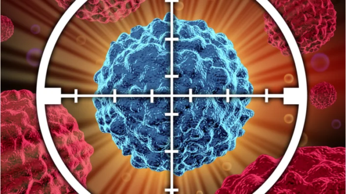
Senolytics 

Cellular senescence is one of the hallmarks of aging. A study in 14 patients with fibrosis showed improved physical benefits from dasatinib and quercetin in 6-minute walking distance, 4-minute gait speed and chair-stand time. However, they did not observe changes in pulmonary function, biomarkers, reported health, and frailty [22]. A study on 11 people with diabetes and kidney dysfunction showed that dasatinib and quercetin resulted in a significant decrease in senescent cell burden in adipose tissue along with senescence markers [23].

A recent trial of senolytics for knee osteoarthritis failed phase 2 trials. While the full results of these trials have not yet been published, there are many questions surrounding the trial design. The authors of this mini-review conclude this section by expressing the need for additional clinical trials involving senescence-reducing therapeutics.

Clinical trials that target mTOR

Promising data has been demonstrated that inhibiting mTOR extends lifespan in animals [24]. However, these results have not yet been reproduced in human trials that use rapamycin to inhibit mTOR [25,26]. Additionally, Dr. Mannick’s human research showed decreased infection rates and upregulated immune function in older adults [26]. A recent article shows how this work has led to industry collaboration on mTOR inhibition by mTOR analogs.

Rapamycin applied as a cream applied showed reduced skin senescence after 8 months of use [27]. Current proposed strategies to treat declining mitophagy include NAD+ supplements, activation of AMPK and/or SIRT1, and mTOR inhibition [28,29]. Urolithin A has been shown to improve in mitochondrial and muscle health [30], and we recently summarized a study showing that urolithin A affects mitochondrial and muscle in older adults. An animal study suggests that the mechanism involves mTOR inhibition [31].

Exercise and eating pattern 

In long-term cross-country skiers, endurance training was associated with reduced systemic and muscle inflammation and improved telomeres [32,33]. A study done in 34 cyclist men at a variety of ages suggests that endurance training reduces inflammation, while senescence increases with age and may be unaffected by endurance training [34].

A different study of sleep, a plant-based diet, and exercise for 8 weeks showed a reduction of DNA methylation age by 3.32 years in 43 middle-aged and older males [35].  In a similar 2-year study with 219 females, DNA methylation was significantly decreased with a plant-based diet and exercise intervention [36]. Additionally, the authors of this review discuss how prior human data suggests that a Mediterranean diet reduces cardiovascular risk [37] by improving immune function and changing gut microbiome composition [38,39].

Conclusion

Beyond the scope of this review, the authors encouraged readers to become informed about other emerging inventions, such as senescence immunotherapy, stem cell reprogramming, nutraceutical inventions, and microbiome alterations.

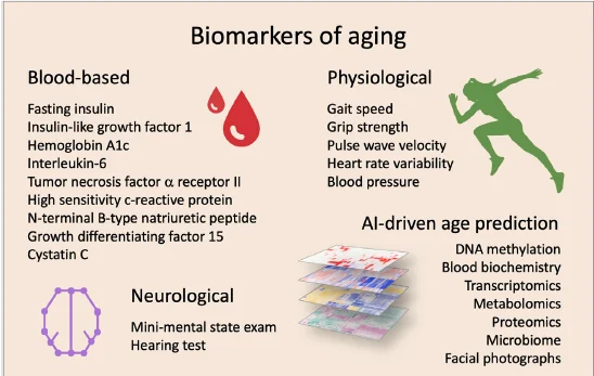
In their concluding paragraph, the authors discuss the need to carefully select biomarkers in clinical trials that target aging in humans. They further go on to explain how AI can help with age and health predictions. In the meantime, the authors include this figure of proposed biomarkers to target and/or monitor in clinical trials targeting aging.

Clinical trials targeting aging
We would like to ask you a small favor. We are a non-profit foundation, and unlike some other organizations, we have no shareholders and no products to sell you. All our news and educational content is free for everyone to read, but it does mean that we rely on the help of people like you. Every contribution, no matter if it’s big or small, supports independent journalism and sustains our future.

Literature

[1] Nielsen, J. L., Bakula, D., & Scheibye-Knudsen, M. (2022). Clinical trials targeting aging. Frontiers in Aging, 3. https://doi.org/10.3389/fragi.2022.820215

[2] Velthuis-te Wierik, E. J., Hoogzaad, L. V., van den Berg, H., & Schaafsma, G. (1994). Effects of moderate energy restriction on physical performance and substrate utilization in non-obese men. International journal of sports medicine, 15(8), 478–484. https://doi.org/10.1055/s-2007-1021091

[3] Loft, S., Velthuis-te Wierik, E. J., van den Berg, H., & Poulsen, H. E. (1995). Energy restriction and oxidative DNA damage in humans. Cancer epidemiology, biomarkers & prevention : a publication of the American Association for Cancer Research, cosponsored by the American Society of Preventive Oncology, 4(5), 515–519.

[4] Weiss, E. P., Racette, S. B., Villareal, D. T., Fontana, L., Steger-May, K., Schechtman, K. B., Klein, S., Holloszy, J. O., & Washington University School of Medicine CALERIE Group (2006). Improvements in glucose tolerance and insulin action induced by increasing energy expenditure or decreasing energy intake: a randomized controlled trial. The American journal of clinical nutrition, 84(5), 1033–1042. https://doi.org/10.1093/ajcn/84.5.1033

[5] Lefevre, M., Redman, L. M., Heilbronn, L. K., Smith, J. V., Martin, C. K., Rood, J. C., Greenway, F. L., Williamson, D. A., Smith, S. R., Ravussin, E., & Pennington CALERIE team (2009). Caloric restriction alone and with exercise improves CVD risk in healthy non-obese individuals. Atherosclerosis, 203(1), 206–213. https://doi.org/10.1016/j.atherosclerosis.2008.05.036

[6] Kraus, W. E., Bhapkar, M., Huffman, K. M., Pieper, C. F., Krupa Das, S., Redman, L. M., Villareal, D. T., Rochon, J., Roberts, S. B., Ravussin, E., Holloszy, J. O., Fontana, L., & CALERIE Investigators (2019). 2 years of calorie restriction and cardiometabolic risk (CALERIE): exploratory outcomes of a multicentre, phase 2, randomised controlled trial. The lancet. Diabetes & endocrinology, 7(9), 673–683. https://doi.org/10.1016/S2213-8587(19)30151-2

[7] Larson-Meyer, D. E., Newcomer, B. R., Heilbronn, L. K., Volaufova, J., Smith, S. R., Alfonso, A. J., Lefevre, M., Rood, J. C., Williamson, D. A., Ravussin, E., & Pennington CALERIE Team (2008). Effect of 6-month calorie restriction and exercise on serum and liver lipids and markers of liver function. Obesity (Silver Spring, Md.), 16(6), 1355–1362. https://doi.org/10.1038/oby.2008.201

[8] Civitarese, A. E., Carling, S., Heilbronn, L. K., Hulver, M. H., Ukropcova, B., Deutsch, W. A., Smith, S. R., Ravussin, E., & CALERIE Pennington Team (2007). Calorie restriction increases muscle mitochondrial biogenesis in healthy humans. PLoS medicine, 4(3), e76. https://doi.org/10.1371/journal.pmed.0040076

[9] Heilbronn, L. K., de Jonge, L., Frisard, M. I., DeLany, J. P., Larson-Meyer, D. E., Rood, J., Nguyen, T., Martin, C. K., Volaufova, J., Most, M. M., Greenway, F. L., Smith, S. R., Deutsch, W. A., Williamson, D. A., Ravussin, E., & Pennington CALERIE Team (2006). Effect of 6-month calorie restriction on biomarkers of longevity, metabolic adaptation, and oxidative stress in overweight individuals: a randomized controlled trial. JAMA, 295(13), 1539–1548. https://doi.org/10.1001/jama.295.13.1539

[10] Meydani, M., Das, S., Band, M., Epstein, S., & Roberts, S. (2011). The effect of caloric restriction and glycemic load on measures of oxidative stress and antioxidants in humans: results from the CALERIE Trial of Human Caloric Restriction. The journal of nutrition, health & aging, 15(6), 456–460. https://doi.org/10.1007/s12603-011-0002-z

[11] Waziry, R., Corcoran, D. L., Huffman, K. M., Kobor, M. S., Kothari, M., Kraus, V. B., Kraus, W. E., Lin, D. T. S., Pieper, C. F., Ramaker, M. E., Bhapkar, M., Das, S. K., Ferrucci, L., Hastings, W. J., Kebbe, M., Parker, D. C., Racette, S. B., Shalev, I., Schilling, B., & Belsky, D. W. (2021). Effect of long-term caloric restriction on DNA methylation measures of biological aging in healthy adults: CALERIE™ Trial Analysis. https://doi.org/10.1101/2021.09.21.21263912

[12] Zhu, X. H., Lu, M., Lee, B. Y., Ugurbil, K., & Chen, W. (2015). In vivo NAD assay reveals the intracellular NAD contents and redox state in healthy human brain and their age dependences. Proceedings of the National Academy of Sciences of the United States of America, 112(9), 2876–2881. https://doi.org/10.1073/pnas.1417921112

[13] Petr, M. A., Tulika, T., Carmona-Marin, L. M., & Scheibye-Knudsen, M. (2020). Protecting the aging genome. Trends in Cell Biology, 30(2), 117–132. https://doi.org/10.1016/j.tcb.2019.12.001

[14] Zhang, H., Ryu, D., Wu, Y., Gariani, K., Wang, X., Luan, P., D’Amico, D., Ropelle, E. R., Lutolf, M. P., Aebersold, R., Schoonjans, K., Menzies, K. J., & Auwerx, J. (2016). NAD? repletion improves mitochondrial and stem cell function and enhances life span in mice. Science (New York, N.Y.), 352(6292), 1436–1443. https://doi.org/10.1126/science.aaf2693

[15] Gomes, A. P., Price, N. L., Ling, A. J., Moslehi, J. J., Montgomery, M. K., Rajman, L., White, J. P., Teodoro, J. S., Wrann, C. D., Hubbard, B. P., Mercken, E. M., Palmeira, C. M., de Cabo, R., Rolo, A. P., Turner, N., Bell, E. L., & Sinclair, D. A. (2013). Declining NAD(+) induces a pseudohypoxic state disrupting nuclear-mitochondrial communication during aging. Cell, 155(7), 1624–1638. https://doi.org/10.1016/j.cell.2013.11.037

[16] Dellinger, R. W., Santos, S. R., Morris, M., Evans, M., Alminana, D., Guarente, L., & Marcotulli, E. (2017). Repeat dose NRPT (nicotinamide riboside and pterostilbene) increases NAD+ levels in humans safely and sustainably: a randomized, double-blind, placebo-controlled study. NPJ aging and mechanisms of disease, 3, 17. https://doi.org/10.1038/s41514-017-0016-9

[17] Jensen, J. B., Dollerup, O. L., Møller, A. B., Billeskov, T. B., Dalbram, E., Chubanava, S., Dellinger, R. W., Trost, K., Moritz, T., Ringgaard, S., Møller, N., Treebak, J. T., Farup, J., & Jessen, N. (2021). A randomized placebo-controlled clinical trial of nicotinamide riboside and Pterostilbene supplementation in experimental muscle injury in elderly subjects. https://doi.org/10.1101/2021.10.04.21264504

[18] Martens, C. R., Denman, B. A., Mazzo, M. R., Armstrong, M. L., Reisdorph, N., McQueen, M. B., Chonchol, M., & Seals, D. R. (2018). Chronic nicotinamide riboside supplementation is well-tolerated and elevates NAD+ in healthy middle-aged and older adults. Nature communications, 9(1), 1286. https://doi.org/10.1038/s41467-018-03421-7

[19] Elhassan, Y. S., Kluckova, K., Fletcher, R. S., Schmidt, M. S., Garten, A., Doig, C. L., Cartwright, D. M., Oakey, L., Burley, C. V., Jenkinson, N., Wilson, M., Lucas, S., Akerman, I., Seabright, A., Lai, Y. C., Tennant, D. A., Nightingale, P., Wallis, G. A., Manolopoulos, K. N., Brenner, C., … Lavery, G. G. (2019). Nicotinamide Riboside Augments the Aged Human Skeletal Muscle NAD+ Metabolome and Induces Transcriptomic and Anti-inflammatory Signatures. Cell reports, 28(7), 1717–1728.e6. https://doi.org/10.1016/j.celrep.2019.07.043

[20] Dollerup, O. L., Christensen, B., Svart, M., Schmidt, M. S., Sulek, K., Ringgaard, S., Stødkilde-Jørgensen, H., Møller, N., Brenner, C., Treebak, J. T., & Jessen, N. (2018). A randomized placebo-controlled clinical trial of nicotinamide riboside in obese men: safety, insulin-sensitivity, and lipid-mobilizing effects. The American journal of clinical nutrition, 108(2), 343–353. https://doi.org/10.1093/ajcn/nqy132

[21] Dollerup, O. L., Chubanava, S., Agerholm, M., Søndergård, S. D., Altintas, A., Møller, A. B., Høyer, K. F., Ringgaard, S., Stødkilde-Jørgensen, H., Lavery, G. G., Barrès, R., Larsen, S., Prats, C., Jessen, N., & Treebak, J. T. (2020). Nicotinamide riboside does not alter mitochondrial respiration, content or morphology in skeletal muscle from obese and insulin-resistant men. The Journal of physiology, 598(4), 731–754. https://doi.org/10.1113/JP278752

[22] Justice, J. N., Nambiar, A. M., Tchkonia, T., LeBrasseur, N. K., Pascual, R., Hashmi, S. K., Prata, L., Masternak, M. M., Kritchevsky, S. B., Musi, N., & Kirkland, J. L. (2019). Senolytics in idiopathic pulmonary fibrosis: Results from a first-in-human, open-label, pilot study. EBioMedicine, 40, 554–563. https://doi.org/10.1016/j.ebiom.2018.12.052

[23] Hickson, L. J., Langhi Prata, L., Bobart, S. A., Evans, T. K., Giorgadze, N., Hashmi, S. K., Herrmann, S. M., Jensen, M. D., Jia, Q., Jordan, K. L., Kellogg, T. A., Khosla, S., Koerber, D. M., Lagnado, A. B., Lawson, D. K., LeBrasseur, N. K., Lerman, L. O., McDonald, K. M., McKenzie, T. J., Passos, J. F., … Kirkland, J. L. (2019). Senolytics decrease senescent cells in humans: Preliminary report from a clinical trial of Dasatinib plus Quercetin in individuals with diabetic kidney disease. EBioMedicine, 47, 446–456. https://doi.org/10.1016/j.ebiom.2019.08.069

[24] ??Harrison, D. E., Strong, R., Sharp, Z. D., Nelson, J. F., Astle, C. M., Flurkey, K., Nadon, N. L., Wilkinson, J. E., Frenkel, K., Carter, C. S., Pahor, M., Javors, M. A., Fernandez, E., & Miller, R. A. (2009). Rapamycin fed late in life extends lifespan in genetically heterogeneous mice. Nature, 460(7253), 392–395. https://doi.org/10.1038/nature08221

[25] Kraig, E., Linehan, L. A., Liang, H., Romo, T. Q., Liu, Q., Wu, Y., Benavides, A. D., Curiel, T. J., Javors, M. A., Musi, N., Chiodo, L., Koek, W., Gelfond, J., & Kellogg, D. L., Jr (2018). A randomized control trial to establish the feasibility and safety of rapamycin treatment in an older human cohort: Immunological, physical performance, and cognitive effects. Experimental gerontology, 105, 53–69. https://doi.org/10.1016/j.exger.2017.12.026

[26] Mannick, J. B., Morris, M., Hockey, H. P., Roma, G., Beibel, M., Kulmatycki, K., Watkins, M., Shavlakadze, T., Zhou, W., Quinn, D., Glass, D. J., & Klickstein, L. B. (2018). TORC1 inhibition enhances immune function and reduces infections in the elderly. Science translational medicine, 10(449), eaaq1564. https://doi.org/10.1126/scitranslmed.aaq1564

[27] Chung, C. L., Lawrence, I., Hoffman, M., Elgindi, D., Nadhan, K., Potnis, M., Jin, A., Sershon, C., Binnebose, R., Lorenzini, A., & Sell, C. (2019). Topical rapamycin reduces markers of senescence and aging in human skin: an exploratory, prospective, randomized trial. GeroScience, 41(6), 861–869. https://doi.org/10.1007/s11357-019-00113-y

[28] Fang, E. F., Scheibye-Knudsen, M., Brace, L. E., Kassahun, H., SenGupta, T., Nilsen, H., Mitchell, J. R., Croteau, D. L., & Bohr, V. A. (2014). Defective mitophagy in XPA via PARP-1 hyperactivation and NAD(+)/SIRT1 reduction. Cell, 157(4), 882–896. https://doi.org/10.1016/j.cell.2014.03.026

[29] Scheibye-Knudsen, M., Mitchell, S. J., Fang, E. F., Iyama, T., Ward, T., Wang, J., Dunn, C. A., Singh, N., Veith, S., Hasan-Olive, M. M., Mangerich, A., Wilson, M. A., Mattson, M. P., Bergersen, L. H., Cogger, V. C., Warren, A., Le Couteur, D. G., Moaddel, R., Wilson, D. M., 3rd, Croteau, D. L., … Bohr, V. A. (2014). A high-fat diet and NAD(+) activate Sirt1 to rescue premature aging in cockayne syndrome. Cell metabolism, 20(5), 840–855. https://doi.org/10.1016/j.cmet.2014.10.005

[30] Andreux, P. A., Blanco-Bose, W., Ryu, D., Burdet, F., Ibberson, M., Aebischer, P., Auwerx, J., Singh, A., & Rinsch, C. (2019). The mitophagy activator urolithin A is safe and induces a molecular signature of improved mitochondrial and cellular health in humans. Nature metabolism, 1(6), 595–603. https://doi.org/10.1038/s42255-019-0073-4

[31] Ryu, D., Mouchiroud, L., Andreux, P. A., Katsyuba, E., Moullan, N., Nicolet-Dit-Félix, A. A., Williams, E. G., Jha, P., Lo Sasso, G., Huzard, D., Aebischer, P., Sandi, C., Rinsch, C., & Auwerx, J. (2016). Urolithin A induces mitophagy and prolongs lifespan in C. elegans and increases muscle function in rodents. Nature medicine, 22(8), 879–888. https://doi.org/10.1038/nm.4132

[32] Beavers, K. M., Brinkley, T. E., & Nicklas, B. J. (2010). Effect of exercise training on chronic inflammation. Clinica chimica acta; international journal of clinical chemistry, 411(11-12), 785–793. https://doi.org/10.1016/j.cca.2010.02.069

[33] Østhus, I. B., Sgura, A., Berardinelli, F., Alsnes, I. V., Brønstad, E., Rehn, T., Støbakk, P. K., Hatle, H., Wisløff, U., & Nauman, J. (2012). Telomere length and long-term endurance exercise: does exercise training affect biological age? A pilot study. PloS one, 7(12), e52769. https://doi.org/10.1371/journal.pone.0052769

[34] Balan, E., De Groote, E., Bouillon, M., Viceconte, N., Mahieu, M., Naslain, D., Nielens, H., Decottignies, A., & Deldicque, L. (2020). No effect of the endurance training status on senescence despite reduced inflammation in skeletal muscle of older individuals. American journal of physiology. Endocrinology and metabolism, 319(2), E447–E454. https://doi.org/10.1152/ajpendo.00149.2020

[35] Fitzgerald, K. N., Hodges, R., Hanes, D., Stack, E., Cheishvili, D., Szyf, M., Henkel, J., Twedt, M. W., Giannopoulou, D., Herdell, J., Logan, S., & Bradley, R. (2021). Potential reversal of epigenetic age using a diet and lifestyle intervention: a pilot randomized clinical trial. Aging, 13(7), 9419–9432. https://doi.org/10.18632/aging.202913

[36] Fiorito, G., Caini, S., Palli, D., Bendinelli, B., Saieva, C., Ermini, I., Valentini, V., Assedi, M., Rizzolo, P., Ambrogetti, D., Ottini, L., & Masala, G. (2021). DNA methylation-based biomarkers of aging were slowed down in a two-year diet and physical activity intervention trial: the DAMA study. Aging cell, 20(10), e13439. https://doi.org/10.1111/acel.13439

[37] Estruch, R., Ros, E., Salas-Salvadó, J., Covas, M. I., Corella, D., Arós, F., Gómez-Gracia, E., Ruiz-Gutiérrez, V., Fiol, M., Lapetra, J., Lamuela-Raventos, R. M., Serra-Majem, L., Pintó, X., Basora, J., Muñoz, M. A., Sorlí, J. V., Martínez, J. A., Fitó, M., Gea, A., Hernán, M. A., … PREDIMED Study Investigators (2018). Primary Prevention of Cardiovascular Disease with a Mediterranean Diet Supplemented with Extra-Virgin Olive Oil or Nuts. The New England journal of medicine, 378(25), e34. https://doi.org/10.1056/NEJMoa1800389

[38] Maijo, M., Ivory, K., Clements, S. J., Dainty, J. R., Jennings, A., Gillings, R., Fairweather-Tait, S., Gulisano, M., Santoro, A., Franceschi, C., Carding, S. R., & Nicoletti, C. (2018). One-Year Consumption of a Mediterranean-Like Dietary Pattern With Vitamin D3 Supplements Induced Small Scale but Extensive Changes of Immune Cell Phenotype, Co-receptor Expression and Innate Immune Responses in Healthy Elderly Subjects: Results From the United Kingdom Arm of the NU-AGE Trial. Frontiers in physiology, 9, 997. https://doi.org/10.3389/fphys.2018.00997

[39] Ghosh, T. S., Rampelli, S., Jeffery, I. B., Santoro, A., Neto, M., Capri, M., Giampieri, E., Jennings, A., Candela, M., Turroni, S., Zoetendal, E. G., Hermes, G., Elodie, C., Meunier, N., Brugere, C. M., Pujos-Guillot, E., Berendsen, A. M., De Groot, L., Feskins, E., Kaluza, J., … O’Toole, P. W. (2020). Mediterranean diet intervention alters the gut microbiome in older people reducing frailty and improving health status: the NU-AGE 1-year dietary intervention across five European countries. Gut, 69(7), 1218–1228. https://doi.org/10.1136/gutjnl-2019-319654

Dina Radenkovic

Dina Radenkovic on the Root Cause of Gender Inequality

Our interviewee today holds that female reproductive aging, or ovarian aging, is accelerated compared to other organs and that it has a deep impact both on the lives and health of women and on our society as a whole. Nevertheless, ovarian aging continues to be underresearched, with only a handful of companies working in the field. Dina Radenkovic, a medical doctor, bioinformatics researcher, and health entrepreneur, heads one such company: Gameto, a startup that recently made waves after securing 23 million dollars in a round of funding.

Could you give us an overview of ovarian aging? What is it, what does it do to the female body, and how exactly can we measure it, considering that we think of it as premature?

The way we talk about it is in terms of function, not just cellular pathways. The reason our company is called Gameto is that we want to change the game of gametogenesis. Spermatogenesis is male gametogenesis, and it occurs throughout the lifetime, whereas female gametogenesis occurs while the woman is still an embryo. However, when women need to start using their ovaries, in the current social environment, this mostly happens relatively late, after they get an education, advance a career, and so on.

Yet, in standard gynecology, ovaries are often characterized as geriatric starting from the woman’s mid-thirties. Women experience a steep decline in the function of this organ. So, we say it’s an aged organ simply because its function is declining. That’s why we have the need for IVF, which only started about 40 years ago, and now it’s one of the largest out-of-pocket industries, and the demand is ever-growing because we’re pushing it to the edge in terms of age.

Organ dysfunction, or organ failure, is one way to characterize aging. In this paradigm, the first milestone is the decline in fertility, and the second is menopause. This is when you lose the function of your ovary and the production of the hormones that control a lot of your behavior and physiology. Menopause is so functionally important in the context of aging that the age of menopause onset is actually associated with life expectancy.

There have been studies that show that if you postpone the age of menopause by one year, you can increase life expectancy by 2-2.6 years. Interestingly, prior to menopause, women are more protected from many health conditions than men – for instance, from coronary heart disease. But after menopause, they start experiencing all those diseases of aging.

It’s like the stereotypical frail old lady with the cane, right? Your bones become weaker. You have increased risk of dementia, heart disease, and many other conditions, and you develop this syndrome of frailty. If you look at the diseases of aging in women, they all start with menopause.

This is what ovarian aging is: you’re 35 and your skin and brain are in perfect order, but your ovaries are already declining in function and that’s something that’s not happening to your male counterparts. Yes, there is this concept of andropause, but it happens relatively in synchrony with the function of other organs.

What we at Gameto are saying is that if this loss of function is the root cause of both infertility and all those diseases that occur with menopause (and there’s also early menopause, this extremely problematic condition called primary ovarian failure, when your ovaries stop working in your twenties and thirties), let’s develop therapeutics that will solve this, that will rescue this function.

To summarize: if you look at ovarian aging, be it in terms of follicular count or the quality of the eggs, ovaries share many aging pathways with other organs, but the function of the ovaries declines faster, and that’s why we say that the ovaries age faster than the rest of your body.

Do we have any idea why ovarian aging is so different?

I wouldn’t say it’s necessarily different. The reason why a lot of the interventions for aging work in the ovaries is because the same pathways are involved, but the actual answer to your question is that we don’t know. For instance, why does the thymus age faster? The truth is we have no idea. If you want me to speculate, using the antagonistic pleiotropy theory of aging or the hyperfunction theory, you could argue that ovaries are hyper-stimulated. We lose many follicles to get the one good mature follicle per month.

It’s not optimized to preserve function, it’s hyper-stimulated to ensure that one good egg is mature, and this one good egg will control all your cycle and physiology. This hyperfunction in the early stages is probably optimal for reproduction, but as the hyperfunction theory suggests, later on, hyperfunction leads to dysfunction.

The reason why it hasn’t been “fixed” by evolution is because evolutionary forces mostly work during the reproductive period. But the society has changed! Today, women can expect to live to 90 or 100, if they don’t smoke and adopt a healthy lifestyle. So, they spend two thirds of their lives infertile, unable to have children, and also half of their life in this state of post-menopausal poor health. And that’s what we are trying to solve.

Because of accelerated ovarian aging, women in the modern society have to accept compromises they may not wish to make, like should I take this job, or should I have a baby? Suppose you want to do both, but biologically, you have to make a choice, and that’s what we hope to change.

How does this accelerated reproductive aging drive gender equality? What would be the societal implications of extending reproductive healthspan in women? 

I would call it the root cause of gender inequality. I would love to see a future where a young couple in their twenties can pursue their careers, travel the world, do all those things they want to do, and then start a family in their forties. Today, most women are unable to do that, so men have the advantage. Leveling the field would make the game fairer, would empower women and allow them more choice.

Childbearing and childcare are also expensive, and not everyone can afford it when they’re young. So, we don’t want to tell people what to do with their lives and when to have children, we just want to give them more choice, that’s our company’s mission.

This year in the United States, we have the lowest fertility rates on record, and that’s not necessarily good because a lot of women are postponing childbirth or deciding not to have children at all because of various difficulties. We want to ensure they can have children when they feel ready, mature, when they feel that they found the right partner or that they’ve done what they wanted in terms of career. To me, this is very important. I think this would enable more women to participate in the workforce, to get education, to contribute to society, which would be good for society as a whole. I would like to have it myself as a woman, and my sister to have that option. I would have wanted my grandma to have that option.

Female aging is known to be underresearched. Is this a part of the broader gender bias in medicine?

I think things are moving slowly in the right direction, but we need to do more. We must have more women in high-profile positions. As there are more women, and as they climb up the corporate ladder, they will talk more about this issue. Before we had representation of women in those positions, these issues were a taboo. Women create the market; women talk about women’s problems. It might be hard to understand women’s issues if you’re not experiencing them yourself. You might not necessarily have bad intentions; people can be simply unaware of problems that are not theirs. That’s why I support diversity and inclusion.

It is also very important to have serious biotech companies in this space to show that this can be backed by real science, that proper therapeutics can be invented, that it can be profitable for investors.

So, is this gender bias still around?

I think it is. For instance, because I’m a woman, people would always think I’m a nurse, not a doctor, and so on. Yes, it is still out there, in some areas more than others. It’s changing slowly, but we need to keep pushing for it.

We need to encourage more people to work in this space, both on the biotech side and in service delivery. We’re talking about half the population that are experiencing these problems, and it’s horrible that we haven’t done anything about this.

For instance, every decade, we add two years to life expectancy, but we haven’t pushed back the onset of female reproductive aging an inch since we started keeping medical records in the 1800s. As long as we keep living longer without changing this, we’re just worsening the gender inequality. This is so unfair! When I started working in the aging field, I thought that this is a problem I must solve.

Looks like ovarian aging today is perceived like organismal aging was perceived until recently – something that we just thought was normal.

Yes, exactly.

Premenopausal hormone levels seem to be protective for women. On the other hand, there are estrogen-related cancers, and hormone replacement therapies have been linked to increased risk of cancer as well. How sure are we that extending this premenopausal hormonal landscape would do more good than bad?

It’s not like we’re building a drug for everyone, and everyone should start taking it. This is not what we’re doing. We’re developing biologics that are going to solve some of the problems like infertility or diseases that occur after menopause. Hopefully, they will get approved and licensed for specific indications, like infertility, primary ovarian failure, and physicians would prescribe them based on the clinical need.

As to the studies, there was this study where the median age of patients was 63, I think, and they found a very marginal increase in the risk of breast cancer. On the other hand, the women also became more fit, they had improvements in some other conditions.

The answer is that it will obviously require a conversation between a patient and their physician, an assessment of what’s best for the patient, for how many years they need to take it, when to stop and so on, based on the clinical picture. It’s very hard to generalize, but I think overall, if we could provide personalized dose delivery and to slow some of the pathological processes that occur with menopause, we could find a way to get the best of both worlds – that is, to prevent diseases without necessarily causing the increased risk of cancer.

You could argue that if you push the age of menopause by continuing hormone exposure, what will happen will be similar to men and prostate cancer. Prostate continues to grow because men don’t go through a quick andropause. They don’t lose 80% of their hormones within one year, so their prostate continues to be constantly exposed to androgens. That’s why there is a saying that most men will die with prostate cancer, but not of prostate cancer. You could argue that a similar picture might occur with women – they won’t die of it because they will be biologically younger, and fitter. But yes, we could see increased incidents of some cancers.

I think, the regulators and the clinicians have the right to be cautious and to give those therapies to women who need them and for the amount of time they need them, because yes, of course, with every therapy you have the upside and the downside, and it’s about the overall benefit.

Let’s talk about Gameto. You haven’t released a lot of details about what you’re going to do and what’s the science behind your strategy. Could you walk us through this? For instance, cellular reprogramming is a vast topic, so what exactly are you going to do with it?

I think we’ve disclosed everything that we are working on right now. We’re a very early-stage company, but we use cellular reprogramming to make ovarian cells and ovaroids. And then we use ovaroids to make biologics for infertility in ovarian diseases. We’ve published a paper and we’ll publish another one, hopefully, this year, once the patents are converted, and we want to keep publishing everything as soon as we are legally able to, because we’re pushing the boundaries of an interesting area of science.

In terms of the biologics, we have two lines that we’re working on. The first one is fertility. We’re testing a biologic for IVF, so that instead of giving stimulation with injections before we extract the eggs, we’d be able to make it a full in vitro experience, making the cell extraction shorter, safer, more effective. That would allow women to undergo IVF for a little bit longer, as long as they’re pre-menopausal. My co-founder and chairman is Martine Ruszkowski, he’s a global leader in the fertility space, and we collaborate with his chain of fertility clinics, the largest in the United States.

Later down the line, we’re working on a biologic for primary ovarian failure, which is early menopause, and that could also help other women with menopause. Right now, we have a sponsored research agreement with George Church’s lab at Harvard. A lot of the basic science in cellular reprogramming comes from Harvard, and then the more clinical work is done within the company. We have more things to tell, and as soon as we are legally allowed to disclose, we surely will.

Are you aware of any competition? I think just a handful of companies are currently working in this space. 

I wouldn’t call that competition. I would like to have more people working in this area. There are so many problems: we have endometriosis, PCOS (polycystic ovarian syndrome). For endometriosis, there’s no therapy. For PCOS, we have seen some use of metformin, but it’s not even licensed for this. In menopause, there’s not much innovation as well. Then, we have IVF. There’s just so much to do, and only a handful of companies, so I would love to have more people in, more talent, both on the science side, and in the industry. This is something that’s affecting half of the population, and we only have three or four companies working in this field. We can’t do it all by ourselves.

Also, in order to grow, you need an ecosystem, a talent pool. Now, it’s easy because we’re a small company. But if we, hopefully, grow and become a big company developing multiple biologics, we will need to hire scientists who are interested in this space and educated in it. Unless there’s relevant funding for academia, where are we going to find them?

I see everybody working in this space as my partners, my collaborators, because we’re trying to establish ourselves as a credible field and to attract top talent, and today, it’s like a nuclear war for talent. It’s really hard to recruit people. I think we’re all helping each other, and I would love to see more companies entering this space.

What are your thoughts about the situation in the longevity field in general? Are you focused solely on female reproductive aging, or are you, let’s say, a longevity enthusiast?

In the company, we’re focused on female reproductive aging, but I have spent a lot of time in the aging field, and I am passionate about it. I think what has happened over the last few years has been mind-blowing. We’ve seen huge organizations taking shape. In the past, if you said you worked in the aging field, people would not understand you. What is longevity medicine? Today, it’s becoming a respectable, interesting, cutting-edge area.

I think this is extremely important, and I’m so happy for the field, but as a company, we will not be able to do everything. So, I want us to stay focused on the diseases caused by ovarian aging. Still, I’m obviously extremely passionate about everything aging-related.

I love to follow all the other aging companies, to read about their work, I cheer for them. And I think we can make an important contribution to the field, because a lot of what we’ve learned about ovarian aging could be translated and shared.

Various lifestyle interventions might currently be our best bet to slow aging. Do you think any of these, such as caloric restriction or exercise, can work against ovarian aging?

Being generally healthy, having good metabolic health, maintaining healthy weight and low levels of inflammation should be just as good for ovaries as for other organs, but sadly, I don’t think these interventions are sufficient to have a marked effect on this accelerated type of aging that results in the loss of ovarian function.

We would like to ask you a small favor. We are a non-profit foundation, and unlike some other organizations, we have no shareholders and no products to sell you. All our news and educational content is free for everyone to read, but it does mean that we rely on the help of people like you. Every contribution, no matter if it’s big or small, supports independent journalism and sustains our future.
Cell targeting

Using Gene Expression to Target Senescent Cells

An open access study published in iScience has discussed the development of senolytics to target particular genes that are upregulated in cellular senescence.

Understanding senescence, cell by cell

In our earlier interview with Simon Melov of the Buck Institute, the lead researcher of this paper, Dr. Melov, explained his lab’s focus on single-cell sequencing. As this paper points out, senescent cells represent only a small fraction of tissue, so bulk analysis is difficult and inefficient.

Instead, the researchers used single-cell RNA sequencing to analyze two cellular types that regenerate muscle tissue: fibro-adipogenic progenitors (FAPs) and muscle stem cells. They performed this sequencing technique on thousands of these cells, comparing gene expression between mice that had received the senolytic drug ABT-263 and mice that had not. Both sets of mice had received doxocirubin, a drug that induces cellular senescence.

The researchers then did further analysis to determine which cells were senescent and what genes they were expressing. The proteins p21, encoded by Cdkn1A, and p16, encoded by Cdkn2A, are widely known to be associated with cellular senescence. Genes that were upregulated with doxocirubin but downregulated with ABT-263 were also closely examined.

Most importantly, the researchers focused on genes that are known to cause apoptosis when silenced or inhibited, comparing that list to genes whose downregulation was spurred by ABT-263. Genes with both qualities, the researchers reasoned, would be critical targets in the development of a new senolytic.

Narrowing down the targets

The researchers found a total of ten targets, four in FAPs and six in stem cells. They found that two of these genes, Cryab and Hmox1, were significantly upregulated in both kinds of cells after administration of doxocirubin. The researchers focused their efforts on these two specific genes.

Interestingly, small molecules that target these genes were already known; they were just never tested as senolytics. While one of these small molecules, NCI-41356, killed both senescent and non-senescent cells, HC25 was found to be a powerful, preferential senolytic against CRYAB expression, and OB24 and QC-308 were found to have slight but significant preference for killing senescent cells.

Testing their new candidate

To determine if this approach had yielded something actually effective, the researchers tested HC25 against the current top dog that they used as their reference senolytic: ABT-263. They found that HC25 was equal to or better than ABT-263 against FAPs, muscle stem cells, and other muscle cells, human skeletal muscle myoblasts. They then went on to test HC25 and their other candidates against multiple other cells, finding that while OB24 and QC-308 were ineffective or had off-target effects, HC25 substantially and significantly caused senescent cells to die while leaving normal cells alone, whether that senescence was caused by doxocirubin or irradiation.

Finally, the researchers tested HC25 in mice. In young mice administered a regimen of doxocirubin, HC25 was able to restore key biomarkers of senescent cells, p21 and Cryab, either slightly above or even somewhat below their original states. In naturally aged mice, these biomarkers were restored to nearly their levels in young mice.

Conclusion

In many ways, this research was a demonstration of a technique more than the development of a senolytic. While it may be possible that HC25 will be an effective therapeutic, which requires substantially more testing to determine, this single-cell technique may find many more, possibly better, targets for the treatment of cellular senescence.

It may also be possible, and even likely, that a cocktail of drugs developed with this gene expression targeting technique will show substantially more effectiveness than individual drugs. If multiple genes that are upregulated only in cellular senescence can be targeted at once, the resulting combination therapy may ultimately lead to a treatment that removes the harmful cells from our bodies – and only those.

We would like to ask you a small favor. We are a non-profit foundation, and unlike some other organizations, we have no shareholders and no products to sell you. All our news and educational content is free for everyone to read, but it does mean that we rely on the help of people like you. Every contribution, no matter if it’s big or small, supports independent journalism and sustains our future.
Guinea pig

Tuberculosis Is Associated with Epigenetic Aging

Publishing in Aging, a team of researchers has explained the relationship between tuberculosis (TB) and multiple aspects of aging, including epigenetic alterations and the senescence-associated secretory phenotype (SASP).

A disease known to cause long-term harm

Tuberculosis, a disease that has proliferated around the world, kills 1.4 million people every year. Previous research has shown that, even if the disease is successfully treated, it decreases life expectancy by at least seven years [1]. With a combination of previous research and newly derived animal results, the researchers sought to provide clear evidence that this is the result of epigenetic alterations, one of the hallmarks of aging.

TB causes hypermethylation in guinea pigs and people

Guinea pigs are commonly associated with animal research, but their actual use is relatively rare; however, they are a common model of tuberculosis infection, as they experience the disease in much the same way as people [2]. The researchers had discovered previously that tuberculosis is associated with DNA hypermethylation in people [3], and this research shows that similar methylation occurs in guinea pigs.

People and guinea pigs both had hypermethylation of the immune system, including CD4, CD8, and CD14 cells. The pathways affected by this methylation included metabolism and signaling processes, such as the mTOR pathway, which is well-known to be associated with longevity in model organisms; calcium signaling; the MAPK pathway, which interprets intercellular signaling; and enzymes that affect chromosomes. Similar results were found in the lungs and spleens of guinea pigs.

The researchers confirmed these results through gene expression analysis, which also found that genes responsible for SASP secretion were upregulated, showing a link between TB and cellular senescence.

TB hypermethylation is associated with aging

In addition to further confirmation that tuberculosis is associated with the SASP, the researchers found that the epigenetic age of TB patients was an average of 12.7 years above their chronological age, according to the Horvath epigenetic clock, and this continued for at least six months after TB therapy had been completed. An RNA sequencing calculator reported an increase of 14.38 years. The researchers suggest that this epigenetic aging may be responsible for the increase in all-cause mortality associated with TB. However, more advanced epigenetic clocks, such as GrimAge, were not used in this analysis.

Conclusion

The researchers point out that while human studies could not prove causation, the guinea pig data makes it clear that TB leads to hypermethylation. However, they also note that TB is not the only disease associated with hypermethylation, epigenetic alterations may still lead to disease susceptibility, and confounding factors that cause hypermethylation, such as smoking, can also increase susceptibility to tuberculosis infection. Additionally, previous research has shown that TB is also associated with telomere attrition, another hallmark of aging [4].

In the wake of the COVID-19 pandemic, this relationship between aging and infectious disease deserves further understanding and investigation. Many of the issues associated with infection, such as systemic inflammation, are also associated with aging. Being able to directly counteract these effects at the cellular level is a plausible method of extending human lives that deserves further exploration.

We would like to ask you a small favor. We are a non-profit foundation, and unlike some other organizations, we have no shareholders and no products to sell you. All our news and educational content is free for everyone to read, but it does mean that we rely on the help of people like you. Every contribution, no matter if it’s big or small, supports independent journalism and sustains our future.

Literature

[1] Lee-Rodriguez, C., Wada, P. Y., Hung, Y. Y., & Skarbinski, J. (2020). Association of mortality and years of potential life lost with active tuberculosis in the United States. JAMA network open, 3(9), e2014481-e2014481.

[2] McMurray, D. N. (2001). Disease model: pulmonary tuberculosis. Trends in molecular medicine, 7(3), 135-137.

[3] DiNardo, A. R., Rajapakshe, K., Nishiguchi, T., Grimm, S. L., Mtetwa, G., Dlamini, Q., … & Mandalakas, A. M. (2020). DNA hypermethylation during tuberculosis dampens host immune responsiveness. The Journal of clinical investigation, 130(6), 3113-3123.

[4] Freimane, L., Barkane, L., Igumnova, V., Kivrane, A., Zole, E., & Ranka, R. (2021). Telomere length and mitochondrial DNA copy number in multidrug-resistant tuberculosis. Tuberculosis, 131, 102144.

Mouse genes

Long-Term Cellular Reprogramming Causes Rejuvenation in Mice

Scientists have shown that in vivo partial cellular reprogramming can be safe and effective in an animal model, although the rejuvenation was mostly limited to long-term treatments and two tissue types [1].

There and back again

Cellular reprogramming sounds like a technology from Star Trek, and it is undeniably futuristic. Our tissues mostly consist of various specialized cells that develop (differentiate) from pluripotent stem cells. Differentiated cells get old, but overexpressing certain regulatory genes makes them revert to their pluripotent state in a process that is accompanied by significant rejuvenation; this is cellular reprogramming. This novel technology can produce stem cells from a person’s somatic cells, which can be very useful against various diseases, but it is also being actively explored by geroscientists for its rejuvenation potential [2].

Obviously, we don’t want all our somatic cells to become stem cells: we need them to support our tissues. Reprogramming somatic cells all the way to their pluripotent state and back also carries a significant risk of carcinogenic mutations and teratomas, ghastly tumors consisting of many types of tissue [3]. This led to the rise of partial reprogramming, in which the reprogramming factors are expressed for just enough time to achieve rejuvenation without pushing the cell all the way to pluripotency.

Partial reprogramming has shown a lot of potential in vitro, but in vivo experiments are scarce, and scientists are still searching for the Goldilocks zone where cells can be rejuvenated without adverse effects. In this new study, the researchers describe a novel protocol for partial reprogramming in vivo that appears safe and at least somewhat effective.

Naturally aging mice

The scientists produced mice that overexpress the reprogramming factors when given the drug doxycycline, which is an established method of in vivo reprogramming. Unlike in some earlier studies, these mice aged naturally without any genetic engineering for accelerated aging. The researchers chose to use the original four OSKM reprogramming factors rather than an alternative version.

The mice were divided into three study groups and several control groups. Two of the study groups underwent long-term reprogramming starting at 12 and 15 months of age, respectively, and continuing to 22 months, which is quite old in mouse years. The third group underwent a much shorter reprogramming protocol, starting at 25 months and continuing for one month. The reprogramming phase consisted of continuous cycles of 2 days on and 5 days off doxycycline.

A mixed bag of results

All the protocols appeared to be safe: no teratomas were formed, and no pathological phenotypes were reported. Following the reprogramming, rejuvenation was assessed in several tissues. First, the researchers used epigenetic clocks that register age-related changes in DNA methylation to calculate biological age. The clocks revealed significant reduction of epigenetic age in the mice that underwent long-term reprogramming, but not in the short-term group. However, epigenetic rejuvenation was only observed in two tissues: the kidney and skin.

Some signs of rejuvenation did appear in the short-treatment group too, including altered gene expression with genes involved in hair growth cycle upregulated and inflammation-related genes downregulated. In both long-term groups, gene expression changes were even more pronounced, with genes involved in inflammation and cellular senescence downregulated. The proliferation rates of cells in the skin of all treated mice were increased compared to the controls, and in the long-term groups, a significant decrease in fibrosis during wound healing was detected.

The researchers also performed a broad analysis of the mice’s blood serum, looking for any changes compared to controls. Here, again, changes in the short-term group were minuscule while changes in the long-term groups were more pronounced. Some metabolites that are elevated in progeroid mice were decreased in those groups, causing the researchers to conclude that some overall rejuvenation on the physiological level did occur.

However, the researchers did not report any changes in average or maximum longevity in the treated mice compared to controls. Moreover, even the long-term treatment did not cause any discernible increase in fitness.

Conclusion

This pioneering study of partial cellular reprogramming in vivo brings mixed results. On one hand, the treatment protocols that were used appear to be safe, and at least some rejuvenation of transcriptional, metabolic, and functional levels was observed. On the other hand, rejuvenation only occurred in two tissues, and only as a result of the long-term treatment, which raises questions about the limits of partial reprogramming. This should not discourage us, though, as in vivo partial reprogramming is still in its infancy, and we can expect things to keep improving.

We would like to ask you a small favor. We are a non-profit foundation, and unlike some other organizations, we have no shareholders and no products to sell you. All our news and educational content is free for everyone to read, but it does mean that we rely on the help of people like you. Every contribution, no matter if it’s big or small, supports independent journalism and sustains our future.

Literature

[1] Browder, K. C., Reddy, P., Yamamoto, M., Haghani, A., Guillen, I. G., Sahu, S., … & Izpisua Belmonte, J. C. (2022). In vivo partial reprogramming alters age-associated molecular changes during physiological aging in mice. Nature Aging, 1-11.

[2] Gill, D., Parry, A., Santos, F., Hernando-Herraez, I., Stubbs, T. M., Milagre, I., & Reik, W. (2021). Multi-omic rejuvenation of human cells by maturation phase transient reprogramming. bioRxiv.

[3] Wuputra, K., Ku, C. C., Wu, D. C., Lin, Y. C., Saito, S., & Yokoyama, K. K. (2020). Prevention of tumor risk associated with the reprogramming of human pluripotent stem cells. Journal of Experimental & Clinical Cancer Research, 39(1), 1-24.

Meat on plate

Meat Consumption Increases Risk of Some Types of Cancer

In a new population study, scientists have found more evidence that consuming a lot of meat might not be a good idea, though the association between meat and cancer depends on multiple factors [1].

Diet and health

Given that lifestyle changes may be the only anti-aging intervention available today, it is important to elucidate the effects that diets have on health. Unfortunately, this is made harder by the variability of dietary habits and numerous other confounding factors. Because it is relatively costly (but possible) to conduct a proper clinical trial of a diet, researchers often make do with  data drawn from larger projects. Thankfully, the information in such databases has become more and more abundant.

UK Biobank is a huge repository of various health data gathered over several years from almost half a million people between the ages of 40 and 70. This data bank is a treasure trove for geroscientists, and it has been featured in our articles more than once. This time, a group of researchers used UK Biobank to find correlations between diet and cancer.

The study encompassed all 472,377 UK Biobank participants who were free from cancer at recruitment. The participants were categorized as regular meat-eaters (52.4%), low meat eaters (43.5%), fish-eaters (2.3%), and vegetarians (almost 2%). The vegetarian group also included 446 vegans, and the average follow-up period was 11.4?years. The researchers assessed the relationship between those dietary habits and the risk of all types of cancer, colorectal cancer, postmenopausal breast cancer, and prostate cancer.

We already know that some connection between diet and cancer exists: due to a growing body of evidence, WHO now considers processed meat to be a proven carcinogen and red meat to be a likely one. A recent study has also found direct evidence in the form of a genetic mutational signature of red meat in colorectal tumors [2].

In population studies, adjusting for possible confounding variables is key. For instance, in one study, the strong correlation between meat consumption and all-cause mortality became statistically insignificant after adjusting for alcohol consumption and smoking [3], while another study found that BMI (body mass index) explains most of the correlation between meat consumption [4].

In this new study, the researchers accounted for multiple potentially important variables, including age, BMI, physical activity, socioeconomic status, education, smoking, alcohol consumption, and diabetes.

Less than straightforward

Compared with regular meat-eaters, being a low meat-eater, fish-eater, or vegetarian were all associated with a slight reduction of overall cancer risk (2%, 10%, and 14%, respectively), but this association was even stronger for smokers and statistically insignificant for non-smokers.

Being a low meat-eater was associated with a 9% lower risk of colorectal cancer in comparison to regular meat-eaters; however, significant sex differences emerged. In men, the risk of colorectal cancer in low meat-eaters, fish-eaters, and vegetarians was significantly reduced compared to regular meat-eaters (by 11%, 29% and 43%, respectively), while in women, the effect became statistically insignificant.

In another interesting finding, vegetarian postmenopausal women had a 12% lower risk of breast cancer, but the relationship stopped being statistically significant after accounting for BMI. For other cancers, the effect of BMI was minuscule.

Another drastic effect was recoded in men: being a fish-eater or a vegetarian was associated with a 20% and 31% lower risk of prostate cancer, respectively.

Variables and more variables

Still, it is virtually impossible to account for all possible factors, and the researchers list many caveats. For instance, they admit that they were unable to adjust for total energy intake because no such question was asked at recruitment. Given how potent caloric restriction has been proven in mitigating health risks, this overlooked factor might be an important one.

As in some other similar studies, no distinction was made between vegetarians and vegans, although this could be relevant as well: evidence exists that a higher intake of dairy products may increase the risk of prostate cancer [5].

Finally, not all meats are the same: other recent studies have shown that red meat, especially processed meat, is significantly less healthy than poultry.

Conclusion

As in many other population studies, this one answers some questions but also raises new ones. Among the notable takeaways is that there are too few vegetarians, especially vegans, in the population for a robust analysis, even when using such humongous databases as UK Biobank. Still, the study seems to confirm at least one previously established association: consuming a lot of meat appears to be harmful in the long run.

We would like to ask you a small favor. We are a non-profit foundation, and unlike some other organizations, we have no shareholders and no products to sell you. All our news and educational content is free for everyone to read, but it does mean that we rely on the help of people like you. Every contribution, no matter if it’s big or small, supports independent journalism and sustains our future.

Literature

[1] Watling, C. Z., Schmidt, J. A., Dunneram, Y., Tong, T. Y., Kelly, R. K., Knuppel, A., … & Perez-Cornago, A. (2022). Risk of cancer in regular and low meat-eaters, fish-eaters, and vegetarians: a prospective analysis of UK Biobank participants. BMC medicine, 20(1), 1-13.

[2] Gurjao, C., Zhong, R., Haruki, K., Li, Y. Y., Spurr, L. F., Lee-Six, H., … & Giannakis, M. (2021). Discovery and features of an alkylating signature in colorectal cancer. Cancer discovery, 11(10), 2446-2455.

[3] Mihrshahi, S., Ding, D., Gale, J., Allman-Farinelli, M., Banks, E., & Bauman, A. E. (2017). Vegetarian diet and all-cause mortality: Evidence from a large population-based Australian cohort-the 45 and Up Study. Preventive medicine, 97, 1-7.

[4] Papier, K., Appleby, P. N., Fensom, G. K., Knuppel, A., Perez-Cornago, A., Schmidt, J. A., … & Key, T. J. (2019). Vegetarian diets and risk of hospitalisation or death with diabetes in British adults: results from the EPIC-Oxford Study. Nutrition & diabetes, 9(1), 1-8.

[5] Clinton, S. K., Giovannucci, E. L., & Hursting, S. D. (2020). The world cancer research fund/American institute for cancer research third expert report on diet, nutrition, physical activity, and cancer: impact and future directions. The Journal of nutrition, 150(4), 663-671.

Sunlight

Vitamin D Dosing Safety in a Randomized, Controlled Trial

New data has been released from the Vitamin D Type 2 Diabetes (D2d) study [1]. This study is a randomized, controlled trial that includes overweight and obese people studied at 22 academic medical centers in the United States.

This data was collected between October 2013 and February 2017. After screening procedures and inclusion criteria, 2423 people with a mean age of 60 were assigned to either a vitamin D3 group or placebo. No significant differences were detected in baseline characteristics.

Few adverse events were reported

Upon initial testing, there were 36 cases of elevated blood calcium levels (hypercalcemia). Upon repeated testing, only ten cases were confirmed: six in the vitamin D3 group and four in the placebo group. Initial testing also showed 21 participants with newly elevated levels of calcium in urine (hypercalciuria), and repeated testing confirmed it in only two participants: one in the vitamin D3 group and one in the placebo group.

One participant in the vitamin D3 group and two participants in the placebo group had kidneys that filtered inadequate amounts of blood. Kidney stones were reported in 28 of the participants in the vitamin D3 group and 24 participants in the placebo group. Nausea and vomiting were reported in 20 participants in the vitamin D3 group and 9 people in the placebo group.

During the follow-up period of the study, 8304 adverse events occurred. Interestingly, the incidence of reporting these was higher in the placebo group than the vitamin D3 group.

Serious adverse events were more frequent in the placebo group

529 serious adverse events were reported during follow-up. The number of serious adverse events reported between the groups was not significantly different. The highest reported serious event was hospitalization, though the groups did not differ in hospitalization rates. However, when investigators examined musculoskeletal events and injuries, the vitamin D3 group reported fewer adverse events and fewer serious adverse events than the placebo group.

Conclusion

In this large, multi-center, randomized control trial, a vitamin D3 dose of 4000 IU per day was well tolerated and did not increase adverse events, serious or not, compared to placebo. This is the first safety study of the tolerable upper limit of vitamin D3 given to prediabetic American adults who are above the recommended BMI. Some of the authors report no conflicts of interest, but some of the authors reported multiple potential conflicts of interest.

The authors mention that further trials are warranted to study the efficiency of higher vitamin D doses. The benefits and risks may be different in different populations due to factors such as disease state, skin pigmentation, weight status, and geographical location. A prior study supplemented healthy participants with vitamin D3 at 10000 IU, 4000 IU, or 400 IU for three years. Participants taking the higher doses, 4000 and 10000 IU, had significantly lower radial bone mineral density [2]. This study did not measure this; instead, it reported data on musculoskeletal adverse events.

According to the USDA 2020-2025 Dietary Guidelines for Americans, “Vitamin D recommendations are harder to achieve through natural sources from diet alone and would require consuming foods and beverages fortified with vitamin D. In many cases, taking a vitamin D supplement may be appropriate especially when sunlight exposure is limited due to climate or the use of sunscreen.” This can also be taken to apply to people whose daily lives seldom expose them to unfiltered sunlight.

Dosing of vitamin D3 has been a controversy in the research field for quite some time. These debates are common after studies on essential nutrients for human health. As more research is published, it may result in physicians creating personalized recommendations based on individual risk for vitamin D deficiency.

Disclaimer

This article is only a very brief summary. It is not intended as an exhaustive guide and is based on the interpretation of research data, which is speculative by nature. This article is not a substitute for consulting your physician about which supplements may or may not be right for you. We do not endorse supplement use or any product or supplement vendor, and all discussion here is for scientific interest.

We would like to ask you a small favor. We are a non-profit foundation, and unlike some other organizations, we have no shareholders and no products to sell you. All our news and educational content is free for everyone to read, but it does mean that we rely on the help of people like you. Every contribution, no matter if it’s big or small, supports independent journalism and sustains our future.

Literature

[1] Johnson, K. C., Pittas, A. G., Margolis, K. L., Peters, A. L., Phillips, L. S., Vickery, E. M., Nelson, J., Sheehan, P. R., Reboussin, D., Malozowski, S., Chatterjee, R., & D2d research group (2022). Safety and tolerability of high-dose daily vitamin D3 supplementation in the vitamin D and type 2 diabetes (D2d) study-a randomized trial in persons with prediabetes. European journal of clinical nutrition, 10.1038/s41430-022-01068-8. Advance online publication. https://doi.org/10.1038/s41430-022-01068-8

[2] Burt, L. A., Billington, E. O., Rose, M. S., Raymond, D. A., Hanley, D. A., & Boyd, S. K. (2019). Effect of High-Dose Vitamin D Supplementation on Volumetric Bone Density and Bone Strength: A Randomized Clinical Trial. JAMA, 322(8), 736–745. https://doi.org/10.1001/jama.2019.11889

Simon Melov

Dr. Simon Melov on Single Cell Genomics

Dr. Simon Melov is a professor at the Buck Institute for Research on Aging. His lab is working on identifying the molecular hallmarks of aging, specifically in the context of cellular senescence. Recently, the lab published a paper in which it announced a discovery of a completely new senolytic compound, 25HC. This work was groundbreaking in several respects, including the implementation of single cell sequencing techniques to discover senolytic targets and the use of the novel senolysis marker developed by Dr. Judy Campisi’s group. We talked to Dr. Melov about this study, the phenomenon of cellular senescence, and the state of affairs in the longevity field in general.

Your group has discovered an entirely new senolytic using cutting-edge methods such as single cell sequencing. Could you tell us more about that?

I think it’s worth emphasizing a couple of key points about senescence without getting too much into the nitty-gritty. We’ve always had difficulty identifying causes and drivers of aging because aging is asynchronous in tissues.

It occurs in one cell at a time; all cells are not in lockstep, and this differentiates the process of aging from development, in the sense that development has a suite of coordinated changes which happen simultaneously. The technology for investigating changes in aging biology has always relied on averaging all cells within tissues.

You might take a sample of tissue, then you grind it up, and you’re, of course, homogenizing all the cells together so that you can get an average signal. There might be a difference with age compared to young tissues. In the end, you might infer something, but you would be incorrect because that average change reflects the sum of all those individual changes.

There may be more important things happening in subsets of cells, which are being obscured by this kind of homogenization. We’ve always had this problem, and the only way of addressing it in the past has been through microscopy, where you’re literally looking at cells in tissue sections. This has been incredibly slow and not particularly impactful, with the exception perhaps of descriptive changes where you can look at the incidence of fibrosis and things like that.

However, over the last decade or so, a slow-rolling revolution has been happening in the methodology for investigating changes at the single cell level. That technology is increasingly being applied to aging biology, and I’d make the argument that single cell genomics, which encompasses multiple different techniques, is perhaps the most impactful technology to hit aging in decades – specifically because aging happens one cell at a time. We need to understand that heterogeneity within a tissue before we can begin to develop rational therapeutics that will impact the functional decline, which is present in almost every tissue of our bodies as we age. Now that we have this tremendous technology, which is yielding enormous volumes of data, we can apply it, and I think it will pay big dividends and insights into aging biology.

We’re just at the “beginning of the beginning” of understanding aging biology. We’d been stymied over the last three decades or so from getting true understanding at the single cell level because of the technological limitations in looking at single cell changes. We’ve had some successes, but they haven’t been dramatic. There’s a lot of hyperbole in the aging field about dramatic breakthroughs. We’re always on the cusp of discovering this or discovering that, the phrase “In five years, we will have this,” but sadly, those five years are always in the future.

I’m very hopeful that single cell technology is going to make those kinds of statements redundant, because we actually will have breakthroughs, and our paper is the beginning of that kind of rational exploitation of a technology to gain fundamental insights into one aspect of aging, which is senescence.

Senescence is not the only driver of aging. I hesitate to say ‘minor’ because it depends on the tissue and on the organism, but it certainly contributes to functional decline. The magnitude of that contribution is still under active investigation. We don’t yet know how much of the functional decline associated with aging is caused by senescence itself. It’s a powerful biology, certainly at the single cell level. When you have a senescent cell, it’s secreting inflammatory factors, which can alter the tissue around it.

This is something we’re trying to enumerate in a large-scale mapping effort, which crosses multiple institutions across the US called the “SenNet”, which is based on single cell technology. We’re applying it heavily at the Buck, focusing on three tissues: muscle, the ovary, and the breast. Other institutions are focusing on different tissues, and that’s just to identify senescent cells in aging tissue, because we don’t yet have a good understanding of the burden of senescent cells in multiple different tissues.

To summarize, our paper is using the tool of single cell biology to discover potential targets which mediate senescence and then exploit those by uncovering new drugs or tool compounds that can attack those targets in senescent cells and hopefully confer beneficial effects.

We’re still in the early days, but we applied this technology to identify a target, which appeared to be important to senescent cells. It’s a protein called CRYAB, and we identified a molecule which seems to interact with that target, resulting in the death of senescent cells. This is the senolytic approach, which is somewhat similar to chemotherapy and cancer (killing the bad cells).

We have looked at this in many different cell types, human and mouse, and we found that the molecule 25-hydroxycholesterol seems to kill senescent cells in multiple different tissue types and species. We’re pretty excited about this as a general approach to drug development: using the technology of single cell profiling to uncover new targets and leverage that in the rational development of therapeutics for aging.

It looks like a black box approach – just look at the genes that are upregulated by senescence and find relevant inhibitors – which makes me think: do we even need to know how things work mechanistically?

We don’t, but certainly if you talk to drug development folks, they always like to have a mechanism. Editors in journals also like to have a mechanism. I’m much more agnostic about this in the sense that if you get a beneficial effect, you can just try and double down on that. I think mechanism is a very subjective terminology, because one person’s mechanism is another person’s hypothesis. For instance, it’s very difficult to unambiguously say you have the mechanism of action on COVID because there’s always something upstream, but the “mechanistic meme” has a lot of impact in different sectors of the community. They like to say that we understand the mechanism, but I think it more fair to say you understand just some of the mechanism. I think that’s true of most drugs.

Do you think that things like single cell techniques and AI are going to make this black box approach widely accepted and probably the primary tool at our disposal?

We’ve been doing single cell biology in the context of aging for more than a decade in my lab, and I’m convinced that the technology of single cell genomics, in particular, is perhaps the best application of that technology for a particular type of biology, which is aging.

You can argue that the return on investment for applying single cell genomics to aging paradigms is going to be enormous, much more impactful than in many other fields. Single cell techniques are also used in cancer, and that has some analogies to the situation with aging, and the methodology is used routinely in developmental work, and in many types of diseases. But, in terms of the bang for the buck, using single cell biology in the context of aging, we are going to make major discoveries which would just not be possible with any other tool.

You can combine it with AI because AI loves big data. Anyone who tells you that they’ve applied AI to n=5 or n=6 – they don’t know what they’re doing. You need zillions of data points for AI to work really well, and that’s exemplified by some of the best use cases of AI, which is probably in advertising, with billions of patterns of repetition in terms of clicks.

A more recent example and quite relevant one is the use of AI in Tesla, in autonomous driving, where they’ve logged millions of miles to try and work out how the car should turn in appropriate spots. That’s a comparatively simple problem, but it is enormously complicated when you try and break it down into individual components.

When you’re talking about genomics and the range of changes which can happen in aging tissue, that also is extremely complicated. For example, we don’t yet have a good sense of the variance across hundreds or even thousands of individuals with regards to gene expression changes, and it’s something which I think can be done in the near future, if not now.

It is expensive, that’s the downside of this. Not only expensive from the laboratory consumable side of things, but also computationally. When you start applying single cell workflows to hundreds of samples, it gets computationally heavy very quickly.

Do you think we might still hit the wall with senolytics somewhere in the future, maybe because we are going to discover serious adverse effects or that the doses that can be safely administered to humans are just not effective enough?

It’s extremely early to say. We’ve yet to see a successful application of aging biology in terms of a rationally designed therapeutic hitting the marketplace. That hasn’t happened yet. There was great optimism around resTORbio sometime ago. Their clinical trials failed for a variety of reasons, maybe not just because the paradigm was wrong. Maybe the design was wrong, that’s often a confounding factor in the success or failure of clinical trials. It’s not necessarily that the central hypothesis is incorrect. Or maybe they just didn’t hit on the right indication. The same thing happened, of course, with UNITY, their failure on their first initiative. They are starting to get some encouraging results in their Phase I data of the eye, which was released just a few weeks ago.

If there is a success story going to come out of the basic biology of aging, it may be senescent cells, the TOR pathway, maybe epigenetic changes being reversed – all of these things are going to take time.

Let’s go back to your research. Do you think your results, particularly on the restoration of muscle mass, can be replicated in humans?

I like to think that the mouse is a miniature human, but I have colleagues who tell me the mouse is a terrible model. It depends on who you talk to. There are many success stories using mice for developing therapeutics, where what you find in the mouse does hold in human beings, but there are also many failures. It depends on the context and I’m not sure we can make a strong prediction one way or the other.

We like to think that the core processes which we’re studying in the mouse will hold over to humans, otherwise we wouldn’t be doing it. We’d be just sitting in our armchairs, talking about what we should do instead of actually doing stuff. I think the mouse is a good model for aging. No model is perfect, all models have downsides. Something I heard recently, “all models are wrong, it’s just a question of how wrong.” So, we hope to use the least wrong model in moving the field forward. You really want to hope that what you’re doing has some relevance to human biology, but ultimately, we don’t know until there is a successful translation of what you find at the mouse model level into a human therapy.

I’m hopeful, because of the dramatic success of speeding up of clinical trials with COVID, which would have been unheard of five years ago, that it is now possible to speed up the clinical trial process if there’s sufficient incentive. You can make the argument that that is the case with aging; there’s an incentive to speed it up just because of the whole ‘silver tsunami’ argument.

I think that’s already happening, but there’s a disconnect between the research, the epidemiology, and the political apparatus, which is weird, because the political apparatus is right in the middle of that tsunami. You would think that the basic biology of aging would be funded at least at the level of Alzheimer’s disease, but it is not. That’s because there’s an effective lobby group for Alzheimer’s disease, but there is not an effective lobby group for the functional decline associated with aging. I don’t understand why those dots are not being connected.

Such groups are beginning to emerge, for instance, an Alliance for Longevity Initiatives, a lobbying group here in the US. Were you personally involved in any conversations with politicians, decision makers about that?

I’m not involved with that at all, so I don’t really have an opinion on it beyond seeing a lack of funding. There has been a steady small increase in funding for the biology of aging, but when you think about it, basically all major medical problems stem from aging, except childhood infectious diseases.

There’s remarkably little funding, considering that basic fact, and it doesn’t make sense. Researchers in this area have bemoaned this situation for many years because to those of us who studied aging, it’s obvious where the results are going to come from in terms of overall improvement of human health – beyond things like basic nutrition, vitamins, and so on, which is a given, and even that’s not particularly well funded.

I know that in the States there’s still an appallingly high level of just basic poverty, which could be solved overnight if Congress got its act together. So, sometimes things are obvious, and it just doesn’t happen for whatever reason politically. I don’t know why aging falls into this category. For some reason, Alzheimer’s disease and cancer resonate with the politicians.

I’m not sure why it’s so hard for some people to understand that aging is a huge problem. I don’t know if it’s solvable, but we at least should try to solve it with all we have.

As someone who’s been in the field for quite a while, we’ve been talking about this for decades. It’s not new. We’ve had politicians at the Buck, we’ve talked to them about this, and there’s nodding of heads and an apparent understanding of the problem and then nothing ever happens. I’m not sure why, one of the reasons might be this inability to accept the fact that aging is a plastic biology. It’s malleable and we can muck around with it in the lab. It’s trivial for us to manipulate lifespan in simple organisms now. Still hard in mice, but in simple organisms, like drosophila and C. elegans, it’s really easy.

When I got started in the field in the early nineties, it was really hard. There were only a couple of labs in the world who were able to do it. My old boss Tom Johnson was one of the first to do it, discovering genes which extended the nematodes’ lifespan. That was unheard of at the time. Today, it’s something that a graduate student can do in a summer, it’s really trivial.

That’s different from what happened with the late politician Arlen Specter. He got cancer, I can’t recall which, and then he made it his mission to increase the budget of the NIH (National Institutes of Health) – as a function of him being ill – and he was dramatically successful in doing that.

We haven’t had the same sort of thing happening with Alzheimer’s; it’s been more of a sustained lobbying effort by multiple groups. As you said, there are groups trying to do this with aging, but it’s hard maybe in part because aging is so poorly defined. What does it mean to be old apart from the passage of years? There’s a lot of debate over it.

On the other hand, it’s something that every person is familiar with or will become familiar with.

But it’s also like accepting the weather. The weather just happens, and you don’t try to do anything about it. You might want to predict where it’s going (which in our case would be equivalent to, say, putting dollars into nursing homes), but you’re not going to try and fundamentally alter the weather. That is, unless it’s about climate change.

That’s an encouraging example. We had seen the same denial with climate change for decades, but then it changed. 

That’s true, but there are still large chunks of the population who don’t accept it. They just don’t believe science basically, that’s the bottom line.

Going back to your research again, what exactly is your target protein CRYAB? Like many other targets of senolytics, it seems to be protecting cells from apoptosis, right?

Yes, that’s correct. It’s a small heat shock protein. Interestingly enough, it has been reported previously to aggregate with age in human skeletal muscle. There is a report from more than a decade ago where they looked at the level of aggregated CRYAB represented by its insoluble form, which accumulates with age in human skeletal muscle quite dramatically.

We’re following up on that in multiple ways at the moment. We’re trying to understand if this target plays into the whole proteostasis argument around aging, which says that there’s a failure to maintain conformation with age and this somehow confers a benefit to senescent cells.

We’re investigating that at this point. We’re having a particular emphasis on skeletal muscle. It’s difficult to talk about apoptosis in skeletal muscle fibers, because they are long syncytial cells. This is a weird tissue to work on in many ways because there’s no single nucleus. They’ve got thousands of nuclei per fiber. They also have support cells and things like that, and maybe that’s where the action is. We are drilling into that at this point.

We know for certain that when you make cells senescent, multiple different cell types, everything from cardiac support cells through to liver cells, they will respond to the molecule (25HC), which we discovered, which somewhat bizarrely is also an anti-viral molecule discovered in the course of the COVID-19 pandemic. It kills senescent cells preferentially. We don’t really understand the interaction, but we’re working on the mechanism of that at the moment.

This brings me to the senolysis marker that was mentioned in your paper. How does it work; what exactly does it measure?

This marker was discovered by Judy Campisi and Chris Wiley early last year. They reported it in the context of a chemotherapeutic model, and we reported it now in the context of aging and treating aged animals with senolytics. It’s produced only in senescent cells, and when the senescent cell is lysed, this molecule is released into the plasma and the urine where it can be detected.

You get an increase in this marker when you’re killing senescent cells – increased signal is good, decreased signal is bad. Interestingly, we found an endogenous signal present in animals not treated with the senolytic, which might represent continuous turnover of senescent cells in aged animals. We were able to show that in old animals compared to young animals, there is a marked increase in the presence of this molecule. Then it goes up further when you treat them with a senolytic.

This marker is very useful because it’s really hard to enumerate senescence or senolysis in vivo. A lot of people are interested in that topic, and this is one way of doing it, which seems to work pretty well.

Since lifestyle changes are probably among the most potent anti-aging interventions that we have now…

The only anti-aging interventions we have now.

Do we have any indication that those lifestyle changes can help specifically with the accumulation of senescent cells or the rate of senolysis?

That’s a great question and one we’re actively interested in investigating. It’s likely going to be a major component in another initiative, which we have at the Buck in partnership with the Astera program.

This is this gigantic project to do single cell mapping on thousands of mice treated with different interventions that are reported to extend lifespan. I suspect some of these will not replicate when we test them. We will be looking at exercise as one of the modalities of improving function across multiple different domains of aging in mice in conjunction with senolytics like 25HC, and then we’re going to be able to answer that question.

It’s not something we have data on yet, but it’s certainly something there’s a lot of interest in, given the profound effects of exercise on slowing the functional decline associated with aging. You can’t stop aging, but exercise and a good diet appear to be the only way we have of slowing things down. You can actually make the philosophical argument that all we’re really doing is optimizing the system instead of fundamentally affecting aging.

It might be more appropriate to say that when you’re not exercising and you’re overeating, you’re just screwing the system up and you’re speeding things up. You’re making the system worse. So, when you exercise and you eat well, all you’re doing is putting the system back to what where it should be.

The question is whether many of the prospective anti-aging interventions work the same way, by simply optimizing the system?

We don’t know the answer to that yet. That’s something we are hoping to answer in the course of the Astera program over the next 5 to 10 years. It’s the largest project that Buck has ever launched. The whole thing is funded by Jed McCaleb, a crypto billionaire. I think it’s going to be tremendously useful to the community at large in the long run.

Do you have any plans or thoughts about human trials of 25HC? 

I have thoughts and plans, but I don’t know if they are realistic at this point. 25HC is being touted as a potential therapeutic for COVID. I don’t know whether that conversation is meaningful, but there’s been almost half a dozen papers published on 25HC and COVID in the last year alone. I think it might move forward in that route, not necessarily as a result of our work.

Still, there’s an intriguing possibility that the mechanism of action of endogenously produced 25HC is to kill senescent cells. In fact, it may be that the cytokine storm, which is very common in severely affected COVID patients, is linked to a high level of senescent cells. I haven’t seen anything on that, it’s pure speculation on my part, but if that’s true, then administration of 25HC might attenuate that storm by killing senescent cells.

Human trials are extremely hard to pull off. Do you think that the system might be broken, that things should be easier, less restrictive?

Yes, it’s just tremendously hard. It should be hard on one hand, I don’t want to say that it should be easy to do clinical trials: the possibility of bad things happening is high when you stick unknown substances into human beings. But there are things we can do.

For instance, I have a collaborator at Boulder (the University of Colorado), an investigator called Doug Seals. He’s really good at small-scale clinical trials, and he does them on an NIH budget, so it’s not true that you need billions of dollars to do a decent clinical trial. His trials are modest but well-designed; he’s assessing functional changes in the cardiovascular system, and he’s been very successful at running these little trials which involve just a couple of dozen people instead of hundreds.

I think it is possible to be creative within the constraints of the system without necessarily having to commit huge sums of money to large scale trials.

What are your thoughts on the situation in the aging field today? Are you more optimistic or more pessimistic? 

I’m optimistic, and it comes back to the point I made initially about technology. I do think the technology is here now to get fundamental insights at the level of a single cell, which we’ve lacked over the last three or four decades.

We can also make the readout at the level of the single cell for thousands, to hundreds of thousands, to maybe millions of cells. We’re going to have an instrument at the Buck fairly soon in conjunction with Astera which allows us to do spatial profiling at the level of the single cell.

It’s true single cell spatial profiling, and it yields enormous volumes of data because you get the positions of the transcripts within the cells, and you’re able to localize them to specific organelles, to quantitate them. It’s going to give tremendous insights into what’s happening at any given instant especially in conjunction with putative therapeutics.

Because we’re so good at measuring function in animal models (we can measure nearly any functional readout you care to name in human beings that we have a good proxy for in mice), we’ve got to be able to link function with therapeutics at the single cell level. That’s going to break down barriers like we’ve never seen before.

We would like to ask you a small favor. We are a non-profit foundation, and unlike some other organizations, we have no shareholders and no products to sell you. All our news and educational content is free for everyone to read, but it does mean that we rely on the help of people like you. Every contribution, no matter if it’s big or small, supports independent journalism and sustains our future.
Immune cell warrior

SENS Researchers Culture Immune Cells to Fight Senescence

Publishing in Aging, a team of researchers from SENS Research Foundation has described a new method of enriching natural killer (NK) cells to fight senescent cells.

Culturing NK cells to fight senescence

Previous experiments have shown that natural killer (NK) cells are partially responsible for the clearance of senescent cells from the human body [1]. While some senescent cells have ways of avoiding detection and clearance [2], NK cells are attracted to certain parts of the SASP, which trigger them to kill the cells expressing it. Techniques are being developed to use this senolytic ability of NK cells as a potential therapy.

The researchers lament two problems with the experiments used to develop some of these techniques: in many cases, the co-culturing lasted only 2 to 6 hours, and there was an extremely high ratio of NK (“effector”) cells to senescent (“target”) cells. Some of these experiments had 20 or more NK cells for every senescent cell [1]. The researchers hold that such experiments are not physiological: they do not match the conditions of the human body.

Enriching and using NK cells

After taking NK cells out of whole blood, the researchers sought to change the distribution of these cells. NK cells express different amounts of CD56 and CD16. NK cells that express high CD56 but low CD16 are immature and secrete interferon-γ; NK cells with low CD56 and high CD16 are responsible for cytotoxicity: the actual killing of other cells. The enrichment process, which involved activating the cells through the cytokine IL-2, substantially increased the percentages of both of these cell types.

These enriched cells were found to be very good at selectively eliminating senescent cells after 16 hours. In an experiment where there was only one NK cell for every senescent cell, 15% of normal fibroblasts and 43% of senescent fibroblasts died. These numbers remained largely the same regardless of how senescence was induced, and endothelial cells yielded similar results to fibroblasts.

Doubling or tripling the number of NK cells did kill more senescent cells; however, it also increased the number of normal cells being killed in the process. Therefore, instead of using more NK cells, the researchers increased the time in co-culture. This proved to be extremely helpful; while the number of normal fibroblasts dying remained low, only 10% of senescent cells survived after four days’ exposure to fresh, enriched NK cells. If the NK cells had been previously frozen (a common storage technique), 30% of senescent cells survived.

While the power of these cells, as measured by expression of cytotoxic factors, varied slightly by donor, all donor cells were significantly more cytotoxic towards senescent cells than normal cells.

Conclusion

The researchers describe the four-day experiment as follows:

a 4-day co-culture in which virtually all senescent cells were killed whereas viability of non-senescent cells with NK cell effectors was visually indistinguishable from negative control cells untouched by effector cells.

Even with this success, it’s easy to see potential ways in which this technique could be improved; it may be possible to adjust the protocol to increase specificity or attack different types of senescent cells. However, even in its current form, this technique could be explored in animal models to ascertain its potential therapeutic value.

Additionally, while this research is specific to this particular problem, the researchers are correct that lab conditions should match the conditions of the human body as closely as possible. Biology is an extremely complicated system, and if what cells experience in cell cultures (or animal models) do not match the experiences of cells in human beings, the results could be useless or misleading.

We would like to ask you a small favor. We are a non-profit foundation, and unlike some other organizations, we have no shareholders and no products to sell you. All our news and educational content is free for everyone to read, but it does mean that we rely on the help of people like you. Every contribution, no matter if it’s big or small, supports independent journalism and sustains our future.

Literature

[1] Sagiv, A., Biran, A., Yon, M., Simon, J., Lowe, S. W., & Krizhanovsky, V. (2013). Granule exocytosis mediates immune surveillance of senescent cells. Oncogene, 32(15), 1971-1977.

[2] Pereira, B. I., Devine, O. P., Vukmanovic-Stejic, M., Chambers, E. S., Subramanian, P., Patel, N., … & Akbar, A. N. (2019). Senescent cells evade immune clearance via HLA-E-mediated NK and CD8+ T cell inhibition. Nature communications, 10(1), 1-13.

Exercise Improves Cognition Depending on Type and Amount

In a new review paper, scientists show that exercise can alleviate age-related cognitive decline, but not all types of exercise are created equal [1].

Watch your head

Lifestyle choices, such as exercise and diet, are the most powerful anti-aging interventions currently available to us, and they might be the only effective ones. Exercise, in particular, has been proven to slow down various age-related changes [2].

One of these changes is cognitive decline. It spares virtually no one, and in some people, it grows into devastating dementias, such as the deadly Alzheimer’s disease. Geroscientists suspect that the reason not all old people become demented is that most die of other causes before age-related dementias can get to them. As life expectancy continues to rise, finding ways to maintain cognitive health becomes extremely important.

Numerous studies have tested the effect of exercise on age-related cognitive decline, mostly with positive results, but there are many confounding variables, such as types of exercise, sex, and BMI (body mass index). In this new review, which encompasses 44 studies with almost 5000 participants, the authors set out to determine  the minimum and optimal amounts of various types of exercise for influencing age-related cognitive decline.

Some exercise is better than nothing

The researchers begin with a brief overview of the current literature, noting that most of it still focuses on aerobic exercise, while other types of exercise have been shown to confer cognitive benefits as well – such as resistance training, dancing, and mind-body practices [3].

In their analysis, the scientists compare various types of exercise using their MET (metabolic equivalent), an established metric of energy expenditure. One MET-minute equals the amount of energy our body consumes during one minute of absolute rest. Brisk walking and vigorous weight training score at 5 MET, bicycling on flat terrain is equivalent to 9 MET, and running is one of the most energy-consuming activities at 11.5 MET. That means one minute of running uses up to 11.5 MET-mins worth of energy.

When measuring only MET equivalents and not type of exercise, the review shows that there is no minimum amount of exercise below which it does not promote cognition, however slightly. This sits well with the saying (officially endorsed the WHO) that some exercise is better than no exercise. However, according to the review, the difference is only perceptible at around 700 MET-minutes per week, which is on the smaller side of WHO recommendations (600-1200 MET-minutes per week).

Starting from 1200 MET-minutes per week, however, the effect becomes weaker, although it never plateaus, which led the researchers to conclude that beyond this point, the further benefits of exercise are unproven. This result aligns well with previous research showing that the benefits of exercise might peak at some point and even be reversed by too much exercise.

Exercise Recommendations

Obesity erases the effect

The differences between various types of exercise were quite significant. Resistance exercise shows a powerful effect, but it peaks at around 400 MET-minutes per week and then rapidly declines as the dose gets higher, producing an inverted U-shape on the chart. Walking and weightlifting had less pronounced effects on cognition than aerobic exercises. A mix of various aerobic exercises yielded good results, but a balanced combination of resistance training, weightlifting, and other exercise may be better. One previous meta-analysis indeed found that a mix of various types of exercise is most beneficial [4].

Finally, obesity seems to drastically change the picture. In people who are overweight, exercise improves cognition only slightly up to about 600 MET-minutes, and any further addition of MET-minutes diminishes the effect. Not only is obesity a major driver of aging, it can dampen the effect of exercise.

The researchers add the obvious observation, which can be taken as a warning, that it might be hard for aging people who had previously led sedentary lifestyles to drastically ramp up their exercise capacity to the recommended levels.

Conclusion

This review provides important insights into the effect that exercise can have on age-related cognitive decline, showing us that not all types of exercise are the same, and answering the question of how much exercise someone actually needs to start improving cognition in old age. It also reminds us that exercise is one of the few interventions available today that can put a dent in aging.

We would like to ask you a small favor. We are a non-profit foundation, and unlike some other organizations, we have no shareholders and no products to sell you. All our news and educational content is free for everyone to read, but it does mean that we rely on the help of people like you. Every contribution, no matter if it’s big or small, supports independent journalism and sustains our future.

Literature

[1] Gallardo-Gómez, D., del Pozo-Cruz, J., Noetel, M., Álvarez-Barbosa, F., Alfonso-Rosa, R. M., & del Pozo Cruz, B. (2022). Optimal Dose and Type of Exercise to Improve Cognitive Function in Older Adults: A Systematic Review and Bayesian Model-Based Network Meta-Analysis of RCTs. Ageing Research Reviews, 101591.

[2] Shephard, R. J., & Shek, P. N. (1995). Exercise, aging and immune function. International journal of sports medicine, 16(01), 1-6.

[3] Biazus-Sehn, L. F., Schuch, F. B., Firth, J., & de Souza Stigger, F. (2020). Effects of physical exercise on cognitive function of older adults with mild cognitive impairment: A systematic review and meta-analysis. Archives of Gerontology and Geriatrics, 89, 104048.

[4] Huang, X., Zhao, X., Li, B., Cai, Y., Zhang, S., Wan, Q., & Yu, F. (2021). Comparative efficacy of various exercise interventions on cognitive function in patients with mild cognitive impairment or dementia: a systematic review and network meta-analysis. Journal of Sport and Health Science.

Gut bacteria

Gut Viruses Found to Improve Cognition in Humans

A study published in Cell Host & Microbe has found that members of the Cuadovirales viral order positively affect cognition in multiple species, including humans.

Viruses in the human body, but not human viruses

Cuadovirales is an order of bacteriophages, which infect bacteria rather than animal cells. Previous research has noted that 80% of bacterial genomes contain viral DNA, which is linked to the diversity of gut bacteria, and therefore a healthy gut microbiome [1].

It has been previously discovered that viral abundance changes with age. Specifically, within the Cuadovirales order, Microviridae goes up and Siphoviridae goes down [2]. This change also occurs in mice fed a high-fat diet [3].

The taxonomy of these viruses is not fully eludicated, and, in fact, has been recently changed; because this change was done after the researchers finished their work, the researchers’ investigation into Siphoviridae now includes two other taxonomic familes as well; however, approximately 90% of the viruses in these three families are still Siphoridae. The researchers refer to members of these three families as “specific Cuadovirales“.

As the gut-brain axis is known to have effects on cognition, the researchers of this study sought to determine the extent of these viruses’ effects, and what they found was substantial.

Effects on bacterial metabolism

Viral infection, as expected, changes how the infected bacteria work. Surprisingly, the researchers found a strong negative association between specific Cuadovirales and multiple aspects of folate metabolism, including DNA repair mechanisms and the use of vitamins B2 and B6 in the folate cycle. However, the researchers also found that many of the bacterial pathways associated with folate metabolism were increased instead.

The researchers point to one specific bacterial gene upregulated by Siphoviridae: thyX, a gene that is known as TYMS in humans and is instrumental in neuroplasticity, neurological development, and memory retention [4]. Specific Cuadovirales also affected bacterial genes associated with multiple other aspects of central nervous system biochemistry, and they were shown to largely infect lactic acid-related bacteria, which are associated with dairy products [5].

Human cognition effects

Using multiple diagnostic tests and a careful examination of gut bacteria, the researchers investigated a discovery cohort of 114 people. Controlling for confounding factors such as age and BMI, the researchers found that specific Cuadovirales were associated with improved performance on the trail-making task B, a measurement of cognitive ability, in women, while Microviridae were associated with decreased performance in both sexes. On the other hand, specific Cuadovirales were associated with the backward digit span test, another measure of cognitive performance, in men but not women.

The researchers then examined a larger validation cohort of 942 people. The results in this cohort were even more pronounced in men; specific Cuadovirales were associated with improved short-term and long-term memory, executive function, and speed of information processing.

Animal results

The researchers also explored the effects of specific Cuadovirales in mice and fruit flies. Transplanting bacteria infected with these viruses into mice improved their scores on the novel object recognition test and increased gene expression associated with cognition; as expected, Microviridae were shown to negatively affect these genes.

In flies, the researchers chose to test specifically Lactococcal 936-type viruses, giving a control group whey without any viruses. Flies given live viruses were found to score better on learning and memory, which, in flies, is represented by an aversive taste test.

Conclusion

This study is extremely detailed with multiple layers of results, and it involves a great many individual viruses, bacteria, proteins, and genes. While some of the contradictory results are not fully elucidated, and the sex-based differences are not explained, this study offers very good insight into these topics, and it provides interesting potential for future development.

This study could be foundational for managing our fecal bacteria, even to the point of genetic manipulation, in order to keep our gut microbiomes functioning as they should to provide our brains with what they need.

We would like to ask you a small favor. We are a non-profit foundation, and unlike some other organizations, we have no shareholders and no products to sell you. All our news and educational content is free for everyone to read, but it does mean that we rely on the help of people like you. Every contribution, no matter if it’s big or small, supports independent journalism and sustains our future.

Literature

[1] Keen, E. C., & Dantas, G. (2018). Close encounters of three kinds: bacteriophages, commensal bacteria, and host immunity. Trends in microbiology, 26(11), 943-954.

[2] Gregory, A. C., Zablocki, O., Zayed, A. A., Howell, A., Bolduc, B., & Sullivan, M. B. (2020). The gut virome database reveals age-dependent patterns of virome diversity in the human gut. Cell host & microbe, 28(5), 724-740.

[3] Schulfer, A., Santiago-Rodriguez, T. M., Ly, M., Borin, J. M., Chopyk, J., Blaser, M. J., & Pride, D. T. (2020). Fecal viral community responses to high-fat diet in mice. Msphere, 5(1), e00833-19.

[4] Heyward, F. D., & Sweatt, J. D. (2015). DNA methylation in memory formation: emerging insights. The Neuroscientist, 21(5), 475-489.

[5] Mahony, J., & Van Sinderen, D. (2014). Current taxonomy of phages infecting lactic acid bacteria. Frontiers in microbiology, 5, 7.

Blood pressure

Carotid Bodies Play a Role in Glucose Regulation

An advance online preprint was recently released in Circulation Research examining carotid bodies’ role in hypertension and diabetes [1]. It will be published in the journal later this week.

Drugs that target the glucagon-like peptide 1 (GLP1) receptor are widely used to treat type 2 diabetes and have also been well-documented for their cardiovascular and antihypertensive benefits. However, the mechanism on how these medications achieve this is lacking.

The investigators of this study mention that despite established clinical protocols in Europe, less than 40% of patients with hypertension and diabetes achieve their treatment targets. Additionally, cardiovascular disease risk remains high for approximately 40% of patients whose treatment is controlled and within normal parameters [2]. Due to poor target achievement rates, the investigators set out to discover additional mechanisms at play that are impacting cardiovascular disease risk.

Background

The GLP1 receptor (GLP1R) stimulates insulin secretion and is found in the beta cells of the pancreas as well as in neurons in the brain.

The sympathetic nervous system (SNS) is responsible for the “fight or flight” response, which can be induced by stress. Carotid bodies (CBs) can be thought of as sensors that control peripheral reflexes. These reflexes are induced by chemical stimuli, which can be caused by environmental or bodily changes. These stimuli act by binding chemoreceptors on chemosensory cells, which can cause changes in the SNS.

The investigators of this study previously showed that hypertension in spontaneously hypertensive rats is dependent on carotid bodies promoting increased SNS activation [3,4]. These rats, which display diabetic traits, are used as a model for cardiometabolic disease, which describes a group of preventable diseases such as diabetes, insulin resistance, heart attack, stroke, and certain liver diseases.

GLP1R in CBs improves insulin signaling and blood glucose in rats

It was demonstrated in this study that the expression of the GLP1 receptor decreased expression of SNS activity in rats with cardiometabolic disease. When the researchers targeted CB with an agonist treatment for the GLP1 receptor, blood pressure was reduced. Additionally, when GLP1 receptors in CBs are activated, blood glucose is decreased. These results show that GLP1 receptors in CBs play a role in insulin signaling and blood glucose regulation.

The researchers then demonstrated that GLP1 receptors were localized in the CB chemosensory cells and a subset of blood vessels supplying the peripheral chemoreceptors.

GLP1 receptors are expressed in human CBs as well

Messenger RNA (mRNA) was isolated from CBs from five human cadavers. As in rats, gene expression of the GLP1 receptor was present in the glomus cells of these CBs. Additionally, with one human cadaver sample, they used an anti-GLP1 receptor antibody to confirm that GLP1 receptors are localized in the glomus cells of the CBs.

Additionally, as the authors provide in this figure, GLP1’s action on CBs is part of SNS regulation. Upon ingestion of food, the rise in blood glucose initiates CBs that cause the excitation of the SNS (sympathoexcitation), which stimulates the release of GLP1 from intestinal L-cells.

GLP1 Chart

Then, GLP1 mediates insulin secretion, which stimulates specific CB receptors. GLP1 can also inhibit chemosensory CB cells by counteracting sympathoexcitation, which tends to elevate levels of glucose and/or insulin, depending on physiological state. A GLP1 receptor agonist acts to reduce the sympathetic activity by suppressing sympathoexcitation activity from CBs.

Multiple receptors involved in energy metabolism

This robust study discussed multiple receptors involved in energy metabolism. Collectively, the results suggest that specific ligands that bind the GLP1 receptor suppress CB-driven SNS activation and that GLP1 may be released in the CBs to modulate its activity.

Conclusion

This novel study demonstrated a signaling circuit in which a GLP1 receptor agonist prevented SNS-induced elevated blood glucose. It does so by inhibiting specific chemoreceptors in response to the body’s physiological response to the increase in blood glucose.

These results suggest that CBs play a role in blood glucose regulation. Therefore, in individuals who have a hypertensive/diabetic condition, CBs could be a therapeutic target for decreasing excess SNS activity by using drugs that target GLP1 receptors.

Determining the mechanisms of action of these therapeutics opens the door for new therapeutics to be created. It will be exciting to see what other clinical research studies come of this work.

We would like to ask you a small favor. We are a non-profit foundation, and unlike some other organizations, we have no shareholders and no products to sell you. All our news and educational content is free for everyone to read, but it does mean that we rely on the help of people like you. Every contribution, no matter if it’s big or small, supports independent journalism and sustains our future.

Literature

[1] Pauza, A. G., Thakkar, P., Tasic, T., Felippe, I., Bishop, P., Greenwood, M. P., Rysevaite-Kyguoliene, K., Ast, J., Broichhagen, J., Hodson, D. J., Salgado, H. C., Pauza, D. H., Japundzic-Zigon, N., Paton, J., & Murphy, D. (2022). GLP1R Attenuates Sympathetic Response to High Glucose via Carotid Body Inhibition. Circulation research, CIRCRESAHA121319874. Advance online publication. https://doi.org/10.1161/CIRCRESAHA.121.319874

[2] Banegas, J. R., López-García, E., Dallongeville, J., Guallar, E., Halcox, J. P., Borghi, C., Massó-González, E. L., Jiménez, F. J., Perk, J., Steg, P. G., De Backer, G., & Rodríguez-Artalejo, F. (2011). Achievement of treatment goals for primary prevention of cardiovascular disease in clinical practice across Europe: the EURIKA study. European heart journal, 32(17), 2143–2152. https://doi.org/10.1093/eurheartj/ehr080

[3] McBryde, F. D., Abdala, A. P., Hendy, E. B., Pijacka, W., Marvar, P., Moraes, D. J., Sobotka, P. A., & Paton, J. F. (2013). The carotid body as a putative therapeutic target for the treatment of neurogenic hypertension. Nature communications, 4, 2395. https://doi.org/10.1038/ncomms3395

[4] Pijacka, W., Moraes, D. J., Ratcliffe, L. E., Nightingale, A. K., Hart, E. C., da Silva, M. P., Machado, B. H., McBryde, F. D., Abdala, A. P., Ford, A. P., & Paton, J. F. (2016). Purinergic receptors in the carotid body as a new drug target for controlling hypertension. Nature medicine, 22(10), 1151–1159. https://doi.org/10.1038/nm.4173

Chloroquine Increases Maximum Lifespan in Rats by 13%

Scientists have shown that chloroquine, a well-known anti-malarial drug and the cousin of hydroxychloroquine, attenuates inflammation and fibrosis while significantly extending median and maximum lifespan in naturally aged rats [1].

Fighting malaria since 1934

Chloroquine (CQ) was developed back in 1934 as an anti-malarial treatment and as such has enjoyed significant success. Since 2020, an analog of CQ – hydroxychloroquine – has attracted a lot of attention as a possible treatment for COVID-19, though its efficacy is heavily disputed [2], and the CDC currently does not recommend its use against the virus.

Over the years, more clinical uses for CQ have been found, including as an anti-inflammatory agent against rheumatoid arthritis. There were even some indications that CQ enhances the effects of chemotherapy and radiotherapy in cancer treatment [3]. More recently, CQ has been shown to reduce cellular senescence [4].

Can CQ extend lifespan?

Over the last few years, dozens of previously known compounds have been tested as life-prolonging interventions in animal models, and now CQ’s turn has arrived.

In this study, the scientists first conducted in vitro experiments to confirm the effect of CQ on cellular senescence. The researchers used human mesenchymal stem cells (hMSCs), multipotent cells present in many bodily tissues. The hMSCs were genetically engineered to recapitulate Werner’s syndrome, a type of progeria – in other words, they became a cellular model of accelerated aging.

Having treated the cells with various concentrations of CQ, the researchers found that in lower concentrations, CQ decreased markers of cellular senescence, but in higher doses, it actually increased them. This is consistent with CQ being known for rather severe side effects. Lower doses of CQ also increased markers of heterochromatin integrity, and heterochromatin destabilization is closely associated with aging and cellular senescence.

Next, the scientists treated naturally aged rats with CQ, sticking to a low and relatively safe dose of 0.1 mg/kg. 24-month-old male rats, approaching the end of their normal lifespan, were treated with CQ twice a week for five months. The treatment resulted in a 6% extension in median lifespan and a 13% extension in maximum lifespan, which is on par with some of the best anti-aging compounds ever tested in murine models.

CQ Chart

Various markers showed a significant decrease in systemic inflammation. Notably, the levels of tumor necrosis factor alpha (TNF-α) and white blood cell count fell to the levels observed in young healthy rats. CQ treatment also attenuated fibrosis in the kidney, liver, heart, small intestine, and lung.

Interestingly, after showing anti-senescence potential in hMSCs, CQ failed to lower the levels of ß-galactosidase, a popular senescence marker, in any of the tissues that were analyzed. On the bright side, although liver and cardiovascular damage are among the most well-documented side effects of CQ, none were detected in the experiments.

Good for the kidneys, not so for the heart

The researchers also conducted a transcriptomic analysis to determine how the CQ treatment changed the expression of genes that are either upregulated or downregulated with age. In all but one tissue, most changes reversed rather than exacerbated the aging phenotype, with the notable exception of the heart, where pro-aging changes were more pronounced. This is consistent with the adverse effect that CQ is known to have on vascular function and warrants caution, despite the lack of visible damage to the heart. Of all the organs, the kidney was the one whose age-related genes benefited the most from CQ.

Among the affected genes, those associated with inflammation and fibrosis were downregulated in most tissues, while those involved in protein homeostasis, glucose metabolism, and fatty acid metabolism were upregulated. Although the treatment did not lead to a noticeable decline in cellular senescence, it downregulated some genes involved in the production of the senescence associated secretory phenotype (SASP), the signature mix of mostly harmful molecules secreted by senescent cells. Moving to higher doses probably would not have helped, since in the in vitro experiments, higher doses were shown to be less effective against senescence.

Conclusion

With this study, CQ joins the growing list of molecules that prolong lifespan in animal models. The idea of repurposing existing drugs, which are usually relatively safe, cheap, and widely available, as anti-aging treatments, is not new, and it holds a lot of potential. Time will tell whether CQ can hold its own against anti-aging stars such as rapamycin and metformin that are already in human trials.

We would like to ask you a small favor. We are a non-profit foundation, and unlike some other organizations, we have no shareholders and no products to sell you. All our news and educational content is free for everyone to read, but it does mean that we rely on the help of people like you. Every contribution, no matter if it’s big or small, supports independent journalism and sustains our future.

Literature

[1] Li, W., Zou, Z., Cai, Y., Yang, K., Wang, S., Liu, Z., … & Zhang, W. (2022). Low-dose chloroquine treatment extends the lifespan of aged rats. Protein & Cell, 1-8.

[2] Chen, Y., Li, M. X., Lu, G. D., Shen, H. M., & Zhou, J. (2021). Hydroxychloroquine/chloroquine as therapeutics for COVID-19: truth under the mystery. International Journal of Biological Sciences, 17(6), 1538.

[3] Solomon, V. R., & Lee, H. (2009). Chloroquine and its analogs: a new promise of an old drug for effective and safe cancer therapies. European journal of pharmacology625(1-3), 220-233.

[4] Sargiacomo, C., Sotgia, F., & Lisanti, M. P. (2020). COVID-19 and chronological aging: senolytics and other anti-aging drugs for the treatment or prevention of corona virus infection?. Aging (Albany NY)12(8), 6511.

Rejuvenation Roundup February

Rejuvenation Roundup February 2022

February might be the shortest month, but it’s still full of exciting research on the path towards ending age-related diseases, including senolytics and new ways of looking at epigenetics. Let’s see what’s happened in these four weeks.

LEAF News

Life NogginLife Noggin Joins the Lifespan.io Family: Today, we are delighted to officially announce that the popular pop-sci Youtube channel, Life Noggin, has joined the Lifespan.io family. To celebrate the official launch, we are pleased to reveal a new episode of Life Noggin that focuses on some unexpected sources of life extension that we interact with every day.

EARD2021

Irina Conboy on Heterochronic Plasma at EARD2021: Irina Conboy discussed the effects of plasma dilution and heterochronic plasma exchange and their relationship to epigenetic alterations.

EARD2021 Greg FahyGrey Fahy on the TRIIM-X Trial at EARD2021: Greg Fahy discussed TRIIM-X, the ongoing continuation of his original TRIIM trial on reversing thymic involution, along with some surprising findings from it.

Lifespan News

WIRED and Rejuvenation: For this episode of Lifespan News, Ryan O’Shea focuses on Morgan Levine’s promotion of rejuvenation biotechnology on WIRED. WIRED, a media company historically associated with their coverage of the digital revolution, has released a video on the science of slowing down aging with Dr. Morgan Levine.

Resveratrol Controversy: On this episode of Lifespan News, Ryan O’Shea focuses on a recent controversy involving David Sinclair and his work regarding the well-known supplement resveratrol.

Rejuvenation Roundup Podcast

Ryan O’Shea of Future Grind hosts this month’s podcast, showcasing the events and research discussed here.

Journal Club

CRISPR activation of Yamanaka Factors in Human Cells: Journal Club returned on February 22nd at 12 PM Eastern time / 5 PM UK and was broadcast live to our Facebook page. This month, Dr. Oliver Medvedik took a look at a new method of partial cellular reprogramming using the gene editing tool CRISPR.

Interviews

Pankaj Kapahi InterviewDr. Pankaj Kapahi: “AGEs are a Trillion-Dollar Industry”: Dr. Pankaj Kapahi is a veteran geroscientist. His laboratory at the famous Buck Institute for Research on Aging is among the few that study advanced glycation end products (AGEs) and the many ways in which they affect aging. Dr. Kapahi’s company, Juvify, produces GLYLO, a supplement for detoxifying AGEs that also reduces food cravings.

Advocacy and Analysis

A Critique of Geroscience: Eric Le Bourg has published a thought-provoking paper in Biogerontology that offers a critique of geroscience. When people discuss the prospects of targeting the aging processes to prevent, halt, or even reverse age-related diseases, their beliefs often fall into two opposing categories: extreme optimism and extreme pessimism.

Research Roundup

Smart mouseSenolytic Navitoclax Rescues Neurogenesis and Memory in Mice: Scientists have shown that clearing senescent neural precursor cells with the senolytic drug navitoclax reverses the age-related decline in neurogenesis and improves spatial memory in mice.

Brain Cell Types Respond Differently to Aging Interventions: A new preprint on bioRxiv shows how researchers have developed transcriptomic, cell type-specific aging clocks from the regenerative zones of mouse brains. While chronological age is straightforward, it doesn’t necessarily capture the underlying biology.

Food DNAHigher Diet Quality Is Linked to Reduced Epigenetic Aging: A study published in the American Journal of Clinical Nutrition examined the relationship between diet quality and epigenetic aging clocks. This study used data from the Framingham Heart Study Offspring Cohort.

Naked Mole Rats Age, but Not Like Other Mammals: A research paper published in Nature by Dr. Vadim Gladyshev and his team has investigated the epigenetic aging of the naked mole rat, an animal whose mortality rate does not appear to increase with age.

ReprogrammingEffective New Method of Cellular Reprogramming Proposed: Scientists have proposed a novel method of cellular reprogramming that is more predictable and effective than traditional methods. Cellular reprogramming is a hot topic in geroscience that is being pursued by many academics and companies, including major players such as Google’s Calico along with Altos Labs.

Mapping the Gene Expression of Senescent Brain Cells: As explained in a paper published in GeroScience, researchers have used spatial transcriptomic analysis to show the inflammatory nodes that are caused by brain cells undergoing senescence.

Aging DNALongevity Allele Slows Immune and Cardiovascular Aging: In a new study, mice transduced with a longevity-associated variant (LAV) of the BPIFB4 gene showed less immunosenescence and healthier vasculature. Longevity-associated alleles prove that aging does not affect everyone equally. On average, centenarians and supercentenarians are more protected from age-related diseases than other people, and this protection is better explained by their genes than by their lifestyle choices.

Senolytics for Cardiac Regeneration in Diabetics: Research published in Diabetes has implicated senescent cardiac stem cells as the link between diabetes and cardiovascular disease. Aging is a major risk factor for diabetes, and individuals with diabetes exhibit several characteristics of aging.

MankaiA Polyphenol-Rich Diet Affects Age-Related Brain Atrophy: A preprint study originating from the DIRECT PLUS trial was recently released from the American Journal of Clinical Nutrition. This study has been accepted into the journal but has not yet been peer reviewed. It focused on a plant called Mankai, a strain of the Wolffia globosa duckweed.

Debunking Myths About Work Stress and Age Acceleration: A study published in Aging that studied the Northern Finland Birth Cohort of 1966 has found some interesting and counterintuitive results regarding work stress and accelerated epigenetic aging. Work stress is widely associated with premature death.

New BiotechNew Partial Reprogramming Drastically Lowers Cellular Age: In a pre-print paper that has not yet been peer reviewed, a group of researchers presented a new highly efficient method of partial cellular reprogramming. Our cells age, and old cells differ from young ones in multiple ways, including gene expression, telomere length, and protein milieu, but old cells turn back into young cells in the embryonic state.

Puerarin Shows Promise in Fighting COPD in Cells: Publishing in Aging, a team of researchers has discovered how puerarin, an extract of kudzu, ameliorates chronic obstructive pulmonary disorder (COPD) signs in cellular cultures by limiting mitophagy: the consumption of mitochondria by cells.

Hand arthritisNew Treatment for Osteoarthritis Proposed: In a paper published in Nature Aging, a group of scientists outlines a previously unknown pathway that leads to osteoarthritis and describes a promising treatment. There are plenty of deadly age-related diseases, but arthritis is one that shortens our healthspan rather than lifespan.

Muscular Oxidative Capacity Predicts Mobility Decline: Researchers publishing in Aging Cell have shown a relationship between mitochondrial dysfunction and mobility decline in older adults. Since 1958, a team of researchers from the National Institute on Aging Intramural Research program has been conducting the Baltimore Longitudinal Study of Aging (BLSA).

Rainbow ClocksA Fresh Examination of Epigenetic Clocks: In a preprint published in bioRxiv, Morgan Levine and colleagues have identified and grouped 5,717 epigenetic CpG sites into 12 different modules, conducting an in-depth examination into how epigenetic clocks work.

Caloric Restriction Promotes Rejuvenation in Human Trials: In a new paper, Yale scientists present encouraging data from an unprecedented human study of caloric restriction, a powerful anti-aging intervention. Despite all the might of today’s science, simple lifestyle choices are still the clearest option for living longer and healthier.

TelomeresA New Understanding of Telomere Attrition: In Nature Cell Biology, a team of researchers has presented a current review of telomeres and how they relate to aging, reflecting modern research into a decades-old topic.

NAD+ Levels Are Correlated with Physical Activity in Humans: In a study published in Nature Aging, a group of scientists has shown that NAD+ levels are correlated not only with age but with physical activity, with elder athletes rivaling normal young adults. Nicotinamide adenine dinucleotide (NAD) is as important as it is tiny.

Skeletal MuscleUrolithin A Affects Muscle and Mitochondria in Older Adults: A study published in the Journal of the American Medical Association has examined the dosage and safety of a urolithin A supplement. This study also examined urolithin A’s effect on muscle endurance and specific health biomarkers.

The Effects of Early Life Rapamycin Administration on Mice: A team of researchers, including Steve Horvath, Leonid Peshkin, and Vadim Gladyshev, has published a preprint on bioRxiv showing the effects of early administration of rapamycin over the lifespans of mice in a placebo-controlled experiment.

Rat LookResveratrol Ameliorates Erectile Dysfunction in Old Rats: Scientists have shown that resveratrol rescues erectile function in aged rats, most probably by activating SIRT1, and that it can work in synergy with the existing erectile dysfunction treatment tadalafil. Resveratrol, a plant-derived chemical of the polyphenol family, was once considered one of the most promising anti-aging molecules.

Combining Epigenetic and Imaging Biomarkers: Cardiologists, epidemiologists, and other researchers publishing in Aging have discovered that epigenetic aging and brain scans can be combined to accurately predict cognitive decline. The ongoing CARDIA study, from which this paper gets its data, was started in 1985 to monitor coronary artery risk development in young adults.

Optimal dose and type of exercise to improve cognitive function in older adults: There was a non-linear, dose-response association between overall exercise and cognition. The researchers found no minimal threshold for the beneficial effect of exercise on cognition.

Resistance exercise as a treatment for sarcopenia: prescription and delivery: The researchers state that two exercise sessions per week, with a combination of upper- and lower-body exercises performed with a relatively high degree of effort for 1-3 sets of 6-12 repetitions, is appropriate as a treatment for sarcopenia.

MIB-626, an Oral Formulation of NMN, Increases Circulating NAD in Middle-aged and Older Adults: MIB-626 1000-mg once daily or twice daily regimens were safe and associated with substantial dose-related increases in blood NAD levels and its metabolome.

Effect of 12-Week Intake of Nicotinamide Mononucleotide on Sleep Quality, Fatigue, and Physical Performance in Older Japanese Adults: Overall, NMN intake in the afternoon effectively improved lower limb function and reduced drowsiness in older adults. These findings suggest the potential of NMN in preventing loss of physical performance and improving fatigue in older adults.

Senolysis induced by 25-hydroxycholesterol targets CRYAB in multiple cell types: 25HC represents a potential class of senolytics, which may be useful in combating diseases or physiologies in which cellular senescence is a key driver.

Pharmacological Depletion of Microglia Leads to a Dose-Dependent Reduction in Inflammation and Senescence in the Aged Murine Brain: These results indicate that increased and detrimental brain inflammation in aged murine brain can be impaired by selectively reducing the microglial cell population with PLX5622.

Mycobacterium vaccae immunization in rats ameliorates features of age-associated microglia activation in the amygdala and hippocampus: Elevated neuroinflammatory priming, as is observed due to aging, is mediated in part by microglia, the primary immunocompetent cell in the CNS. Microglia in the amygdala and hippocampus appear most responsive to the anti-inflammatory effects of M. vaccae immunization, protecting against some age-associated microglia morphological changes.

Caudovirales bacteriophages are associated with improved executive function and memory in flies, mice, and humans: Supplementation of the Drosophila diet with the 936 group of lactococcal Siphoviridae bacteriophages resulted in increased memory scores and upregulation of memory-involved brain genes. Thus, bacteriophages warrant consideration as novel actors in the microbiome-brain axis.

Differences in Cognitive Functioning in Two Birth Cohorts Born 20 Years Apart: Data from the Interdisciplinary Longitudinal Study of Ageing: After correcting for age, a group born in the early 1950s significantly outperformed a group born in the early 1930s in all domains except concentration and verbal fluency.

Clinical Trials Targeting Aging: This review mentions caloric restriction, NAD+, senolytics, mTOR, and exercise. The future of clinical trials targeting aging may be phase 2 and 3 studies with larger populations if safety and tolerability of investigated medication continues not to be a hurdle for further investigations.

Cellular reprogramming and the rise of rejuvenation biotech: The researchers focus on an often-asked question: Can the ‘young’ science of rejuvenation, currently mostly based on in vitro studies, drive a new biotech field toward clinical applications?

The selection force weakens with age because ageing evolves and not vice versa: The researchers of this mathematical paper attempt to explain in evolutionary terms why aging exists.

Evidence that dog ownership protects against the onset of disability in an older community-dwelling Japanese population: Dog ownership appears to protect against incident disability among older Japanese adults. Additional benefits are gained from ownership combined with regular exercise. Daily dog care may have an important role to play in health promotion and successful aging.

Cannabis Use and Resting State Functional Connectivity in the Aging Brain: These findings suggest that future studies should examine both the potential risks of cannabinoids, as well as a potential benefits, on cognition and brain health for older adults.

We would like to ask you a small favor. We are a non-profit foundation, and unlike some other organizations, we have no shareholders and no products to sell you. All our news and educational content is free for everyone to read, but it does mean that we rely on the help of people like you. Every contribution, no matter if it’s big or small, supports independent journalism and sustains our future.
Brain Image

Combining Epigenetic and Imaging Biomarkers

Cardiologists, epidemiologists, and other researchers publishing in Aging have discovered that epigenetic aging and brain scans can be combined to accurately predict cognitive decline.

Two entirely different measurements

The ongoing CARDIA study was started in 1985 to monitor coronary artery risk development in young adults (who are no longer young). This large cohort has been repeatedly analyzed with multiple metrics, and this study took an interest in two measurements of epigenetic aging (taken in 2000 and 2005) along with two brain scans done in 2010 and 2015.

Fortunately, it is possible to use modern epigenetic analyses on older epigenetic data. While other clocks were also examined, the researchers focused most prominently on the well-known GrimAge, a predictor of all-cause mortality that has also been found to be associated with brain health in previous research [1].

The researchers also used an entirely different biomarker, Spatial Pattern of Atrophy for Recognition of Brain Age (SPARE-BA), which is a machine learning algorithm that uses imaging to detect brain deterioriation. As expected, this marker is associated with verbal fluency and attention problems [2].

When used to measure the acceleration of biological aging compared to chronological aging, these biomarkers are termed GrimAA and SPARE-BAA. Despite being associated with brain aging, these two biomarkers are not strongly correlated with each other, and this surprising finding led the researchers of this study to combine these two biomarkers into a unified analysis.

To measure cognition, the researchers chose the well-known Stroop test, which uses words in different colors than the words represent, the RAVLT long-delay recall test, and the Digit Symbol Substitution Test (DSST), which maps numbers to symbols.

Each marker is already powerful

Analyzing GrimAA findings compared to cognitive decline, the researchers found that the 2005 GrimAge results were strongly associated with the 2010 cognitive testing results and that the 2000 GrimAge results were, interestingly, even more strongly associated with the 2015 cognitive testing on both the Stroop and DSST cognitive biomarkers.

Similarly, the 2010 SPARE-BAA results were able to significantly predict 2015 measurements on Stroop and DSST, and cross-sectional analyses also significantly correlated SPARE-BAA to cognitive decline, particularly the 2015 results.

Combining the 2005 GrimAA results and the 2015 SPARE-BAA results yielded the best measurement that the researchers were able to find. Other combinations offered no benefit over GrimAge alone.

Conclusion

The researchers make a number of conclusions related to epigenetic aging and physical brain aging. Importantly, they point out that changes to brain synapses are governed by epigenetic alterations [3], which can explain a direct association between GrimAge and the cognitive results. Other potential correlations, such as inflammation, were also suspected.

While the CARDIA study used a large and wide-ranging cohort, the researchers note that epigenetic measurements were not taken at younger ages and that unmeasured cofactors might have biased the results, a common concern for longitudinal studies such as this one. Despite these potential issues, however, this study inspires confidence that it may be possible to develop combined biomarkers that examine both epigenetics and physical morphology in order to get a better picture of aging.

We would like to ask you a small favor. We are a non-profit foundation, and unlike some other organizations, we have no shareholders and no products to sell you. All our news and educational content is free for everyone to read, but it does mean that we rely on the help of people like you. Every contribution, no matter if it’s big or small, supports independent journalism and sustains our future.

Literature

[1] Hillary, R. F., Stevenson, A. J., Cox, S. R., McCartney, D. L., Harris, S. E., Seeboth, A., … & Marioni, R. E. (2021). An epigenetic predictor of death captures multi-modal measures of brain health. Molecular psychiatry, 26(8), 3806-3816.

[2] Eavani, H., Habes, M., Satterthwaite, T. D., An, Y., Hsieh, M. K., Honnorat, N., … & Davatzikos, C. (2018). Heterogeneity of structural and functional imaging patterns of advanced brain aging revealed via machine learning methods. Neurobiology of aging, 71, 41-50.

[3] Barter, J. D., & Foster, T. C. (2018). Aging in the brain: new roles of epigenetics in cognitive decline. The Neuroscientist, 24(5), 516-525.