×

The Blog

Building a Future Free of Age-Related Disease

Rat Look

Resveratrol Ameliorates Erectile Dysfunction in Old Rats

Scientists have shown that resveratrol rescues erectile function in aged rats, most probably by activating SIRT1, and that it can work in synergy with the existing erectile dysfunction treatment tadalafil [1].

A molecule of discord

Resveratrol, a plant-derived chemical of the polyphenol family, was once considered one of the most promising anti-aging molecules. David Sinclair’s lab at Harvard University showed that resveratrol works by activating SIRT1 [2], a protein thought to be protective against various diseases of aging [3].

Sinclair’s research eventually led to a huge 720-million-dollar deal with GlaxoSmithKline, but a few years later, GSK reluctantly shut the program down due to various problems. Currently, there is an open controversy about resveratrol, involving several prominent figures in the field, the gist of which you can get from this Lifespan News video.

Yet, resveratrol was not abandoned, and numerous studies have documented its various beneficial effects, some of which may be unrelated to SIRT1 [4]. This should not come as a surprise, since many other polyphenols, such as quercetin and curcumin, have shown strong anti-aging potential.

It would be safe to say that the question of whether resveratrol indeed activates SIRT1, and whether such activation is beneficial for lifespan and healthspan, is currently debated. In this new study, a group of Chinese scientists contributes to the discussion in an interesting way.

Rats have the same problem

The researchers decided to analyze the effect of SIRT1 activation by resveratrol on erectile dysfunction in aged rats (apparently, humans are not the only mammals suffering from age-related loss of erection). Sixty-eight 20-month-old rats were divided into four groups: a control group, a resveratrol group, a group that was given the SIRT1 inhibitor nicotinamide (NAM), and a group that was given a combination of resveratrol and tadalafil, a known treatment for erectile dysfunction.

In this study, 8-week-long resveratrol supplementation led to a significant amelioration of erectile dysfunction and coincided with an increase in SIRT1. In the NAM group, SIRT1 levels were lower, and erectile dysfunction was more severe than in the controls. Finally, the fourth group showed the best results, which led the researchers to believe that resveratrol and tadalafil work in unison against erectile dysfunction.

The researchers also analyzed histological changes in the rats’ penises. Resveratrol supplementation led to a significant increase in smooth muscle and endothelial tissue in the corpus cavernosum, which is indicative of better function. Yet again, NAM supplementation delivered the opposite results, while tadalafil reinforced the resveratrol-related gains.

Nitric oxide (NO) levels are negatively correlated with erectile dysfunction. In this study, resveratrol supplementation increased NO levels. This might be the mechanism behind the synergy between resveratrol and tadalafil, since drugs like tadalafil need a minimum amount of NO in order to work. One study has shown that SIRT1 promotes vascular relaxation (hence, erectile function) by activating the NO-producing enzyme nitric oxide synthase, which might also have contributed to the overall effect [5].

Erectile dysfunction has also been linked to oxidative stress. The researchers checked for two markers of oxidative stress: the antioxidant enzyme SOD, which eliminates free radicals, and MDA, which is indicative of tissue damage, and found that resveratrol supplementation had a positive effect on the levels of both (that is, it increased SOD levels and decreased MDA levels). In the context of oxidative stress, the advantage of the combination treatment was less pronounced than in other experiments but still significant. Overall, the benefits of resveratrol were shown to be statistically significant across all the experiments.

A safer ED drug?

This is not the first study of its kind. A previous one found that resveratrol reduces erectile dysfunction in rats that was induced by diabetes [6], another problem that these rodents seem to share with humans. According to the researchers, those previous findings, as well as their own, point at resveratrol as a potentially effective treatment for erectile dysfunction that is also safer than the current ones.

This study has some limitations. First, although the researchers did employ a SIRT1 inhibitor, it would be interesting to also see SIRT1 activation by other means, in order to be sure that resveratrol indeed works via this pathway. Second, a group treated with tadalafil alone could have provided a deeper insight into the supposed synergy between tadalafil and resveratrol.

Conclusion

This interesting study appears at a time when resveratrol’s beneficial effects and its ability to activate SIRT1 are under discussion. Although this cannot be considered an ironclad proof, and more research is needed, this study shows that in a rodent model, resveratrol does activate SIRT1, which leads to the amelioration of erectile dysfunction.

We would like to ask you a small favor. We are a non-profit foundation, and unlike some other organizations, we have no shareholders and no products to sell you. All our news and educational content is free for everyone to read, but it does mean that we rely on the help of people like you. Every contribution, no matter if it’s big or small, supports independent journalism and sustains our future.

Literature

[1] Yu, W., Wang, J., Dai, Y. T., Wang, B., Xu, Y., Gao, Q. Q., & Xu, Z. P. (2022). Modulation of SIRT1 expression improves erectile function in aged rats.

[2] Baur, J. A., Pearson, K. J., Price, N. L., Jamieson, H. A., Lerin, C., Kalra, A., … & Sinclair, D. A. (2006). Resveratrol improves health and survival of mice on a high-calorie diet. Nature444(7117), 337-342.

[3] Elibol, B., & Kilic, U. (2018). High levels of SIRT1 expression as a protective mechanism against disease-related conditions. Frontiers in endocrinology, 614.

[4] Pollack, R. M., Barzilai, N., Anghel, V., Kulkarni, A. S., Golden, A., O’Broin, P., … & Crandall, J. P. (2017). Resveratrol improves vascular function and mitochondrial number but not glucose metabolism in older adults. Journals of Gerontology Series A: Biomedical Sciences and Medical Sciences72(12), 1703-1709.

[5] Mattagajasingh, I., Kim, C. S., Naqvi, A., Yamamori, T., Hoffman, T. A., Jung, S. B., … & Irani, K. (2007). SIRT1 promotes endothelium-dependent vascular relaxation by activating endothelial nitric oxide synthase. Proceedings of the National Academy of Sciences104(37), 14855-14860.

[6] Fukuhara, S., Tsujimura, A., Okuda, H., Yamamoto, K., Takao, T., Miyagawa, Y., … & Okuyama, A. (2011). Vardenafil and resveratrol synergistically enhance the nitric oxide/cyclic guanosine monophosphate pathway in corpus cavernosal smooth muscle cells and its therapeutic potential for erectile dysfunction in the streptozotocin-induced diabetic rat: Preliminary findings. The journal of sexual medicine, 8(4), 1061-1071.

Running Mice

The Effects of Early Life Rapamycin Administration on Mice

A team of researchers, including Steve Horvath, Leonid Peshkin, and Vadim Gladyshev, has published a preprint on bioRxiv showing the effects of early administration of rapamycin over the lifespans of mice.

Rapamycin before and after weaning

In this placebo-controlled experiment, the researchers selected the UMHET3 strain of mice, as it is a crossbreed that does not suffer the negative effects of inbreeding. 130 newborn mice were selected for a lifespan study, and another 40 were selected for a healthspan study. The mothers of the newborns selected for treatment were administered rapamycin, which was shown to be transmitted to the newborns through their milk. This continued until the mice were weaned at three weeks, after which they were administered rapamycin until they were 45 days old.

In line with previous research [1], the pups administered rapamycin were substantially smaller than their untreated counterparts, even within a few days. Protein content and milk consumption were the same between the groups, showing that the difference was caused by rapamycin rather than a nutritional deficit. Rapamycin was also shown to delay the onset of sexual maturity, and it was shown to significantly reduce the size of the spleen, liver, heart, kidney, and brain.

Effects on murine lifespan

Early life rapamycin was shown to significantly increase lifespan only in male mice. As this research shows, male mice do not normally live as long as female mice, but rapamycin administration brought their lifespan nearly to that of females, who were only slightly affected by rapamycin. The reasons why the mice died were the same between both groups; rapamycin simply delayed the mortality of age-related diseases.

Gait disorders, body condition, distended abdomen, and a loss of forelimb grip strength, all of which are regularly chosen aging biomarkers for mice, were shown to be delayed with early life administration of rapamycin. Rapamycin significantly improved glucose and insulin tolerance only in males.

The effects on epigenetics largely matched the effects on lifespan. Male mice maintained a comparatively youthful transcriptome, but this did not hold true for females, particularly in the liver. Many of the effects brought about by early life rapamycin were attenuated with age in both sexes.

To confirm their findings, the researchers examined the effects of early life rapamycin on the commonly studied Daphnia species, receiving similar results to their murine experiment. Daphnia given a very small dose of rapamycin eventually reached the body size of the control group while living significantly longer.

Conclusion

The researchers offer several hypotheses and explanations for their findings. The somatic mutations that drive cancer later in life may be suppressed by early life rapamycin [2]. Other research has shown that rapamycin has lifelong benefits for the chromatin in the mouse intestine [3].

The researchers also note that they used only one dose of rapamycin in this study, and, given the Daphnia results, it may have been too large. There may be a rapamycin dose that has substantial effects on murine lifespan while having reduced effects on body weight.

However, it must be noted that this preprint study is simply a scientific curiosity that may lead to other research, not a proposed intervention. Using such a treatment on human newborns is obviously a very bad idea, not least because this research shows that rapamycin shrinks the organs, including the brains, of baby mice.

We would like to ask you a small favor. We are a non-profit foundation, and unlike some other organizations, we have no shareholders and no products to sell you. All our news and educational content is free for everyone to read, but it does mean that we rely on the help of people like you. Every contribution, no matter if it’s big or small, supports independent journalism and sustains our future.

Literature

[1] Siegmund, S. E., Yang, H., Sharma, R., Javors, M., Skinner, O., Mootha, V., … & Schon, E. A. (2017). Low-dose rapamycin extends lifespan in a mouse model of mtDNA depletion syndrome. Human molecular genetics, 26(23), 4588-4605.

[2] Williams, N., Lee, J., Mitchell, E., Moore, L., Baxter, E. J., Hewinson, J., … & Nangalia, J. (2022). Life histories of myeloproliferative neoplasms inferred from phylogenies. Nature, 1-7.

[3] Lu, Y. X., Regan, J. C., Eßer, J., Drews, L. F., Weinseis, T., Stinn, J., … & Partridge, L. (2021). A TORC1-histone axis regulates chromatin organisation and non-canonical induction of autophagy to ameliorate ageing. Elife, 10, e62233.

Skeletal Muscle

Urolithin A Affects Muscle and Mitochondria in Older Adults

A study published in the Journal of the American Medical Association has examined the dosage and safety of a urolithin A supplement [1]. This study also examined urolithin A‘s effect on muscle endurance and specific health biomarkers.

A randomized, controlled clinical trial 

This study is a follow-up to prior work done by the Amazentis company, with collaborators at the University of Washington Medical center and the Red Hutchinson Cancer Research center in Seattle [2]. This placebo-controlled, double-blinded study examined 50 women and 16 men ranging in age from 65 to 90. All of the participants identified as White, and all of them could perform daily activities without assistance.

Participants took four gel caps, totaling 1000 milligrams of urolithin A (Mitopure®), in the morning on an empty stomach with 1 cup of water between March 2018 to July 2020. 15 individuals in the placebo group and 16 in the urolithin A group reported adverse events, but none were medically serious.

Improvements in function

120 days into this study, the placebo and urolithin A groups walked the same distance on a flat and hard surface in six minutes; however, both groups had significantly improved in this regard during that time. At month two, the urolithin A group enjoyed significant improvements in hand and leg skeletal muscles compared to placebo, and this improvement in the urolithin A group was further increased at month four.

Biomarker improvements

The researchers investigated whether taking urolithin A improved mitochondrial capacity in aged skeletal muscle. They did this by measuring maximal ATP production via magnetic resonance spectroscopy (MRS) and by examining the recovery rate of phosphocreatine. From baseline to 4 months, there was no significant difference in the maximal ATP production of skeletal muscles in the hand and leg.

In the urolithin A group, higher levels of urolithin A and its conjugate, urolithin A glucuronide, were detected 4 months after supplementation began. At two and four months, the urolithin A group also had significant reductions in several acylcarnitines and ceramides, which, in this study, represented improved mitochondrial function and decreased inflammation, respectively. Inflammation was directly examined by measuring the ratio of plasma levels of C-reactive protein from the start of the study to 4 months. The urolithin A group had significant reductions in plasma C-reactive protein during this time, but the placebo group did not.

There were no other significant differences between the placebo and urolithin A groups regarding blood chemistry parameters, vital signs, urinalysis, and hematology.

Conclusion 

This study was limited in several ways. Daily physical activity was not monitored in this study, the participants were demographically homogenous, and the 6-minute walking distance did not differ between the two groups. The researchers suspect that this may be due to the study motivating some individuals to exercise. They suggest that future studies should have longer intervention times than 6 months, track daily physical activity, and select participants with reduced physical performance.

This study was funded by Amazentis, and the authors disclosed many conflicts of interest. Nevertheless, these results still provide pioneering insight on urolithin A supplement tolerability, and the results of this study suggest that it may be a promising approach to combat age-associated muscle decline. More research studies with larger sample sizes and a wider variety of participants are needed to confirm these results.

Disclaimer

This article is only a very brief summary. It is not intended as an exhaustive guide and is based on the interpretation of research data, which is speculative by nature. This article is not a substitute for consulting your physician about which supplements may or may not be right for you. We do not endorse supplement use or any product or supplement vendor, and all discussion here is for scientific interest.

We would like to ask you a small favor. We are a non-profit foundation, and unlike some other organizations, we have no shareholders and no products to sell you. All our news and educational content is free for everyone to read, but it does mean that we rely on the help of people like you. Every contribution, no matter if it’s big or small, supports independent journalism and sustains our future.

Literature

[1] Liu, S., D’Amico, D., Shankland, E., Bhayana, S., Garcia, J. M., Aebischer, P., Rinsch, C., Singh, A., & Marcinek, D. J. (2022). Effect of Urolithin A Supplementation on Muscle Endurance and Mitochondrial Health in Older Adults: A Randomized Clinical Trial. JAMA network open, 5(1), e2144279. https://doi.org/10.1001/jamanetworkopen.2021.44279

[2] Singh, Anurag, D’Amico, D., Andreux, P. A., Dunngalvin, G., Kern, T., Blanco-Bose, W., … Rinsch, C. (2021). Direct supplementation with Urolithin A overcomes limitations of dietary exposure and gut microbiome variability in healthy adults to achieve consistent levels across the population. European Journal of Clinical Nutrition. https://doi.org/10.1038/s41430-021-00950-1

Exercising man

NAD+ Levels Are Correlated with Physical Activity in Humans

In a study published in Nature Aging, a group of scientists has shown that NAD+ levels are correlated not only with age but with physical activity, with elder athletes rivaling normal young adults [1].

NAD: small, crucial, age-related

Nicotinamide adenine dinucleotide (NAD) is as important as it is tiny. As its name suggests, this molecule consists of just two nucleotides joined via their phosphate groups. Despite that, NAD performs many functions in the cell, the most important among them being carrying electrons around to facilitate redox (reduction/oxidation) reactions. Therefore, NAD exists in two forms: NAD+ acts as an oxidizing agent that takes electrons away from molecules, becoming NADH – the reducing agent that can donate electrons. With this “shuttle diplomacy”, NAD plays a crucial role in mitochondrial energy production.

NAD+ levels are known to decline with age, and this decline has been linked to multiple age-related diseases [2]. In animal models, NAD+ supplementation has been shown to provide health benefits, although the science is less clear regarding its effect on lifespan [3-5]. Yet, human data is hard to come by. To complicate things further, various tissues display different age-related NAD+ dynamics.

Aging incurs many metabolic changes to the body, with skeletal muscles taking a heavy toll. Age-induced loss of skeletal muscle mass and function, called sarcopenia in its most extreme form, limits a person’s mobility and ability to exercise, and it can cause frailty and falls. Despite mitochondrial dysfunction being linked to sarcopenia, the age-related dynamics of NAD+ in muscle have not been well-understood.

Regular exercise keeps your NAD+ up

This new important study analyzes what happens with NAD+ in our muscles as we age. The researchers recruited 52 people, dividing them into four groups: 12 young people (20-30), 17 old people (65-80) reporting normal levels of physical activity, 17 “trained” old people with above-average levels of physical activity, and 12 old people categorized as physically impaired with abnormally low physical activity levels.

“Trained” older adults reported having completed at least 3 one-hour exercise sessions a week for the last year, “normal” adults complete one session a week, and the physically impaired were people who scored less than 9 on the SPPB (Short Physical Performance Battery) test, a usual metric of fitness in such trials.

In addition to self-reporting, the researchers measured physical activity levels for five days after the recruitment. During that period, both the young adults and the “normal” older adults averaged around 10 thousand steps daily, while “trained” older adults were considerably more active, clocking more than 13 thousand daily steps on average.

The scientists then conducted a deep metabolomic analysis, recording the levels of more than a hundred various metabolites. They found that NAD+ was among the metabolites most downregulated with aging. Not only were NAD+ levels correlated with aging, they also depended on the amount of physical activity. While there was a considerable difference in NAD+ levels between the young adults and the normally active older adults, trained older adults were almost on par with the young group. The physically impaired older adults fared the worst, with the lowest levels of NAD+ recorded. The researchers also plotted NAD+ levels against physical activity in individual participants and found a strong correlation between the two.

Correlation or causation?

NAD+ was not the only metabolite correlated with physical activity. In fact, according to the researchers, their results imply that “most metabolic changes that occur with age in muscle can be reversed with regular exercise training.”

While NAD+ was negatively correlated with aging and positively correlated with physical activity, many metabolites exhibited an opposite behavior, including markers of oxidative stress. This led the researchers to believe that the decline in NAD+ occurs in parallel with increased reactive oxygen species (ROS) production.

Despite the results strongly suggesting that physical activity can rescue NAD+ levels, this is still not hard proof. As the authors note, the cross-sectional design of their study (that is, the participants were asked about their recent physical activity levels, and NAD+ was measured once) makes it difficult to establish a causal relationship. For this, multiple measurements across a period of time are required.

Conclusion

It has been known for a while that NAD+ levels decline with aging, and scientists have been searching for ways to replenish it. By showing the correlation between NAD+ levels and physical activity, this study suggests that exercise could be an effective way to boost your NAD+, at least in skeletal muscle. Still, more research is needed to establish a causal relationship between physical activity and NAD+ levels.

We would like to ask you a small favor. We are a non-profit foundation, and unlike some other organizations, we have no shareholders and no products to sell you. All our news and educational content is free for everyone to read, but it does mean that we rely on the help of people like you. Every contribution, no matter if it’s big or small, supports independent journalism and sustains our future.

Literature

[1] Janssens, G. E., Grevendonk, L., Perez, R. Z., Schomakers, B. V., de Vogel-van den Bosch, J., Geurts, J. M., … & Hoeks, J. (2022). Healthy aging and muscle function are positively associated with NAD+ abundance in humans. Nature Aging, 1-10.

[2] Garrido, Amanda, and Nabil Djouder. “NAD+ deficits in age-related diseases and cancer.” Trends in cancer 3, no. 8 (2017): 593-610.

[3] Mitchell, S. J., Bernier, M., Aon, M. A., Cortassa, S., Kim, E. Y., Fang, E. F., … & de Cabo, R. (2018). Nicotinamide improves aspects of healthspan, but not lifespan, in mice. Cell metabolism27(3), 667-676.

[4] Miao, Y., Cui, Z., Gao, Q., Rui, R., & Xiong, B. (2020). Nicotinamide mononucleotide supplementation reverses the declining quality of maternally aged oocytes. Cell reports32(5), 107987.

[5] Hou, Y., Wei, Y., Lautrup, S., Yang, B., Wang, Y., Cordonnier, S., … & Bohr, V. A. (2021). NAD+ supplementation reduces neuroinflammation and cell senescence in a transgenic mouse model of Alzheimer’s disease via cGAS–STING. Proceedings of the National Academy of Sciences118(37).

Pankaj Kapahi Interview

Dr. Pankaj Kapahi: “AGEs are a Trillion-Dollar Industry”

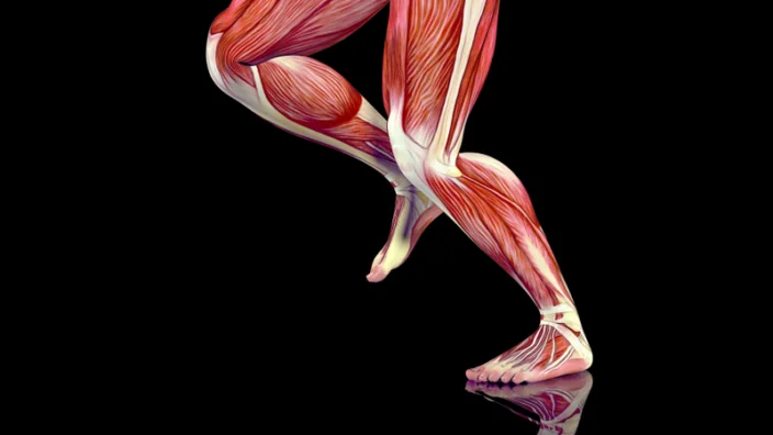
Dr. Pankaj Kapahi is a veteran geroscientist. His laboratory at the famous Buck Institute for Research on Aging is among the few that study advanced glycation end products (AGEs) and the many ways in which they affect aging. Dr. Kapahi’s company, Juvify, produces GLYLO, a supplement for detoxifying AGEs that also reduces food cravings. In this conversation, we discuss how it works, how sound the science behind it is, and whether supplements are inferior to drugs.

Could you give us a quick overview of AGEs and of their role in aging?

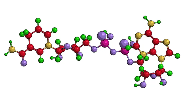
The history of advanced glycation end products starts with the discovery of the Maillard reaction in 1912. The gist of it is that when sugars and amino acids come together at high heat, they react to form irreversible products, which are called advanced glycation end products, or AGEs.

That is what drives the flavor upon cooking food. As an example, when you put toast in the toaster, because of the sugars and amino acids in the bread, you form more than 50 new flavors.

Funny enough, I just did this half an hour ago, and it made me think of AGEs.

It’s ubiquitous. It’s the chemical reaction most commonly conducted chemical reaction, and yet, if you ask someone, they probably won’t have heard of it.

Just the act of cooking and frying creates thousands of chemicals. Later it was realized that advanced glycation end products also build up in your body, and we know this is important because several enzymes are in place to deal with this. One of the things I find fascinating is that every cell that uses glucose can’t avoid those byproducts of metabolism, which are reactive, like the one called methylglyoxal. I’ll stick to that one today because there are many sources of AGEs and precursors, but this is the one we have been most fascinated with because it comes from glycolysis and is a ubiquitous reaction that is conserved from bacteria to us.

Every time a cell breaks down glucose into two, three carbon atoms, you make methylglyoxal non-enzymatically. There are many interesting questions to ask, like “at what rate does this methylglyoxal form, what can regulate that?” We liken it to a leaky tap, whereby during glycolysis, you leak some methylglyoxal, but these things are not well understood, though some of those steps have been identified.

What is important to remember is that these are non-enzymatic reactions, so they have been ignored by biochemists. Biochemists mostly studied metabolic pathways in the past, as this was thought to be the important stuff. Stuff that is happening at low rates, non-spontaneously, was not of interest, but it becomes of great interest when you are talking about aging, because we are now studying not how the system functions but rather how it falls apart.

We view AGEs are one of the drivers of it. Research into AGEs was popular a while back but unfortunately, I would say, fell out of favor, I think because there weren’t any drugs to target them. Some, like aminoguanidine, were successful in animals, but they didn’t make it through clinical trials. Let me give you the background. They used what are called AGE blockers to prevent the formation of AGEs by trapping the precursors. This was done more with rational design, by chemists, who were asking the question, how do you block this reaction? Turned out that had some side effects, and that’s why the clinical trials didn’t go forward.

Our approach has been different: we screen for compounds that enhance endogenous defenses, for example, by activating the TRPA1 channel. It’s a very interesting drug target but not really tackled yet. We published some work, and we have shown that a commonly used supplement, lipoic acid, activates TRPA1 to lower methylglyoxal, a major precursor of AGEs, by activating Nrf2.

Several AGEs have been discovered, the list keeps growing, and we still don’t understand the chemistry around them, because like I said, they’re going to be present at small levels, being non-enzymatic, and each one is going to react with a variety of different amino acids, proteins and lipids.

Methylglyoxal has been shown to induce DNA damage and mutations, and it also reacts with lipids and amino acids, especially arginine and lysine, but also cysteine to some degree.

The use of AGEs is also a trillion-dollar industry. For example, cooking one of those fake meat burgers is a very interesting experience. When you cook vegetables at home, it takes a while to do this, but you put one of those burgers on the skillet, and it goes brown in no time. I think they have added things, played with glycation chemistry to make that patty resemble meat, to create the familiar flavor.

Hundreds of millions of dollars are spent to give our cornflake just the right taste with the right roasting. There are myriads of possible combinations, and that’s why it’s a huge thing to get it right.

What is less talked about is that we consume AGEs from outside as well. There are studies showing that you increase the risk of diabetes by consuming AGEs from outside. This is what our lab is studying as well, the intake of AGEs from outside, but more importantly, what are the endogenous defenses?

In a few words, how do AGEs do this damage to our body?

Think of a piece of bread. Once it’s become brown, you can’t get it back into its original form, and that’s what’s happening in the body. When amino acids react with methylglyoxal or other precursors, they form advanced glycation end products, where the word “advanced” means that the product does not get reversed anymore, theoretically. As more of these are created, you will end up with more inert stuff in the cell, making it less viable.

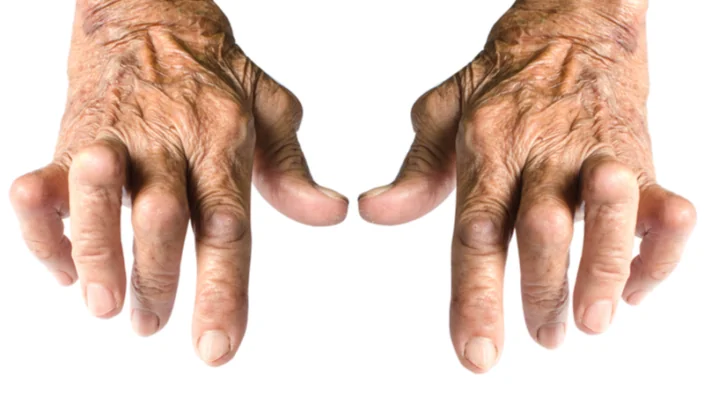
The kidneys are what normally clear a lot of these AGEs. AGEs are present in our cells and in biofluids, and they are constantly being removed, and it’s one of the problems. AGEs have been linked to chronic kidney disease because they cause inflammation, and if the kidney is not doing its job getting rid of them; this creates a positive feedback loop, leading to more inflammation.

Do our endogenous defenses fail with age, is that what’s happening?

First, those defenses do decline with age. Second, they vary between us depending on our genetics and diets. There are people who have diabetes for 50 years but don’t show any pathology. How do they avoid that? In fact, many animals do too. Diabetic animals can be long-lived. We work on flies that have high glucose, but they somehow manage to avoid this damage.

The other very interesting point is that there’s a battery of enzymes in place to deal with AGEs, and we know that if you knock one out, it’s not that you just die. Animals can survive without one or two of these enzymes, because there is a redundancy built in, but as you knock more of them, that’s when you start seeing the collapse. You’ve got changes with age, diet, genetic variance, insults, et cetera, and the whole system is imperfect and tuned differently in different organisms.

If I understand correctly, formation of AGEs is inherent to our metabolism, so we will probably have to do mitigation. What would the possible strategies be?

We have multiple thoughts on strategies. First, let’s think about where these are coming from. Like I said, we focus more on methylglyoxal, so that’s already keeping us busy. This major AGE comes from glycolysis, so every time you reduce glycolysis, you will reduce methylglyoxal production. Now, you can see how fasting makes sense. Under fasting, you would make much less of it.  If it’s coming from glucose, whenever you shift utilization of fat, which is what you do by fasting, you would reduce the methylglyoxal load.

In general, many of these AGEs are derived from metabolism, so that would be one way to tackle this. Then there is detoxification. We are conducting screens to identify compounds and understand the endogenous enzymes that are involved. A number of those have been identified, and polymorphisms in those have been shown to be linked to diseases. Looking for pharmacological strategies to activate them would be a second avenue. Following the research we have done recently, we came up with a cocktail of compounds that we call glycation lowering compounds under the name GLYLO. That’s another one of our strategies.

There is also a third one. As you can imagine, once an AGE is formed, it goes and binds many things. For example, the most studied one is the RAGE, receptor for advanced glycation end-products, but it’s not the only one. One thing people have been trying to do is to produce antibodies that prevent binding. By the way, in my view, the field has been over-focused on RAGE. We think there are multiple receptors for AGEs, and we have identified a number of them using mass spectrometry.

What would the dietary recommendations for mitigating damage from AGEs be?

I have to say that as a result of this research, I began paying much more attention to my glucose and sugar intake. I do intermittent fasting. The reason is, if you just look at your glucose level after each meal, it’s going to take a couple of hours to come down.

If you have only two meals a day, you’re giving the body more of a chance to go into using fat at the expense of glycolysis, so when and what you eat makes a difference. You can go towards low-glycemic diets, more whole food-based, cutting down your free sugars. Those are very simple things anyone can do to decrease AGE burden.

Especially in plant-based diets, it could be hard to drastically cut on carbs.

You can eat whole grains since they do not increase the glycemic load as much. Fiber plus carbs always helps in maintaining glucose levels. Having a high-fiber diet is the way to go.

What about the AGEs that are created by cooking?

Studies indeed show that diabetics on a raw food diet do a lot better, maybe for several reasons. You just feel better on a raw diet. We try to choose our food very carefully, but nobody ever asks what happens when you cook those products. Some types of cooking should be considered quite dangerous. If you look at the metabolomic analysis, when you fry something, you are making more than 5,000 chemicals. We have no idea what they do.

To lower AGEs, one  advice is to cut on dry cooking. You should try to do more steam cooking, that’s good. Adding more acid also reduces the production of AGEs.

Let us talk about your supplement, GLYLO. How does it work? Specifically, at which stage does it intervene with AGEs?

It started with us publishing a study in C. elegans where we had a mutant that created a lot of AGEs, and it was short-lived and developed neuropathy. We then screened for chemicals that would reduce the AGEs there. We were agnostic about where it would lower AGEs. It could be doing it by multiple paths, but we know at least one of the pathways is detoxification of AGEs, so upregulation of NRF2, the pathway that activates GLO1 and detoxifies methylglyoxal. We were very pleasantly surprised to see that alpha-lipoic acid, which we found as a hit in worms, is actually given for diabetic complications, even in humans.

Alpha-lipoic acid is clinically prescribed in many countries and has been shown to mitigate diabetic neuropathy and other diabetic symptoms and lower blood sugar. In terms of the mechanism, we think that it also shifts the animal towards fat utilization and reduced sugar intake, which is very interesting, because when we give the animals this combination, they eat less when they’re on a high-sugar diet. There is also the increase in detoxification, and maybe there’s some effect on how AGEs bind certain targets as well. We think there are multiple mechanisms at play, and that’s what we are trying to decipher now in the lab.

There are other ingredients as well, and you say that they work synergistically. Could you explain how this happens?

What we found was that when we combine these compounds, there is synergy between them and the effects are not one plus one. We did an unbiased screen, and we tried combining all the products that came up in all possible combinations. Adding a fifth of each of the components was much better than using any of the single compounds alone.

We reduced it to these five ingredients, and when we added them together, the effect became multi-fold rather than additive. For example, our data suggests that it is way better to take this combination than nicotinamide alone or alpha-lipoic acid alone. Maybe, when we add them together, they’re targeting multiple pathways including methylglyoxal in different ways.

The effect of reduced cravings – was there something you anticipated, or was it discovered serendipitously?

It was indeed serendipitous, but it kind of makes sense in hindsight that the animals that make more AGEs eat more. The way I like to think of it is that sugar addiction had a protective role in the evolutionary sense for human beings and that we wouldn’t have made it this far without it.

In the past, when resources were limited, one can imagine what would have happened. When we encountered a food source, a sugar source, there would be a mechanism in place to tell you, “Eat more of this because you don’t know when you’re going to get the next meal.” Everyone has this mechanism built in because it ensures survival. Perhaps that’s why we love to cook our food – in part, because cooking makes it a lot easier to take in more calories. In hindsight, it makes sense that AGEs mediate food addiction.

The supplement route that you took has both advantages and problems. Why did you choose it?

First of all, as I said, we screened a lot of compounds, and we found multiple hits, including among some supplement-grade compounds in that library that had demonstrated very strong effects.

Second, we obviously wanted an easier path to the clinic; we wanted to test if this would work in humans. At the end of the day, you want to help people, and it’s just faster. If it’s a drug, the path is much longer, and you need much more money.

I would say, I see two kinds of people. If you talk to clinicians and patients, they would love something like this, but if you talk to the pharma, to the people on the business side of it, they will tell you that it’s not the right way to go.

Also, what is a prescribed drug or a supplement is somewhat arbitrary, and it varies across countries. In some countries, lipoic acid is a clinically prescribed drug, and in others, it’s considered a supplement. The lines between these two classes of chemicals are very fine. I feel comfortable with it being a supplement, as there is good clinical safety there, and that is accessible to everyone.

Take rapamycin, for example. It has been proven in multiple model systems, but I can’t go and take rapamycin. Despite its potential benefits, it’s not easily available to people.

Do you think that supplements requiring less rigorous research might hurt the longevity field’s reputation?

First, AGEs are not just about aging. They are very relevant for diabetes, for insulin sensitivity. Second, I would argue that if you want something for aging and diabetes, with a majority of the people affected, it should be very safe, which is why a supplement where all the ingredients have already been proven safe and its easily accessible is more appealing to me.

After all, both a drug and a supplement are chemical entities, right? It’s about the work you do on them, the amount of work that the scientists will put into it. This is what makes it more or less rigorous, and this is what we should be talking about. If you look at the aging field, some of the best chemicals that have come up are supplements, such as nicotinamide.

As I said, I see it more as a divide between what the pharma wants and what the patients and the doctors want. These are the market forces we have created. You can’t make a billion dollars off a supplement, so there is less interet. You can make a billion-dollar drug, but that doesn’t guarantee that it will be more effective than some supplements. For instance, omega-3 fatty acids and vitamin D are just as powerful as many things out there.

What are your plans for clinical trials?

We are working hard to publish our paper and seek funding for the clinical trial. We have a number of physicians interested. The first clinical trial we are planning to do would be a three- to six-month trial to see if that would influence insulin sensitivity and weight gain. We’re starting there to follow up on what we’re seeing in the preclinical studies.

As a geroscientist, how do you see the field today? Which directions are most promising, most exciting? What are we doing wrong?

I think the aging field is definitely at its most exciting time. Exciting discoveries are being made in multiple areas in the fields of metabolism, senescence, epigenetics, and stem cells. This suggests that there’s a lot of hope for the future.

Scientifically, I think there’s a lot of things coming together, but much more needs to be understood. I highly value the importance of basic science, and we ourselves are trying to understand the genetics of advanced glycation end-products, which is a neglected area. I think the next step is the translation of all this.

What are we doing wrong?

I would say the problem is with the market forces. The way the FDA works, the way the insurance works, they are not quite ready to embrace performance-improving, health-improving technologies. Like many people have said, what we have is a sickcare system. It only rewards treating the sick. We need incentives for keeping the average person healthy.

For instance, just imaging the benefits of simply asking people to move more. An attempt was made to do this during the Obama administration, but a lot more needs to be done. The impact would be enormous. Those things have to become the culture.

We were able to give up smoking. I always use this as an example. As a country, the US did very well in curtailing smoking. This can be extended to other healthy behaviors. We just have to create the right culture and the incentives.

We would like to ask you a small favor. We are a non-profit foundation, and unlike some other organizations, we have no shareholders and no products to sell you. All our news and educational content is free for everyone to read, but it does mean that we rely on the help of people like you. Every contribution, no matter if it’s big or small, supports independent journalism and sustains our future.
VitaDAO Collaboration

New Partnership Between Molecule, Apollo, and VitaDAO

Combining a Web3 IP market, venture capital, and a decentralized autonomous organization (DAO), three companies have banded together to create a new decentralized science ecosystem. The full press release is included here.

Feb. 21, 2022, Switzerland — Molecule, a biotech IP Web3 marketplace, announces a tripartite partnership with Apollo Health Ventures, a leading longevity-focused venture capital fund, and VitaDAO, a decentralized autonomous organization funding early-stage longevity biotech (“LongBio”) R&D.

VitaDAO and Molecule collaboratively source geroscience assets and finance R&D at universities, institutes, and biotech companies. VitaDAO has a worldwide network of >3,000 community members engaged in sourcing and evaluating early stage longevity biotech assets. Apollo and VitaDAO will collaborate on financing and building the LongBio ecosystem. By connecting a leading LongBio VC to the DeSci (decentralized science) movement, this partnership is a major milestone for the greater decentralized ecosystem.

There are inefficiencies in biopharma R&D and university tech transfer that new Web3 marketplace tools will address. Molecule has pioneered a new type of liquid asset class, the IP-NFT (a non fungible token that holds intellectual property) that is one such tool. With new forms of governance via DAOs, such as VitaDAO, and valuation of IP, such as IP-NFTs, moves early-stage intellectual property into Web3 to allow for greater liquidity, discoverability, and reduced legal complexity by standardizing licensing terms.

About Apollo Health Ventures

Apollo Health Ventures is a transatlantic venture capital firm specialized in developing and investing in data-driven biotechnology and health tech ventures. Apollo Health Ventures invests in game-changing companies at the seed or early stage and builds companies within the aging sector. Apollo’s team consists of entrepreneurs, seasoned biotech investors and scientists with remarkable track records in life science investments and venture creation.

About Molecule

Molecule is a decentralized biotech protocol building a web3 marketplace for research-related IP and scalable frameworks to build biotech DAOs. They enable quick and easy funding for academics and biotech companies globally, while enabling patient, researcher, and investor communities to directly fund, govern, and own research-related IP. Molecule democratizes biopharma research and development.

About VitaDAO

VitaDAO is a DAO collective for community-governed, decentralized drug development. Its core mission is the acceleration of research and development (R&D) in the longevity space and the extension of human life and healthspan. To achieve this, VitaDAO funds and digitizes research and the resulting assets using IP-NFTs and other innovative financialization approaches.

For media enquiries, contact:

Heinrich Tessendorf

heinrich@molecule.to

Apollo Health Ventures Socials:

Medium

Twitter

Crunchbase

VitaDAO Socials:

Discord

Twitter

Medium

Molecule Socials

Discord

Twitter

Medium

We would like to ask you a small favor. We are a non-profit foundation, and unlike some other organizations, we have no shareholders and no products to sell you. All our news and educational content is free for everyone to read, but it does mean that we rely on the help of people like you. Every contribution, no matter if it’s big or small, supports independent journalism and sustains our future.
Telomeres

A New Understanding of Telomere Attrition

In Nature Cell Biology, a team of researchers has presented a current review of telomeres and how they relate to aging, reflecting modern research into a decades-old topic.

Telomeres and cellular senescence

This paper touches upon senescent cells and the senescence-associated secretory phenotype (SASP) that they excrete. As this paper explains, cellular senescence is initiated by a constant DNA damage response (DDR): DNA repair mechanisms, which are capable of fixing DNA damage caused by other methods, are ineffective against DNA damage caused by shortened telomeres [1]. Additionally, the SASP itself drives DDR against telomere-associated DNA foci [2].

The researchers also report that telomere DNA is uniquely sensitive to oxidative damage. This process, called TelOxidation [3], harms the activity of the telomere-lengthening enzyme telomerase [4] and telomere-binding proteins, thus leading to further telomere damage [5].

Multiple other hallmarks of aging are mentioned as being affected by telomere attrition, leading the researchers to suggest that this hallmark is more prominent in aging than otherwise thought [6].

Diseases related to telomere dysfunction

This review includes a lengthy list of age-related diseases that research has shown are relevant to telomeres. This includes lung diseases such as idiopathic pulmonary fibrosis [7] and chronic obstrucive pulmonary disorder (COPD) [8], both of which are also strongly related to cellular senescence. There is also evidence suggesting a causal relationship between aplastic anemia and telomere attrition in people [9], and short telomeres may be a cause of kidney fibrosis [10].

The researchers also point to evidence of telomere attrition being involved in many other disorders, including a syndrome related to stem cell exhaustion, cardiovascular diseases, diabetes and other metabolic issues, arthritis, atherosclerosis, and brain disorders such as Alzheimer’s and Parkinson’s.

Conclusion

To cap off this review, the researchers discuss the possibility of telomere lengthening as a therapy. They name the telomere lengthening molecule TERT, suggesting that cells can be encouraged to increase its production and that it can be delivered, itself, as a drug. Of course, this approach might have side effects [11], so the clinical trial process is necessary to ensure safety.

The importance of telomere attrition is a somewhat divisive topic in the longevity community, with some schools of thought holding it to be much more important than others. As the hallmarks of aging build upon and strongly interact with one another, it is very unlikely that telomerase reactivation would be completely ineffective, and it is equally unlikely that it would be a complete cure for telomere-related diseases. To deal with these intertwined hallmarks, a combination approach that simultaneously deals with telomerase and cellular senescence, along with other aspects of aging, is much more likely to be effective; the more completely and thoroughly we can directly and safely target the hallmarks of aging, the better.

We would like to ask you a small favor. We are a non-profit foundation, and unlike some other organizations, we have no shareholders and no products to sell you. All our news and educational content is free for everyone to read, but it does mean that we rely on the help of people like you. Every contribution, no matter if it’s big or small, supports independent journalism and sustains our future.

Literature

[1] Hewitt, G., Jurk, D., Marques, F. D. M., Correia-Melo, C., Hardy, T., Gackowska, A., & Anderson, R. 818 Taschuk, M., Mann, J., and Passos, JF (2012) Telomeres are favoured targets of a persistent DNA 819 damage response in ageing and stress-induced senescence. Nature communications, 3, 708-820.

[2] Victorelli, S., Lagnado, A., Halim, J., Moore, W., Talbot, D., Barrett, K., … & Passos, J. F. (2019). Senescent human melanocytes drive skin ageing via paracrine telomere dysfunction. The EMBO journal, 38(23), e101982.

[3] Jacome Burbano, M. S., Cherfils-Vicini, J., & Gilson, E. (2021). Neutrophils: mediating TelOxidation and senescence. The EMBO Journal, 40(9), e108164.

]4] Fouquerel, E., Lormand, J., Bose, A., Lee, H. T., Kim, G. S., Li, J., … & Opresko, P. L. (2016). Oxidative guanine base damage regulates human telomerase activity. Nature structural & molecular biology, 23(12), 1092-1100.

[5] Opresko, P. L., Fan, J., Danzy, S., Wilson III, D. M., & Bohr, V. A. (2005). Oxidative damage in telomeric DNA disrupts recognition by TRF1 and TRF2. Nucleic acids research, 33(4), 1230-1239.

[6] Chakravarti, D., LaBella, K. A., & DePinho, R. A. (2021). Telomeres: history, health, and hallmarks of aging. Cell, 184(2), 306-322.

[7] Lee, S., Islam, M. N., Boostanpour, K., Aran, D., Jin, G., Christenson, S., … & Bhattacharya, M. (2021). Molecular programs of fibrotic change in aging human lung. Nature communications, 12(1), 1-10.

[8] Ahmad, T., Sundar, I. K., Tormos, A. M., Lerner, C. A., Gerloff, J., Yao, H., & Rahman, I. (2017). Shelterin telomere protection protein 1 reduction causes telomere attrition and cellular senescence via sirtuin 1 deacetylase in chronic obstructive pulmonary disease. American journal of respiratory cell and molecular biology, 56(1), 38-49.

[9] Townsley, D. M., Dumitriu, B., Liu, D., Biancotto, A., Weinstein, B., Chen, C., … & Young, N. S. (2016). Danazol treatment for telomere diseases. New England Journal of Medicine, 374(20), 1922-1931.

[10] Saraswati, S., Martínez, P., Graña-Castro, O., & Blasco, M. A. (2021). Short and dysfunctional telomeres sensitize the kidneys to develop fibrosis. Nature Aging, 1(3), 269-283.

[11] Martínez, P., & Blasco, M. A. (2011). Telomeric and extra-telomeric roles for telomerase and the telomere-binding proteins. Nature Reviews Cancer, 11(3), 161-176.

Life Noggin has joined the Lifespan.io family.

Life Noggin Joins the Lifespan.io Family

Today, we are delighted to officially announce that the popular pop-sci Youtube channel, Life Noggin, has joined the Lifespan.io family. To celebrate the official launch, we are pleased to reveal a new episode of Life Noggin that focuses on some unexpected sources of life extension that we interact with every day.

Life Noggin is an animated science show in which Blocko, the show’s mascot, answers questions related to human life, Earth science, and the universe.

I’m thrilled that Life Noggin has found a new home in Lifespan. We finally have access to a team that’s dedicated to science and technology and understands the importance of educational content online. This collaboration is going to allow Life Noggin to make even more videos, videos that we’ve always wanted to make but didn’t have the bandwidth for. I’m looking forward to a new year of exciting projects!

– Pat Graziosi

Existing fans of the show should also be happy to learn that joining the Lifespan.io family means that there will be more Life Noggin episodes to enjoy. Don’t worry, we won’t be changing things much, but we will be making sure Blocko has everything he needs to make more awesome episodes!

Working with celebrated YouTube channels such as Life Noggin has led to some of the most impactful initiatives we have ever done in the past – both in terms of positively shifting public perception and attracting significant resources to the field at large. I’m overjoyed that Life Noggin is officially joining our team at Lifespan.io, and excited for the incredible opportunities this affords all of us to educate and inspire the public regarding extending healthy human lifespan.

– Keith Comito

Because we love all kinds of science, we are not planning to fill Life Noggin entirely with content about rejuvenation biotechnology, but we hope that people will enjoy some more shows that touch upon biology, aging, and rejuvenation. For example, you may recall that we did a collaboration about aging research with Life Noggin back in September 2018:

It is also important to note that almost any topic that people passionately care about can be related directly to the subject of life extension in one way or another, and having the capability to speak to this point, and collaborate with some of the largest content creators in the world, can be an amazing opportunity for our field.

You can send us your questions for the #dearblocko episodes, where Blocko answers audience questions. You can ask a question by sending Blocko an email and including the phrase “Dear Blocko” in the title. You can also suggest questions by commenting on the videos with #dearblocko followed by your question.

Finally, if you would like to sponsor an episode of Life Noggin, please get in touch with Pat Graziosi to discuss.

Fat and skinny

Caloric Restriction Promotes Rejuvenation in Human Trials

In a new paper, Yale scientists present encouraging data from an unprecedented human study of caloric restriction, a powerful anti-aging intervention [1].

Eat less, live more

Despite all the might of today’s science, simple lifestyle choices are still the clearest option for living longer and healthier. These choices include being physically active, eating better, and eating less, as simple calorie count matters a lot. A healthy diet might be able to confer the benefits of caloric restriction (CR).

CR has been doing wonders in extending the lives of animals [2], but it is not devoid of problems. Results like this require drastic 25%-40% CR, and cutting that many calories involves certain trade-offs. For instance, while being generally healthier and living longer, calorically restricted mice have suffered from an increased severity of viral and parasitic infections [3]. Furthermore, such regimens are just too harsh to be translatable to humans.

In this new paper, scientists from Yale University describe the first controlled CR trial in humans, Comprehensive Assessment of Long-term Effects of Reducing Intake of Energy (CALERIE). Around 200 participants were divided into a study group and a control group. Those in the study group were asked to reduce their calorie intake by about 14% for the two-year duration of the study. This amount of CR was generally well tolerated by the participants. The question is whether such mild CR is good for anything.

CR helps the immune system

Since it is much harder to measure lifespan in humans, who thankfully are pretty long-lived species, researchers have to use various proxies. In this case, the authors started with analyzing thymic function.

The thymus is a small organ where immune T cells are produced and matured. Unlike most organs, the thymus does not continue working for the entire lifespan. Its function peaks during puberty, and then the thymus shuts down its T cell production and turns into fat. This decline in thymic function, called thymic involution, is thought to be a major aspect of aging. One of the most impressive levels of rejuvenation in humans to date was achieved by restoring the thymus [4].

The two-year-long CR significantly increased thymic mass and volume in the study group, while in the control group, no change was detected. The number of cells that had been recently produced by the thymus was increased as well.

CR lowers glucose utilization and promotes oxidation of fatty acids as the body switches to burning fat for energy. Consequently, CR usually results in weight loss, and CALERIE participants experienced a reduction of fat mass. The researchers then decided to dig deeper by analyzing the impact of CR on gene expression in fat.

They found that CR did alter the adipose tissue transcriptome, with 233 genes upregulated and 131 downregulated compared to the control group. Interestingly, this change happened during the first year of CR, and then the transcriptome stabilized, with no changes during the second half of the study detected. This CR-induced transcriptional reprogramming included pathways that are known to regulate the production of energy by mitochondria, anti-inflammatory responses, and longevity.

The researchers also note that the changes in gene expression resembled the ones that follow bariatric surgery. Similar expression patterns were observed in a study of twins with different levels of physical activity. This hints at similarities in the ways CR and physical activity affect our body.

The CR gene?

One of the genes most drastically inhibited as a result of CR caught the scientists’ attention. It codes for the protein PLA2G7, which is produced by macrophages. The researchers tried deleting this gene in mice to test whether this would recapitulate any effects of CR. The deletion indeed resulted in a decrease in thymic atrophy and provided some protection against diet-induced weight gain and age-related inflammation.

The researchers then discovered that PLA2G7 activates the NLRP3 inflammasome, a protein complex also produced in macrophages that triggers an inflammatory immune response. The NLRP3 inflammasome has been shown to contribute to various age-related diseases via the persistent low-grade inflammation known as inflammaging [5]. All this makes the inhibition of PLA2G7 a therapeutic avenue worth exploring.

Conclusion

The molecular underpinnings of CR are not entirely understood, so the discovery of a protein that may be responsible for some of its effects is major news. Geroscientists have been looking for CR mimetics for quite some time, since taking a pill instead of going on a diet would be an easy way – and for some people, the only way – to benefit from CR. Still, most people can just start enjoying the real thing.

We would like to ask you a small favor. We are a non-profit foundation, and unlike some other organizations, we have no shareholders and no products to sell you. All our news and educational content is free for everyone to read, but it does mean that we rely on the help of people like you. Every contribution, no matter if it’s big or small, supports independent journalism and sustains our future.

Literature

[1] Spadaro, O., Youm, Y., Shchukina, I., Ryu, S., Sidorov, S., Ravussin, A., … & Dixit, V. D. (2022). Caloric restriction in humans reveals immunometabolic regulators of health span. Science375(6581), 671-677.

[2] Pifferi, F., Terrien, J., Marchal, J., Dal-Pan, A., Djelti, F., Hardy, I., … & Aujard, F. (2018). Caloric restriction increases lifespan but affects brain integrity in grey mouse lemur primates. Communications biology1(1), 1-8.

[3] Kristan, D. M. (2007). Chronic calorie restriction increases susceptibility of laboratory mice (Mus musculus) to a primary intestinal parasite infection. Aging cell6(6), 817-825.

[4] Fahy, G. M., Brooke, R. T., Watson, J. P., Good, Z., Vasanawala, S. S., Maecker, H., … & Horvath, S. (2019). Reversal of epigenetic aging and immunosenescent trends in humans. Aging cell18(6), e13028.

[5] Gritsenko, A., Green, J. P., Brough, D., & Lopez-Castejon, G. (2020). Mechanisms of NLRP3 priming in inflammaging and age related diseases. Cytokine & Growth Factor Reviews55, 15-25.

Rainbow Clocks

A Fresh Examination of Epigenetic Clocks

In a preprint published in bioRxiv, Morgan Levine and colleagues have identified and grouped 5,717 epigenetic CpG sites into 12 different modules, conducting an in-depth examination into how epigenetic clocks work.

What is a CpG site?

Methylation cannot occur everywhere in the genome. The researchers explain that for a mammalian gene to be silenced through methylation, a methyl group must bind to a specific section of DNA, which consists of cytosine followed by guanine (two of the DNA ‘letters’) and is known by the shorthand CpG [1]. The genome then wraps around that methyl group, thus making it inaccessible [2].

The researchers selected these particular 5,717 sites because they are well researched and their methylation states are known to change with aging; they appear in 15 different epigenetic clocks along with five new tissue-specific clocks that the researchers developed by selecting CpGs that were common between mice and people..

Then, they grouped these CpG sites based on their behaviors during both aging and epigenetic reprogramming. Methylation does not affect all CpG sites in the same way; different sites age at different rates. Sites that acted similarly were given the same ‘color’ as a means of identification.

A rainbow of reprogramming analysis

The researchers found that epigenetic reprogramming through administration of the Yamanaka factors did not entirely reset the epigenetic clock back to a youthful state. The researchers determined.that this did occur in many regions, particularly the green-yellow module, which lost methylation with aging and gained methylation upon reprogramming. This module consisted mostly of sites that had a low density of CpG regions.

The light blue behaved in a reverse, but expected, manner; sites there gained methylation with age and lost methylation with reprogramming. However, the cyan module went completely against the pattern. Sites in this module that lost methylation with aging also lost methylation with reprogramming.

The researchers also contended with technical concerns. Locations that were almost always methylated, or almost never methylated, were difficult to analyze as moving in one direction or the other. The researchers claim that due to the imprecision of current analysis techniques, any signal got lost in the noise.

Analyzing clocks with this new information

With their module analysis in hand, the researchers then turned their eyes towards current clocks, both old and new. They found that, while all of these clocks contain multiple different modules, the more advanced clocks are more strongly weighted towards CpGs that change more dramatically with age. The researchers suggest that many older clocks are weaker because they contain CpG sites that are less strongly, or even inversely, correlated with aging and mortality.

The researchers also analyzed other information associated with these clocks and found unsurprising results. For example, according to the GrimAge clock, smoking strongly affected CpG sites that the researchers placed in the yellow and red modules, and GrimAge also strongly associated epigenetic alterations in the yellow module with all-cause mortality.

There were also differences between tumor and normal cells. While many clocks have had trouble distinguishing from normal cells and cells undergoing uncontrolled division, this study found that cancerous, rapidly dividing cells increase their epigenetic aging in the navy and light blue sites.

Conclusion

As with other preprints published on bioRxiv, this paper hasn’t been peer reviewed. However, this isn’t research into whether or not a potential therapeutic works in an animal or cellular model; this is a paper that describes how DNA methylation sites can be grouped in order to better understand epigenetic aging and create more descriptive, more accurate measurements of it. As such, it is best understood as a look into the inner workings of how epigenetic clocks are developed and what makes these clocks tick.

However, if one thing can be clearly taken from this study, it is that epigenetic changes with aging are much, much more complicated than “younger” or “older”. Going beyond similarity-based, color-coded modules in order to understand which of these thousands of changes are harmless and which changes are ultimately dangerous is obviously a tremendous challenge; however, it is one that might be necessary to undertake.

We would like to ask you a small favor. We are a non-profit foundation, and unlike some other organizations, we have no shareholders and no products to sell you. All our news and educational content is free for everyone to read, but it does mean that we rely on the help of people like you. Every contribution, no matter if it’s big or small, supports independent journalism and sustains our future.

Literature

[1] Jabbari, K., & Bernardi, G. (2004). Cytosine methylation and CpG, TpG (CpA) and TpA frequencies. Gene, 333, 143-149.

[2] Derreumaux, S., Chaoui, M., Tevanian, G., & Fermandjian, S. (2001). Impact of CpG methylation on structure, dynamics and solvation of cAMP DNA responsive element. Nucleic acids research, 29(11), 2314-2326.

Cambrian Logo

Novartis Licenses mTOR Analogs to Cambrian Biopharma

Following the efforts of Adicet Bio, formerly resTORbio, to create therapies based on the mTOR pathway, Cambrian Biopharma is working with Novartis to use this pathway in order to increase healthspan and lifespan. The full press release is included here.

NEW YORK, Feb. 16, 2022 /PRNewswire/ — Cambrian Biopharma, a multi-asset longevity biotech, today announced a licensing agreement with Novartis to advance novel, selective compounds designed and characterized by Novartis to target the mechanistic target of rapamycin (mTOR) pathway. The licensed assets are structural analogs of the FDA-approved drug rapamycin, which has been shown to prevent or reverse multiple age-related health deficits in mice and extend their average lifespan by up to 31%.

The assets will be developed by new Cambrian subsidiary Tornado Therapeutics led by industry and Novartis veteran Joan Mannick, M.D., as CEO. The most advanced asset is now moving into IND-enabling studies, while a second asset is undergoing preclinical efficacy testing.

Under the terms of the agreement, Cambrian acquired exclusive, worldwide rights to the assets, while Novartis received an upfront payment and is entitled to royalties and milestone payments for successfully commercialized medicines. Financial terms of the agreement were not disclosed.

“Pioneers in longevity therapeutics have seen the potential of next-generation mTOR inhibitors for years,” said James Peyer, CEO of Cambrian Biopharma. “This partnership, led by Dr. Mannick and combining Cambrian’s development capabilities with the foundational work done at Novartis, creates a fantastic opportunity to bring a new class of potentially safer, more effective mTOR inhibitors to patients.”

“Although mTOR inhibitors are the best validated therapeutic targeting aging biology, their potential benefits for human aging are just beginning to be explored,” said Dr. Mannick, CEO of Tornado Therapeutics. “The assets we have in-licensed from Novartis will allow us to do a thorough assessment of the safety and efficacy of mTOR inhibitors in aging-related conditions in humans with the ultimate goal of extending healthy lifespan.”

Dr. Mannick joins Tornado Therapeutics from Life Biosciences, where she served as Head of Research and Development. Prior to joining Life Biosciences, she was the Co-Founder and Chief Medical Officer of resTORbio, now Adicet Bio. Previously, she served as Executive Director of the New Indications Discovery Unit at the Novartis Institutes for BioMedical Research, where she led clinical studies of mTOR inhibitors to improve immune function in older adults. Prior to Novartis, Dr. Mannick served as Medical Director at Genzyme working in multiple therapeutic areas and was faculty member at Harvard Medical School and University of Massachusetts Medical School. She received an A.B. from Harvard College and an M.D. from Harvard Medical School and completed her residency at Brigham and Women’s Hospital, followed by an infectious diseases fellowship as part of the Harvard Combined Infectious Diseases Program.

About mTOR Inhibitors

The FDA-approved drug rapamycin and its analogues (rapalogs) are inhibitors of the mechanistic target of rapamycin (mTOR). mTOR inhibitors are an exceptionally well-explored therapeutic class, encompassing three FDA-approved medications evaluated in more than 3,000 clinical trials. Recent studies have demonstrated that mTOR inhibitors extend health and lifespan in multiple organisms, including yeast, worms, flies and mice, establishing them as the best validated class of longevity therapeutics. The next generation mTOR inhibitors in-licensed by Cambrian are predicted to have improved safety and efficacy as compared to currently approved rapalogs.

About Cambrian Biopharma

Cambrian Biopharma is building the medicines that will redefine healthcare in the 21st century – therapeutics to lengthen healthspan, the period of life spent in good health.

As a Distributed Development Company (or DisCo), Cambrian is advancing multiple scientific breakthroughs each targeting a biological driver of aging. Our approach is to develop interventions that treat specific diseases first, then deploy them as preventative medicines to improve overall quality of life as we age. To date, Cambrian has 17 novel therapeutics in development across its pipeline. For more information, please visit www.cambrianbio.com or follow us on Twitter @CambrianBio and LinkedIn.

About Tornado Therapeutics

Tornado Therapeutics is a majority-owned pipeline company of Cambrian Biopharma focused on the development of next-generation mTOR inhibitors to improve efficacy and safety through greater selectivity. The company is currently operating in stealth mode.

We would like to ask you a small favor. We are a non-profit foundation, and unlike some other organizations, we have no shareholders and no products to sell you. All our news and educational content is free for everyone to read, but it does mean that we rely on the help of people like you. Every contribution, no matter if it’s big or small, supports independent journalism and sustains our future.
Seniors walking

Muscular Oxidative Capacity Predicts Mobility Decline

Researchers publishing in Aging Cell have shown a relationship between mitochondrial dysfunction and mobility decline in older adults.

A changing longitudinal study

Since 1958, a team of researchers from the National Institute on Aging Intramural Research program has been conducting the Baltimore Longitudinal Study of Aging (BLSA). The BLSA, which has been used for other research, continuously enrolls volunteers free of chronic conditions from a variety of age groups.

The organization conducts visits every one to four years, depending on age. Participants younger than 60 were seen every four years, participants between 60 and 79 were seen every two years, and participants who were at least 80 were seen every year.

Major revisions were made in 2003 to the BLSA study to add phenotypic measurements and molecular biomarkers. Due to the longitudinal nature of the study, changes in technology have occurred during its duration, and Dr. Luigi Ferruci and colleagues explain that extensive efforts have been made over time to control for these changes in analysis.

Linking oxidative capacity to mobility

A new analysis has been published from the BLSA study [1] using data collected between 2013 and 2019. The researchers examined 380 participants who were at least 60 years old and cognitively normal, had good posture and walking speed, and were free from strokes and Parkinson’s disease. The participants also had to be mobile enough, according to specific measurements, to qualify.

To determine muscular oxidative capacity (the ability to use oxygen) after exercise, the researchers measured the recovery rate of phosphocreatine (kPCr). A higher oxidative capacity represents more favorable muscle health, and a primary function of phosphocreatine is to maintain adenosine triphosphate (ATP) levels after muscle use.

Gait speed, a predictor of functional decline and mobility, was measured in four different scenarios at two different times. This study used multiple speed scenarios based on a prior study that measured fitness in older adults [2], including usual and rapid speeds during a 2.5-minute walk and a 6-minute walk. Another scenario included a 400-meter walk.

Muscular oxidative capacity is correlated with age

Lower levels of kPCr was associated with a higher body mass index (BMI) and lower muscle strength. Women had lower kPCr scores than men, and black participants had lower kPCr than white participants.

When the investigators ran analysis at one specific time point (cross-sectionally), a lower kPCr was significantly associated with a slower 6-minute walk at usual gait speed, a slower 2.5-minute walk at usual gait speed, and a slower 400-meter walk. There was no association observed between kPCr and the 6-minute rapid gait speed.

When the investigators ran a longitudinal analysis, a lower kPCr at the start of the study was significantly associated with a larger decline in all the walking scenarios. These analyses were adjusted for baseline sex, age, phosphocreatine depletion during exercise, and BMI over time.

The investigators ran additional analyses after adjusting for the covariates of longitudinal thigh muscle strength, baseline thigh muscle strength, total lean mass, and total fat mass. This data can be seen in their supplementary data tables, and many of these results further support their conclusions.

Conclusion

Collectively, these results suggest that worse mitochondrial function, as shown by a lower kPCr, contributes to mobility decline. Additional studies are needed to verify these results.

Future research is needed to examine the loss of muscle fibers. Such loss, specifically of type II muscle fibers, can lead to sarcopenia [3]. It is known that excess sedentary time decreases mitochondrial density and surface area of muscle fibers. Taking the results of this study into account along with exercise physiology research, there is data to support the idea that aerobic and resistance exercise increase muscular oxidative capacity and thus ameliorate age-related muscle decline.

We would like to ask you a small favor. We are a non-profit foundation, and unlike some other organizations, we have no shareholders and no products to sell you. All our news and educational content is free for everyone to read, but it does mean that we rely on the help of people like you. Every contribution, no matter if it’s big or small, supports independent journalism and sustains our future.

Literature

[1] Tian, Q., Mitchell, B. A., Zampino, M., Fishbein, K. W., Spencer, R. G., & Ferrucci, L. (2022). Muscle mitochondrial energetics predicts mobility decline in well-functioning older adults: The baltimore longitudinal study of aging. Aging cell, e13552. Advance online publication. https://doi.org/10.1111/acel.13552

[2] Simonsick, E. M., Montgomery, P. S., Newman, A. B., Bauer, D. C., & Harris, T. (2001). Measuring fitness in healthy older adults: the Health ABC Long Distance Corridor Walk. Journal of the American Geriatrics Society, 49(11), 1544–1548. https://doi.org/10.1046/j.1532-5415.2001.4911247.x

[3] Jones, T. E., Stephenson, K. W., King, J. G., Knight, K. R., Marshall, T. L., & Scott, W. B. (2009). Sarcopenia–mechanisms and treatments. Journal of geriatric physical therapy (2001), 32(2), 83–89. https://pubmed.ncbi.nlm.nih.gov/20039588/

Hand arthritis

New Treatment for Osteoarthritis Proposed

In a paper published in Nature Aging, a group of scientists outlines a previously unknown pathway that leads to osteoarthritis and describes a promising treatment [1].

Less than a killer, more than a nuisance

There are plenty of deadly age-related diseases, and then there are others that shorten our healthspan rather than lifespan, making the last parts of our lives miserable. These include osteoarthritis (OA), the age-related degradation of cartilage that causes pain, inflammation, and frailty and can severely limit freedom of movement. This last part means that OA indirectly affects mortality by keeping people from being physically active [2].

Despite being the most prevalent degenerative joint disease, with 237 million people affected worldwide, and the leading cause of chronic disability among the elderly, OA has no FDA-approved treatments, and its molecular mechanisms are poorly understood [3].

Is arthritis caused by a pathway?

Cartilage is a peculiar tissue. Just like bone, it consists mostly of extracellular matrix (ECM) with rare inclusions of cells – in this case, chondrocytes – that excrete the molecules that the ECM is built from. Chondrocytes are relentlessly building and repairing cartilage, but their function, just like that of other cell types, gets disrupted as we age.

Cell-ECM relations are regulated by numerous proteins, including kindlins, an obscure family of proteins discovered in the 1990s. One of them, kindlin-2, plays an important role in the formation of focal adhesions: large molecular assemblies that transmit mechanical force and chemical signals between the cell and the ECM. Kindlin-2 is abundant in healthy articular chondrocytes, but its levels dwindle with age, even more so in OA-affected joints. In chondrocytes that reside in calcified cartilage, kindlin-2 expression is all but lost.

Two other proteins have been linked to OA: p-Stat3 and Runx2. Previous research suggested that those three proteins might constitute a pathway important to the development of OA.

To investigate the effect of kindlin-2 loss on the development of OA, and its relationships with p-Stat3 and Runx2, the researchers started by creating the first kindlin-2-knockdown strain of mice in the world. Kindlin-2 deficiency in this strain is conditional and can be turned on by a drug.

Knockdown of kindlin-2 increased protein levels of both p-Stat3 and Runx2 in articular chondrocytes and caused a dramatic loss of cartilage, leading to the accelerated development of OA in the mice.

The levels of kindlin-2 were reduced and the levels of p-Stat3 and Runx2 also increased in aged wild-type mice, thus confirming the crucial role of aging in OA development. Moreover, the severity of aging-induced OA was highly correlated with the magnitude of kindlin-2 downregulation as well as with p-Stat3 and Runx2 upregulation.

The researchers then looked at two types of double-mutants: mice with both kindlin-2 and p-Stat3 knocked out as well as mice with both kindlin-2 and Runx2 knocked out. In both cases, despite kindlin-2 deficiency, the accelerated development of OA did not happen.

Understanding the pathway

A picture of a pathway began to emerge. It had already been established that the loss of kindlin-2 caused both p-Stat3 and Runx2 upregulation. Blocking p-Stat3 expression in chondrocytes both prevented Runx2 accumulation and decreased the OA damage caused by kindlin-2 deletion. On the other hand, blocking Runx2 reversed OA symptoms as well but without downregulating p-Stat3. The scientists arrived at the conclusion that kindlin-2 deficiency causes OA by upregulating Runx2 in a p-Stat3-dependent manner. This discovery of a pathway enables scientists to effectively choose a target, and some proteins might be easier, safer, and more effective to target than others.

Finally, the researchers treated mice suffering from OA with kindlin-2. They injected a cocktail of viral vectors carrying a kindlin-2-coding DNA sequence directly into OA-affected cartilage. A single treatment noticeably alleviated OA symptoms. The scientists admit that they did not follow up on the mice long enough to determine the long-term efficacy of the treatment, but the results are very encouraging nonetheless.

Conclusion

OA is a highly prevalent disease of aging that adversely affects health and quality of life in old age. It is also a known comorbidity for other, deadlier age-related diseases. Given its prevalence, finding a cure for OA is a pressing problem. This study suggests a previously unknown molecular mechanism of OA development and even outlines a possible treatment. The authors promise to test it on primates soon, which means an effective cure for OA might be not that far away.

We would like to ask you a small favor. We are a non-profit foundation, and unlike some other organizations, we have no shareholders and no products to sell you. All our news and educational content is free for everyone to read, but it does mean that we rely on the help of people like you. Every contribution, no matter if it’s big or small, supports independent journalism and sustains our future.

Literature

[1] Wu, X., Lai, Y., Chen, S., Zhou, C., Tao, C., Fu, X., … & Xiao, G. (2022). Kindlin-2 preserves integrity of the articular cartilage to protect against osteoarthritis. Nature Aging, 1-16.

[2] Hochberg, M. C. (2008). Mortality in osteoarthritis. Clinical & Experimental Rheumatology26(5), S120.

[3] Chen, D., Shen, J., Zhao, W. et al. Osteoarthritis: toward a comprehensive understanding of pathological mechanism. Bone Res 5, 16044 (2017).

Pueraria

Puerarin Shows Promise in Fighting COPD in Cells

Publishing in Aging, a team of researchers has discovered how puerarin, an extract of kudzu, ameliorates chronic obstructive pulmonary disorder (COPD) signs in cellular cultures by limiting mitophagy: the consumption of mitochondria by cells.

When autophagy isn’t a good thing

Mitophagy is a subset of autophagy, the consumption of any organelle by a cell. This is a maintenance process that cells use to remove malfunctioning components, and most longevity-oriented research is oriented towards enhancing it, not suppressling it.

However, this process, like many other biological processes, can go out of control. The researchers point to prior studies showing that in COPD, mitophagy is substantially increased due to the increase in oxidative stress [1,2]. The mitochondria are the powerhouses of the cell, and these powerhouses are being destroyed in COPD.

As the kudzu root has been used in traditional Chinese medicine to treat age-related conditions similar to COPD, these researchers sought to discover what actual biochemical effects that its active compound, puerarin, has on cells.

Stopping cells from having too much FUN

Prior research has shown that the FUN14 domain protein 1 (FUNDC1) gene is strongly linked to the enhanced mitophagy associated with COPD [3] and that silencing this gene ameliorates the symptoms of COPD in cellular cultures [4]. The researchers put together a series of experiments linking puerarin to FUNDC1 inhibition.

First, they administered cigarette smoke extract (CSE) to human bronchial epithelial cells (HBECs) alongside differing concentrations of puerarin and examined multiple biochemical markers, including cellular apoptosis (death). Their results show that treatment with puerarin restored these markers back to that of the control group. There was a smooth dose-response curve; the more puerarin the researchers administered, the more the cells looked like cells that had never been exposed to CSE at all.

Looking more closely, the researchers found that puerarin was having the desired physiological effects: the concentration of reactive oxygen species (ROS), which was dramatically increased with CSE, was decreased to approximately the level of the untreated control group with the highest dose of puerarin administered. Proteins associated with mitophagy were similarly decreased, and ATP generation was restored.

Next, the researchers investigated the expression of FUNDC1 and a related protein, DRP1. Similar to the previous results, FUNDC1 and DRP1, increased by CSE, were decreased back to normal levels with puerarin on a smooth dose-response curve. The phosphorylated form of FUNDC1, however, was decreased with CSE and increased with puerarin. These results were further explored and confirmed with the additional administration of PH0321, a protein phosphatase inhibitor that directly counteracted many of the positive effects of puerarin.

The mTOR pathway and mitophagy

To confirm the effects of mitophagy, the researchers also compared the effects of puerarin with those of the mitophagy inhibitor Mdivi. They found that the two compounds performed nearly identically in multiple metrics, including apoptosis and protein expression.

In their very last experiment, the researchers examined the pathways responsible. Pl3K, AKT, and mTOR are part of an important biochemical pathway that is inhibited by CSE and restored by puerarin. By blocking the PI3K/AKT/mTOR signaling pathway with a compound called 3-MA, the researchers were able to completely stop the positive effects of puerarin as shown by other metrics.

Conclusion

While these results are very promising, even to the point of being suspiciously too good, this is simply an intriguing cellular culture study that shows increased performance on specific cellular metrics. Puerarin has not been shown to completely ameliorate the long-term effects of COPD, even in cells, and this study needs to be confirmed by other research laboratories and followed up by animal and human studies before any real conclusions can be drawn about its effectiveness as a treatment.

This study also serves as an implicit public health reminder. COPD and cigarette smoking are so tightly linked that the researchers of this study, just as many previous researchers did [5], used CSE to induce it. As any long-time Star Trek fan knows, even smoking in early life can cause lingering damage that is not naturally healed, thus depleting both healthspan and lifespan. if you truly want to live long and prosper, staying away from things that will increase cellular damage is an obvious step towards that goal.

We would like to ask you a small favor. We are a non-profit foundation, and unlike some other organizations, we have no shareholders and no products to sell you. All our news and educational content is free for everyone to read, but it does mean that we rely on the help of people like you. Every contribution, no matter if it’s big or small, supports independent journalism and sustains our future.

Literature

[1] Jiang, Y., Wang, X., & Hu, D. (2017). Mitochondrial alterations during oxidative stress in chronic obstructive pulmonary disease. International journal of chronic obstructive pulmonary disease, 12, 1153.

[2] Ryter, S. W., Rosas, I. O., Owen, C. A., Martinez, F. J., Choi, M. E., Lee, C. G., … & Choi, A. M. (2018). Mitochondrial dysfunction as a pathogenic mediator of chronic obstructive pulmonary disease and idiopathic pulmonary fibrosis. Annals of the American Thoracic Society, 15(Supplement 4), S266-S272.

[3] Yao, R. Q., Ren, C., Xia, Z. F., & Yao, Y. M. (2021). Organelle-specific autophagy in inflammatory diseases: a potential therapeutic target underlying the quality control of multiple organelles. Autophagy, 17(2), 385-401.

[4] Wen, W., Yu, G., Liu, W., Gu, L., Chu, J., Zhou, X., … & Lai, G. (2019). Silencing FUNDC1 alleviates chronic obstructive pulmonary disease by inhibiting mitochondrial autophagy and bronchial epithelium cell apoptosis under hypoxic environment. Journal of Cellular Biochemistry, 120(10), 17602-17615.

[5] Chen, X., Li, Y., Hua, C., Jia, P., Xing, Y., Tian, X., … & Xie, F. (2019). Establishment of rapid risk assessment model for cigarette smoke extract exposure in chronic obstructive pulmonary disease. Toxicology letters, 316, 10-19.

New Biotech

New Partial Reprogramming Drastically Lowers Cellular Age

In a pre-print paper that has not yet been peer reviewed, a group of researchers presented a new highly efficient method of partial cellular reprogramming [1].

Young from old

Our cells age, and old cells differ from young ones in multiple ways, including gene expression, telomere length, and protein milieu. However, old germline cells can give rise to a shining new organism. The reason is that during the early stages of embryogenesis, germ cells revert to a youthful state [2]. In 2006, a groundbreaking study showed that similar “reprogramming” can be achieved for somatic cells: forced overexpression of just four genes can turn them into rejuvenated cells called iPSCs (induced pluripotent stem cells).

A milder version of this technique, called partial or transient reprogramming, can rejuvenate cells while preserving their cellular identity. Halfway to pluripotency, the expression of the reprogramming factors is stopped, and the cells revert to their normal, differentiated state. As exciting as it sounds, this approach is tricky, and it fails to achieve the same rejuvenation that full reprogramming does.

All the way to the maturation phase

The authors of this new paper claim to have developed a new partial reprogramming technique that is superior to existing analogs. Reprogramming can be divided into three phases: initiation, maturation, and stabilization, with all previous attempts at partial reprogramming being halted during the initiation phase. The researchers call their technique maturation phase transient reprogramming (MPTR) since they ventured deeper into the reprogramming process before bringing the cells back.

Previous research shows that during reprogramming, which can take some 50 days, cellular rejuvenation becomes substantial at around day 10. Day 17 is just about the final frontier where reprogramming can still be reversed before cells reach pluripotency. This was the window that the scientists focused on.

The expression of Yamanaka factors was made transient by conditioning it on the presence of the drug doxycycline. As soon as the researchers withdrew the drug, the expression stopped. In their experiments, the researchers used cultured human fibroblasts sourced from middle-aged donors, subjecting groups of cells to reprogramming for different periods of time before withdrawal: 10, 13, 15, or 17 days.

Drastic rejuvenation

The first thing the scientists noticed was that in all groups, the reprogramming caused dramatic changes to the fibroblasts’ morphology. After the withdrawal of doxycycline, the cells regained their original shape. When this happens in cell culture, there’s no harm done, but cells changing their morphology in vivo might pose a problem.

On the bright side, the age reversal in the best group was very substantial: 30 years on average, according to the Horvath epigenetic clock, which measures aging-associated DNA methylation. Similar results were achieved with the more advanced 2018 Horvath clock [3]. The researchers estimate that MPTR lowers cellular age about three times more than previous partial reprogramming techniques.

Interestingly, the most substantial age reversal was achieved when the reprogramming stopped at day 13. The researchers hypothesize that a return from more advanced reprogramming stages, such as at day 17, is too stressful for the cell, and this stress eats into the rejuvenation gains.

We need rejuvenated somatic cells so that they can work properly for longer. Therefore, the authors decided to see whether the reprogramming caused restoration of cellular function. One of fibroblasts’ major roles is to produce collagen, a protein indispensable not just for young looks but also for the health of multiple organs, such as our vasculature. Sadly, collagen production declines with age.

The experiments showed that the reprogramming boosted the fibroblasts’ collagen-producing abilities, both in terms of expression of relevant genes and of actual collagen quantity. Several other tests confirmed that after reprogramming, most cells became active and healthy fibroblasts again – just younger.

No telomere rejuvenation

Two results were less encouraging. First, one of the symptoms of cellular aging is the shortening of telomeres with every division. While full reprogramming extends telomeres, no such effect was observed in this case. Further investigation is probably needed to determine just how serious this drawback is.

Second, only 25% of the cells were successfully reprogrammed to the maturation stage. 35% failed to reach it, while for the rest, the results were inconclusive. This might signal a limit for partial reprogramming. On the other hand, even the cells that had failed to reach the maturation stage showed signs of rejuvenation.

Conclusion

Partial reprogramming has been shown to cause cell rejuvenation both in vitro and in vivo, but this technology is still in its infancy, and many hurdles remain before it can be translated into clinical use. Studies such as this one deepen our understanding of how reprogramming works, and the hope is that we will eventually be able to perform it safely and efficiently.

We would like to ask you a small favor. We are a non-profit foundation, and unlike some other organizations, we have no shareholders and no products to sell you. All our news and educational content is free for everyone to read, but it does mean that we rely on the help of people like you. Every contribution, no matter if it’s big or small, supports independent journalism and sustains our future.

Literature

[1] Gill, D., Parry, A., Santos, F., Hernando-Herraez, I., Stubbs, T. M., Milagre, I., & Reik, W. (2021). Multi-omic rejuvenation of human cells by maturation phase transient reprogramming. bioRxiv.

[2] Kerepesi, C., Zhang, B., Lee, S. G., Trapp, A., & Gladyshev, V. N. (2021). Epigenetic clocks reveal a rejuvenation event during embryogenesis followed by aging. Science Advances, 7(26), eabg6082.

[3] Horvath, S., & Raj, K. (2018). DNA methylation-based biomarkers and the epigenetic clock theory of ageing. Nature Reviews Genetics, 19(6), 371-384.