×

The Blog

Building a Future Free of Age-Related Disease

Rectal cancer

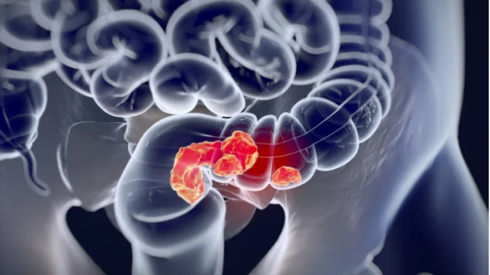
Antibody Therapy Alone Eliminates Certain Rectal Cancers

Researchers publishing in the New England Journal of Medicine have been able to completely eliminate stage 2 and 3 rectal tumors with a single monoclonal antibody drug [1].

Therapy comes with a price

If we leave out skin cancers, colorectal cancer is the third most commonly diagnosed cancer in the US. According to the American Cancer Society, in 2022, more than 106,000 Americans will get colon cancer and 46,000 will get rectal cancer.

On the brighter side, the prevalence of colorectal cancer has declined in recent decades, probably due to early detection and extraction of polyps before they can actually turn into tumors. Early diagnostics and better treatment have also significantly bolstered the survival rate.

However, immediate survival is not the end of the story. Chemo- and radiotherapy negatively affect patients’ quality of life and decrease their healthspan and lifespan, while rectum excision surgery is highly invasive and has harmful long-term consequences.

Evading detection

Immune therapy, including PD-1 blockade, has recently emerged as a supplementary treatment for certain cancers [2]. PD-1 (programmed cell death protein 1) is a receptor on immune cells that dampens their activity when it binds to a ligand (PD-L1 or PD-L2) on the surface of a non-immune cell. This mechanism prevents an autoimmune reaction, but it is often hijacked by tumor cells. By expressing the ligand, tumor cells fool immune cells into misrecognizing them as benign, law-abiding members of the cellular community.

Blocking PD-1 with monoclonal antibodies suppresses this mechanism and boosts anti-tumor immune response (although it also increases autoimmune reaction). Some cancer types are known to respond better to PD-1 blockade, including a subset of rectal cancer called mismatch-repair deficient rectal cancer. Mismatch repair is one of the mechanisms that cells use to repair DNA damage, and its deficiency leads to a higher rate of mutations in the cell. Interestingly, mismatch-repair deficient cancers are usually more resistant to chemo [3], which makes PD-1 blockade a weapon of choice against them.

Complete response

In this new phase 2 study, 16 patients were recruited, and 12 completed the treatment. 15 participants had stage 3 rectal cancer, and one had stage 2. At those stages, which represent the majority of cases, cancer is still mostly localized to the rectum and adjacent tissues, and the five-year survival rate is around 70%. Current protocols usually begin with chemo- and radiotherapy and then proceed to surgery. The participants received dostarlimab, a PD-1 blocking monoclonal antibody agent, intravenously every three weeks for six months.

The researchers hoped that the drug would improve the participants’ chances, but they certainly were not ready for what was to come: after receiving the antibody treatment, all patients demonstrated so-called “clinical complete response”, which means that there was no evidence of tumor as measured by MRI, PET, endoscopic evaluation, digital rectal examination, or biopsy. According to the researchers, such wall-to-wall success is unprecedented in cancer research. Even six months later, none of the patients had required additional therapy. The well-known side effects of monoclonal antibody therapy were mostly mild.

Needs replication

As spectacular as those results are, usual caveats apply. First, the results will need to be replicated in a larger study. Second, only 5-10% of all colorectal cancers are mismatch-repair deficient. Third, the endpoint used (clinical complete response) does not equal total elimination of cancer, and the follow-up period was not nearly enough to conclude that all patients have become cancer-free. Finally, metastatic colorectal cancer remains a huge problem: while it now accounts for just 22% of all cases, the five-year survival rate is only 14%.

The researchers attribute their success to several factors, including the relatively long duration of the treatment. It is also obvious that for reasons largely unknown, this particular type of cancer is especially sensitive to PD-1 blockade. The authors also discuss the important question of why metastatic colorectal cancer responds to the same treatment poorly, despite having the same molecular characteristics. They arrive at an interesting conclusion that the gut microbiome augments the treatment’s efficacy, which would be in line with some previous research [4].

In our study, single-agent dostarlimab was remarkably effective in mismatch repair–deficient, locally advanced rectal cancer, providing a clinical complete response in all 12 patients who have completed treatment to date. The study also provides a framework for evaluation of highly active anticancer therapies in the neoadjuvant context, wherein patients would potentially be spared from chemoradiotherapy and surgery while their tumor is treated when it is most likely to respond — namely, before exposure to other agents that might select for cells with a resistant phenotype.

Conclusion

Immunotherapy for cancer is quickly becoming more effective and sophisticated. This study demonstrates for the first time that immunotherapy alone might be enough in some cases to suppress cancer, eliminating the need for more traditional therapies that can be taxing on patients.

We would like to ask you a small favor. We are a non-profit foundation, and unlike some other organizations, we have no shareholders and no products to sell you. All our news and educational content is free for everyone to read, but it does mean that we rely on the help of people like you. Every contribution, no matter if it’s big or small, supports independent journalism and sustains our future.

Literature

[1] Cercek, A., Lumish, M., Sinopoli, J., Weiss, J., Shia, J., Lamendola-Essel, M., El Dika, I. H., Segal, N., Shcherba, M., Sugarman, R., Stadler, Z., Yaeger, R., Smith, J. J., Rousseau, B., Argiles, G., Patel, M., Desai, A., Saltz, L. B., Widmar, M., Iyer, K., … Diaz, L. A., Jr (2022). PD-1 Blockade in Mismatch Repair-Deficient, Locally Advanced Rectal Cancer. The New England journal of medicine, 10.1056/NEJMoa2201445. Advance online publication.

[2] Kuchroo, J. R., Hafler, D. A., Sharpe, A. H., & Lucca, L. E. (2021). The double-edged sword: Harnessing PD-1 blockade in tumor and autoimmunity. Science Immunology6(65), eabf4034.

[3] Cercek, A., Fernandes, G. D. S., Roxburgh, C. S., Ganesh, K., Ng, S., Sanchez-Vega, F., … & Stadler, Z. K. (2020). Mismatch Repair–Deficient rectal cancer and resistance to neoadjuvant chemotherapy. Clinical Cancer Research26(13), 3271-3279.

[4] Gopalakrishnan, V., Spencer, C. N., Nezi, L., Reuben, A., Andrews, M. C., Karpinets, T. V., … & Wargo, J. (2018). Gut microbiome modulates response to anti–PD-1 immunotherapy in melanoma patients. Science359(6371), 97-103.

Lit mouse on wheel

Senolytic Interventions Ameliorate Radiation Damage in Mice

In their publication in eLife Sciences, researchers at Newcastle University in the UK have illustrated how radiation-induced damage can be somewhat ameliorated with senolytics. These researchers focused on navitoclax along with the well-known combination of dasatinib and quercetin.

A focus on cancer survivors

The researchers begin this paper with a discussion of cancer treatments. They point out that, while modern medicine has made many cancers much easier to cure, cancer survivors often suffer from premature aging. Part of this stems from the fact that successful methods of defeating cancer often involve sending the cancerous cells into a senescent state [1].

Turning these cells senescent usually puts a stop to their deadly, uncontrolled growth, but it opens the door to other dangers, and leaving these senescent cells as they are can even lead to the recurrence of cancer [2]. This makes senolytics an attractive proposition as an additional treatment [1].

Senolytics, delivered early and late

The researchers irradiated 5-month-old male mice with sublethal doses. As in previous experiments [3], this caused the mice to suffer from premature aging, including such symptoms as frailty, decreased coordination, memory problems, and increased mortality.

Administering senolytics soon after this irradiation aided the mice in multiple respects, decreasing whisker loss, body weight, and body condition. Navitoclax also decreased the numbers of tumors formed and improved fur color.

After three months, mice on early senolytics had reduced alanine transaminase, a measurement of liver damage. They were able to hang from their forelimbs for longer and stay on a rotating rod at greater speeds. They behaved smarter in a maze test and remained tumor-free for longer.

Early senolytics did not reverse the frailty caused by irridation, but they slowed its progression compared to other irradiated mice. While dasatinib and quercetin aided the mice in statistically significant ways, navitoclax was found to be more potent than that combination in nearly all the tests conducted in this part of the study. Hanging grip, frailty accumulation, and liver damage were also ameliorated by metformin, which reduces the accumulation of senescent cells.

However, later senolytics, delivered at the age of 11 months and tested at 14 months, did not help nearly as much. Navitoclax did protect against tumors in this cohort of mice. Dasatinib and quercetin improveed forelimb strength more than navitoclax in this cohort, and the rate of frailty decrease was similar. Memory was somewhat improved, but liver damage was largely unaffected, as was the mice’s performance on the rod rotation test.

Conclusion

While this study’s findings were significant, its results were limited and possibly disappointing to anyone who expected serious rejuvenative effects. While removing senescent cells does ameliorate radiation-induced damage, senolytic treatment does not reverse such damage, nor does it restore the frailty of radiation-affected mice to that of their unaffected counterparts.

However, this study does show that senolytics may be an effective treatment for slowing the accelerated aging of cancer patients, either shortly after such therapies or during them; early treatment is obviously a significant factor here. As always, human studies are required to know if this will truly be effective in people.

We would like to ask you a small favor. We are a non-profit foundation, and unlike some other organizations, we have no shareholders and no products to sell you. All our news and educational content is free for everyone to read, but it does mean that we rely on the help of people like you. Every contribution, no matter if it’s big or small, supports independent journalism and sustains our future.

Literature

[1] Short, S., Fielder, E., Miwa, S., & von Zglinicki, T. (2019). Senolytics and senostatics as adjuvant tumour therapy. EBioMedicine 41: 683–692.

[2] Saleh, T., Tyutyunyk-Massey, L., & Gewirtz, D. A. (2019). Tumor cell escape from therapy-induced senescence as a model of disease recurrence after dormancy. Cancer research, 79(6), 1044-1046.

Gene scissors

Modifiying Retrons for Better Gene Editing

A new study published in Nature Chemical Biology has described an advancement in the use of retrons, a novel method of genetic engineering that may become superior to CRISPR technology.

A new solution for an old problem

The researchers begin their paper by pointing out some of the ongoing problems with existing genetic engineering technology. Modifying DNA in a consistent way between cells is difficult and inconsistent, with ongoing technical challenges that can cause severe genetic damage [1].

They also mention other recent work done in the field of retrons. Retrons, which are naturally used by bacteria as a part of phage defense [2], can be used to produce DNA within target cells [3]. This DNA can then be integrated within the cells’ genome through other biotechnologies, such as the well-known “gene scissors” Cas9.

However, previous work with retrons has mostly been done only in bacteria, with only one test done in yeast and a small test done in mammalian cells [4]. The researchers also note that retrons have only been edited to the minimum extent necessary to allow them to be used as editing tools.

A modification for more genomic content

As the researchers explain, a functional part of a retron (an operon) contains three elements: non-coding RNA (ncRNA) that provides a template, a reverse transcriptase (RT) that transforms this ncRNA into RT-DNA, and accessory proteins that assist in this process. As with previous work, the researchers showed that retrons can be used to substantially increase expression of the targeted in bacteria.

They also made modifications to the ncRNA. This RNA contains a long stem with a loop on top, and both of these pieces can be changed. The researchers found that stems between 12 and 30 base pairs were the most effective. Except for a strange spike at 28 base pairs, loops of only 5 base pairs work best, although much longer loops can still produce RT-DNA.

The genetic structures of bacteria and other cells are different, so the researchers had to make substantial modifications, inverting the operon from its normal state. However, once these modifications were made, both the Eco1 and Eco2 operon variants were successful in causing yeast cells to express the desired RT-DNA.

Human cells were much less strongly affected, although Eco2 was much more effective than Eco1. Extending the stem from 12 to 27 base pairs, which increased its effectiveness in yeast, harmed its effectiveness in human cells.

Combining RT-DNA and Cas9

The researchers then went on to test the effectiveness of their technique by incorporating it into a Cas9 genetic modification platform. The approach worked as intended, with the RT-DNA being inserted into the desired locations within a couple of days after injection into yeast. This approach was much more effective than Cas9 plasmids alone, yielding a substantially greater number of precise, on-target edits in multiple genetic loci.

However, in human cells, the on-target effects were still relatively small. This approach, at best, yields an astounding 74:1 ratio of on-target to off-target cells in yeast but a dismal 1:15 ratio in humans: a thousand times worse. Clearly, as the researchers themselves note, substantial modifications need to be made to this approach before we will see any benefits in mammals.

Conclusion

This technology is definitely in its infancy and requires significantly more work to be useful in human cells, let alone in living human beings. The limited number of base pairs is also a serious limitation of this technology, as it greatly restricts the potential proteins that can be encoded through a single use. This is doubly true in light of cytomegalovirus-based gene therapies that might allow for much larger sequences to be expressed in cells.

However, these technical problems are not necessarily insurmountable. If the differences between yeast and human cells can be sorted out and the technology properly modified, it may be feasible to drastically increase the on-target effects in human cells and make retron technology useful as a therapy. Ultimately, retrons may become an integral and safe part of the genetic modification landscape.

We would like to ask you a small favor. We are a non-profit foundation, and unlike some other organizations, we have no shareholders and no products to sell you. All our news and educational content is free for everyone to read, but it does mean that we rely on the help of people like you. Every contribution, no matter if it’s big or small, supports independent journalism and sustains our future.

Literature

[1] Kosicki, M., Tomberg, K., & Bradley, A. (2018). Repair of double-strand breaks induced by CRISPR–Cas9 leads to large deletions and complex rearrangements. Nature biotechnology, 36(8), 765-771.

[2] Millman, A., Bernheim, A., Stokar-Avihail, A., Fedorenko, T., Voichek, M., Leavitt, A., … & Sorek, R. (2020). Bacterial retrons function in anti-phage defense. Cell, 183(6), 1551-1561.

[3] Farzadfard, F., & Lu, T. K. (2014). Genomically encoded analog memory with precise in vivo DNA writing in living cell populations. Science, 346(6211), 1256272.

[4] Mirochnitchenko, O., Inouye, S., & Inouye, M. (1994). Production of single-stranded DNA in mammalian cells by means of a bacterial retron. Journal of Biological Chemistry, 269(4), 2380-2383.

NMN pills

NMN Human Trial Results Published

The data from a multicenter NMN clinical trial have been published with some very modest but still interesting results [1].

What is NMN?

NMN is short for nicotinamide mononucleotide, a naturally occurring molecule present in all species. It is a precursor of nicotinamide adenine dinucleotide (NAD+), a molecule that may be useful in slowing down some aspects of aging. NAD+ serves many critical functions in our cells, such as electron transport, cell signaling, facilitating cellular energy production, and DNA repair.

A growing amount of evidence suggests that as we age, our levels of NAD+ decline, increasing our risk of age-related diseases. This is why some researchers have proposed that restoring NAD+ levels in older people could help to slow down aging and keep them healthy.

NMN is marketed as a supplement and as a NAD+ booster, even though the data supporting its efficacy in humans was, until now, limited. There have been multiple animal studies suggesting that NMN slows down aging, but the evidence for human use has previously been lacking.

An NMN anti-aging human study

The goal of this study was to determine if NMN has any influence on aging in humans. NAD+ was chosen as its primary endpoint, and its increase was used as an indication of an anti-aging effect.

A higher amount of NAD+ present in cells has been correlated with higher energy levels. This makes sense given that NAD+ helps to produce ATP, which is a universal form of cellular energy.

Higher levels of NAD+ may also convey improved DNA repair, as its presence facilitates that [2]. This could also have an influence over genomic instability, which is thought to be a primary reason we age.

The study also chose the 6m walking test as a secondary endpoint. The researchers believed that NMN would improve energy levels and thus the walking capacity of participants.

The NMN study results

The study involved 66 healthy subjects aged between 40 and 65 years. Participants were given two capsules (containing 150 mg of NMN or starch powder) once a day following breakfast for a period of 60 days.

The primary efficacy parameter, NAD+/NADH levels in the serum, had increased by 11.3% in the active group (Uthever group) at day 30, whereas no change was observed in the placebo group at all. At the end of the study (day 60), the NAD+/NADH levels were increased further by 38% from baseline in the Uthever group, compared to a mere 14.3% rise in the placebo group. The increase in the placebo group may be attributed to the placebo effect in this study. Although the difference between the active and placebo groups is not statistically significant, the results indicate that Uthever does increase the NAD+ levels in the serum after 2 months of duration as well.

This suggests that the NMN is entering the cells to increase energy levels and could be interpreted as an anti-aging effect, albeit it relatively small in this case. That said, the NAD+ levels also increased in placebo participants not taking NMN which needs further investigation.

At the end of the study, the mean HOMA IR Index showed a rise of 0.6% among the NMN group and 30.6% among the Placebo group from baseline.

Homeostatic Model Assessment of Insulin Resistance (HOMA IR) shows how much insulin your pancreas needs to regulate blood sugar levels. Developed in the 1980s, it is an indirect measure that is calculated from fasting glucose and fasting insulin levels. While it does have its limitations, it is widely used in clinical research.

The HOMA score rose in the placebo group given starch powder, while there was little change in the group taking NMN. The researchers suggest that this demonstrates an age-slowing effect and that without NMN, HOMA became worse.

The walking endurance increased by 4.3% in the NMN group and 3.9% in the placebo group on day 30 of the treatment. So no effective difference was seen on day 30 of the treatment for walking endurance. When the same treatment was continued up to day 60, the NMN group showed a rise of 6.5% whereas for the placebo group, it remained the same, i.e., 3.9%.

While this is a small increase, it would be interesting to see if the difference between the placebo and test groups increases with longer-term use.

The analysis of the data obtained in this study did not find any statistically significant changes in efficacy outcomes between the active and placebo groups. However, the increase in NAD+/NADH levels in serum, the improvement in overall health and walking endurance, are clinically significant.

Conclusion

Given the mechanisms by which NAD+ has been shown to work in mice and other animals, there is some reason to believe they may work in a similar way in humans, but so far, conclusive results in people have not been obtained.

This trial was conducted by an employee of Effepharm (Shanghai) Co., Ltd and that the company supplied the NMN. While this does not necessarily mean that the data and their interpretation should be discounted, it should be considered when evaluating the results.

The other caveat is that the size of the trial was only 66 people, which is quite small. The larger the trial, the less prone results are to statistical noise and the more reliable they tend to be.

This study does suggest that NMN may have potential in humans though a longer study, increased dosage, and why the placebo group saw increased NAD+ levels should ideally be explored.

We would like to ask you a small favor. We are a non-profit foundation, and unlike some other organizations, we have no shareholders and no products to sell you. All our news and educational content is free for everyone to read, but it does mean that we rely on the help of people like you. Every contribution, no matter if it’s big or small, supports independent journalism and sustains our future.

Literature

[1] Huang, D. A Multicentre, Randomized, Double Blind, Parallel Design, Placebo Controlled Study to Evaluate the Efficacy and Safety of Uthever (NMN Supplement), an Orally Administered Supplementation in Middle Aged and Older Adults. Frontiers in Aging, 26.

[2] Li, J., Bonkowski, M. S., Moniot, S., Zhang, D., Hubbard, B. P., Ling, A. J., … & Sinclair, D. A. (2017). A conserved NAD+ binding pocket that regulates protein-protein interactions during aging. Science, 355(6331), 1312-1317.

AVEA Founders

Maximon Startup AVEA Closes 2.5M CHF Funding Round

The longevity company builder Maximon has concluded a financing round for AVEA at this year’s World Economic Forum. The press release is included here.

ZUG, SWITZERLAND — AVEA, the longevity supplement startup with the aim to improve healthspan, announces the closing of a 2.5 million CHF seed financing round led by Maximon, The Longevity Company Builder and its Longevity Co-Investment Fund. The deal was made public in Davos during this year’s “World Economic Forum” where Maximon hosted an event for investors in the field of longevity.

Dr. Tobias Reichmuth, serial entrepreneur and founding partner of Maximon, states: “The global longevity market is worth $8 trillion. The longevity field is the future of healthcare and we believe AVEA will play an important role in the industry.”

“As we are seeking to build and launch longevity companies with Maximon, Avea was a clear case for us to invest in. We are at an inflection point where people not only look at supplements to boost health and vitamin levels, but start to actively invest in a longer healthy lifespan. This is the opportunity that Avea is well positioned to capture and we are excited to be part of this story“ adds Caroline Wagner, chairwoman of the Board of Avea and founding partner of Maximon.

This first funding will support the company’s rapid growth across Switzerland, Germany, Austria and the UK, power scientific research, propel the development of innovative longevity-focused supplements and help establish the brand as a leader in Europe.

After a year of research, AVEA launched its first products in February 2022. The first formulation is a synergistic blend of ingredients which reflect the latest research in ageing and healthspan. AVEA has enlisted award-winning scientists and researchers in the field of longevity to produce the highest quality, comprehensive and scientifically-backed supplement line on the European market for those who wish to optimize their long-term health as well as slow and even reverse some of the signs of ageing.

“Long-term health is an investment, not an expense. Next to a balanced diet, regular exercise, hot and cold therapy and quality sleep, a longevity supplement regimen is essential to support and optimize the healthy ageing process”, says Scientist and Co-founder Sophie Chabloz.

AVEA’s strategy is to leverage a science-based approach to help customers make sense of the overwhelming health and wellness supplement market. The main idea behind AVEA’s products is to focus on ingredients that act in synergy to slow ageing at the cellular level, because when your cells work better, your whole body works better.

AVEA has been named by Sifted one of 13 longevity startups to watch according to top investors.

About AVEA

AVEA’s mission is to support people’s health and longevity goals with research-backed longevity supplements, providing effective healthy ageing and rejuvenation solutions to help people feel energized and youthful for as long as possible. AVEA believes that everyone deserves to achieve vibrant health throughout their years so they can keep learning, achieving new goals, and enjoying life without feeling limited by their age. AVEA’s supplements are available for order at https://avea-life.com/.

About Maximon

Maximon is a longevity company builder based in Switzerland with the mission to become the leading player in the field of longevity and healthy lifespan by empowering entrepreneurs to build impactful, science-based and scalable companies, which provide healthy ageing and rejuvenation solutions.

More information about Maximon can be found on https://www.maximon.com/

Contact:

Teresa Budetta – Chief Marketing Officer Avea.

Email: teresa@avea-life.com

Phone number: +33 671 366297

Mouse test

Fighting Alzheimer’s Disease with Increased Autophagy

In a new study published in Pharmacology Biochemistry and Behavior, researchers have shown that a combination of two compounds targeting different autophagy pathways is effective in fighting the pathological mechanism of Alzheimer’s disease in a mouse model [1].

Cleaning up is a big deal

The accumulation of “junk”, such as misfolded proteins and damaged organelles, inside and outside the cells in an organism is considered to be one of the key processes underlying aging. Thus, increasing autophagy, the natural process that cells use to get rid of cellular debris, is actively being studied as a way to slow down aging and to treat some age-associated diseases.

The most widely known way of inducing autophagy is by inhibiting the mTOR pathway, such as with rapamycin. However, mTOR-independent autophagy, which can be induced by lithium among other compounds, is also actively being researched as a defense against aging. Targeting both pathways might be more beneficial, particularly for combating neurodegenerative diseases.

Alzheimer’s disease is a classic example of a neurological condition characterized by the accumulation of nonfunctional and harmful materials that overwhelm the cell’s ability to clear them away.

The cytotoxic aggregates of amyloid-β peptide and tau protein in the brain are considered the core molecular mechanisms of Alzheimer’s. Nevertherless, it is still not clear if these aggregates are the primal cause of the disease.

Previous research has shown that autophagy, including mitophagy, the clearance of damaged mitochondria, is compromised in Alzheimer’s disease. Stimulating mTOR-dependent autophagy proved effective in animal models of the disease: it decreased amyloid accumulation and prevented cognitive decline [2].

In this study, the researchers sought to investigate if targeting mTOR-independent autophagy with trehalose in addition to the mTOR-dependent pathway with rapamycin would bring additional benefit in a mouse model of Alzheimer’s disease.

Cleaning tools matter

To induce Alzheimer’s-like pathology, the researchers injected amyloid-β into the ventricles of the brains of 2-month-old male mice. The mice were then divided into several groups, including animals treated with a combination of trehalose and rapamycin along with animals treated with individual drugs.

The treated animals were compared to a control group injected with water and to the untreated, amyloid-β-injected group. This was an important part of the study design because amyloid-β administration itself induces autophagy.

To assess autophagic activity, the researchers first quantified the protein expression of an autophagy marker, LC3-II, in several brain structures that are notably affected in Alzheimer’s disease. Trehalose treatment significantly upregulated autophagy in the hippocampus and frontal cortex, while the combined treatment was more efficient in inducing autophagy than monotherapies in the amygdala and dentate gyrus.

Analysis of mRNA expression of several autophagy-related genes in the hippocampus showed that trehalose was the most efficient in upregulating all the genes tested.

Meanwhile, the accumulation of amyloid-β in the hippocampus and frontal cortex was reduced in trehalose-only, rapamycin-only, and combination-treated groups without a significant difference between them. Likewise, either alone or in combination, the two compounds brought down the activation of microglia (neuroinflammation).

Neuronal density staining revealed that amyloid-β administration led to neuronal loss in the hippocampus. Inducing autophagy with either individual compound, or their combination, prevented neuronal death, as evidenced by the comparison of these groups to the control.

In addition to histological examination of the brain tissue, the researchers conducted several behavioral tests. The combined treatment proved most efficient in reducing anxiety, while all three autophagy-inducing treatments restored the memory and learning impairment brought on by the amyloid-β administration.

Conclusion

This study shows that the induction of autophagy by trehalose alone or in combination with rapamycin in general is more beneficial than rapamycin-only treatment in an animal model of AD. This points out the importance of activating the mTOR-independent pathway to address neurodegeneration.

The authors argue that unlike rapamycin, which acts through mTOR, trehalose affects the expression of several transcription factors, including FOXO1 and TFEB, which, in turn, regulate the expression of autophagy genes. This leads to an increased production of lysosomes.

Three steps underly autophagy: the formation of autophagosomes, debris-containing vesicles; their fusion with lysosomes, organelles filled with digestive enzymes; and, ultimately, degradation [3]. Trehalose might improve one or several of these processes, thus leading to a neuroprotective effect.

It would be exciting to see if this approach can be translated into clinical practice and reverse molecular and cognitive impairments in people with Alzheimer’s disease as effectively as in mice.

We would like to ask you a small favor. We are a non-profit foundation, and unlike some other organizations, we have no shareholders and no products to sell you. All our news and educational content is free for everyone to read, but it does mean that we rely on the help of people like you. Every contribution, no matter if it’s big or small, supports independent journalism and sustains our future.

Literature

[1] Pupyshev, A. B. et al. Combined induction of mTOR-dependent and mTOR-independent pathways of autophagy activation as an experimental therapy for Alzheimer’s disease-like pathology in a mouse model. Pharmacol. Biochem. Behav. 173406 (2022).

[2] Spilman, P. et al. Inhibition of mTOR by rapamycin abolishes cognitive deficits and reduces amyloid-beta levels in a mouse model of Alzheimer’s disease. PLoS One 5, e9979 (2010).

[3] Zhang, X.-J., Chen, S., Huang, K.-X. & Le, W.-D. Why should autophagic flux be assessed? Acta Pharmacol. Sin. 34, 595–599 (2013).

Rejuvenation Roundup May

Rejuvenation Roundup May 2022

Two days ago, the United States honored its fallen veterans. How many of those heroes came home, having survived the horrors of war, only to have their own bodies betray them decades later? Here’s some of what’s been done to fight aging in May.

LEAF News

Lifespan News

LSN Learned HelplessnessLearned Helplessness: We discuss the idea that people have learned to accept something that they don’t need to accept anymore on this episode of Lifespan News.

Plastic Nanoparticles: This episode is on how plastic nanoparticles might lead to at least one hallmark of aging being accelerated.

LSN CBDCBD and Autophagy: On this episode of Lifespan News, Ryan O’Shea discusses how CBD promotes autophagy, our cells’ ability to clear out damaged components.

Young Cerebrospinal Fluid: Researchers have exchanged cerebrospinal fluid between young and old mice in order to determine if it has the same longevity effects that blood exchange has.

Interviews

Yuri Deigin on Cellular Reprogramming in Humans: In a recent press release, YouthBio Therapeutics announced that it has left stealth mode. YouthBio is a self-proclaimed longevity biotech company with a focus on developing gene therapies that reverse the epigenetic alterations that cause us to age.

Richard MillerProf. Richard Miller on the Intervention Testing Program: Richard A. Miller is a Professor of Pathology at the University of Michigan and the Director of Michigan’s Paul F. Glenn Center for Biology of Aging Research. He is also a driving force behind the ITP, the Interventions Testing Program, created in the early 2000s to study the effect of various drugs on lifespan in mice.

João Pedro de Magalhães on Reprogramming and Aging Theories: Professor João Pedro de Magalhães leads the Genomics of Ageing and Rejuvenation Lab at the Institute of Inflammation and Ageing in the University of Birmingham. He is also CSO of YouthBio Therapeutics, a US-based biotech company that develops rejuvenation gene therapies based on partial reprogramming by Yamanaka factors.

Crowd Funded CuresDiscussing Crowd Funded Cures with Savva Kerdemelidis: We have written extensively about VitaDAO, a collective dedicated to community-governed, decentralized drug development. We asked Savva Kerdemelidis a few questions for a deeper dive into the world of DAOs, PFS contracts, and intellectual property on the blockchain.

Reprogramming Cells with Vittorio Sebastiano of Turn.bio: Vittorio Sebastiano is an Associate Professor (Research) of Obstetrics and Gynecology at Stanford University and one of the most prominent scientists in the emerging field of cellular reprogramming. He is also co-founder and Scientific Advisory Board Chairman of Turn Biotechnologies, a cellular rejuvenation company based on the research done in Sebastiano’s Stanford lab.

Rejuvenation Roundup Podcast

Ryan O’Shea of Future Grind hosts this month’s podcast, showcasing the events and research discussed here.

Journal Club

Fecal transfer between young and aged mice reverses hallmarks of aging: On this month’s Journal Club, Dr. Oliver Medvedik explored a recent paper where researchers showed that transfering fecal microbiota from young to old mice reversed some aspects of aging.

Research Roundup

Broken boneSenescent Cells Slow Bone Healing: A study published in the Journal of Clinical Investigation has reported that senescent cells are largely responsible for slow bone healing in aged animals and that senolytics, which remove these harmful cells, can speed bone regeneration.

Longevity in Centenarians Linked to Lower Ribosomal Activity: Scientists have discovered a possible mechanism that protects extremely long-lived people from aging.

Naked Mole Rat SideWhy The Skin of Naked Mole Rats Ages Slowly: A study published in Aging has shown that the skin of the naked mole rat retains nearly all of its physical and biochemical properties as these animals chronologically age, providing new insight into how and why these animals live so long.

Cannabidiol Increases Lifespan and Healthspan in Worms: Scientists have shown that an active ingredient of cannabis significantly upregulates autophagy, extending both lifespan and healthspan in C. elegans nematode worms.

Mouse eatingThe Gut Microbiome Affects the Brain, Eyes, and Gut in Mice: Publishing in Microbiome, a team of researchers has ascertained multiple physical effects of aging gut flora in mice.

NMN Boosts Effectiveness of Immunotherapy in Mice: In a pre-print paper, scientists have shown that treatment with NMN increases the survival and anti-cancer efficacy of CAR-T cells. T cells are a central element of the adaptive immune system, and some of them can be cytotoxic: they have the ability to kill other cells.

Drug combinationDrug Cocktail Delays Aging in Mice: Scientists have shown that a combination of rapamycin, acarbose, and phenylbutyrate has a synergetic rejuvenation effect when administered to 20-month-old mice for three months.

Walking Pace Correlated with Increased Telomere Length: Publishing in Nature Communications, Dr. Tom Yate, Dr. Neliesh J. Samani, and colleagues used data from approximately 400,000 people in the UK Biobank in order to examine the relationship between walking pace and telomere length.

Gene pillUsing an Endemic Virus as a Gene Therapy for Life Extension: In a study printed in PNAS, researchers have shown that telomerase reverse transcriptase (TERT) can be given to cells in living mice through a viral vector, taking the idea of life-extending gene therapies from science fiction to reality.

Caloric Restriction, Feeding Times Lengthen Mouse Lifespan: In a new study published in Science, researchers show that caloric restriction and time-restricted feeding have an additive effect on lifespan in mice. Caloric restriction is considered the first intervention to reliably show that aging is a malleable phenomenon.

Mouse DNAFOXM1 Induction Extends Lifespan in Mice: In a new study published in Nature Aging, researchers have shown that inducing a truncated FOXM1 gene extends lifespan in both progeric and naturally aging mice. Forkhead box (FOX) genes are transcription factors: genes that drive the expression of other genes.

The Role of Mitochondrial Antioxidants in Longevity: A study published in Redox Biology has reported that the upregulation of thioredoxin, a fundamental part of mitochondrial defense against reactive oxygen species, is associated with longevity in mutant C. elegans worms.

Healthy Food“The Perfect Diet” May Increase Lifespan by 13 Years: Scientists from Norway have built a model that predicts the effect of various dietary changes on human lifespan. Diet is obviously a major health factor, but quantifying its impact is not easy.

Biological Aging Fluctuates with Stress: A preprint published in bioRxiv has demonstrated that the second-generation aging clocks PhenoAge, DunedinPACE, and GrimAge show increased but reversible age acceleration in mice and people exposed to significant stress.

OligodendrocytesYoung Cerebrospinal Fluid Improves Memory in Mice: Scientists have found that infusing old mice with cerebrospinal fluid obtained from young mice improves their memory by increasing the proliferation and differentiation of oligodendrocyte progenitor cells.

Lithium Use Might Lower Risk of Dementia: A study published in Plos Medicine has shown that lithium decreases the risk of developing dementia and some of its subtypes, including Alzheimer’s disease and vascular dementia.

Blood pressureBlood Pressure Linked to Dementia, Mortality in Older People: A recent study from the Journal of the American College of Cardiology, conducted by Dr. Wuxiang Xie and colleagues, examined the association of blood pressure with cognitive decline, dementia, and mortality.

Visually Identifying Senescent Cells with an Algorithm: A team of researchers publishing in Aging has developed a method of identifying senescent cells through their physical morphology, potentially making future senescence research much easier. Before modern biomarkers of senescence were eludicated, cell size was considered to be one of its defining features.

Cellular membranePlasmalogens Alleviate Age-Related Cognitive Decline in Mice: Scientists have learned that plasmalogens, obscure but important lipids, serve as a mediator of neurogenesis and synaptic health and can reverse age-related cognitive decline in mice. Plasmalogens are a subtype of phospholipids, the molecules that cellular membranes are mostly made of.

Chloroquine Increases Lifespan in Male Mice: Researchers publishing in Aging have found that the anti-malarial drug chloroquine increases lifespan in the males of a standard breed of mice. The reasons seem to be contradictory.

Antique clockNot All Hallmarks of Aging Contribute to Epigenetic Age: Scientists have shown that only some of the hallmarks of aging affect methylation clocks, which are widely used to measure biological age.

Impact of modifiable healthy lifestyle adoption on lifetime gain from middle to older age: The findings imply the importance of improving the one’s lifestyle for an increased lifespan, even among older patients and/or those with multimorbidity.

Association of Serum Antioxidant Vitamins and Carotenoids With Incident Alzheimer Disease and All-Cause Dementia Among US Adults: Incident all-cause dementia was inversely associated with serum lutein+zeaxanthin and β-cryptoxanthin levels.

Astaxanthin Influence on Health Outcomes of Adults at Risk of Metabolic Syndrome: Results show marginal effects of astaxanthin on reduction in total cholesterol and systolic blood pressure, and a significant attenuating effect on low-density lipoprotein cholesterol.

A Controlled Study to Evaluate the Efficacy and Safety of an NMN Supplement in Middle Aged and Older Adults: The primary efficacy parameter, NAD+/NADH levels in the serum, had increased by 11.3% in the active group (Uthever group) at day 30, whereas no change was observed in the placebo group at all.

Long-term low-dose acetylsalicylic shown to protect against vascular dementia and Alzheimer’s disease in patients with coronary heart disease: The protective potential of low-dose acetylsalicylic for these diseases seems to strongly depend on pre-existing coronary heart disease and the willingness of patients to take it for a minimum of ten years.

Vitamin D supplementation is associated with slower epigenetic aging: Intake of vitamin D supplements is associated with reduced epigenetic age acceleration in participants with vitamin D deficiency.

Urolithin A improves muscle strength, exercise performance, and biomarkers of mitochondrial health in a randomized trial in middle-aged adults: Levels of plasma acylcarnitines and C-reactive proteins are significantly lower with Urolithin A, indicating higher mitochondrial efficiency and reduced inflammation.

Effects of Spermidine Supplementation on Cognition and Biomarkers in Older Adults With Subjective Cognitive Decline: In this randomized clinical trial, longer-term spermidine supplementation in participants with subjective cognitive decline did not modify memory and biomarkers compared with placebo. Exploratory analyses indicated possible beneficial effects on verbal memory and inflammation that need to be validated in future studies at higher dosage.

Heterochronic parabiosis induces stem cell revitalization and systemic rejuvenation across aged tissues: Hematopoietic stem and progenitor cells are one of the most responsive cell types to young blood exposure, from which a continuum of cell state changes across the hematopoietic and immune system emanate.

The Protective Effects of Osteocyte-Derived Extracellular Vesicles Against Alzheimer’s Disease Diminished with Aging: This study uncovers the role of OCY-EV as a regulator of brain health, suggesting a novel mechanism in bone-brain communication.

Analysis of senescence in gingival tissues and gingival fibroblast cultures: The potential of senolytic drugs to modify aging-related changes in the gingiva was shown.

Short senolytic or senostatic interventions rescue progression of radiation-induced frailty and premature ageing in mice: This study suggests that the progression of adverse long-term health and quality-of-life effects of radiation exposure, as experienced by cancer survivors, might be rescued by short-term adjuvant anti-senescence interventions.

We would like to ask you a small favor. We are a non-profit foundation, and unlike some other organizations, we have no shareholders and no products to sell you. All our news and educational content is free for everyone to read, but it does mean that we rely on the help of people like you. Every contribution, no matter if it’s big or small, supports independent journalism and sustains our future.
Antique clock

Not All Hallmarks of Aging Contribute to Epigenetic Age

Scientists have shown that only some of the hallmarks of aging affect methylation clocks that are used to measure biological age [1].

The big black box

Epigenetic clocks that measure biological age based on aging-related changes in genome methylation have been a great success. They enable researchers to analyze the effects of various interventions on lifespan without waiting for a lab animal to die, and they work even on cells in vitro.

Yet, despite the strong correlation between existing epigenetic clocks and various aspects of aging, we still hardly know how those clocks work – in particular, what specific biological changes drive them. For now, this ignorance does not preclude scientists from using the clocks, but there are several reasons why discovering their biological underpinnings is important, as it can provide a more nuanced understanding of aging itself.

In this new study led by Ken Raj of Cambridge university and Altos Labs, and Steve Horvath, the world’s foremost authority on epigenetic clocks, the researchers investigated the relationship between epigenetic clocks and several hallmarks of aging [2]. The group used Horvath’s second-generation multi-tissue clock [3], which is known for its accuracy, and several types of human cells, including keratinocytes and fibroblasts, taken from 14 human donors of various ages.

Cellular senescence

First, the researchers showed that the clock does not correlate well with cellular senescence. Senescence was induced in cells by three different stressors: irradiation, oncogene activation, and replication. Oncogene activation and irradiation led to senescence quickly, after about two weeks, but it did not increase epigenetic age compared to non-senescent cells of the same chronological age. Conversely, it took cells up to six months to reach replicative senescence, which was duly reflected by an increase in epigenetic age.

Telomere attrition

Replicative stress is accompanied by telomere attrition, another hallmark of aging in which telomeres get shorter as the cells edge towards senescence. When the researchers added telomerase reverse transcriptase (TERT), an enzyme that keeps telomeres from shortening, the cells continued to proliferate without reaching senescence, and their epigenetic age continued to increase. This means that telomere attrition, like senescence, does not contribute to epigenetic aging.

DNA instability

Another important hallmark of aging is DNA instability. The researchers used irradiation protocols known to induce some amount of double-strand DNA breaks without affecting cellular viability. The cells, immortalized with TERT to avoid replicative senescence, lived on and kept proliferating, while their epigenetic age continued to rise on par with non-irradiated controls, showing that genomic instability had no effect on the epigenetic clock.

Deregulation of nutrient sensing

Interestingly, rapamycin administered to immortalized cells after 80 passages effectively stopped further increase in epigenetic age. Since rapamycin mediates nutrient sensing, the researchers concluded that deregulated nutrient sensing, also a hallmark of aging, does contribute to epigenetic age.

Mitochondrial dysfunction

To understand how mitochondrial dysfunction, another hallmark of aging, contributes to epigenetic aging, the researchers treated cells with a compound that inhibits mitochondrial activity, which greatly accelerated their rate of epigenetic aging. Treatment with Bezafibrate, a promoter of mitochondrial activity, reversed this increase. The researchers suggest that the two hallmarks (nutrient sensing and mitochondrial activity) that showed relevance to the epigenetic clock might be linked to each other.

Stem cell exhaustion

To investigate this hallmark of aging, the researchers divided tissue samples in two. They increased the ratio of stem cells, which are characterized by a minuscule rate of epigenetic aging, in one part and depleted stem cells from the other part. As a result, the stem cell-enriched fraction aged slower than the stem cell-depleted fraction.

However, can differentiated cells age at different rates? To answer this question, the researchers took cell samples from two donors, both with an epigenetic age of 23. While their cells clonally expanded, their epigenetic age trajectories diverged significantly from the shared baseline. This divergence could not be explained by the much smaller variability of the clock, confirming that an epigenetic age of a tissue is an average of the epigenetic ages of its cells, which could differ substantially from each other.

Aging and longevity

While rapamycin blocked the increase of epigenetic age in immortalized cells, nicotinamide adenine dinucleotide (NAD), nicotinamide riboside (NR), and metformin all extended the cells’ lifespan but without significantly altering their rate of epigenetic aging. The researchers suggest that, in line with previous research, some perturbations can affect both the rate of epigenetic aging and lifespan, while others can affect lifespan without slowing the rate of epigenetic aging, “indicating that aging and longevity, although intimately associated, may nevertheless be distinct”. Interestingly, in the Intervention Testing Program (ITP) trials, rapamycin did substantially increase lifespan in mice, while both metformin and NR failed to do so.

At a higher level of consideration, the innate nature and inevitability of epigenetic aging contrasts with the stochasticity of wear and tear, which is presumed to exert a measurable aging effect only later in life when damage outstrips repair. This, however, does not argue against the relevance of wear and tear and cellular senescence. Instead, these distinct stochastic processes are likely to synergize with epigenetic aging in manifesting the overall phenotypical features of aging. If a successful strategy against aging is to be found, then these distinct and parallel aging mechanisms must be addressed; for example, by the removal of senescent cells, together with the retardation of epigenetic aging.

Conclusion

This important paper is an attempt to look inside the black box that epigenetic clocks have mostly been so far. It raises many intriguing questions that should be addressed in future research. The researchers did not investigate the two other hallmarks of aging, loss of proteostasis and altered cellular communication, though they cite some previous research that links those hallmarks to epigenetic aging.

We would like to ask you a small favor. We are a non-profit foundation, and unlike some other organizations, we have no shareholders and no products to sell you. All our news and educational content is free for everyone to read, but it does mean that we rely on the help of people like you. Every contribution, no matter if it’s big or small, supports independent journalism and sustains our future.

Literature

[1] Kabacik, S., Lowe, D., Fransen, L., Leonard, M., Ang, S. L., Whiteman, C., … & Raj, K. (2022). The relationship between epigenetic age and the hallmarks of ageing in human cellsNature Aging, 1-10.

[2] López-Otín, C., Blasco, M. A., Partridge, L., Serrano, M., & Kroemer, G. (2013). The hallmarks of aging. Cell153(6), 1194-1217.

[3] Horvath, S., Oshima, J., Martin, G. M., Lu, A. T., Quach, A., Cohen, H., … & Raj, K. (2018). Epigenetic clock for skin and blood cells applied to Hutchinson Gilford Progeria Syndrome and ex vivo studiesAging (Albany NY)10(7), 1758.

Lab mouse in hand

Chloroquine Increases Lifespan in Male Mice

Researchers publishing in Aging have found that the anti-malarial drug chloroquine increases lifespan in male NRMI mice. The NRMI, like the Black 6, is a standard breed widely used in research, and these mice were not directly genetically modified.

Substantial positive lifespan effects

We have previously reported a study showing that choloroquine increases longevity in rats, and this study’s results were similar. However, these researchers did not expect such results here: the purpose of this study was to rule out a potentially negative effect on lifespan rather than to find a positive one.

A 50 mg/kg dose of chloroquine was administered at day 500 of lifespan. While the mortality rates of the treatment and control groups began similarly, there was a long period in which the treated mice stopped dying. Slightly before 700 days after the mice were born, all of the untreated group had died while half of the treatment group was still alive; the last treated mouse died a hundred days later.

Food intake did not decrease in the treated group, although liquid intake and body weight were both slightly decreased.

Toxicity

This study showed that chloroquine can be toxic to the liver. Although the 50 mg/kg dose did not lead to significant pathological changes, the 100 mg/kg and 200/kg doses led to hydropic degeneration and even hepatocyte necrosis, the death of functional liver cells.

The heart results, as expected, also showed signs of toxicity, although the toxicity was not significant until higher doses. However, even the 50 mg/kg dose was associated with mild interstitial edema and a loss of striations, two signs of muscle damage, and these results only worsened with increasing doses.

This toxicity was accompanied by a decrease in proteostasis-related activity. The proteosome, which is responsible for recycling unwanted proteins, was significantly downregulated in the liver at 100 mg/kg and in the heart at 200 mg/kg.

Of mice and autophagy

Previous research has shown that chloroquine significantly interferes with the fundamental mechanisms behind autophagy, the process by which cells clear out unwanted components [1]. Autophagy is normally seen as a good thing, as it removes waste; however, the self-cannibalism of cells can obviously have a harmful aspect as well.

The researchers hypothesize that the lifespan results, which fly in the face of the toxicity results, are due to the modulation of autophagy, a reduction in proteostasis mechanisms, and an alteration in the way the cells process glycogen. However, these are only hypotheses; further research is needed to confirm them.

Conclusion

This study adds to a body of conflicting and contradictory research, and while it answers some questions, many others only still have hypothetical answers. Things like autophagy and even protein recycling are not always good nor bad. “The dose makes the poison” is well known in medicine, and it has been once again shown here.

This was a rodent study, and chloroquine, by itself, is unlikely to move the needle much on human longevity. However, what this study does tell us, and further studies will hopefully elucidate in detail, is that the low-level biology of cellular aging can be successfully interfered with.

There are two broader, critical questions that are downstream of this line of research. The first is the inverse of this study: whether or not autophagy-enhancing interventions that increase lifespan have downsides that can be removed to enhance their effectiveness. The second is whether or not these sorts of interventions are any more effective than basic lifestyle choices in lengthening human lifespan.

We would like to ask you a small favor. We are a non-profit foundation, and unlike some other organizations, we have no shareholders and no products to sell you. All our news and educational content is free for everyone to read, but it does mean that we rely on the help of people like you. Every contribution, no matter if it’s big or small, supports independent journalism and sustains our future.

Literature

[1] Mauthe, M., Orhon, I., Rocchi, C., Zhou, X., Luhr, M., Hijlkema, K. J., … & Reggiori, F. (2018). Chloroquine inhibits autophagic flux by decreasing autophagosome-lysosome fusion. Autophagy, 14(8), 1435-1455.

Cellular membrane

Plasmalogens Alleviate Age-Related Cognitive Decline in Mice

Scientists have learned that plasmalogens, obscure but important lipids, serve as a mediator of neurogenesis and synaptic health and can reverse age-related cognitive decline in mice [1].

Plasmalogens and membranes

Plasmalogens are a subtype of phospholipids, the molecules that cellular membranes are mostly made of. Plasmalogens are especially abundant in brain, heart, and immune cells, where they constitute as much as 20% of the total phospholipids in cell membranes. Their function is not well understood, but it is becoming evident that plasmalogens protect cells from stress and that their depletion decreases cellular viability. Diet-wise, plasmalogens can be found mostly in seafood, such as scallops and mussels; a limited number of commercial plasmalogen supplements is already available.

The levels of plasmalogens decline with age, especially in patients with Alzheimer’s disease [2]. Plasmalogen supplementation has been tested before in a handful of studies, including one double-blind, placebo-controlled trial in humans that showed that two-month plasmalogen supplementation significantly improves measures of memory in people with Alzheimer’s and mild cognitive impairment, especially in women [3].

Prettier and smarter

For this new study, aimed at improving our understanding of how plasmalogens work, researchers used 40 16-month-old naturally aging female mice. The mice were divided evenly into two groups, and the study group received plasmalogens with drinking water for two months. Additional 15 mice were used as young controls.

The first thing the researchers noticed was an improvement in the mice’s appearance. Plasmalogen-fed mice looked healthier than their similarly aged controls, with glossier and thicker body hair. The researchers even detected new hair growth:

After the treatment, the mice’s cognitive abilities were assessed via a water maze test, where they are required to navigate a maze filled with water until they find the platform they can use to get out. Healthy mice should quickly learn the location of the platform, as the young controls did. The learning ability was significantly impaired in old controls but restored by the plasmalogen treatment, almost to youthful levels.

Healthy synapses

The researchers then dived deeper into the workings of plasmalogens. It has been previously suggested that plasmalogens are abundant in synapses where they help to form synaptic vesicles, the tiny bubbles that carry neurotransmitters between neurons [4]. Synaptic structures were equally abundant in young controls and in old plasmalogen-treated mice, much more than in old controls. The number of synaptic vesicles in plasmalogen-fed mice was lower than in young controls but much higher than in old controls. These results strongly suggest that plasmalogens alleviate age-related synaptic loss.

This was confirmed by transcriptomic analysis that revealed significant upregulation of synapse-related genes in the study group. Other upregulated genes were related to neural stem cell proliferation, neurogenesis, and production of neurotrophins, which are molecules that affect synaptic plasticity – a quality that makes learning possible. In particular, the expression of synaptophysin, a key mediator of synaptic plasticity, was significantly decreased in old controls compared to young controls in two regions of the hippocampus but almost completely rescued in the study group:

Plasmalogens 2

Less neuroinflammation, more neurogenesis

Neuroinflammation is thought to be a major cause of age-related cognitive decline [5]. It is mostly promoted by overactivation of microglia, the immune cells of the brain. When activated, microglia produce a cocktail of pro-inflammatory cytokines. The researchers analyzed the levels of three of them: TNF-α, IL-1β and IL-6. For all three, the levels shot up with age but were almost completely reversed by the treatment. Activated microglia also morphologically differ from quiescent ones, as they are larger and have fewer extremities. Those differences were mostly rolled back by plasmalogens as well.

Plasmalogens 3

Neurogenesis, the creation of new neurons, occurs in a few discrete niches in mammalian brains, but its rate steadily declines with aging [6]. Keeping the production of new neurons up is very important for preserving cognitive ability. In the study, the number of neuronal stem cells that express Sox2, a marker of differentiation, was significantly reduced in old controls versus young controls, showing an age-related decrease in neurogenesis that was partially alleviated by the treatment:

Plasmalogens 4

Conclusion

Plasmalogen deficiency looks increasingly interesting as a target for treating Alzheimer’s disease and other types of age-related cognitive decline. Plasmalogen levels are so well-correlated with Alzheimer’s that scientists have proposed using them as a diagnostic tool. While Phase 1 human studies are required, oral supplementation of plasmalogens is likely to be safe and, as we learn from studies like this one, effective. Since some anti-aging interventions work in a sex-specific manner, we hope to see a study in mice of both sexes. It would also be interesting to know whether plasmalogen supplementation has any effect on lifespan.

We would like to ask you a small favor. We are a non-profit foundation, and unlike some other organizations, we have no shareholders and no products to sell you. All our news and educational content is free for everyone to read, but it does mean that we rely on the help of people like you. Every contribution, no matter if it’s big or small, supports independent journalism and sustains our future.

Literature

[1] Gu, J., Chen, L., Sun, R., Wang, J. L., Wang, J., Lin, Y., … & Fu, L. (2022). Plasmalogens Eliminate Aging-Associated Synaptic Defects and Microglia-Mediated Neuroinflammation in Mice. Frontiers in Molecular Biosciences, 159.

[2] Han, X., Holtzman, D. M., & McKeel Jr, D. W. (2001). Plasmalogen deficiency in early Alzheimer’s disease subjects and in animal models: molecular characterization using electrospray ionization mass spectrometry. Journal of neurochemistry77(4), 1168-1180.

[3] Fujino, T., Yamada, T., Asada, T., Tsuboi, Y., Wakana, C., Mawatari, S., & Kono, S. (2017). Efficacy and blood plasmalogen changes by oral administration of plasmalogen in patients with mild Alzheimer’s disease and mild cognitive impairment: a multicenter, randomized, double-blind, placebo-controlled trial. EBioMedicine17, 199-205.

[4] Dorninger, F., Forss‐Petter, S., & Berger, J. (2017). From peroxisomal disorders to common neurodegenerative diseases–the role of ether phospholipids in the nervous system. FEBS letters591(18), 2761-2788.

[5] Sartori, A. C., Vance, D. E., Slater, L. Z., & Crowe, M. (2012). The impact of inflammation on cognitive function in older adults: implications for health care practice and research. The Journal of Neuroscience Nursing44(4), 206.

[6] Babcock, K. R., Page, J. S., Fallon, J. R., & Webb, A. E. (2021). Adult hippocampal neurogenesis in aging and Alzheimer’s disease. Stem Cell Reports16(4), 681-693.

Cells Under Microscope

Visually Identifying Senescent Cells with an Algorithm

A team of researchers publishing in Aging has developed a method of identifying senescent cells through their physical morphology, potentially making future senescence research much easier.

Senescent cells look different

Before modern biomarkers of senescence were eludicated, cell size was considered to be one of its defining features, and the researchers cite a 1969 paper to that effect [1]. This excessive growth has been shown to have a causal relationship with senescence, with larger cells having more diluted cytoplasm that contributes to the condition [2]. Other prior work has found links between senescence and nuclear size and shape [3], and a common staining method shows less visual intensity in senescent cells, as they tend to be flatter under a microscope [4].

These visual differences have largely fallen out of favor among the research community as identifiers, being replaced by well-known chemical biomarkers. However, modern image processing algorithms, specifically the high-content analysis (HCA) used in this study, may make it easier to look at senescent cells instead of biochemically analyzing them.

An analysis with a lot of variables

Throughout this study, the researchers primarily examined four different populations of senescent cells: a line of human fetal lung fibroblasts with oncogene-induced senescence (OIS), the same line of fibroblasts with SASP-induced senescence, human mammary fibroblasts (HMFs) with replicative senescence, and human dermal fibroblasts (HDFs) with replicative senescence.

The researchers also used 62 separate biomarkers to determine the differences between proliferating and senescent cells, based both on Z-score analysis, which uses defined controls as reference points, and standard normalization, which uses averages from the given sample. In an effort to simplify future analyses, the researchers also used an algorithmic technique called exploratory factor analysis to generalize these 62 factors into eight “latent factors”, such as nucleus size and cell shape.

Heterogeny and morphology

There were substantial differences between the types of senescent cells. Cells that had reached their replicative limit and became senescent due to telomere attrition were less visually distinguishable from their proliferating counterparts, and the researchers attribute this to the gradual nature of replicative senescence.

Cells that became senescent through oncogenes and cells that became senescent due to the SASP had more substantial differences from proliferating cells. However, these two groups also differed somewhat from each other: the OIS cells had more differences in cell shape, while the SASP-induced group had more differences in visual intensity.

There were also differences from the cells within each model. Cells in OIS group were emitting the SASP to each other, and the researchers hold that some cells may have been more affected by this process than by the original oncogenes, which would logically lead to variances in the cells’ morphology.

Visualizing the data through heat maps corroborated these findings. The OIS and SASP-influenced groups were in distinct clusters from proliferating cells, while the replication-induced groups’ results had fuzzier borders.

An in vivo analysis

The researchers also confirmed their findings through an in vivo analysis. They grew tumors of human cells in mice, induced senescence with a drug, and tested the cells for the senescence biomarker p21 and examined their cellular morphology. The researchers’ algorithms reported 27 substantial differences in morphology between cells that were positive and negative for this biomarker.

Conclusion

Image analysis is continuously being refined as a diagnostic tool in medicine, and visual identification simply requires placing the cells under a suitable microscope rather than biochemistry of any kind. Using physical differences might let researchers better distinguish between the types of senescent cells, possibly leading to a greater understanding of the nature of these cells. If such an algorithm can be refined and proven reliable, it might become a new, superior standard for the identification of senescent cells.

We would like to ask you a small favor. We are a non-profit foundation, and unlike some other organizations, we have no shareholders and no products to sell you. All our news and educational content is free for everyone to read, but it does mean that we rely on the help of people like you. Every contribution, no matter if it’s big or small, supports independent journalism and sustains our future.

Literature

[1] Cristofalo, V. J., & Kritchevsky, D. (1969). Cell size and nucleic acid content in the diploid human cell line WI-38 during aging. Pharmacology, 19(6), 313-320.

[2] Neurohr, G. E., Terry, R. L., Lengefeld, J., Bonney, M., Brittingham, G. P., Moretto, F., … & Amon, A. (2019). Excessive cell growth causes cytoplasm dilution and contributes to senescence. cell, 176(5), 1083-1097.

[3] Sadaie, M., Dillon, C., Narita, M., Young, A. R., Cairney, C. J., Godwin, L. S., … & Narita, M. (2015). Cell-based screen for altered nuclear phenotypes reveals senescence progression in polyploid cells after Aurora kinase B inhibition. Molecular biology of the cell, 26(17), 2971-2985.

[4] Zhao, H., Halicka, H. D., Traganos, F., Jorgensen, E., & Darzynkiewicz, Z. (2010). New biomarkers probing depth of cell senescence assessed by laser scanning cytometry. Cytometry Part A, 77(11), 999-1007.

Blood pressure

Blood Pressure Linked to Dementia, Mortality in Older People

A recent study from the Journal of the American College of Cardiology, conducted by Dr. Wuxiang Xie and colleagues, examined the association of blood pressure with cognitive decline, dementia, and mortality [1]. This study included two cohorts: the English Longitudinal Study of Ageing (ELSA) and the Health and Retirement Study (HRS).

In this analysis, 7,566 participants with healthy cognition from ELSA and 9,294 from the HRS were included. When combined, the average age was 63.5 years old. The ELSA cohort was 55.2% female, and the HRS cohort was 59.8% female. Blood pressure was collected at three different study visits for the ELSA cohort and at two different visits for the HRS cohort.

Blood pressure and cognitive decline

Higher systolic and pulse pressures were associated with increased global cognitive decline, while diastolic blood pressure was, in general, inversely associated with cognitive decline. However, people in the ELSA cohort with high diastolic blood pressure had a higher risk of cognitive decline than people with approximately 80 mm Hg of diastolic pressure.

Blood pressure and dementia

588 people in ELSA and 1,764 people in the HRS were reported to have developed dementia during these studies. Increased systolic blood pressure and pulse pressure were associated with an increased risk of dementia. An inverse association was shown with diastolic blood pressure, although a level of above approximately 80 mm Hg showed no benefit in either cohort.

Blood pressure and all-cause mortality

459 deaths in ELSA and 1629 deaths in the HRS were reported. Higher pulse pressure was associated with higher mortality rates in both cohorts. An inverse association was shown between cumulative diastolic blood pressure and mortality, but as with many of the previous results, higher diastolic pressure than approximately 80 mm Hg was not shown to have any benefit.

Conclusion

This study shows that blood pressure is linked to cognitive outcomes and that people with consistently elevated systolic and total pulse pressure are more likely to experience accelerated cognitive decline. The authors note that it is worthwhile to track pulse pressure over time.

The results of this study regarding diastolic pressure differ from the CARDIA study done in younger adults. This may be explained by atrial stiffness and left ventricular hypertrophy, which can occur with consistently elevated blood pressure and lead to lower diastolic pressure [2,3].

The authors list multiple limitations of their study. The participants from both cohorts were predominately white, so its results may not be applicable to all demographic groups. The authors also note that some of the cognitive tasks may have lacked sensitivity in detecting small cognitive changes. Between the two cohorts, there were some discrepancies between measurements of executive function, which may have affected the total cognition results. Finally, due to the observational nature of this study, causal relationships cannot be determined.

The authors conclude their the study with:

We found that long-term cumulative BP was associated with subsequent cognitive decline, risk for incident dementia, and all-cause mortality among cognitively healthy adults >50 years of age. For middle-aged and older individuals, efforts may be required to control long-term systolic blood pressure and simultaneously maintain adequate diastolic blood pressure, with additional attention on controlling long-term pulse pressure warranted; this could be beneficial to both neurocognition and longevity.

We would like to ask you a small favor. We are a non-profit foundation, and unlike some other organizations, we have no shareholders and no products to sell you. All our news and educational content is free for everyone to read, but it does mean that we rely on the help of people like you. Every contribution, no matter if it’s big or small, supports independent journalism and sustains our future.

Literature

[1] Li, C., Zhu, Y., Ma, Y., Hua, R., Zhong, B., & Xie, W. (2022). Association of Cumulative Blood Pressure With Cognitive Decline, Dementia, and Mortality. Journal of the American College of Cardiology, 79(14), 1321–1335. https://doi.org/10.1016/j.jacc.2022.01.045

[2] Jefferson, A. L., Cambronero, F. E., Liu, D., Moore, E. E., Neal, J. E., Terry, J. G., … & Carr, J. J. (2018). Higher aortic stiffness is related to lower cerebral blood flow and preserved cerebrovascular reactivity in older adults. Circulation, 138(18), 1951-1962.

[3] Lauer, M. S., Anderson, K. M., & Levy, D. (1991). Influence of contemporary versus 30-year blood pressure levels on left ventricular mass and geometry: the Framingham Heart Study. Journal of the American College of Cardiology, 18(5), 1287-1294.

Lithium Pills

Lithium Use Might Lower Risk of Dementia

A study published in Plos Medicine has shown that lithium decreases the risk of developing dementia and some of its subtypes, including Alzheimer’s disease and vascular dementia [1].

Enigmatic but effective

According to WHO, Alzheimer’s disease and other dementias were the second-leading cause of death in high-income countries and the seventh globally in 2019. To date, there are no preventive or disease-modifying treatments available for dementia.

In recent years, lithium has emerged as a potential therapeutic for several neurodegenerative diseases, including dementia [2], although randomized, controlled studies assessing its effectiveness have yet to arrive.

Lithium has long been known for its mood-stabilizing properties: it is the first-line treatment for bipolar disorder (mania) and major depression [3]. The neuroprotective effect of lithium is not completely understood but seems to involve several mechanisms of action, such as the induction of autophagy and the reduction of oxidative stress [4].

On the molecular level, lithium (Li+) competes with magnesium (Mg2+) and can thus interfere with several pathways that rely on the activity of magnesium-dependent enzymes. Specifically, lithium inhibits glycogen synthase kinase-3 beta (GSK-3β) and inositol monophosphatase (IMP), modulating autophagy.

Does lithium fight dementia?

In this retrospective study, the researchers analyzed the clinical data of 29,618 patients collected over a 15-year period in the UK. The patients were ≥ 50 years of age (mean = 73.9 years) and were treated for mental health conditions. 548 of these patients were exposed to lithium as concluded from either a prescription or a documented blood level. 53 (9.7%) and 3,244 (11.2%) patients developed dementia in the lithium-exposed and unexposed groups, respectively.

The patients in the lithium group were subdivided into several categories based on the exposure duration: short-term (≤1 year), long-term (>5 years), and medium-term (1-year-long periods in between 1 and 5 years of exposure). All the patients were followed up until either the first record of dementia, the final record, or death.

The researchers controlled for several confounding variables: age, sex, marital status, ethnicity, smoking status, medications, and physical and mental comobidities. Among the latter, the presence of bipolar disorder was of particular importance, because it is a known risk factor for dementia and the most common reason for a lithium prescription. Indeed, 73% of the patients in the lithium group had this condition.

The study shows that the patients in the lithium group were more likely to have a partner, be a smoker, use antipsychotics, and have depression, bipolar disorder, diabetes, high blood pressure, central vascular disease, or a high cholesterol level. All these factors, except marital status, are associated with a higher risk of developing dementia.

Meanwhile, the most important finding of the study is a significantly lower risk of dementia in people exposed to lithium. Although several dementia subtypes were included in this study, the researchers also analyzed Alzheimer’s disease and vascular dementia incidents separately and confirmed that the risk of these two was also lower in the lithium group.

Next, the researchers show that both short-term and long-term lithium exposure is associated with lower risk of dementia. Medium-term exposures, on the other hand, were not that beneficial. However, this could be attributed to a smaller number of patients in these subgroups and/or a shorter follow-up period. Longer lithium exposure seems best at decreasing the risk of developing all dementias and Alzheimer’s disease but not vascular dementia.

Enlightening but limited

This study had quite a few limitations. First, there were many confounding variables that are difficult to control for. Second, the sample size of people using lithium was rather small. Third, the reason for, and level of, lithium exposure were not always documented. Fourth, both mental and physical comorbidities might have been underestimated. Finally, there was no proof of causation.

Nevertheless, the researchers acknowledge and address some of these limitations by including sensitivity tests to confirm the results obtained from the initial analysis. For example, they analyzed the data with the assumption that all the lithium-exposed patients were suffering from bipolar disorder or excluding all the patients diagnosed with depression.

Abstract

Dementia is the leading cause of death in elderly Western populations. Preventative interventions that could delay dementia onset even modestly would provide a major public health impact. There are no disease-modifying treatments currently available. Lithium has been proposed as a potential treatment. We assessed the association between lithium use and the incidence of dementia and its subtypes.

Conclusion

This study, like many others, raises more questions than answers. Does the anti-dementia effect of lithium apply to the general population as opposed to people with mental health conditions analyzed in this study? Is there an optimal level of lithium and an ideal exposure duration for protecting against dementia? Can lithium decrease the risk of developing other dementias, such as Parkinson’s disease?

Overall, the results of this study are in line with previous research demonstrating the neuroprotective role of lithium, including in animal models of dementia and studies of clinical data conducted in other countries. This warrants the need to further investigate if lithium could be used as a disease-modifying treatment for dementia in randomized, controlled clinical trials.

We would like to ask you a small favor. We are a non-profit foundation, and unlike some other organizations, we have no shareholders and no products to sell you. All our news and educational content is free for everyone to read, but it does mean that we rely on the help of people like you. Every contribution, no matter if it’s big or small, supports independent journalism and sustains our future.

Literature

[1] Chen, S., Underwood, B. R., Jones, P. B., Lewis, J. R. & Cardinal, R. N. Association between lithium use and the incidence of dementia and its subtypes: A retrospective cohort study. PLoS Med. 19, e1003941 (2022).

[2] Hampel, H. et al. Lithium as a Treatment for Alzheimer’s Disease: The Systems Pharmacology Perspective. J. Alzheimers. Dis. 69, 615–629 (2019).

[3] Volkmann, C., Bschor, T. & Köhler, S. Lithium Treatment Over the Lifespan in Bipolar Disorders. Front. Psychiatry 11, 377 (2020).

[4] Forlenza, O. V., De-Paula, V. J. R. & Diniz, B. S. O. Neuroprotective effects of lithium: implications for the treatment of Alzheimer’s disease and related neurodegenerative disorders. ACS Chem. Neurosci. 5, 443–450 (2014).

Oligodendrocytes

Young Cerebrospinal Fluid Improves Memory in Mice

Scientists have found that infusing old mice with cerebrospinal fluid obtained from young mice improves their memory by increasing proliferation and differentiation of oligodendrocyte progenitor cells (OPCs) [1].

The fountain of brain youth

It has been known for years that blood from young animals can alleviate or even reverse some age-related changes, including cognitive changes, in old animals [2]. However, very little research has been done done relating to cerebrospinal fluid (CSF), which helps nourish and maintain our brain cells.

Similarly to blood, CSF composition changes with age [3], but scientists still know very little about the contribution of these changes to aging. One study has shown that in humans, CSF from young healthy donors increases neuronal viability [4]. Another one found that CSF from people with multiple sclerosis is toxic to neurons in vitro [5].

Oligodendrocytes: the electricians of the brain

In this new study, researchers infused 20-month-old mice with CSF drawn from young mice. Three weeks later, the researchers analyzed the mice’s memory by using a simple test that checks how well the mice remember stimuli associated with discomfort in the past. The researchers found that the mice who received a transfusion of young CSF performed significantly better than controls.

The scientists then ran a transcriptomic analysis of the hippocampus and identified 271 genes that were differentially expressed following the procedure. They found that genes related to oligodendrocytes, especially to their differentiation, were the most significantly upregulated.

Oligodendrocytes create myelin sheaths that envelop the axons of brain neurons. These sheaths provide insulation (just like with electric wires), reducing ion leakage and ensuring rapid signal conduction. Myelionogenesis mostly happens during early development but continues throughout life, and it is thought to be extremely important for memory and learning.

Oligodendrocytes, in turn, differentiate from OPCs. One recent study showed that oligodendrogenesis is drastically reduced with age and that increasing it can improve learning and memory in aged mice [6].

Serum response factor and the cytoskeleton

The researchers found that young CSF induced a 2.35-fold surge in the percentage of proliferating OPCs in the hippocampus. Further experiments revealed an expected increase in the number of myelinated axons. An increase in proliferation was also observed in vitro, in cultured cells treated with young CSF.

One of the proteins most upregulated by young CSF was serum response factor (SRF), a ubiquitous transcription factor that is present not just in the brain but also in skeletal muscle and the heart. Wherever it is expressed, SRF is known to increase cell motility, proliferation, and differentiation by promoting the formation of actin filaments that the cytoskeleton is mostly made from. The researchers found that CSF treatment significantly improves cytoskeleton building in OPCs.

SRF knockout, on the other hand, eliminated the gains in OPC proliferation induced by young CSF. These results strongly suggest that the effect of young CSF is mediated by SRF and specifically by SRF-induced cytoskeleton growth.

The missing link

Something in young CSF was boosting the production of SRF, but there are hundreds of proteins that could be responsible for this. After another battery of experiments, the researchers formed a list of 35 potential SRF inducers and then narrowed their search down to fibroblast growth factor 17 (Fgf17), which responded most strongly to changes in the dose of young CSF. Interestingly, the levels of Fgf17 in human CSF decrease with age.

Fgf17 supplementation induced proliferation and differentiation of OPCs in vitro, just like the treatment with young CSF did. Similar results were then obtained in vivo, in aged hippocampi. Fgf17 was also shown to improve memory in mice. Infusing young mice with Fgf17-blocking antibodies impaired their performance in cognitive tests. In vitro, blocking Fgf17 inhibited the increase in proliferation caused by young CSF.

Conclusion

While the rejuvenating properties of young blood are more widely known, the finding that oligodendrogenesis and memory function in the aged brain can be improved by young CSF opens an entirely new avenue of research into brain aging. Importantly, although both blood and CSF consist of hundreds of components, scientists can isolate the ones responsible for particular outcomes (such as Fgf17) and turn them into therapeutic targets. Unfortunately, only one type of memory test was used in this study, and more research will be required to determine how effective this approach is on other types of cognition.

We would like to ask you a small favor. We are a non-profit foundation, and unlike some other organizations, we have no shareholders and no products to sell you. All our news and educational content is free for everyone to read, but it does mean that we rely on the help of people like you. Every contribution, no matter if it’s big or small, supports independent journalism and sustains our future.

Literature

[1] Iram, T., Kern, F., Kaur, A., Myneni, S., Morningstar, A. R., Shin, H., … & Wyss-Coray, T. (2022). Young CSF restores oligodendrogenesis and memory in aged mice via Fgf17. Nature, 1-7.

[2] Villeda, S. A., Plambeck, K. E., Middeldorp, J., Castellano, J. M., Mosher, K. I., Luo, J., … & Wyss-Coray, T. (2014). Young blood reverses age-related impairments in cognitive function and synaptic plasticity in mice. Nature medicine20(6), 659-663.

[3] Chen, C. P., Chen, R. L., & Preston, J. E. (2012). The influence of ageing in the cerebrospinal fluid concentrations of proteins that are derived from the choroid plexus, brain, and plasma. Experimental gerontology47(4), 323-328.

[4] Schwarz, N., Hedrich, U., Schwarz, H., PA, H., Dammeier, N., Auffenberg, E., … & Koch, H. (2017). Human Cerebrospinal fluid promotes long-term neuronal viability and network function in human neocortical organotypic brain slice cultures. Scientific reports7(1), 1-12.

[5] Schwarz, N., Hedrich, U., Schwarz, H., PA, H., Dammeier, N., Auffenberg, E., … & Koch, H. (2017). Human Cerebrospinal fluid promotes long-term neuronal viability and network function in human neocortical organotypic brain slice cultures. Scientific reports7(1), 1-12.

[6] Wang, F., Ren, S. Y., Chen, J. F., Liu, K., Li, R. X., Li, Z. F., … & Mei, F. (2020). Myelin degeneration and diminished myelin renewal contribute to age-related deficits in memory. Nature neuroscience23(4), 481-486.

Chaotic clocks

Biological Aging Fluctuates with Stress

A preprint published in bioRxiv has demonstrated that the second-generation aging clocks PhenoAge, DunedinPACE, and GrimAge show increased but reversible age acceleration in mice and people exposed to significant stress.

What clocks are for

As the researchers remind us, epigenetic clocks measure “how old you are” in a very specific context. Two of the clocks used to test the humans in this study, PhenoAge and GrimAge, are focused on mortality: that is, if someone has a GrimAge of 60, the clock is saying that this person is as likely to die from age-related causes as the average 60-year-old. DunedinPACE is similar, with a focus on age acceleration and the rate of aging. This particular clock was presented at last year’s Ending Age-Related Diseases conference.

in this paper, the researchers show that these biomarkers of increasing mortality rise with certain stresses but return to baseline once the stresses have passed.

Heterochronic parabiosis, revisited

It is well-known in the longevity field that heterochronic parabiosis, the joining of the circulatory systems of old and young animals, leads to the reversal of multiple aspects of aging in the older animal [1]. On the other hand, it also causes stress to the body of the younger animal.

The researchers joined three-month-old mice to 20-month-old mice for three months, with a control group of three-month-old mice joined together in the same way, and then allowed two months for recovery. The liver, which filters the blood, was chosen as the organ for this testing, and the researchers used six separate clocks, including pan-tissue clocks and clocks specifically geared to the liver.

Both groups suffered some epigenetic stress from the procedure itself, as shown by their recovery following the procedure; however, the heterochronic group substantially increased in biological age, which was then reverted after the recovery period. These results make it clear that the young mouse in this scenario is actually aged in a quantifiable, but reversible, way.

Humans age from some stresses, not others

The researchers tested the methylation of older people undergoing surgery at three time points: immediately before, the morning after, and four to seven days later before discharge from the hospital. An emergency hip surgery group had 9 people, an elective hip surgery group had 10, and an elective colorectal surgery group had 11.

In the emergency hip surgery group, biological aging was increased in nearly all the patients and then decreased after surgery, according to all three human clocks. There were outliers in this group; in particular, one person had much greater age acceleration than the others, which continued even after surgery. However, the results were still significant.

However, this did not occur in the elective hip surgery group, and colorectal surgery, which is much less physically traumatic than hip surgery, had no discernible effect on aging according to these clocks. Therefore, the researchers conclude that only large-scale physical trauma is enough for this stress to affect epigenetic clocks.

The researchers also tested two major, well-known sources of stress: COVID-19 infection and pregnancy. In the case of COVID-19, the patients’ age was already accelerated by the time they were admitted to the ICU; therefore, the researchers expected to see a decrease in aging according to multiple clocks.

The results were very interesting: females, but not males, showed a decrease in epigenetic age during recovery, particularly after discharge from the ICU. The researchers also analyzed the effects of three well-known interventions common at the time the study was conducted: hydroxychloroquine, remdesivir, and tocilizumab. While the first two were not significantly connected with a decrease in biological age, patients who took tocilizumab were reported as being younger after discharge, according to all three clocks.

The researchers also analyzed longitudinal data from multiple studies of human pregnancy among multiple demographics. While first-generation clocks did not show these results, these second-generation clocks reported that human mothers gradually increase in biological age during their pregnancies, with reversal after birth.

Conclusion

Probably the largest potential concern is what constitutes aging in the biological sense. If something can be, and is, reversed as the result of normal biological processes, is it truly aging? The researchers hold that biological aging, while it has a gradually upward trajectory, does naturally fluctuate in this way. Therefore, these clocks report biological changes that constitute real epigenetic aging, which has downstream biological consequences.

If this is the case, it may be possible to develop interventions that harness the inherent malleability of the short-term fluctuations in epigenetic aging, keeping them lower for longer periods of time and thus slowing or perhaps reversing multiple other aspects of aging.

We would like to ask you a small favor. We are a non-profit foundation, and unlike some other organizations, we have no shareholders and no products to sell you. All our news and educational content is free for everyone to read, but it does mean that we rely on the help of people like you. Every contribution, no matter if it’s big or small, supports independent journalism and sustains our future.

Literature

[1] Conboy, I. M., Conboy, M. J., Wagers, A. J., Girma, E. R., Weissman, I. L., & Rando, T. A. (2005). Rejuvenation of aged progenitor cells by exposure to a young systemic environment. Nature, 433(7027), 760-764.