×

The Blog

Building a Future Free of Age-Related Disease

Senolytic Helps Anti-Osteoarthritis Potential in Stem Cells

Scientists have improved the potential effectiveness of a stem cell therapy for osteoarthritis by targeting senescent cells [1].

Stem cell exhaustion and osteoarthritis

Osteoarthritis is an age-related disease that affects joints by degrading cartilage. While not fatal in itself, osteoarthritis negatively affects mobility and quality of life, and it can decrease lifespan and healthspan. Several studies link osteoarthritis to higher mortality, especially from cardiovascular diseases [2]. Osteoarthritis patients may require drastic measures such as knee replacement, while less invasive treatments have limited effectiveness.

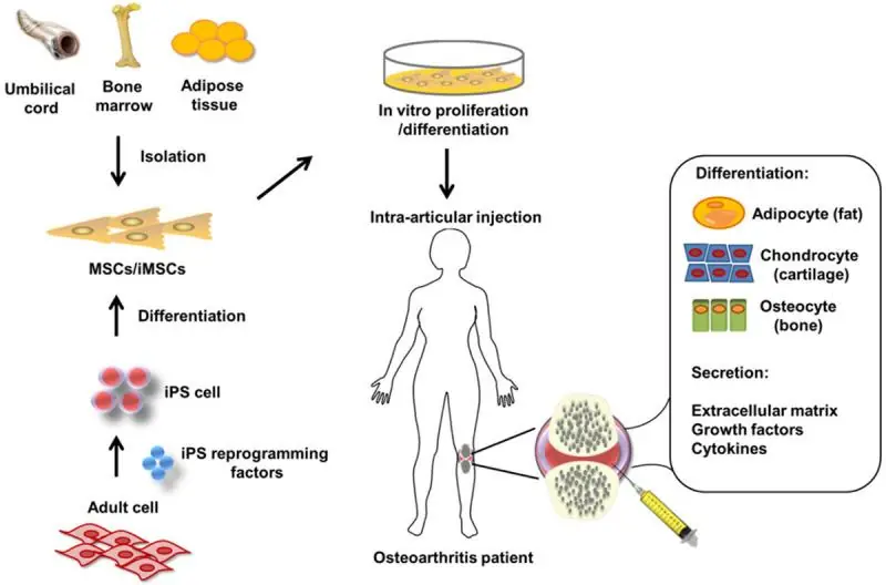
Osteoarthritis can be viewed as a loss of balance between the degradation and regeneration of joint cartilage. This is partially caused by the age-related exhaustion of mesenchymal stem cells (MSCs) that are supposed to differentiate into healthy cartilage-producing cells (chondrocytes). Emerging stem cell therapies for osteoarthritis work by harvesting MSCs from the patient, culturing them in vitro to increase their number and injecting them directly into the affected joint [3].

These therapies, already available in several countries, though not approved in the US, are still limited by the age-related decline in MSC function. Producing brand-new MSCs from induced pluripotent stem cells (iPSCs), which undergo rejuvenation during reprogramming, is a promising direction [3] but is still years away from clinical use.

Senescent MSCs 1

High levels of senescence

In this new study, Japanese scientists discovered that MSCs derived from osteoarthritis patients contain a large fraction of senescent cells, which might be an important cause of the decline in their regenerative potential. Not only are senescent stem cells unable to differentiate into chondrocytes, but they also damage neighboring stem cells by secreting the senescensce-associated secretory phenotype (SASP), a cocktail of mostly pro-inflammatory molecules.

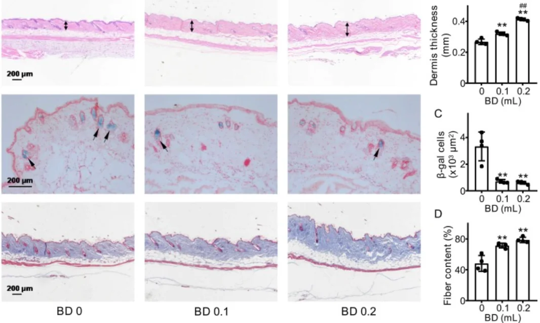
In search of a remedy, the researchers derived synovial MSCs from five donors and treated them with the senolytic drug ABT-263 for one day. Seven days later, senescence levels, colony-forming potential, and multipotency were assessed. The average percentage of senescent (β-galactosidase-positive) cells at baseline was a whopping 55%. Treatment with ABT-263 lowered this percentage to 18% in a uniform manner across all samples:

Senescent MSCs 2

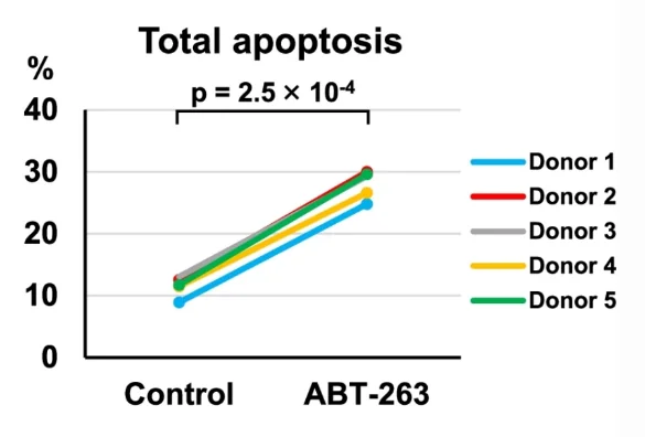
The researchers then confirmed that ABT-263 cleared senescent cells by inducing apoptosis (cellular death). This was done by assessing the levels of caspase-3, a central element of the apoptotic pathway:

Senescent MSCs 3

MSC therapy works by harvesting MSCs and making them grow into colonies. In the study, the researchers assessed colony-forming potential by culturing the cells for 14 days. Cells treated with ABT-263 produced many more colonies, and the colonies were significantly larger than those produced by controls. In the culture, the researchers found enlarged and flattened cells that did not produce colonies. This morphology is characteristic of senescent cells. The percentage of those dysfunctional cells was much lower in the treated group, in line with the hypothesis that ABT-263 eliminates senescent cells.

Improved regenerative potential

Treated cells were also much better than controls in forming cartilaginous pellets, which is a measure of MSCs’ ability to differentiate into chondrocytes and regenerate cartilage. The mean pellet weight was more than twice as large in the treatment group, and the pellets were richer in essential ECM elements such as glycosaminoglycan. The treated cells also produced the “right” collagen: there was less collagen type I, which is mostly present in the skin, tendons, ligaments, and bones, and more collagen type II, the staple protein in cartilage.

As expected, the treatment also decreased the production of SASP, including the collagen-degrading enzyme MMP-13 and the pro-inflammatory cytokine IL-6.

Conclusion

This proof-of-concept study shows a way to increase the regenerative potential of stem cells, which might not be limited to treating osteoarthritis. It also highlights the prevalence of cellular senescence in stem cells from aging donors as one of the problems that must be solved on the way to developing effective stem cell therapies for age-related diseases. As the researchers note, one of the limitations of their study was that the therapeutic effect of the senolytic treatment was not investigated in vivo. Hopefully, follow-up studies will be able to address this question.

We would like to ask you a small favor. We are a non-profit foundation, and unlike some other organizations, we have no shareholders and no products to sell you. All our news and educational content is free for everyone to read, but it does mean that we rely on the help of people like you. Every contribution, no matter if it’s big or small, supports independent journalism and sustains our future.

Literature

[1] Miura, Y., Endo, K., Komori, K., & Sekiya, I. (2022). Clearance of senescent cells with ABT-263 improves biological functions of synovial mesenchymal stem cells from osteoarthritis patients. Stem Cell Research & Therapy13(1), 1-15.

[2] Veronese, N., Cereda, E., Maggi, S., Luchini, C., Solmi, M., Smith, T., … & Stubbs, B. (2016, October). Osteoarthritis and mortality: a prospective cohort study and systematic review with meta-analysis. In Seminars in arthritis and rheumatism (Vol. 46, No. 2, pp. 160-167). WB Saunders.

[3] Zhu, C., Wu, W., & Qu, X. (2021). Mesenchymal stem cells in osteoarthritis therapy: A review. American journal of translational research13(2), 448.

Strong mouse

Stem Cells for Fighting Sarcopenia

In their publication in Stem Cell Research & Therapy, a team of researchers has shown how effective mesenchymal stromal cells (MSCs) are in a mouse model.

Multipotent cells with multiple uses

As multipotent stem cells, MSCs can differentiate into several different cell types, including fat cells, neurons, and bone cells [1]. The researchers point to prior studies showing how MSCs have been investigated for the treatment of age-related diseases such as frailty [2]. As the umbilical cord is considered medical waste, umbilical cord-derived cells (here, UC-MSCs) pose no ethical problems in their harvesting and are widely considered a top candidate with which to develop stem cell therapies [3].

Despite their history, the effects of MSCs on diseases such as sarcopenia have never been fully elucidated. This work studied the effects of human cells on SAMP10, a mouse model that is prone to accelerated senescence, to pave the way towards an understanding.

Effects over months

In this research, 24-week-old SAMP10 mice were injected with either one million UC-MSCs or saline solution at the base of the tail. At 28, 32, and 36 weeks, grip strength and treadmill running time were tested. Muscle samples were also taken at 36 weeks, and tests for gene expression and a wide variety of intercellular signaling compounds, such as mTOR and AMPK, were conducted.

Both the grip strength and treadmill results were conclusive. Interestingly, treadmill distance improved in the control mice, but not nearly to the extent of the treatment group; by 36 weeks of age, the treated mice were able to run for one and a half times the distance of their untreated counterparts. In both groups, grip strength also increased at 28 and 32 weeks, and then decreased at 36 weeks; however, the treated mice enjoyed a very large boost to their performance at 28 weeks, which continued throughout the experiment.

Two key muscles, the gastrocnemius and the soleus, were significantly larger in the treated mice than the untreated mice. The treatment made the mice’s muscles visibly thicker and without the voids found in the control group. Their mitochondria were similarly healthier, with fewer visibly damaged mitochondria and lipid droplets.

Compound analysis told a similar story. Among increases in other signaling compounds, the treated mice had more AMPK, which increases NAD+, and mTOR, which, in this context, promotes metabolism and muscular fortitude. They had much less of the inflammatory marker TNF-α and similar biomarkers of inflammaging, along with a reduced expression of compounds that are related to cellular death through apoptosis.

Many other tests told the same story. Every biomarker that the researchers investigated showed signs of the same basic fact: mice given this stem cell therapy had muscles that were significantly stronger and healthier than those of the control group.

Conclusion

One of this study’s limitations was that the researchers were unable to directly discover if the human UC-MSCs had engrafted themselves into the mice’s musculature. However, one examination that they were able to perform, a test for human dystrophin, suggested that this was not the case. Instead, the researchers hypothesize that the positive effects were paracrine in nature: they were due to signals sent by the stem cells instead of the cells themselves being put to work.

While this was a study in a mouse model, the across-the-board positive results suggest a strong potential for therapeutic use in human beings. If these cells, or the paracrine signals they secrete, can be shown to be effective, they might signal a new standard of care for the treatment of sarcopenia and frailty.

We would like to ask you a small favor. We are a non-profit foundation, and unlike some other organizations, we have no shareholders and no products to sell you. All our news and educational content is free for everyone to read, but it does mean that we rely on the help of people like you. Every contribution, no matter if it’s big or small, supports independent journalism and sustains our future.

Literature

[1] Hsieh, J. Y., Fu, Y. S., Chang, S. J., Tsuang, Y. H., & Wang, H. W. (2010). Functional module analysis reveals differential osteogenic and stemness potentials in human mesenchymal stem cells from bone marrow and Wharton’s jelly of umbilical cord. Stem cells and development, 19(12), 1895-1910.

[2] Golpanian, S., DiFede, D. L., Khan, A., Schulman, I. H., Landin, A. M., Tompkins, B. A., … & Hare, J. M. (2017). Allogeneic human mesenchymal stem cell infusions for aging frailty. The Journals of Gerontology: Series A, 72(11), 1505-1512.

[3] Nagamura-Inoue, T., & He, H. (2014). Umbilical cord-derived mesenchymal stem cells: their advantages and potential clinical utility. World journal of stem cells, 6(2), 195.

Heart and Brain

Aspirin Affects Alzheimer’s Outcomes in Cardiac Patients

A study published in Alzheimer’s Research & Therapy has shown that low-dose aspirin affects Alzheimer’s disease and vascular dementia only in the presence of coronary heart disease.

A well-known preventative

Acetylsalicylic acid, more commonly known as aspirin, is often taken at a steady, low dose of 100 to 300 milligrams a day in order to protect against cardiovascular diseases [1]. It is a non-steroidal anti-inflammatory drug (NSAID), which places it in a class of drugs that are often sold over-the-counter as pain relievers. However, aspirin in particular has been well documented in its positive effects on vasculature, particularly when taken over the long term.

Previous research has shown that long-term aspirin might be effective against Alzheimer’s disease and vascular dementia [2], but only when given over a very long time period. Two other studies excluded patients with cardiovascular disease, and these studies did not show significant results. As cardiovascular events are linked to cognitive decline [3], the researchers of this study sought to determine if this was the defining factor in aspirin’s effectiveness against dementias.

Two different cohorts

To better ensure that their results would apply to multiple populations, the researchers applied their analysis to two different cohorts: the German ESTHER cohort and the UK Biobank. Over 5,000 people in ESTHER and more than 300,000 people in the UK Biobank were eligible to be analyzed in this study. Because of the different nature of these cohorts, exclusions, such as for pre-existing dementia, could not be processed in the same way.

The results were clear. The use of low-dose aspirin for over ten years, in people with a history of coronary heart disease, made them much less likely to get Alzheimer’s disease, vascular dementia, or all-cause dementia than people with this same symptom who did not take low-dose aspirin.

Interestingly, this strong signal was only found in the ten-year group. Participants who had taken low-dose aspirin for more than five years but less than ten years did not enjoy statistically significant benefits. People who did not have coronary heart disease also did not significantly benefit from this treatment.

Conclusion

Discussing their results, the researchers note some limitations regarding this study, many of which apply to such cohort studies more generally. There was no placebo group. People who take long-term, low-dose aspirin often begin taking it after a diagnosis of coronary heart disease or some other cardiovascular disease, which, of course, is a risk factor for dementia. The researchers accounted for this fact in their proportional hazard model, and it is also the reason they excluded people who have been taking low-dose aspirin for less than five years.

In total, the evidence seems fairly conclusive that any effect that aspirin may have against Alzheimer’s disease and vascular dementia is primarily due to its well-known effects against cardiovascular diseases. While there may be some biochemical effects, the researchers hypothesize that instead of having much of an effect against Alzheimer’s itself, low-dose aspirin could be decreasing the apparent symptoms of Alzheimer’s disease largely due to its influence on vascular dementia, which frequently accompanies Alzheimer’s. This phenomenon is known as mixed dementia [4].

These results, while disappointing for people looking for an anti-inflammatory treatment for Alzheimer’s, open the door to another line of questioning: how much is there to be gained by focusing on vasculature when looking for ways to reduce dementia? If low-dose aspirin’s effects can be replicated with a drug that shows effects in less than ten years, it may prove useful in keeping people healthier for longer.

We would like to ask you a small favor. We are a non-profit foundation, and unlike some other organizations, we have no shareholders and no products to sell you. All our news and educational content is free for everyone to read, but it does mean that we rely on the help of people like you. Every contribution, no matter if it’s big or small, supports independent journalism and sustains our future.

Literature

[1] Smith, S. C., Benjamin, E. J., Bonow, R. O., Braun, L. T., Creager, M. A., Franklin, B. A., … & Taubert, K. A. (2011). AHA/ACCF secondary prevention and risk reduction therapy for patients with coronary and other atherosclerotic vascular disease: 2011 update: a guideline from the American Heart Association and American College of Cardiology Foundation endorsed by the World Heart Federation and the Preventive Cardiovascular Nurses Association. Journal of the American college of cardiology, 58(23), 2432-2446.

[2] Breitner, J. C., Baker, L. D., Montine, T. J., Meinert, C. L., Lyketsos, C. G., Ashe, K. H., … & ADAPT Research Group. (2011). Extended results of the Alzheimer’s disease anti-inflammatory prevention trial. Alzheimer’s & Dementia, 7(4), 402-411.

[3] Xie, W., Zheng, F., Yan, L., & Zhong, B. (2019). Cognitive decline before and after incident coronary events. Journal of the American College of Cardiology, 73(24), 3041-3050.

[4] Custodio, N., Montesinos, R., Lira, D., Herrera-Pérez, E., Bardales, Y., & Valeriano-Lorenzo, L. (2017). Mixed dementia: A review of the evidence. Dementia & neuropsychologia, 11, 364-370.

Ovary follicle

Targeting Fibrosis Reverses Ovarian Aging in Mice

Scientists have successfully treated ovarian aging in mice with compounds that target fibrosis and promote mitochondrial health [1].

Why do ovaries age faster?

Ovarian aging is fascinating because one bodily system completely shuts down while the rest of the body is still in good health. Therefore, studying ovarian aging could provide us with valuable insights into aging in general.

More importantly, female reproductive aging is a huge problem that must be solved. The decline in fertility begins long before menopause, when women are in their 30s, while exhaustion of oocytes only occurs in their late 40s. In today’s world, this is the time window when many women would like to have children, but creeping ovarian aging makes it hard and, for some, impossible.

A possible culprit: fibrosis

It is still not entirely certain why fertility begin to decline while there are still plenty of healthy oocytes in a woman’s body. One theory blames fibrosis, a phenomenon tightly linked to aging. Ovarian follicles are embedded within stromal tissue and surrounded by the extracellular matrix (ECM). Fibrosis affects the ECM in many tissues, such as lung and liver, making it stiffer.

Fibrosis is believed to develop in response to accumulating tissue stress, damage, and inflammation [2]. Responding to pro-inflammatory signals, macrophages stimulate collagen production by resident fibroblasts, which leads to fibrosis, a process akin to wound healing.

Recent research has found that ovaries of postmenopausal women and animal models of reproductive aging show increased collagen deposition [3]. In this study, the researchers set out to investigate whether the ECM, stiffened by age-related fibrosis, constrains follicle growth, barring the release of otherwise healthy oocytes.

Obesity, which also increases fibrosis, negatively affects fertility, with symptoms resembling those of ovarian aging. The scientists showed that obese and reproductively aged mice from the same colony exhibit remarkably similar levels of fibrosis. The ovaries of young controls contained many corpora lutea, bodies that appear in the ovary after the oocyte is released. However, both in aging and obese mice, ovaries contained unruptured follicles, in which oocytes were produced but could not be released.

Just two approved anti-fibrosis drugs, pirfenidone and nintedanib, exist. Both are used to treat advanced pulmonary disease and work by inhibiting pro-inflammatory signaling, which slows fibrosis progression. Aged or obese mice were treated with either pirfenidone or nintendanib for two weeks. As expected, untreated aged mice produced no oocytes, and untreated obese mice produced very few, but more than half of the mice on pirfenidone did ovulate. The oocytes were viable and developed into normal blastocysts following in vitro fertilization. Analysis showed that pirfenidone but not nintendanib significantly reduces fibrosis.

The researchers found that fibrosis was already widespread in the ovaries of 12-month-old mice (corresponding to about 35 human years), which is considerably earlier than previously thought. In those younger mice, pirfenidone was also able to restore the dwindling ovarian function. However, it did not influence ovulation in even younger mice – probably because those animals were not burdened by fibrosis to begin with.

Mitochondrial health as an upstream cause

While fibrosis can be directly caused by cellular stress and inflammation, upstream of those factors lies mitochondrial dysfunction [4], one of the hallmarks of aging. Treating aged and obese mice with the molecule BGP-15, which stimulates mitochondrial activity, resulted in effects similar to those of pirfenidone, increasing the number of oocytes and reducing ovarian fibrosis. BGP-15 did not affect ovarian function in young mice.

Studying ovarian stromal cells in vitro, the researchers confirmed that cells from reproductively old females had impaired energy metabolism as measured by mitochondrial respiration, glycolysis, and fatty acid oxidation. BGP-15 treatment partially reversed those changes.

In an additional experiment, the researchers treated mice with two more molecules that are known to improve mitochondrial function: metformin and MitoQ. Metformin is already used against the loss of ovulation associated with insulin resistance. Both metformin and MitoQ reduced ovarian fibrosis in aged and obese mice but could only significantly improve ovulation in obese animals.

Interestingly, treatment with BGP-15 did not reduce the expression of collagen-producing genes, which are upregulated in both old and obese mice, but it increased the levels of matrix metalloproteinase 13 (MMP13), an enzyme that cleaves collagen. This probably means that the effects of BGP-15 involve removing excessive collagen rather than inhibiting its production. BGP-15 also decreased some markers of inflammation and oxidative stress.

Finally, the researchers tried giving young healthy mice rotenone, a molecule that impairs mitochondrial activity. This treatment caused symptoms of ovarian dysfunction similar to those observed in obese and aging mice, including elevated fibrosis.

Conclusion

This study shows that ovarian dysfunction is linked to fibrosis and can be successfully treated by targeting inflammation and/or mitochondrial dysfunction. If confirmed in humans, these results can signal a breakthrough in treating accelerated ovarian aging and possibly other types of aging.

We would like to ask you a small favor. We are a non-profit foundation, and unlike some other organizations, we have no shareholders and no products to sell you. All our news and educational content is free for everyone to read, but it does mean that we rely on the help of people like you. Every contribution, no matter if it’s big or small, supports independent journalism and sustains our future.

Literature

[1] Umehara, T., Winstanley, Y. E., Andreas, E., Morimoto, A., Williams, E. J., Smith, K. M., … & Robker, R. L. (2022). Female reproductive life span is extended by targeted removal of fibrotic collagen from the mouse ovary. Science Advances8(24), eabn4564.

[2] Henderson, N. C., Rieder, F., & Wynn, T. A. (2020). Fibrosis: from mechanisms to medicines. Nature587(7835), 555-566.

[3] Amargant, F., Manuel, S. L., Tu, Q., Parkes, W. S., Rivas, F., Zhou, L. T., … & Duncan, F. E. (2020). Ovarian stiffness increases with age in the mammalian ovary and depends on collagen and hyaluronan matrices. Aging Cell19(11), e13259.

[4] Li, X., Zhang, W., Cao, Q., Wang, Z., Zhao, M., Xu, L., & Zhuang, Q. (2020). Mitochondrial dysfunction in fibrotic diseases. Cell death discovery6(1), 1-14.

Omega 3 B Vitamin Foods

B Vitamins and Omega-3 Fatty Acids Against Cognitive Decline

Researchers publishing in the European Journal of Nutrition have discovered that a combination of B vitamins and the omega-3 fatty acid DHA is linked to decreased cognitive decline in older individuals.

A post-hoc analysis of previous results

The researchers came to their conclusion after analyzing the results of the B-proof trial, a Dutch study conducted between October 2008 and March 2013. This study was originally geared towards discovering the effectiveness of folic acid and vitamin B12 supplements; the researchers obtained their omega-3 results by analyzing deep-frozen blood plasma samples taken at the time of the study.

The researchers were able to collect the data they needed from nearly 200 people in total, approximarly half of which were in the placebo group. The average age of this group was about 71 and a half, although people in the treatment group were an average of two years younger than people in the placebo group.

This study focused on two specific omega-3 fatty acids: eicosapentaenoic acid (EPA) and docosahexaenoic acid (DHA). For each of these measurements, and for a combined measurement, the participants in each group were divided into three tertiles: low, middle, and high.

The researchers describe the cognitive measurements conducted in this test as an “extensive battery”, as they included short-term memory tasks such as digit and word memorization, long-term memory tasks such as letter-word recall, and detailed cognitive tests such as the trail-making test and the symbol-to-digit marking test. It also included the famous Stroop color-word test, in which a word written represents a different color than the ink it’s written in, such as the word “green” written in yellow.

At the beginning of the study, the baseline cognitive ability of participants in the low, middle, and high omega-3 fatty acid groups did not differ significantly.

One particular fatty acid shows a difference

Vitamin B supplementation appeared to be slightly associated with higher overall scores on cognitive tests after two years, although the difference was not statistically significant. This was independent of how much EPA was in the bloodstream.

However, the results were much different when examining DHA. In the middle and low DHA groups, cognitive ability was much less affected with vitamin B12 supplements. People with high levels of DHA did much better than the other two tertiles in the vitamin B12 group; in this analysis, a combination of vitamin B12 supplements and high levels of the omega 3 fatty acid DHA was shown to be significantly correlated with cognitive health.

Omega 3 B Vitamins Cognitive Decline

This combination was correlated with scores on all of the cognitive tests; there was no individual subcomponent of cognitive performance that particularly stood out.

Conclusion

The researchers compare their results to those of other studies. One study, VITACOG, also showed a significant link between B vitamin supplements and omega-3 fatty acids in reducing cognitive decline [1] and brain atrophy [2]. A different post-hoc study showed that a combination of low homocysteine, an amino acid that is broken down by B vitamins, and omega-3 supplementation is associated with reduced cognitive decline [3]. These studies focused on people with cognitive impairment, while this new study was on cognitively healthy adults.

Pulling from other prior research, the researchers offer multiple biochemical explanations as to why DHA might be so important, such as ways in which B vitamin supplementation spurs the transfer of omega 3 fatty acids to the brain [4].

However, as a post-hoc analysis, this particular study can only show correlation, not prove causation, and the original cohorts were not powered for this analysis. As the researchers point out, a multifactorial clinical study is required to prove that vitamin B12 and DHA interact to improve cognitive performance in healthy older adults.

We would like to ask you a small favor. We are a non-profit foundation, and unlike some other organizations, we have no shareholders and no products to sell you. All our news and educational content is free for everyone to read, but it does mean that we rely on the help of people like you. Every contribution, no matter if it’s big or small, supports independent journalism and sustains our future.

Literature

[1] Oulhaj, A., Jernerén, F., Refsum, H., Smith, A. D., & de Jager, C. A. (2016). Omega-3 fatty acid status enhances the prevention of cognitive decline by B vitamins in mild cognitive impairment. Journal of Alzheimer’s Disease, 50(2), 547-557.

[2] Jernerén, F., Elshorbagy, A. K., Oulhaj, A., Smith, S. M., Refsum, H., & Smith, A. D. (2015). Brain atrophy in cognitively impaired elderly: the importance of long-chain ω-3 fatty acids and B vitamin status in a randomized controlled trial. The American journal of clinical nutrition, 102(1), 215-221.

[3] Jernerén, F., Cederholm, T., Refsum, H., Smith, A. D., Turner, C., Palmblad, J., … & Freund-Levi, Y. (2019). Homocysteine status modifies the treatment effect of Omega-3 fatty acids on cognition in a randomized clinical trial in mild to moderate Alzheimer’s disease: the OmegAD study. Journal of Alzheimer’s Disease, 69(1), 189-197.

[4] Selley, M. L. (2007). A metabolic link between S-adenosylhomocysteine and polyunsaturated fatty acid metabolism in Alzheimer’s disease. Neurobiology of Aging, 28(12), 1834-1839.

Naked mole rats natural habitat

Skin Aging of the Naked Mole Rat

Naked mole rats are long-lived rodents famous for their resistance to cancer and sustained healthy lifespan. In a new study published in the Journal of Investigative Dermatology, researchers have shown that the skin of older members of this species is characterized by high expression of longevity-associated and tumor-suppressing genes [1].

Getting under the skin 

Skin is the first visual clue we get about a person’s age, which is why a myriad of skin care products and cosmetic procedures are marketed to supposedly make people look young again. In humans, smooth, plump, even-colored skin is associated with youth, whereas dry, droopy, wrinkled skin is an indicator of old age.

Several processes underlie these changes, including a decrease of epidermal thickness (outer skin layer), a decline of specific cell types and dermal volume, and a breakdown of the extracellular matrix components such as collagen and elastin. However, these age-driven skin changes might not be universal among all mammals.

In this study, researchers set to investigate the skin aging of naked mole rats, which could be useful to understand human skin aging. Unlike short-lived rats and mice, naked mole rats are a long-lived species on par with humans. In addition, the genome of naked mole rats shows a greater similarity to the human genome than that of many other rodent species.

On the other hand, a recent study showed that unlike human skin, increased epidermal thickness could be the only difference between young and old naked mole rats [2]. As even young naked mole rats are wrinkly, their subterranean environment lacks damaging UV rays, and their skin is heavily resistant to cancer, it’s hardly surprising that their skin aging differs from ours. Nevertheless, in this study, several common features have been discovered.

Morphological skin changes due to aging

First, the researchers compared the visual appearance and moprhology of the back skin of four young (2- to 4.5-year-old) and three aged (19- to 23-year-old) naked mole rats of both sexes. They observed that the skin of the older animals was thinner due to the reduced epidermal thickness, and it was more translucent and less pigmented.

The aged skin of naked mole rats was also characterized by a reduced proliferation of keratinocytes (the most common skin cell type) and a decreased number of Merkel cells (sensory cells) and pigment-producing melanocytes, which was in line with the observed difference in skin pigmentation between younger and older animals.

Age-associated changes in the skin’s immune cells were also observed. The skin of old naked mole rats decreased in T cells, antigen-presenting cells, and mast cells, and there was an increase in senescent cells.

Age-associated molecular changes in the skin

The researchers then conducted a comparative transcriptomic analysis of the skin of two 4-year-old and two 19-year-old animals. They detected an increased expression of some extracellular matrix remodeling enzymes (Mmp9, Mmp11) and a decrease of collagen genes (Colla1, Colla2) in the old skin. These results were consistent with the previously performed immunostaining experiments.

To get an insight of how naked mole rate skin aging correlates with that of mice and people, the researchers compared the transcriptomic data they obtained with the datasets available for the other two species. Surprisingly, only a small portion of genes showed a similar age-associated expression change in all three species.

A comparison of this study’s transcriptomic data with the Human Aging Genomic Resources and the Cancer Gene Census database revealed that the skin of old naked mole rats increases its expression of such longevity-associated genes as Igfbp3, Igf2bp3, and Ing2 and decreases its expression of cancer-related genes.

None of the longevity-associated and cancer suppressing genes upregulated in the old skin of naked mole rats were elevated in the old skin of mice or humans. This might explain the remarkable resistance of naked mole rats to skin cancer. This should be further validated with a functional analysis of these genes in the naked mole rat along with a comparison to their human homologs.

Abstract

Naked mole rats (NMRs, Heterocephalus glaber) are long-lived mammals that possess a natural resistance to cancer and other age-related pathologies, maintaining a healthy life span for >30 years. Here, using immunohistochemical and RNAseq analyses, we compare skin morphology, cellular composition and global transcriptome signatures between young and aged (3-4 versus 19-23-year-old) NMRs. We demonstrate that similar to human skin, aging in NMRs is accompanied by a decrease of epidermal thickness, keratinocyte proliferation, and a decline in the number of Merkel cells, T-cells, antigen-presenting cells and melanocytes. Similar to human skin aging, expression levels of dermal collagens are decreased, while Mmp-9 and Mmp-11 levels increased in aged versus young NMR skin. RNAseq analyses reveal that in contrast to human or mouse skin aging, the transcript levels of several longevity-associated (Igfbp3, Igf2bp3, Ing2) and tumor-suppressor genes (Btg2, Cdkn1a, Cdkn2c, Dnmt3a, Hic1, Socs3, Sfrp1, Sfrp5, Thbs1, Tsc1, Zfp36) are increased in aged NMR skin. Overall, these data suggest that specific features in the NMR skin aging transcriptome might contribute to the resistance of NMRs to spontaneous skin carcinogenesis and provide a platform for further investigations of NMRs as a model organism for studying the biology and disease resistance of human skin.

Conclusion

This study shows that the morphological changes of the aging skin of the naked mole rat are similar to those of human skin, including the decrease of thickness and increase of senescence. These results seem contradictory to the results obtained in a previous study performed in middle-aged 11-year-old animals [2]. This could be explained by naked mole rat skin aging occurring later in life and being detected in this study.

At the molecular level, the skin aging of naked mole rats differs significantly from human skin aging. Longevity-promoting genes are upregulated, possibly protecting the animal from developing cancer and other age-associated skin pathologies. This could give researchers a clue for further investigations to find methods to truly rejuvenate the skin of people.

We would like to ask you a small favor. We are a non-profit foundation, and unlike some other organizations, we have no shareholders and no products to sell you. All our news and educational content is free for everyone to read, but it does mean that we rely on the help of people like you. Every contribution, no matter if it’s big or small, supports independent journalism and sustains our future.

Literature

[1] Fatima, I., Chen, G., Botchkareva, N. V., Sharov, A. A., Thornton, D., Wilkinson, H. N., … & Botchkarev, V. A. (2022). Skin Aging in Long-Lived Naked Mole-Rats is Accompanied by Increased Expression of Longevity-Associated and Tumor Suppressor Genes. Journal of Investigative Dermatology.

[2] Savina, A., Jaffredo, T., Saldmann, F., Faulkes, C. G., Moguelet, P., Leroy, C., … & Fontaine, R. H. (2022). Single-cell transcriptomics reveals age-resistant maintenance of cell identities, stem cell compartments and differentiation trajectories in long-lived naked mole-rats skin. Aging (Albany NY), 14(9), 3728.

Blood donation

Blood Donation Slows Skin Aging in Mice

Scientists have shown that blood donation strongly affects skin aging in mice, probably by lowering the levels of toxic free iron [1].

The good bloodshed

Before the dawn of modern medicine, bloodletting was one of the favorite arrows in medieval doctors’ quivers. Of course, it was a savage and unscientific instrument that probably did more harm than good, especially in gravely ill patients, but could the idea have a grain of truth to it?

Today, bloodletting is no longer practiced, but many people voluntarily donate their blood, saving the lives of others. There have been attempts by scientists to understand how blood donations affect the donor’s health. One study links blood donation to a reduced risk of myocardial infarction [2] and another one to a lower incidence of cancer [3]. A single blood donation is equivalent to burning 500 calories. Finally, blood donation makes donors feel better about themselves, which can affect health as well. On the other hand, frequent blood donations may slightly increase the risk of coronary heart disease [4].

However, these results were gathered from mostly uncontrolled population studies that could be heavily contaminated by healthy donor bias: people who donate blood are usually healthier than their age-matched peers, and they get screened for various health problems. This makes mouse studies unusually valuable, because scientists can easily match the study group and controls in terms of health and age.

Thick-skinned mice

In this new study, the researchers investigated the effect of blood donation on skin aging. To imitate blood donation, 64-week-old aging mice were relieved of 100 μL or 200 μL of blood once every two weeks. As a result, the donors became much leaner than controls, with decreased body weight and fat percentages. However, there was no significant difference in the weight of major organs, except for a notable increase in spleen mass, probably due to intensified spleen hematopoiesis.

The treatment also decreased the percentage of fat in the liver and the size of adipocytes; adipocyte hypertrophy is a sign of fat tissue dysfunction. No serious side effects were observed. In all the experiments, significant effects were achieved in both the 100 μL and the 200 μL groups, although some results were dose-dependent.

Blood donation also improved skin condition. The thickness of the dermis, the collagen content in the skin, and the production of type I and type III collagen substantially increased, while the number of β-galactosidase-positive senescent cells plummeted.

Blood donations skin

The researchers then analyzed changes in gene expression caused by blood donation. 63 DEGs (differently expressed genes) were identified, including genes participating in many age-related pathways, such as estrogen, mTOR, and NF-kB signaling pathways.

The treatment also resulted in significant metabolic alterations in pathways involved in crucial processes such as Krebs cycle and amino acid metabolism. Levels of several metabolites that are known to protect from oxidative stress, such as glutathione, increased significantly.

Lose that iron

Blood donation predictably led to a decrease in iron concentration in serum and the liver, but also in the skin. Iron plays an important role in aging. In general, this chemical element is toxic since it causes oxidative stress via increased production of reactive oxygen species; when you are saying you have become rusty, this might be more than just a metaphor. Therefore, iron is mostly stored in the body as non-toxic ferritin, and it accumulates with age. However, stress factors such as UV radiation can release iron, exacerbating oxidative stress. It has been shown that exposure to the sun leads to a release of iron in the skin [5].

Interestingly, women can get rid of excess iron via menstruation; iron and ferritin levels increase past menopause, which coincides with rapid skin aging [6]. Blood donation can be seen as emulating this process. This might be the reason why one study recommends that older people continue donating blood into later life [7].

Blood donation is associated with many benefits, including decreases in blood pressure, lower risk of cardiovascular disease, and improved plasma lipid profiles. In our present study, we observed that blood donation had impacts on lipid metabolism with decreased body fat mass rate and liver lipid levels. Blood donation also led to improvement in skin aging. We investigated the effect of blood donation on skin aging by analyzing histological and clinical changes in old mice and observed significant increases in dermis thickness and collagen content, and reduction in the number of senescent cells in mice with blood donation, indicating blood donation improves skin aging dramatically.

Conclusion

Interventions involving blood, such as plasma dilution, have been shown to have geroprotective effects. Blood donation fits well into this picture. The current study poses a lot of intriguing questions, and it provides encouraging answers regarding the ability of blood donation to slow aging and the mechanisms involved. Hopefully, well-controlled blood donation studies in humans will be undertaken and reveal more details.

We would like to ask you a small favor. We are a non-profit foundation, and unlike some other organizations, we have no shareholders and no products to sell you. All our news and educational content is free for everyone to read, but it does mean that we rely on the help of people like you. Every contribution, no matter if it’s big or small, supports independent journalism and sustains our future.

Literature

[1] Liu, J., Chen, T., Zhao, Y., Ding, Z., Ge, W., & Zhang, J. (2022). Blood donation improves skin aging through the reduction of iron deposits and the increase of TGF-β1 in elderly skin. Mechanisms of Ageing and Development, 111687.

[2] Salonen, J. T., Tuomainen, T. P., Salonen, R., Lakka, T. A., & Nyyssonen, K. (1998). Donation of blood is associated with reduced risk of myocardial infarction: the Kuopio Ischaemic Heart Disease Risk Factor Study. American journal of epidemiology148(5), 445-451.

[3] MERK, K., MATTSSON, B., MATTSSON, A., HOLM, G., GULLBRING, B., & BJÖRKHOLM, M. (1990). The incidence of cancer among blood donors. International journal of epidemiology19(3), 505-509.

[4] Bani-Ahmad, M. A., Khabour, O. F., Gharibeh, M. Y., & Alshlool, K. N. (2017). The impact of multiple blood donations on the risk of cardiovascular diseases: Insight of lipid profile. Transfusion Clinique et Biologique24(4), 410-416.

[5] Pourzand, C., Watkin, R. D., Brown, J. E., & Tyrrell, R. M. (1999). Ultraviolet A radiation induces immediate release of iron in human primary skin fibroblasts: the role of ferritin. Proceedings of the National Academy of Sciences96(12), 6751-6756.

[6] Pelle, E., Jian, J., Zhang, Q., Muizzuddin, N., Yang, Q., Dai, J., … & Huang, X. (2013). Menopause increases the iron storage protein ferritin in skin. Journal of cosmetic science64(3), 175-179.

[7] Davison, T. E., Masser, B. M., & Thorpe, R. (2019). Growing evidence supports healthy older people continuing to donate blood into later life. Transfusion59(4), 1166-1170.

Fly eye

Dietary Restriction Delays Eye Aging in Flies

A new study published in Nature has reported on how caloric restriction, circadian rhythms, and molecular clocks interact to affect the ocular health and lifespan of fruit flies.

Circadian rhythms, clock genes, and photoreceptors

The researchers begin their paper by discussing the concept of circadian rhythms, which are the 24-hour cycles that organisms have evolved to live on Earth [1]. In Drosophila fruit flies, this is largely controlled by feedback loops: two transcription factors, Clock (CLK) and Cycle (CYC), cause the activation of their own repressors, Period (PER) and Timeless (TIM) [2]. This is true both for central clocks and for peripheral tissue clocks, such as the ones in the eye that are the focus of this study [3].

Previous research has shown that dietary restriction, a well-known method of extending lifespan, has effects on the circadian rhythms of fruit flies, promoting their proper function during aging [4]. Similarly, mice on a high-fat diet have their circadian rhythms disrupted in a way that promotes aging [5]. That research, however, was not focused specifically on the eye.

Targeted effectiveness

In this paper, the researchers defined an ordinary, freely fed (ad libitum) diet as consisting of 5% yeast, while flies under dietary restriction consumed a tenth as much yeast.

This research is specifically on endogenous clocks, rather than light-driven biofeedback. To narrow down which genes and pathways were involved, the researchers employed mutant flies that lacked any endogenous clocks and whose rhythms only directly responded to light.

The effects of dietary restriction on wild-type flies were substantial. Oscillating genes related to eye function were substantially upregulated in the restricted group of wild-type flies, and their rhymic oscillation was substantially enhanced: a barely visible signal in the ad libitum group was strongly visible in the restricted group.

However, while some genes were upregulated in clockless flies, dietary restriction failed to lead to any such rhythmic effect in them. In that respect, this intervention must have only had its effects on the endogenous clock.

The researchers also created flies that had the core clock factor CLK downregulated, identifying which particular genes affected by dietary restriction were related to this specific gene. As expected, these flies had significant inflammation and did not live nearly as long as their unmodified counterparts, even when the knockdown was only in the eye. Similarly, knocking down the particular genes affected by CLK caused significant decline, both in photoreceptor viability and in lifespan.

A direct relationship between photoreceptors, CLK, and eye health

The researchers created a fly model with CLK upregulated and compared them to CLK-downregulated and wild-type flies in both ad libitum and dietary restriction conditions. The findings were as expected. Flies age quickly, but the CLK-downregulated flies had their visual systems rapidly destroyed, especially the ones fed an ad libitum diet. The CLK-upregulated flies fared even better than the wild-type flies in this respect.

Dietary restriction substantially improved outcomes in all three groups. After two weeks of dietary restriction, the CLK-downregulated flies were doing almost as well as the ad libitum group, and the wild-type flies were doing almost as well as the CLK-upregulated group.

The researchers also performed experiments relating to photoreceptor activation and immune health. The ion regulator ATPα is affected by circadian regulation [6], and its knockdown drives age-dependent degeneration [7]. These researchers found that dietary restriction had no effect on this fact.

However, dietary restriction did have an effect on normal, light-induced eye stress. Flies on an ad libitum diet lived longer in complete darkness, but flies under dietary restriction did not live any longer than such flies normally do. Therefore, as the researchers reason, dietary restriction must be protecting the flies from the negative effects of such stress. Further experiments with mutants confirmed this finding.

Conclusion

As this is a fly study, it has yet to be proven that these findings are applicable to people at all. However, the researchers point out previous research showing that fruit flies and mammals show commonalities in how their eyes internally regulate their circadian clocks [8]. Therefore, it is reasonable to believe that a mouse study, and possibly human clinical trials, can result from this exploratory research.

The researchers arrive at an interesting hypothesis in their conclusion. They believe that vision, itself, may cause significant inflammatory harm to an organism as it ages. This is an example of antagonistic pleiotropy, a way in which functional improvements lead to shorter lifespans. They note prior research showing that many long-lived organisms with regressed visual systems, such as naked mole rats and cave fish, often live much longer than their surface-dwelling counterparts [9].

If their hypothesis is correct and the degradation of vision is directly related to shorter lifespans, it makes interventions to slow or prevent such decline that much more important.

Literature

[1] Rijo-Ferreira, F., & Takahashi, J. S. (2019). Genomics of circadian rhythms in health and disease. Genome medicine, 11(1), 1-16.

[2] Patke, A., Young, M. W., & Axelrod, S. (2020). Molecular mechanisms and physiological importance of circadian rhythms. Nature reviews Molecular cell biology, 21(2), 67-84.

[3] Ogueta, M., Hardie, R. C., & Stanewsky, R. (2018). Non-canonical phototransduction mediates synchronization of the Drosophila melanogaster circadian clock and retinal light responses. Current Biology, 28(11), 1725-1735.

[4] Katewa, S. D., Akagi, K., Bose, N., Rakshit, K., Camarella, T., Zheng, X., … & Kapahi, P. (2016). Peripheral circadian clocks mediate dietary restriction-dependent changes in lifespan and fat metabolism in Drosophila. Cell metabolism, 23(1), 143-154.

[5] Honma, T., Shinohara, N., Ito, J., Kijima, R., Sugawara, S., Arai, T., … & Ikeda, I. (2012). High-fat diet intake accelerates aging, increases expression of Hsd11b1, and promotes lipid accumulation in liver of SAMP10 mouse. Biogerontology, 13(2), 93-103.

[6] Damulewicz, M., Rosato, E., & Pyza, E. (2013). Circadian regulation of the Na+/K+-ATPase alpha subunit in the visual system is mediated by the pacemaker and by retina photoreceptors in Drosophila melanogaster. PLoS One, 8(9), e73690.

[7] Luan, Z., Reddig, K., & Li, H. S. (2014). Loss of Na+/K+-ATPase in Drosophila photoreceptors leads to blindness and age-dependent neurodegeneration. Experimental neurology, 261, 791-801.

[8] Felder-Schmittbuhl, M. P., Buhr, E. D., Dkhissi-Benyahya, O., Hicks, D., Peirson, S. N., Ribelayga, C. P., … & Tosini, G. (2018). Ocular clocks: adapting mechanisms for eye functions and health. Investigative ophthalmology & visual science, 59(12), 4856-4870.

[9] Rétaux, S., & Casane, D. (2013). Evolution of eye development in the darkness of caves: adaptation, drift, or both?. EvoDevo, 4(1), 1-12.

The NOVOS company logo in orange

NOVOS Reports Effectiveness Against DNA Damage

NEW YORK – June 15, 2022 – NOVOS, a company that develops science-based products to slow down aging, announced results of two studies demonstrating that the combination of ingredients in its products, NOVOS Core and NOVOS Boost, protect against DNA damage and cellular aging (senescence).

A study found that a combination of the 12 ingredients in NOVOS Core and the nicotinamide mononucleotide (NMN) in NOVOS Boost significantly protects cells against DNA damage over a wide dose range.

The study was done by Ichor Life Sciences, an independent third-party US-based lab specialized in DNA damage, cellular senescence, and aging.

In the study, HUVEC cells (human umbilical vein endothelial cells) were treated with the 13-ingredient NOVOS formulation and then irradiated. Irradiation typically causes pronounced DNA damage. However, the cells expos to NOVOS before irradiation had significantly less DNA damage after being exposed to radiation, as measured via 8-hydroxy-2′-deoxyguanosine (8-OHdG or 8-oxodG) levels. 8-OHdG is a measure of oxidative DNA damage. When DNA gets damaged, 8-OHdG is formed. Treating cells with NOVOS was able to reduce DNA damage as measured by 8-OHdG by up to 77% and on average 68% compared to controls (figure 1).

NOVOS DNA Damage

Irradiated cells treated with the NOVOS formulation (orange bars; different concentrations of NOVOS ranging from 50.0uM to 0.2 uM) showed significantly less DNA damage (as measured by 8-OHdG levels; the lower the bars, the less DNA damage) versus irradiated cells that weren’t treated with the NOVOS formulation (gray bars).

Researchers demonstrated that the NOVOS formulation protects against DNA damage over a wide range of doses, an effect mediated by DNA protective effects of multiple NOVOS ingredients at different doses and synergy between ingredients.

Another study was conducted at the Ageing Research Laboratories at Newcastle University (UK), specialized in cellular senescence and aging. The study found that the combination of multiple NOVOS ingredients could help the body better deal with senescent cells. The study was completed under the guidance of Dr. Thomas von Zglinicki, Professor for Cell Gerontology at Newcastle University.

Senescent cells occur during aging and play an important role in the aging process. These cells accumulate various forms of damage, including DNA and epigenetic damage, and secrete substances that harm healthy surrounding cells.

Research has shown that countering senescent cells using senostatic drugs (which suppress senescent cells) can support the body to deal better with aging. In the UK study, researchers using cell size as marker for the senescent phenotype found that specific NOVOS ingredients had a senostatic effect, while not impacting normal, non-senescent cells.

A senostatic effect comprising a reduction in senescent cell size has also been observed with rapamycin, a prescription drug associated with increased longevity in multiple animal models.

Normal non-senescent cells were not impacted by the NOVOS formulation, demonstrating that specific NOVOS ingredients used in a synergistic combination can impact senescent cells while not impacting healthy cells — a common pitfall of “senolytic” molecules which seek to destroy senescent cells.

“As a data-centric, science-driven company, these early results are a great step forward for NOVOS, further contributing to the validation of our formulations that improve human longevity by targeting the biological causes of aging,” said Chris Mirabile, founder and CEO of NOVOS. “We look forward to seeing results from additional studies in our pursuit of effective longevity formulations validated by science.”

To learn more about NOVOS, its products, and the ingredients analyzed in these studies, visit www.novoslabs.com.

About NOVOS

NOVOS’ mission is to help people live longer, healthier lives by developing science-based products that slow down aging and is the first company to address 10 hallmarks of aging. NOVOS is a global team of longevity scientists, MDs, and practitioners. (www.novoslabs.com).

Disclosure

A portion of the profits and equity from NOVOS are being donated to nonprofits working in the longevity science space, which includes us here at Lifespan.io.

Our Vice President, Dr. Oliver Medvedik, is also a scientific consultant, putting him in good company with Dr. Joao Pedro Magalhaes, Dr. Pamela Maher, Dr. Avi Rosenbaum, and Dr. Matt Kaeberlein, names with whom regular readers may be familiar.

We would like to ask you a small favor. We are a non-profit foundation, and unlike some other organizations, we have no shareholders and no products to sell you. All our news and educational content is free for everyone to read, but it does mean that we rely on the help of people like you. Every contribution, no matter if it’s big or small, supports independent journalism and sustains our future.
Breast cancer

Scientists Discover Therapy Against Aggressive Breast Cancer

Publishing in Nature, scientists from the University of Texas report the discovery of a small molecule that can stop an especially aggressive subtype of breast cancer [1].

The deadliest kind

Breast cancer remains the fourth deadliest type of cancer, claiming more than 40 thousand victims a year, almost exclusively women, in the US alone. Like other cancers, breast cancer has various subtypes, with triple-negative breast cancer (TNBC) being especially aggressive and insensitive to treatment. The name means that this subtype tests negative for estrogen receptors, progesterone receptors, and HER2 protein. Consequently, TNBC’s growth is not fueled by these proteins, and they cannot be used as therapeutic targets [2]. TNBC constitutes around 15% of all breast cancer cases, but it has the highest mortality rate of all subtypes and results in about 150,000 deaths annually worldwide.

A fortuitous discovery

This group of researchers had previously identified several small molecules that target estrogen and progesterone receptors, which TNBC lacks. While working on optimizing those molecules, the researchers made what they candidly call “a serendipitous discovery”: a molecule that surprisingly showed effectiveness against TNBC.

The researchers tested the molecule, ERX-41, in vitro on 21 cell lines representing all molecular subtypes of TNBC and showed that it significantly impairs the proliferation of these cancer cells but does not affect normal human mammary epithelial cells. Within 30 hours after the treatment, ERX-41 induced death in more than 90% of all TNBC cells.

The researchers found that ERX-41 had no clear signs of toxicity in mice and could be administered both intraperitoneally and orally. The molecule significantly reduced the growth of xenografts (human tumors grown in mice) of all TNBC subtypes but did not affect the mice’s weight – another sign of it being relatively safe. Histological analysis of various organs did not show any significant changes, suggesting that ERX-41 does not catalyze an immune response.

How does it work?

To understand the mechanism behind the obvious effect of ERX-41, the researchers performed RNA sequencing in two TNBC cell subtypes. It revealed that the genes most upregulated by ERX-41 relate to endoplasmic reticulum (ER) stress. The ER has numerous functions in the cell, most notably facilitation of protein folding [3].

It didn’t take the scientists long to confirm, using electron microscopy, that ERX-41 does indeed induce severe ER stress in TNBC cells but not in healthy cells. Being very aggressive, TNBC relies on fast growth, and it needs to produce proteins in large quantities to support this growth.

To identify the molecular target of ERX-41, the researchers performed a knockout screen: they used the CRISPR-Cas9 system to knock out specific genes and discover which mutant cells were best at resisting ERX-41. The LIPA gene that encodes the LAL protein turned out to be the only one fitting the profile.

LAL was previously thought to be a lysosomal acid lipase – i.e., a protein that facilitates the breakdown of lipids in lysosomes. However, this could not explain its ERX-41-related activity, which clearly affected the endoplasmic reticulum. The researchers were able to show that LAL is mostly present in the ER and not in lysosomes, which calls for a re-evaluation of the current view of this protein.

The researchers then showed that LAL was significantly overexpressed in most TNBC tumors compared to normal breast tissue. In multiple tissues, including the uterus, liver, kidney, heart, lung, and spleen, LAL levels were also much lower than in tumor tissue.

Gene ontogeny and proteomic analyses showed that binding LAL with ERX-41 specifically affects the protein maturation and folding function of the ER and drastically reduces protein production, leading to cellular death. The lipase function of LAL was neither affected by ERX-41 nor required for its anti-tumor activity.

ERX-41 remained effective when tested on actual human primary TNBC tumors in so-called patient-derived explant (PDE) cultures. Those cultures retain the native tissue architecture and better recapitulate the heterogeneity of human TNBC.

Importantly, the researchers tested ERX-41 in vitro on several other cancer cell lines, including glioblastoma, pancreatic, and ovarian cancer, and in vivo on xenografts of estrogen receptor-positive breast cancer and ovarian cancer. In all cases, the cancers showed strong response to ERX-41, proving that the molecule’s therapeutic potential is not limited to TNBC.

Conclusion

In our age of increasingly complex anti-cancer therapies, the world of small molecules can still surprise us with serendipitous discoveries such as this one. If confirmed, the anti-tumor action of ERX-41 can become a game changer for certain cancer types and save countless lives. More generally, it also shows that inducing ER stress is a plausible anti-cancer strategy worth further exploration.

We would like to ask you a small favor. We are a non-profit foundation, and unlike some other organizations, we have no shareholders and no products to sell you. All our news and educational content is free for everyone to read, but it does mean that we rely on the help of people like you. Every contribution, no matter if it’s big or small, supports independent journalism and sustains our future.

Literature

[1] Liu, X., Viswanadhapalli, S., Kumar, S., Lee, T. K., Moore, A., Ma, S., … & Raj, G. V. (2022). Targeting LIPA independent of its lipase activity is a therapeutic strategy in solid tumors via induction of endoplasmic reticulum stress. Nature Cancer, 1-19.

[2] Foulkes, W. D., Smith, I. E., & Reis-Filho, J. S. (2010). Triple-negative breast cancer. New England journal of medicine363(20), 1938-1948.

[3] Schwarz, D. S., & Blower, M. D. (2016). The endoplasmic reticulum: structure, function and response to cellular signaling. Cellular and molecular life sciences73(1), 79-94.

Old woman in sunlight

Vitamin D Supplements Linked to Slower Epigenetic Aging

Using a Berlin cohort, a study published in GeroScience has uncovered a link between Vitamin D supplementation and a reduction of epigenetic age.

On the back of previous work

This study’s cohort consisted of 60- to 85-year-old participants in the Berlin Aging Study II (BASE-II). The researchers had previously used data from this cohort to determine a relationship between a lack of vitamin D and an accelerated epigenetic age. Their hypothesis was confirmed by the use of 7-CpG, a novel epigenetic clock that is strongly associated with chronological age [1]. That study showed that people who are deficient in Vitamin D have an epigenetic age that is nearly a year more than average [2].

This new study set out to answer a different question: Does restoring Vitamin D through supplementation also reverse the effects on epigenetic age?

A long-term longitudinal study

To answer this question, this study’s authors utilized over a thousand participants who were approximately 68 years old at baseline and re-examined at an average age of 75. This group was roughly evenly split between men and women.

At baseline, nearly half of the participants were deficient in Vitamin D, and only 7% were taking supplements; in the follow-up portion, only a quarter were deficient, and a fifth were taking supplements. Both times, about three-fifths of the participants received their blood draws during the sunnier months of the year, when Vitamin D deficiency is less likely.

The researchers note only 55 people whose increases in Vitamin D can neither be explained by supplements nor by season. They also note that, while some people who were originally taking Vitamin D supplements had stopped, a full 82% of the supplement takers in this study had only begun after their baseline examination.

A quasi-interventional study

In order to analyze the effects of Vitamin D supplements, the researchers noted 63 people who had previously been deficient in Vitamin D but who became sufficient after supplementation. They then matched these participants (based on demographic data) to 63 people who were deficient and unsupplemented, marking them as the untreated group. Finally, after matching, another 63 unsupplemented but healthy people were utilized as controls.

The results were clear only for two of the five clocks: Participants who went from deficiency to sufficiency through vitamin D supplements were shown to be epigenetically younger by more than two and a half years according to the 7-CpG clock and a year and a quarter younger according to the Horvath epigenetic clock, which is also associated with chonological age. However, while some effects were visible on the Hannum, GrimAge, and Levine clocks, these differences were not statistically significant.

As expected, there were also no significant differences between the Vitamin D supplemented group and the people with naturally healthy levels of Vitamin D.

Conclusion

Despite the researchers’ efforts, this is still a longitudinal study and not a true, blinded effectiveness study. There was no placebo group, and the treatment, untreated, and healthy groups were established after the fact. The researchers also note multiple potential confounding factors, most notably that people taking Vitamin D supplements might have attempted to improve their health through other means as well.

With that in mind, it is likely that an ongoing lack of vitamin D is having an effect on epigenetics, but to prove the existence of such a causal relationship, a double-blinded study must be conducted. Fortunately for such future research, a double-blinded study has already been conducted on the safety of Vitamin D supplements.

We would like to ask you a small favor. We are a non-profit foundation, and unlike some other organizations, we have no shareholders and no products to sell you. All our news and educational content is free for everyone to read, but it does mean that we rely on the help of people like you. Every contribution, no matter if it’s big or small, supports independent journalism and sustains our future.

Literature

[1] Vetter, V. M., Meyer, A., Karbasiyan, M., Steinhagen-Thiessen, E., Hopfenmüller, W., & Demuth, I. (2019). Epigenetic clock and relative telomere length represent largely different aspects of aging in the Berlin Aging Study II (BASE-II). The Journals of Gerontology: Series A, 74(1), 27-32.

[2] Vetter, V. M., Spira, D., Banszerus, V. L., & Demuth, I. (2020). Epigenetic clock and leukocyte telomere length are associated with vitamin D status but not with functional assessments and frailty in the Berlin aging study II. The Journals of Gerontology: Series A, 75(11), 2056-2063.

Fish Heavy Diet

Mediterranean Diet Associated with Lifespan and Healthspan

Scientists have shown yet again that adherence to the Mediterranean diet is positively correlated with longer and healthier lives – this time in a large-scale population study spanning 130 countries [1].

Olive oil in, meat mostly out

The so-called Mediterranean diet is one of the most well-researched in the world. It is heavy on olive oil, fruits, vegetables, legumes, whole grains, and to a certain extent fish and wine, but it is light on dairy and meat. The Mediterranean diet has been linked to better health outcomes and increased longevity in numerous epidemiological studies and some large, randomized trials [2].

On a global scale

Do we need another epidemiological study of the Mediterranean diet, then? We might, because this new one, performed by Japanese scientists, uses an intriguing methodology. First, it investigates the association between the Mediterranean diet and both lifespan and healthspan, the latter measured as health-adjusted life expectancy (HALE), which roughly means years spent without a major illness.

Despite overall life expectancy continuing to rise in most of the world (with the United States being a notable exception), the increase in HALE has been smaller and more due to decreasing mortality than to more years lived without disability. Meanwhile, the prevalence of non-communicable diseases, such as cardiovascular disease and diabetes, has been growing [3]. It can be reasoned that while the world on average is getting richer, giving many people access to modern medicine and sanitation, those people are also taking up unhealthy habits, some of which relate to food consumption.

Instead of studying individuals, the researchers calculated the Mediterranean diet score (MDS), a metric of adherence to the Mediterranean diet, for whole countries by analyzing their average per capita consumption of the relevant foods. While being sort of a blunt instrument, this approach allows the researchers to detect worldwide trends and account for heterogeneity (including genetic) among populations.

To improve the robustness of the study, the researchers controlled for an impressive array of covariates, including average BMI (body mass index), obesity rate, current smoking rate, physical activity, years of education, the percentage of the population over 65 years of age, and GDP per capita. They even accounted for air pollution and the percentage of the Muslim population, which follows religious dietary restrictions. The data was obtained for 130 countries with populations of 1 million or more.

Strong correlation

The researchers analyzed data at baseline in 2009 and how it changed between 2009 and 2019. For 2009, Japan was ranked first in HALE (72.7 years) for both males and females, with Sweden and Switzerland sharing second place. Japan and Switzerland also led in overall life expectancy (82.9 and 82 years, respectively).

MDS is calculated on a scale of 1 to 9, with 9 signifying a complete adherence to the Mediterranean diet. Surprisingly, actual Mediterranean countries, excluding Morocco, were not among the leaders. Angola sported the highest score of 8, while both Greece and Italy only received 4. Japan, where some eating habits, such as fish consumption, but not others (low consumption of olive oil and wine) resemble the Mediterranean diet, also received 4. One possible factor could be that developed countries move away from their traditional diets and towards increased consumption of meat and heavily processed food.

Nevertheless, after controlling for confounding variables, the 2009 MDS showed unmistakable correlation with both life expectancy and HALE. Interestingly, the association with HALE was much stronger (p=0.011) than with overall life expectancy (p=0.037). While modern medicine is better at increasing the number of years lived rather than ensuring that those years are disease-free, the Mediterranean diet seems to work in the opposite direction.

In the longitudinal analysis of changes from 2009 to 2019, the correlation was even stronger: p=0.004 for HALE and p=0.03 for overall life expectancy. During that decade, life expectancy increased in most parts of the world, but its correlation with particular MDS scores – i.e., 1, 4, or 7 – remained mostly unchanged, showing that the added value provided by the Mediterranean diet is independent of advances in medicine and life quality.

The results of this study revealed a significant positive association between the MDS and average LE and HALE through a global comparative study using publicly available international data. The MDS was positively associated with LE and HALE in the cross-sectional analysis of data from 2009 and the longitudinal analysis of the period between 2009 and 2019, even after adjusting for variables such as socio-economic status and health indicators. In the longitudinal analysis, no significant interaction effect was found between the MDS and year, suggesting that the MDS continues to have a positive impact on LE and HALE.

Conclusion

Dietary interventions are among the handful that have a proven record of increasing healthspan and lifespan, with the Mediterranean diet being probably the most well-researched. This important study shows just how powerful at least some elements of this diet are in different conditions and populations across the globe.

We would like to ask you a small favor. We are a non-profit foundation, and unlike some other organizations, we have no shareholders and no products to sell you. All our news and educational content is free for everyone to read, but it does mean that we rely on the help of people like you. Every contribution, no matter if it’s big or small, supports independent journalism and sustains our future.

Literature

[1] Sezaki, A., Imai, T., Miyamoto, K., Kawase, F., Shirai, Y., Abe, C., … & Shimokata, H. (2022). Association between the Mediterranean Diet Score and Healthy Life Expectancy: A Global Comparative Study. The journal of nutrition, health & aging, 1-7.

[2] Martinez-Gonzalez, M. A., & Martin-Calvo, N. (2016). Mediterranean diet and life expectancy; beyond olive oil, fruits and vegetables. Current opinion in clinical nutrition and metabolic care19(6), 401.

[3] Benziger, C. P., Roth, G. A., & Moran, A. E. (2016). The global burden of disease study and the preventable burden of NCD. Global heart11(4), 393-397.

Exercising Elderly

Urolithin A Improves Muscle Strength in Middle-Aged Adults

A new study published in Cell Reports Medicine shows that urolithin A supplementation improves muscle strength, fitness, and mitochondrial health in overweight, middle-aged adults [1].

Mitophagy in aging and in disease

Mitochondrial dysfunction is one of the hallmarks of aging. It is associated with age-related skeletal muscle deterioration, which leads to such functional impairments as slow walking speed, loss of muscle strength, muscle fatigue, and atrophy.

Numerous genetic diseases that are caused by mutations in mitochondrial genes or genes that interact with mitochondria are characterized by the same symptoms, which worsen with age. Among these are Charcot-Marie-Tooth neuropathy and Duchenne muscular dystrophy.

Inducing mitophagy, the clearance of damaged mitochondria, is being explored as a way to improve mitochondrial health and, hence, muscle health in aging and hereditary diseases. It could also potentially bring down inflammation, a nasty partner of both.

Exercise activates mitophagy and is the go-to advice for elderly people and people with diseases that lead to skeletal muscle dystrophy.

Maintaining a regular exercise regime, however, is a catch-22 for these two groups: engaging failing muscles hardly seems like a rewarding endeavor. Therefore, dietary interventions are being explored as a way to induce mitophagy. One such compound is urolithin A, which has already been shown to improve mitophagy and muscle strength in the animal models of Duchenne muscular dystrophy [2].

This study was conducted by the Amazentis company, which has previously reported positive results of urolithin A supplementation in older adults [3].

The study design

Healthy overweight untrained middle-aged adults between 40 and 64 years old were enrolled in ATLAS, a randomized, double-blind, placebo-controlled study conducted at the KGK Science clinical site in Canada. Most subjects were Western Europeans, with females prevailing.

The participants were divided into three groups of approximately 30 people each. For four months, one group received a low daily dose of 500 mg of urolithin A (Mitopure®) softgels, another received a high daily dose of 1000 mg, and another group received placebo. No major side effects were reported.

Importantly, the study participants maintained low physical activity throughout the study. Their lower- and upper-body muscle strength was assessed using dynamometry at the beginning and end of the study. The researchers also conducted a VO2max test and a 6-meter walking test to assess aerobic endurance and physical performance.

Functional improvements

First and foremost, the participants in both urolithin A groups showed increased leg muscle strength. The researchers did not observe a significant difference in hand-grip strength in the experimental groups compared to placebo, although the high dose of urolithin A showed a trend for improvement.

In addition, the high dose of urolithin A significantly improved both endurance and physical performance, with increases in distance traveled and walking speed.

Although there was no significant difference in peak power output, the primary endpoint of the study, both peak VO2 and VO2max showed a trend of improvement at the high dose.

Molecular changes

The researchers also analyzed the effect of Urolithin A at the molecular level. Plasma biomarker analysis showed improved fatty-acid oxidation along with reduced levels of C-reactive protein and some pro-inflammatory cytokines, indicating reduced inflammation in the high-dose group.

Transcriptomic analysis of the skeletal muscle samples showed that low-dose Urolithin A activated, mitochondrial genes among others, which is in line with previous research.

At the proteomic level, urolithin A supplementation at both doses activated glycogen metabolism and improved mitochondrial function and mitophagy. Proteins related to fatty-acid oxidation, the electron transport chain, the mitochondrial tricarboxylic acid cycle, and oxidative phosphorylation were enriched in the high-dose group, but mitochondrial remodeling proteins were not affected.

Abstract

Targeting mitophagy to activate the recycling of faulty mitochondria during aging is a strategy to mitigate muscle decline. We present results from a randomized, placebo-controlled trial in middle-aged adults where we administer a postbiotic compound Urolithin A (Mitopure), a known mitophagy activator, at two doses for 4 months (NCT03464500). The data show significant improvements in muscle strength (∼12%) with intake of Urolithin A. We observe clinically meaningful improvements with Urolithin A on aerobic endurance (peak oxygen oxygen consumption [VO2]) and physical performance (6 min walk test) but do not notice a significant improvement on peak power output (primary endpoint). Levels of plasma acylcarnitines and C-reactive proteins are significantly lower with Urolithin A, indicating higher mitochondrial efficiency and reduced inflammation. We also examine expression of proteins linked to mitophagy and mitochondrial metabolism in skeletal muscle and find a significant increase with Urolithin A administration. This study highlights the benefit of Urolithin A to improve muscle performance.

Conclusion

It appears that Urolithin A supplementation could be a valid strategy to increase muscle strength in older people who are sedentary and overweight. Whether it is superior or could be complementary to regular exercise is still questionable. In addition, some dose-specific molecular responses were reported in this study, which calls for further investigation.

Also note that in the declaration of interests A.S., D.D., P.A.A., A.M.F., W.B.-B., and C.R. are employees, P.A. and C.R. are board members, and J.A. and P.A. are members of the Scientific Advisory Board of Amazentis SA, who is the sponsor of this clinical study and who also produce this supplement. While this alone does not invalidate the data presented, this should be noted while evaluating the results.

We would like to ask you a small favor. We are a non-profit foundation, and unlike some other organizations, we have no shareholders and no products to sell you. All our news and educational content is free for everyone to read, but it does mean that we rely on the help of people like you. Every contribution, no matter if it’s big or small, supports independent journalism and sustains our future.

Literature

[1] Singh, A. et al. Urolithin A improves muscle strength, exercise performance, and biomarkers of mitochondrial health in a randomized trial in middle-aged adults. Cell Rep Med 3, 100633 (2022).

[2] Luan, P. et al. Urolithin A improves muscle function by inducing mitophagy in muscular dystrophy. Sci. Transl. Med. 13, (2021).

[3] Liu, S. et al. Effect of Urolithin A Supplementation on Muscle Endurance and Mitochondrial Health in Older Adults: A Randomized Clinical Trial. JAMA Netw Open 5, e2144279 (2022).

Weight Loss

Drug Leads to Drastic Weight Loss With Diet and Exercise

A large Phase 3 study published in the New England Journal of Medicine shows that an existing drug combined with a healthy diet and mild exercise leads to an average weight loss of 20% [1].

Diet and exercise are not always enough

Being a known comorbid factor in many diseases of aging, obesity is tightly linked to both healthspan and lifespan [2]. Other than drastic and potentially harmful interventions, counting calories and engaging in exercise are not always enough to cause weight loss. Evidence has been mounting that diet and exercise prompt counterbalancing biological reactions that make weight reduction and maintenance hard to achieve. In sum, our bodies resist our attempts to lose weight [3].

Medication can enhance the effectiveness of diet and exercise, and scientists continue discovering new candidate drugs in this relatively new field of study. Previous research has highlighted two closely related hormones: GLP-1 (glucagon-like peptide 1) and GIP (glucose-dependent insulinotropic polypeptide). Both belong to the incretin class of molecules that are released by the body after a meal and boost the secretion of insulin.

GLP-1 receptor agonists have already been approved for the treatment of diabetes and long-term weight management. Interestingly, these drugs also seem to improve cardiovascular function and reduce inflammation, highlighting the importance of glucose homeostasis for overall health. Some other anti-diabetes drugs, most notably metformin and acarbose, are among the leading life-prolonging drug candidates, due to similar organism-wide actions.

Novel combination therapy

The authors of this new study hypothesized that a combination treatment targeting both GLP-1 and GIP pathways could have a synergistic effect on weight loss. Luckily, such a medication, tirzepatide, already exists and was approved just last month to treat diabetes.

Eli Lilly sponsored this large-scale, double-blind, randomized, controlled trial, which was run simultaneously at 119 sites in nine countries. 2539 adults participated in the trial, almost all of them with BMIs of greater than 30, the clinical threshold for obesity. The mean weight of the participants was 104.8 kg, and the mean age was 45.

The participants were divided into four equally sized groups: the placebo group and the 5 mg, 10 mg, and 15 mg study groups. Diabetes, bariatric surgery, and other weight-reducing medications were some of the reasons for exclusion. Importantly, all the participants were also subjected to lifestyle interventions that included diet and lifestyle counseling, a deficit of 500 calories per day, and at least 150 minutes of physical activity per week. The treatment duration was 72 weeks, and the drug was administered weekly.

Surgery-like results

Despite doing all the right things, the placebo group saw only mild average weight loss that quickly plateaued at 3.1% of body mass. While a small number of placebo patients did achieve substantial weight reduction, many more failed, showing just how hard it is to shed pounds. The outcomes in the study groups were quite different: the average weight loss was 15% in the 5 mg group, 19.5% in the 10 mg group, and 20.9% in the 15 mg group. The 10 mg and 15 mg groups closely resembled each other in most other metrics as well.

Highlighting the impressive impact of the treatment, a full 56.7% of participants in the 15 mg group achieved at least 20% weight reduction, compared to just above 3% in the placebo group. 96.3% in the 15 mg group and only 27.9% in the placebo group achieved their personalized weight loss targets. Contrary to the placebo group, weight loss in the high-dose groups continued throughout the treatment period, slowing but not plateauing.

Diabetic Weight Loss

The treatment also brought benefits in waist circumference, blood pressure, fasting insulin level, and lipid levels. 95.3% of the participants with pre-diabetes at baseline regained normal glucose levels. Interestingly, this also happened to 61.9% of pre-diabetic participants in the placebo group, showing that healthy diet and mild exercise can affect glucose levels even when not accompanied by weight loss.

Overall, only a fraction of participants suffered from serious adverse effects. The effects that were significantly more prevalent in the study groups vs the placebo group included nausea, diarrhea, constipation, and vomiting, and were mostly observed during the initial stage of the study when the participants were getting used to their dose. Overall, the treatment was deemed safe at all doses.

To put these results into perspective, currently approved anti-obesity medications yield only about 3-8% weight reduction on average. Bariatric surgery can cause a 25-30% weight loss over a 1–2-year period, but it is highly invasive and carries long-term risks.

Conclusion

This is a highly robust Phase 3 study that repurposes an already approved anti-diabetes medication for weight loss, which means that the road to clinical use should be short. The study’s spectacular results show yet again that lifestyle changes are usually not enough to overcome obesity, but science can help. Still, healthy diet and exercise are highly beneficial even without weight loss.

We would like to ask you a small favor. We are a non-profit foundation, and unlike some other organizations, we have no shareholders and no products to sell you. All our news and educational content is free for everyone to read, but it does mean that we rely on the help of people like you. Every contribution, no matter if it’s big or small, supports independent journalism and sustains our future.

Literature

[1] Jastreboff, A. M., Aronne, L. J., Ahmad, N. N., Wharton, S., Connery, L., Alves, B., … & Stefanski, A. (2022). Tirzepatide Once Weekly for the Treatment of Obesity. New England Journal of Medicine.

[2] Kitahara, C. M., Flint, A. J., Berrington de Gonzalez, A., Bernstein, L., Brotzman, M., MacInnis, R. J., … & Hartge, P. (2014). Association between class III obesity (BMI of 40–59 kg/m2) and mortality: a pooled analysis of 20 prospective studies. PLoS medicine11(7), e1001673.

[3] Aronne, L. J., Hall, K. D., M. Jakicic, J., Leibel, R. L., Lowe, M. R., Rosenbaum, M., & Klein, S. (2021). Describing the weight‐reduced state: Physiology, behavior, and interventions. Obesity29, S9-S24.

Missed target

Spermidine Human Trial Results Fail to Impress

The results of a human clinical trial for spermidine against cognitive decline have recently been published, but the results are not positive.

What is spermidine?

Spermidine is a polyamine, meaning it has two or more primary amino groups. It is naturally occurring and is widely encountered in ribosomes and living tissues. It plays a critical role in cell function and survival.

The spermidine clinical trial

Researchers set out to test if supplementation with spermidine has a beneficial effect on memory and potential effects against age-related cognitive decline.

In this randomized trial, 100 adults aged 60 to 90 years were given either spermidine or a microcrystalline cellulose placebo. The test and placebo groups were of equal size. The main criteria for trial participation was subjective cognitive decline (SCD).

SCD describes a feeling of persistent cognitive decline in cognitively healthy older persons. In a nutshell, it is the self-reported perception of worsening of memory or, more frequently, confusion. It is a form of cognitive impairment and thought to be an early sign of Alzheimer’s disease and similar dementias.

The primary endpoint was a change in memory performance when compared to pre-trial baseline levels. The researchers also looked at additional neuropsychological, behavioral, and physiological parameters as a secondary way to determine positive changes.

Unfortunately, the researchers concluded that supplementation with spermidine did not modify memory performance or biomarkers compared with the placebo group. They noted that “Exploratory analyses indicated possible beneficial effects on verbal memory and inflammation that need to be validated in future studies at higher dosage”.

What could have gone wrong?

The researchers do propose potential reasons that these results failed to impress.

The daily dose of 0.9 milligrams may have been too low to elicit significant effects on memory or the secondary biomarkers examined.

The test subjects had SCD, but at that point, cognitive function is essentially normal. It is possible that spermidine does not act as a memory booster but instead protects against memory decline. If this turns out to be the case, then that would explain the lack of results in SCD participants.

The other consideration is that biomarkers for Alzheimer’s disease, such as amyloid, tau, and phosphorylated tau, were not examined or required for trial participation. The researchers suggest that many of the participants did not have amyloid or tau pathology. That could mean that their cognitive decline may be due to other reasons and may not be amenable to dietary interventions.

Abstract

Importance: Developing interventions against age-related memory decline and for older adults experiencing neurodegenerative disease is one of the greatest challenges of our generation. Spermidine supplementation has shown beneficial effects on brain and cognitive health in animal models, and there has been preliminary evidence of memory improvement in individuals with subjective cognitive decline.

Objective: To determine the effect of longer-term spermidine supplementation on memory performance and biomarkers in this at-risk group.

Outcome: In this randomized clinical trial, longer-term spermidine supplementation in participants with subjective cognitive decline did not modify memory and biomarkers compared with placebo. Exploratory analyses indicated possible beneficial effects on verbal memory and inflammation that need to be validated in future studies at higher dosage.

Conclusion

While the results of this particular study and its design were not positive, further studies are still needed to address the issues that it raised.

We would like to ask you a small favor. We are a non-profit foundation, and unlike some other organizations, we have no shareholders and no products to sell you. All our news and educational content is free for everyone to read, but it does mean that we rely on the help of people like you. Every contribution, no matter if it’s big or small, supports independent journalism and sustains our future.