×

The Blog

Building a Future Free of Age-Related Disease

Kaeberlein

Dr. Matt Kaeberlein on WormBot and the Dog Aging Project

Dr. Matt Kaeberlein is a heavy hitter in the longevity field. While his name has become associated with the Dog Aging Project that he started a few years ago, his research interests span much wider. Besides an update on the Dog Aging Project, which is still recruiting dogs, we wanted to talk with Dr. Kaeberlein about his other brainchild, WormBot, a new project that enables scientists to perform mind-boggling numbers of simultaneous experiments in C. elegans nematodes. WormBot has already delivered its first results, and they are exciting.

Let’s start with WormBot.

Conceptually, this project stems from my observation that the field of geroscience has become narrower than it used to be. In part, that’s driven by the fact that we’ve learned enough to be able to formalize some mechanisms of aging, as demonstrated by the hallmarks of aging. We now have things that we can put names on where we sort of understand the mechanisms, but an unanticipated consequence of that is that it narrowed the thinking in the field.

When I first came to the field, people were doing unbiased genetic screens, trying to understand what are the genes that affect lifespan and healthspan in order to get to the mechanisms. We learned a lot from that, obviously, but we don’t do it anymore, in part because of this misconception that we understand aging. It’s probably a natural consequence of formalizing the mechanisms of aging. We’ve created this structure, and it becomes psychologically hard to look outside it.

At any conference on aging, I very much doubt you can find any talks that aren’t directly tied to the hallmarks. One explanation for that could be that we understand it all, but I don’t think that’s true. I think there’s a lot we don’t understand about aging. Then, the question becomes, if you believe there’s a lot more out there that we don’t know, how do you deal with it? The approach that I gravitate towards is to go back to unbiased studies, to look for the things that affect the biology of aging without preconceived notions.

We might think of it as a side effect of specialization.

Absolutely. The more you know about something, the easier it is to continue studying what you know. That works; we’ve learned a lot. Don’t get me wrong:  I’m not criticizing the hallmarks of aging. They have been very important and powerful, and we’ve made a lot of progress in the field, but the fundamental question is, do we know everything? Obviously, we don’t. So, how do we look for new things?

If you’re trying to do that for aging, you’re almost stuck using invertebrate models, because you can’t do thousands or millions of experiments in an animal model like a mouse, certainly not in people. I don’t think you can do it using AI only, because the training sets will be constrained to what you already know. So, which invertebrate models make more sense? We’ve done some work in fruit flies, and they’re hard to work with. You can do this in yeast, but we ultimately decided C. elegans was a good starting point.

We wanted to use lifespan and healthspan as endpoints, because they are most informative for the biology of aging. So, is there a way to do a million lifespan and healthspan experiments? We knew about the flatbed scanner approaches that other people have developed in worms. Those are smart approaches and conceptually similar to ours: a flatbed scanner takes timelapse images of worms on plates. Conceptually, that makes a lot of sense, but technology-wise, it’s not very robust. First, flatbed scanners are obsolete; nobody’s developing new flatbed scanner technologies. Second, the software is proprietary, so it’s difficult to change it, to adapt it to your needs.

We thought, “What would be another solution that would take this approach and bring it to the 21st century?” The answer was relatively simple: webcams. Instead of putting the plates on a flatbed scanner and running the scanner beneath them, the robot moves the camera, takes a picture, or we can take a movie if we want to, every ten minutes. The other advance that we made is using neural networks to help with identification of the animals and then software that would tell us about movement activity and, ultimately, death over the entire experiment.

The way that the robot physically works, it’s a device that, in principle, you can put as many wells as you want into. We put 144 wells, 12 times 12, 30 animals per well. The robot takes a picture of each well, reaches the end and then starts over, and we compile those timelapse image series for each well. Then we use the neural network and the software to track each individual animal over its lifespan. We’ve already figured out how to tell that the worm has died, but we’re still working on the movement tracking part. It already works well enough to do lifespan experiments.

So, with 144 wells and 30 animals per well, we’re at around 4000 individual animal lifespans per experiment. A worm’s lifespan is a month, so you can do the math to figure out how many devices you need. The nice thing about this technology is that it’s almost infinitely scalable.

WormBot

Which invites the question, is there a place for citizen science here? I’m talking about schools, maybe individuals.

There certainly could be. One way to do this would be to distribute the robots to thousands of different schools around the world so that they could do their own experiments and collect data. That’s doable, but the challenge would be upkeep and maintenance of the machines. That’s a solvable problem but not trivial.

The other idea we toyed with is that we build a lot of devices, and people would be able to send in their ideas and even their compounds that they want us to test for their lifespan extension potential. It’s kind of a crazy idea, but we thought it was worth the shot. So, you have a centralized facility, but people from anywhere can suggest things to be tested.

Have you thought of offering people a chance to bet on particular interventions or animals?

That’s an interesting idea. We might have to move the factory offshore if we are to bring in gambling. But yes, we could have them bet on worms, right? Which worm is going to live longer? That’s an interesting funding model.

Anyway, you have to start somewhere, and you chose to start from a library of FDA-approved drugs, right?

We have, I think, five devices up and running in the lab right now. Our goal with the company, Ora Biomedical, is to have within 18 months the capacity to test a hundred thousand interventions per year, but we already have a certain capacity to be able to start asking questions.

The concept I suggested was, if we take the unbiased approach, maybe we could find surprising things with large effect sizes. That’s the other thing that has me concerned about the longevity field in general: that the most effective non-genetic intervention we know about is caloric restriction. We’ve known about that since the 1930s. So, why in almost a hundred years haven’t we found anything better than caloric restriction? That, to me, is disappointing.

I guess that’s because we keep bumping into the same pathways.

I think that’s a big part of the answer. Almost everything we know that affects lifespan acts within the same network. Is that because that’s all of aging or because that’s what we’ve been focusing our attention on? Anyway, I’m disappointed that we haven’t found interventions with an even larger effect size and that rapamycin is the most effective intervention we’ve got for increasing lifespan in mice other than caloric restriction.

By the way, what do you think about the latest results on rapamycin and acarbose?

Yes, it looks like there’s some additive effect in combining them. I think it’s great. The disappointing part of it was that the controls weren’t built into the same experiment. That’s a criticism that’s always going to be raised, much like with the rapamycin and metformin experiment, that you didn’t have rapamycin alone in the same concentration. Even still, I think it’s encouraging from the perspective of combinatorial interactions, which is also where we looked first.

There are a few interesting questions. One, if you had tested a million compounds, would you find something better than rapamycin? I would be shocked if you wouldn’t, but that’s an unknown at this point. Another question is, “What happens when we start combining more than one thing that affects lifespan?”

There’s reason to believe that at least in some cases, combining two or three things could be beneficial, and we wanted to be able to explore that. This is where we decided to start with WormBot, because we felt like we had enough capacity to test individual drugs, and, in select cases, combinations.

The strategy we settled on was to start from FDA-approved drugs because not only are they approved for human use, but, usually, something is known about their mechanisms of action. They are also usually quite potent, so the chance to get a sizeable effect is higher.

Second, we said we can’t do an n-by-n matrix of all possible interactions, but we can take one FDA-approved drug that we know to affect aging and then combine it with individual FDA-approved drugs that we know nothing about aging-wise. So, we settled on metformin, because in worms, metformin is very consistent, it gives them about 15% increase in lifespan.

So, it’s not the largest increase, but it’s consistent?

That’s right. We designed a very simple experiment: take several drugs and test each drug by itself and in combination with metformin. In every run, there was vehicle control, drug alone, metformin alone, and their combination. Another change that we made is that instead of starting with wild-type worms, we started with the strain called GMC-101, which is very short-lived, because it expresses amyloid beta in body wall muscles. We did that to be able to screen through the library faster. It’s a legitimate criticism to question how much those worms can tell us about aging, but I think it’s a pretty good predictor of lifespan in wild-type worms. This is something we are testing now.

What we found was that metformin was rock-solid, it would give a 10-20% increase in lifespan in 95% of the experiments, but we had some completely unexpected interactions. We identified new FDA-approved drugs that are way more effective than metformin: 50-70% effect. Most interestingly, we identified synergistic interactions that extend lifespan by 300%: huge effect sizes. At least in some cases, we know the biochemical mechanism of action of the second drug, and it makes sense to us.

First, some specific interactions are super-interesting, and it obviously is going to be something that the company pursues going forward. We’ll publish all of this as well, but what I think is fundamentally more interesting is the type and frequency of the interactions that we see between metformin and these other drugs.

We have, I think, three cases now where we get these big synergistic interactions. I’m very encouraged by the fact that we are seeing effects that are larger than anything we’ve seen from single drugs or genetic alterations. It’s telling me that this approach has merit and that we have to scale those experiments up. I’m confident we’re going to find some interesting stuff.

Where does it stand now? I understand that you tried to get funding from the NIA and decided to take the commercial route instead.

It’s very hard, nearly impossible, to get NIH funding for a tool that does unbiased screening. As soon as you start talking about anything that even smells like unbiased screening, the reviewer says, “that’s a fishing expedition, you don’t have a mechanistic hypothesis.” This will kill your grant right away.

But someone has to do it. This is frustrating.

I think it’s a culture that’s evolved there. This isn’t unique to NIA, but it’s something that happens when the field becomes more mechanistic and specialized. Yes, mechanistic studies are critically important, and we do such studies in my lab, but the culture evolves where if it doesn’t fit into that framework, you can’t fund it anymore.

So, I could continue banging my head against the wall writing grant after grant, but we decided that there’s an opportunity to spin this out as a company. It’s not an easy sell as a company because it doesn’t fit into the culture of traditional biotech either.

Right, because what’s the product?

That’s a good question. I am certain that we can come up with interventions, combinations with FDA-approved drugs or new drugs, that are at least twice or even an order of magnitude more effective than the interventions we have today that target the biology of aging. I think that’s a pretty goddamn good product.

I certainly will. But there’s a major problem with repurposing drugs, isn’t it? Little commercial incentive here.

I’d say this is an open question. There’s certainly a perception that there’s no path to profitability with repurposing drugs. I’m not sure it’s true. It’s an unusual path, but at least in some cases, you can get some kind of unique IP around it. Anyway, you don’t have to start with FDA-approved drugs. Or you start with them and then, if you know what the target is, in theory, you could develop or identify new drugs that work on the same target.

For example, I mentioned this one drug that interacts with metformin. We know what the biochemistry is, what the target is, and we have this synergistic effect. Our prediction is that other drugs that hit that biochemical target will show the same synergy with metformin. If that’s true, there are other drugs out there that have IP around them.

Yes, the company has absolutely gotten that question, “what’s the product,” and that’s a challenge. You have to convince the investor that the idea is solid enough and that it’s important, that identifying interventions that are significantly better than what we have today is important. We’ll see how successful we are.

Maybe the platform itself can be the product.

It could be, the company could sell bots, but that’s not the path we’re taking for various reasons. I think the data itself could be a product. Imagine a database with one hundred thousand longevity experiments. Is there value in a proprietary database of tested compounds and combinations? I think so.

The one thing that gives me optimism is that there are funders in the longevity space in particular who are mission-oriented and who recognize what some of the outstanding questions in the field are. The company’s been successful in getting initial funding, and the seed funding round will hopefully be closed early next year. Then it’s off to the races.

Let’s switch gears and talk about the Dog Aging Project. Last time we talked, early last year, it was about to begin.

Well, it’s begun. To refresh, there are two parts to this project. There’s the longitudinal study of aging, which is not interventional. We now have survey data on more than 40,000 dogs. We’ve started publishing papers, including in Nature earlier this year. The first data release is now publicly available. We are analyzing data and making it available to the scientific community, which feels really good.

Dog Aging Project

DAP founders Dr. Matt Kaeberlein, Dr. Kate Creevy, and Dr. Daniel Promislow with University of Washington mascot Dubs at the 2022 Dog Aging Project Annual Meeting

Do you have any insights to share that might be valuable for dog owners?

It’s very early, but we have some interesting stuff. First, we did a study on feeding frequency. The idea behind it came to me when I was writing a review on caloric restriction and anti-aging diets that was published in Science at the end of 2021. To reiterate, caloric restriction is the most robust and effective way to increase lifespan, at least in rodents. It doesn’t always work – some genetic backgrounds are harmed by caloric restriction – but across the board, it’s the most effective intervention we’ve got.

There are many variants of caloric restriction that became popular, like intermittent fasting, time-restricted feeding, and ketogenic diet. It wasn’t clear to me that all those things in the absence of caloric restriction per se provide any benefit at all, at least for lifespan. My intuition is that intermittent fasting has a tiny effect, maybe 7-8%, but it’s really the caloric restriction that gives you most of the benefits. So, it occurred to me that dogs provide a great opportunity to study this.

I think any dog owner would agree.

Absolutely. Some feed their dogs three times a day, some twice a day, some once a day. So, we’ve got this population where different dogs are fed different numbers of times each day, and we can ask a very simple question: is there any correlation with age-related diseases?

The statisticians delved into the data and came up with this simple design, where we bin dogs into two groups – those fed once a day and all the others – and look across nine different diseases and cognitive function. I didn’t think this was going to work, because I didn’t really believe in time-restricted feeding.

But, in every case, the directionality was clear: dogs fed once a day were less likely to have been diagnosed with a particular disorder. In six of the nine cases, this was statistically significant, and also for cognitive function. I think it’s interesting, although it doesn’t prove causality, and at least one of the possibilities is that dogs fed once a day are just less likely to be obese, they have better metabolic health.

Did you control for this?

We will be able to do this in the future, when we have more information about diet. We don’t know a lot about the metabolic health of those dogs, but at least we’ll be able to get a better look at obesity. It allows us to develop certain testable hypotheses. I’m interested both in obesity and in diet composition.

Another interesting thing was to look for correlations between cognitive function and environmental metrics. Not shockingly, exercise has a huge role in the risk of developing dementias. Dogs that are regularly exercised have up to sevenfold risk reduction. It’s also reassuring to see that what we know about humans in this respect also holds up for dogs. This allows us to go back and do more quantitative studies. We only had crude owner-reported data, but now we’d like to put activity monitors on some dogs and do more precise measurements of cognitive function.

Is there any dog equivalent of exercising the brain?

I would speculate that a variety of different types of play would probably fall into that category, or maybe dogs that are true working dogs, but I don’t know if anybody has ever formalized that, and I would be very surprised if anybody has ever looked at the impact of that as an intervention. If you spend 20 minutes a day playing a game with your dog that forces them to use different aspects of cognition, would that have a protective effect on dementia? It’s a super interesting question

 One of the people on our team, Evan MacLean, is a neuroscientist, and he’s developed a series of games owners can play with their dogs that measure different aspects of cognitive function, so we’re incorporating those tests into some of our more specialized studies, including the clinical trial of rapamycin. So, it would be interesting to think about it.

Where does TRIAD, the rapamycin arm of the Dog Aging Project, currently stand?

That trial is under way. Several of our first dogs have already completed their first six-month appointments, and a few are nearing their one year appointments. Just to remind you, this is a double-blind, placebo-controlled, randomized clinical trial, the goal of which is to determine whether rapamycin can impact lifespan and healthspan in companion dogs. The primary endpoint of the trial is lifespan, and then secondary endpoints include a bunch of metrics such as cardiac function, neurological function, cognitive function, activity, disease incidence.

Initially, the size was 350 dogs, and then we got a donation to expand it to 580. This is enough to give the study the power to detect a 9% change in lifespan. The dogs have to be at least seven years old to participate in the study, because we want a middle-aged population, and 40 to 110 pounds because larger dogs age faster. They also have to be of normal health status without preexisting diseases.

The dogs come to our sites. Right now, we have nine sites, all in veterinary teaching hospitals, but we are close to finalizing an expansion to 20-30 sites, hopefully, in the next three months.

Are you still recruiting? We’d love to get the word out.

Absolutely. The problem has been that we only have those nine sites, and many veterinary teaching hospitals, for historical reasons, are located in rural areas. Our expansion will also include some private clinics in urban centers. We hope this will help with better recruitment. We have about a hundred dogs in the study right now, so we still have a long way to go to meet our recruitment goals.

My realistic estimate is that we’ll complete recruitment by the end of 2023. The design is a one-year treatment, two-year follow-up. Hopefully we’ll have all dogs through the treatment phase by end of 2024, and the study will be completed by end of 2026. It’s frustrating to me how long it takes, but if you think about the same study in humans, we’re a couple of decades ahead of it.

Do you have an elevator pitch for dog owners? Why should they participate?

Their dogs are part of their families, so the goal here is to extend healthy longevity for a family member. While we can’t guarantee that your dog will be receiving the drug, and we don’t know if rapamycin is going to work, we have good reasons to believe that it will. We need to do this trial to find out. If it’s successful, the potential impact on the quality and quantity of life of those family members will be immense: maybe a 15% increase in healthy longevity. So, it’s important for the dogs, their owners, and for moving forward with similar studies in humans.

You are a well-known enthusiast of rapamycin. Has your opinion about it changed lately?

I think it’s still a fact that we don’t know what the risk profile of rapamycin use is in people who are of normal health status, because it’s usually used for organ transplantation. That’s a question we simply need to get an answer to, otherwise it’s going to be hard to overcome rapamycin’s bad reputation. Certainly, from my own personal experience and anecdotal information collected from people who are using rapamycin, I am confident that rapamycin has beneficial effects for some people.

You do have an interesting anecdotal experience with rapamycin, right?

In my case, within a few weeks of starting rapamycin, I noticed a significant improvement in a condition called adhesive capsulitis, or frozen shoulder. It’s fairly common in middle-aged people, it’s extremely painful, and there’s little that can be done.

It got to the point where I couldn’t go outside and throw a ball with my son, I couldn’t sleep well at night because of the pain. After doing some research, I realized it was an inflammatory condition. Rapamycin is known to have beneficial effects on age-related sterile inflammation, so I thought I’d give it a try. In two to four weeks, I noticed a significant improvement in range of motion, in pain. In the end of the ten-week cycle, I got maybe 90% of motion range back and very little pain. Sure, it could be a placebo effect, but it was so painful and debilitating, so I would be shocked if it was placebo effect.

But we have good anti-inflammatory drugs, why would rapamycin work better?

We do have good anti-inflammatory drugs, but we don’t have drugs that work precisely the same way that rapamycin does. I’m not an immunologist, so it may be naïve, but it’s my intuition from reading a lot of literature that rapamycin seems to be very effective against age-related sterile inflammation.

It probably has something to do with its action on different subsets of T cells. So, it’s not a general anti-inflammatory drug, it doesn’t work in the same way. Why would it be better for this particular condition? I don’t know, I don’t even know it is, this is my n = 1 story. But I think this has something to do with the mechanisms that underline this sterile inflammation that goes with aging, and it is probably due to the fact that MTOR is a potent regulator of aging biology.

There’s been some interesting research about early-life rapamycin treatment, with “rapamycin memory” probably working via autophagy.

I think this is very early. Those are studies in drosophila and mice. We already knew you can get a persistent effect from a single treatment with rapamycin. What we did in my lab was take 20-month-old mice, treat them for 12 weeks, and do follow-up. We have persistent effects on cancer and also longevity benefits. The new data says that you can have some effect even from a very early life treatment. I don’t know if those are the same mechanisms or not. My intuition is that these are different mechanisms, but I think we just don’t know yet at this point. I probably should say they are overlapping but not identical. But, yes, it’s very intriguing.

I think it’s an interesting basic science question, but I don’t see direct therapeutic use. I don’t think any serious person would consider giving rapamycin to pregnant women or kids. If we can understand the biology there, potentially there is therapeutic application. I think epigenetics is the obvious place to look, it’s the easiest explanation, but we’ll have to do more experiments to find out.

Which directions in geroscience are currently underrated or overrated?

I would say I’m both excited by epigenetic reprogramming and I think it’s currently overrated. This is probably the most exciting intervention we have on the table today that could be better in terms of effect size than rapamycin and caloric restriction. I also think it’s been greatly oversold, given where the data is today. We just need to do more experiments. Show me that you can double lifespan in a mouse, and I’d be really excited.

Anything else you are excited about?

Conceptually, I’m excited about what else is out there; what don’t we know? I hope people will start exploring this idea a bit more. There’s so much cool stuff to be discovered, but we have to start looking for it. Another thing I’m excited about is that we’re actually starting to see serious science-based efforts to take the biology of aging and what we know about it and target it in clinical studies. These are really early days, but we’re getting there, and serious people are starting to do real clinical trials or apply science-based longevity medicine in people, and in dogs! I think the next ten years are going to be very exciting.

We would like to ask you a small favor. We are a non-profit foundation, and unlike some other organizations, we have no shareholders and no products to sell you. All our news and educational content is free for everyone to read, but it does mean that we rely on the help of people like you. Every contribution, no matter if it’s big or small, supports independent journalism and sustains our future.
Basic mouse

Extracellular Vesicles from Young Stem Cells Rejuvenate Mice

Scientists have shown that injection of extracellular vesicles secreted by young adipose-derived stem cells increases fitness and promotes tissue rejuvenation in aged mice [1].

Mode of communication

Small extracellular vesicles are one of the ways in which cells communicate with each other [2]. Basically, cells engulf molecules, such as proteins, DNA, and RNA, in pieces of their cellular membranes and let those vesicles float until they bump into another cell and unload their cargo.

Aging involves the dysregulation of multiple functions, and intercellular communication is not an exception. Aging cells start sending out botched signals, contributing to further dysregulation in a feedback loop. Altered intercellular communication is one of the hallmarks of aging [3], and recent studies have found that injecting extracellular vesicles produced by young cells into older animals seems to be beneficial in various ways [4].

Increased fitness

In this new study, the researchers chose young adipose-derived (taken from fat tissue) stem cells as their donor cells. Two doses of small extracellular vesicles secreted by those cells were injected into naturally aged mice one week apart, and data was collected during the one-month follow-up period.

The treated mice showed greatly improved fitness, as measured by grip strength, motor coordination, and the composite frailty score developed by the researchers. Mice in the control group had higher average frailty scores at baseline, but the results were still very convincing: while control mice became much frailer during the follow-up period, frailty in the study group was all but eradicated by the treatment. Treated mice also showed considerable hair regrowth compared to balding controls.

Vesicle effects

The researchers also performed histological analyses of kidneys and muscle. In the kidneys, they looked for signs of tubular atrophy and interstitial fibrosis, two changes that are associated with aging and known to cause loss of renal function. The treated mice fared much better in tubular dilation and density, and they showed mild improvement in renal fibrosis, although this was not statistically significant. This pointed to renal regeneration that could have been driven by proliferating cells. Indeed, the proliferation of tubular epithelial cells, the workhorses of the kidneys, increased almost four-fold.

In line with improved fitness, the researchers detected significantly higher protein content in muscles of treated mice vs controls. Cross-sectional muscle area increased as well. Treated animals also showed less fat deposition in muscles, which indicates better function.

Inflammation and senescence

With age, the levels of inflammatory cytokines in the blood increase driving inflammaging, the age-related, organism-wide inflammation that contributes to many other age-related conditions [5]. The researchers measured the levels of two such cytokines, IL-6 and IL-1ß, and found that both were drastically decreased in both the kidneys and muscles of the treated mice.

Both cytokines are abundant in the senescence-associated secretory phenotype (SASP), a mix of mostly harmful molecules released by senescent cells. Therefore, the researchers checked for markers of cellular senescence, and, sure enough, those were much lower in the treated mice. Interestingly, in a pattern similar to that of cytokines, the decrease was more noticeable in the kidneys than in muscle.

The intrigued researchers performed an in vitro experiment on muscle progenitor cells (myoblasts). First, they induced cellular senescence using the drug palbociclib. After a while, the newly senescent cells were treated with the same extracellular vesicles that had been used in the in vivo experiments. As a result, senescence markers in the culture dropped sharply, while proliferation increased. Notably, this approach was not a senolytic, as it did not kill the senescent cells; instead, it was a senomorphic, as it reverted their senescent phenotype.

The researchers also measured the epigenetic age of the tissues using several murine clocks. A score composed from the readings of all the clocks showed robust age reversal in kidneys but not in muscle. Finally, quantification of 87 metabolites in blood plasma samples revealed that the treatment shifted the mice’s metabolome towards that of young mice.

Conclusion

The current study confirms that extracellular vesicles from young stem cells can have a considerable rejuvenating effect both in vitro and in vivo and suggests their senomorphic action as a possible mechanism. Therapy-wise, extracellular vesicles might have some important advantages over stem cells: for instance, they are more palatable for the host’s immune system, and they can be continuously harvested from cultured stem cells. The researchers note that the treatment’s effect is finite and seems to disappear after about two months, so if this technology ever makes it to the clinic, periodic treatments might be needed.

We would like to ask you a small favor. We are a non-profit foundation, and unlike some other organizations, we have no shareholders and no products to sell you. All our news and educational content is free for everyone to read, but it does mean that we rely on the help of people like you. Every contribution, no matter if it’s big or small, supports independent journalism and sustains our future.

Literature

[1] Sanz-Ros, J., Romero-García, N., Mas-Bargues, C., Monleón, D., Gordevicius, J., Brooke, R. T., … & Borrás, C. (2022). Small extracellular vesicles from young adipose-derived stem cells prevent frailty, improve health span, and decrease epigenetic age in old mice. Science advances, 8(42), eabq2226.

[2] Ludwig, A. K., & Giebel, B. (2012). Exosomes: small vesicles participating in intercellular communication. The international journal of biochemistry & cell biology, 44(1), 11-15.

[3] Fafián-Labora, J. A., & O’Loghlen, A. (2020). Classical and nonclassical intercellular communication in senescence and ageing. Trends in Cell Biology, 30(8), 628-639.

[4] Lai, R. C., Arslan, F., Lee, M. M., Sze, N. S. K., Choo, A., Chen, T. S., … & Lim, S. K. (2010). Exosome secreted by MSC reduces myocardial ischemia/reperfusion injury. Stem cell research, 4(3), 214-222.

[5] Ferrucci, L., & Fabbri, E. (2018). Inflammageing: chronic inflammation in ageing, cardiovascular disease, and frailty. Nature Reviews Cardiology, 15(9), 505-522.

Rats

Gene Therapy to Delay Reproductive Senescence

A team of Spanish researchers publishing in Aging have reported that they can delay reproductive senescence in female rats by stimulating the production of insulin growth factor 1 (IGF-1) through gene therapy.

A well-studied approach

IGF-1 is a well-known anti-inflammatory compound, having shown positive effects in human cells [1] and in mouse models [2]. In previous studies, this research lab has shown that IGF-1 gene therapy aids rats in multiple areas: it somewhat restores motor skills [3], assists microglial performance [4], and fights against the inflammatory effects of lipopolysaccharide [5]. These researchers have even previously proven the basic conclusion of this paper: that gene therapy extends the period in which female rats remain reproductively viable [6].

This research builds upon that previous work, delving deeper into the mechanisms of action, specifically the activities of neurons that release kisspeptin and gonadotropin-releasing hormone (GnRH) along with their resident microglia.

Some aspects of aging begin in the brain

Previous work has shown that neurological inflammation affects GnRH-expressing neurons and that the lack of GnRH is associated with multiple ailments relating to aging, including frailty, memory loss, and skin aging [7]. The activity of GnRH neurons has also been found to be associated with kisspeptin-secreting neurons, but here, the location of the neurons makes a difference. Kisspeptin neurons in the arcurate nucleus (Arc) of the hypothalamus have a negative feedback effect relating to estrogen that discourages GnRH production, while kisspeptin neurons in the anteroventral periventricular nucleus (AVPV) have a positive feedback effect and encourage it [8]. Some research has suggested that these particular neurons are among the first to significantly age [9], making them a primary target for potential therapies.

IGF-1 affects how neurons behave

The researchers began by administering IGF-1 gene therapy to eight-month-old female rats and examining them for 120 days alongside a control group. They found that this approach significantly and substantially prolongs cyclical estrus, delaying this aspect of aging while maintaining the health of ovarian follicles. Estradiol levels were significantly increased in the control group and much less increased in the treatment group.

As expected, GnRH neurons were substantially and positively affected by IGF-1. While the number of neurons was unchanged, their activity, as measured by immunoreactivity, was significantly upregulated.

However, the effects on kisspeptin neurons were, by far, the most dramatic. Kisspeptin neurons in the negative-feedback Arc area were significantly more active in the control group than in the treatment group. On the other hand, active kisspeptin neurons in the positive-feedback AVPV area were nearly nonexistent in the control group and abundant in the treatment group.

Interestingly, IGF-1 gene therapy affected microglial activation substantially and positively in both hypothalamus areas, and it was also shown to decrease the proportion of senescent astrocytes in the hypothalamus as a whole.

Conclusion

As always with this sort of research, there are caveats. Most notably, this particular research paper is very sex-specific in nature, and the results found in female rats may also not apply to human women. The effects on microglia were also not fully elucidated.

However, this research still eludicates a pathway by which IGF-1 gene therapy might be able to have substantial positive effects on people, and it adds to a large body of previous research showing similar results. If this genetic anti-inflammatory approach can be proven to work in human clinical trials, it may become an accessible and easy-to-administer method for delaying frailty, osteoporosis, memory loss, and menopause.

We would like to ask you a small favor. We are a non-profit foundation, and unlike some other organizations, we have no shareholders and no products to sell you. All our news and educational content is free for everyone to read, but it does mean that we rely on the help of people like you. Every contribution, no matter if it’s big or small, supports independent journalism and sustains our future.

Literature

[1] Suh, H. S., Zhao, M. L., Derico, L., Choi, N., & Lee, S. C. (2013). Insulin-like growth factor 1 and 2 (IGF1, IGF2) expression in human microglia: differential regulation by inflammatory mediators. Journal of neuroinflammation, 10(1), 1-12.

[2] Sukhanov, S., Higashi, Y., Shai, S. Y., Vaughn, C., Mohler, J., Li, Y., … & Delafontaine, P. (2007). IGF-1 reduces inflammatory responses, suppresses oxidative stress, and decreases atherosclerosis progression in ApoE-deficient mice. Arteriosclerosis, thrombosis, and vascular biology, 27(12), 2684-2690.

[3] Nishida, F., Morel, G. R., Hereñú, C. B., Schwerdt, J. I., Goya, R. G., & Portiansky, E. L. (2011). Restorative effect of intracerebroventricular insulin-like growth factor-I gene therapy on motor performance in aging rats. Neuroscience, 177, 195-206.

[4] Falomir-Lockhart, E., Dolcetti, F. J. C., García-Segura, L. M., Hereñú, C. B., & Bellini, M. J. (2019). IGF1 gene therapy modifies microglia in the striatum of senile rats. Frontiers in Aging Neuroscience, 11, 48.

[5] Bellini, M. J., Hereñú, C. B., Goya, R. G., & Garcia-Segura, L. M. (2011). Insulin-like growth factor-I gene delivery to astrocytes reduces their inflammatory response to lipopolysaccharide. Journal of neuroinflammation, 8(1), 1-13.

[6] Rodríguez, S. S., Schwerdt, J. I., Barbeito, C. G., Flamini, M. A., Han, Y., Bohn, M. C., & Goya, R. G. (2013). Hypothalamic IGF-I gene therapy prolongs estrous cyclicity and protects ovarian structure in middle-aged female rats. Endocrinology, 154(6), 2166-2173.

[7] Zhang, G., Li, J., Purkayastha, S., Tang, Y., Zhang, H., Yin, Y., … & Cai, D. (2013). Hypothalamic programming of systemic ageing involving IKK-β, NF-κB and GnRH. Nature, 497(7448), 211-216.

[8] Harter, C. J., Kavanagh, G. S., & Smith, J. T. (2018). The role of kisspeptin neurons in reproduction and metabolism. Journal of Endocrinology, 238(3), R173-R183.

[9] Zhang, J., Yang, L., Lin, N., Pan, X., Zhu, Y., & Chen, X. (2014). Aging-related changes in RP3V kisspeptin neurons predate the reduced activation of GnRH neurons during the early reproductive decline in female mice. Neurobiology of aging, 35(3), 655-668.

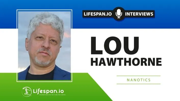
Lou Hawthorne

Lou Hawthorne, NaNotics on Cleaning Blood with Nanoparticles

Lou Hawthorne is the founder and CEO of NaNotics, a biotech company that creates nanoparticles that can soak up harmful molecules in circulation. This paradigm-shifting technology can be used to fight cancer as well as various inflammatory disorders, such as sepsis, which kills more people than cancer does. Other potential targets, familiar to everyone in the longevity field, are age-related sterile inflammation (inflammaging) and cellular senescence. Amazingly, Lou is an autodidact who earned his place in the field with his ingenuity and relentless pursuit of knowledge.

What is your story? What was your journey to the longevity field? 

Before I got into science almost 30 years ago, I was a professional graphic designer, cinematographer, and editor, mostly in the Bay Area. In addition to creative work for various clients, I also consulted on highly technical feasibility studies on the integration of digital media and broadcast video, which in the 1980s and early 90s involved a number of challenges that no longer exist.

I got drawn into science through a documentary I worked on for a couple of years about the Biosphere 2 project in Arizona, a self-contained biological habitat. Making a documentary involves such an enormous amount of time and effort that you naturally become an expert in the subject matter, so this documentary was a crash course in botany, animal physiology, atmospheric geology, and many other scientific topics. That was my first very deep dive into science aside from an immunology course in college. I surprised myself not just with how much I loved the scientific side of this project but also by my facility at absorbing the material.

Not long after Dolly the sheep was cloned, I was hired to explore the feasibility of cloning the pet dog of a billionaire. I had no particular knowledge of cloning, but I had some basic scientific knowledge and I knew how to organize a feasibility study. I wasn’t a bad choice for this role; there wasn’t anyone in particular you could call at the time to clone your dog!

I was originally hired to do a three-month feasibility study on dog cloning. After speaking to various experts – including at Roslin Institute in Scotland, where Dolly was cloned – I delivered a report to my client stating that just because sheep had been cloned, that didn’t mean dogs could be cloned, given that their reproductive physiology was quite different. Even assuming dogs could be cloned, it would require years of very expensive work. I figured that delivering this report would be the end of my involvement, but much to my surprise I was hired to perform the next step: assembling a team of experts capable of completing the work.

Over the next several years, I became something of a cloning expert. That was my first real domain of expertise in science. Cloning is very much about cellular signaling, given that the embryo is in constant molecular communication both with itself and with the surrogate that carries it. Much of this communication is immunologic. The surrogate’s immune system is naturally on guard against foreign invaders of all types. When you insert a cloned embryo into the recipient’s uterus, the goal is to create immunologic “privilege” – meaning the surrogate’s immune system allows the cloned embryo to persist and grow inside her, despite being genetically unrelated to her. All pregnancies – even with non-cloned embryos – actually face a similar problem, given that an embryo produced sexually is only half the mother and thus should be destroyed by her immune system as a parasite.

I became very interested in the molecular mechanisms of immune privilege – how they work and why they sometimes don’t work.

Along the way, I would often hear cloning scientists speculate that the insights we were gaining in cloning might someday lead to cures for cancer. I gradually began to realize that cloning animals and treating cancer are actually mirror processes from an immunologic standpoint. When you clone, you’re trying to create immunologic privilege: to trick the recipient’s immune system into tolerating a foreign embryo. Cancer cells are also abnormal, yet the immune systems of cancer patients tolerate their tumors, because tumors create the same type of immune privilege that a successful embryo creates. When you treat cancer, you’re not trying to create immune privilege as you are with cloning; you’re trying to strip it away.

I ended up spending more than a decade founding and leading various companies developing cloning technology for multiple species: cat, dog, horse, and cattle. The world’s first clone pet dog was actually a clone of my mom’s dog. She became my beloved companion.

Wait, you actually had a clone dog as a pet? 

Yes, for twelve and a half years till she died of old age, just a couple of years ago. Her name was Mira. She was an absolute sweetheart. I adored her. Mira was a clone of my mom’s dog Missy. We made four clones of Missy (we referred to them as the “near Missies”). Mira was the first born.

I just have to ask: did they differ in character, in personality?

The clones were of course physically identical, or as close to that as identical twins. They all started out with very similar behavioral tendencies because part of what gets cloned is the brain, so their neurological wiring was very similar. They responded to various stimuli in very similar ways; for instance, how they reacted to sounds. When they ran, they would run like a flock of birds. They would stop at the same moment, turn their heads at the same moment. They were all very curious and very good with language, picking up new words very quickly. They didn’t necessarily obey commands, because another similarity they had with each other and their genetic donor is that they were all very strong-willed. But they definitely understood what their humans were saying.

As they got older, their personalities started to diverge from each other and the “original” Missy (who had died years before they were born). The Missy clones were raised by different people with different lifestyles, which accelerated their divergence.

I was raising my son Skye at the same time. He had known the original, Missy, as a toddler, and he grew up surrounded by clones of her. It was a wonderful experience for all of us, a nature-vs-nurture experiment in our own home.

Along the way, I continued to study science, gradually moving beyond embryology but with a consistent interest in cellular signaling, which seemed to connect biology to information science, which I had been exposed to in my prior career in digital media.

I shifted my whole focus to disease and aging, with a particular emphasis on the role of pathogenic cell signals and signal inhibitors. The history of medicine began by focusing on organs, which you can easily see with the naked eye, then tissue, then cells. We’ve been able to see cells for 300 years since the invention of the microscope, and this has led to almost a fetish for cells as the locus of disease.

Over the last few decades, we’ve started to be able to detect what’s going on at scales below that of the cell, including the molecular signals that pass between cells, especially cytokines, the signaling molecules of the immune system. Once you investigate these intercellular signals, you realize that they play just as big a role if not bigger than any pathology that happens inside a cell.

In 2008, a dear friend of mine, one of my mentors in cellular signaling, a PhD endocrinologist named Chris Heward, developed stage 4 metastatic esophageal cancer. My investor was also his investor in a company called Kronos, which was focused on human longevity. I believe Kronos was the first really well-capitalized longevity research company, formed around 2000. They were focused on both age-related diseases and mechanisms of aging and published some important findings.

We all loved Chris. He was brilliant, funny, and outrageous. Our investor gave us a blank check “to find something that would save Chris.” That was how I came across a technology in Germany called Immune Pheresis, which treats cancer by depleting tumor-generated immune inhibitors from blood, using apheresis, an extracorporeal method of filtering blood, basically a more advanced version of dialysis.

The specific molecules that Immune Pheresis was depleting were soluble tumor necrosis factor (TNF) receptors. TNF is the body’s main “death signal”, normally delivered by immune cells directly to abnormal cells. TNF instructs an abnormal cell to commit suicide, a process called apoptosis. To protect themselves against TNF, cancer cells, like the human placenta, cleave off the extracellular portion of the TNF receptor, so it’s no longer connected to a cell. This extracellular receptor portion is then called an sTNF-R, the “s” meaning “soluble”, i.e. dissolved in blood. sTNF-Rs intercept and neutralize incoming TNF before it can reach a target cell and induce apoptosis.

We were too late to save Chris, who died just three months after his diagnosis. We were all just devastated.

However, we witnessed some phenomenal regressions induced by Immune Pheresis in patients who had already failed standard of care, meaning no improvement by any means would be expected. About 60% of the patients undergoing this treatment had significant regression of disease, including a surprising number of complete remissions. Even after Chris died, our investor encouraged me to dig more deeply into this data. Further studies confirmed that Immune Pheresis worked, that you could get regression and sometimes even full remission just by taking something out of their blood without adding anything at all.

We wanted to learn more about these inhibitors. We confirmed that the Immune Pheresis device can routinely deplete them by about 70% (NaNots can deplete them by 95%) over a 3- to 4-hour treatment. This alone could make the difference between tumor progression and regression in a patient with multiple kilograms of tumor. We measured the aggregate mass of the sTNF-Rs being depleted in a single treatment, which turned out to be 10 to 12 micrograms. That’s it. By depleting this minute amount of inhibitors, you tilt the battle in cancer away from the tumor and toward the immune system, ultimately leading to regression. You have to repeat the process several times, but it’s still a phenomenal result, entirely without drugs and the associated toxicity.

A profound question came out of these studies: Why do you need a refrigerator-sized device just to deplete 12 millionths of a gram of immune inhibitors from blood? Surely we could design something injectable that would do the same thing? This question triggered the creative process that ultimately led to the invention of NaNots.

This is a fantastic story, and I am sorry about your friend and mentor. So, you accomplished all of this without formal training in biology? 

That’s correct. I’ve been interested in science and science fiction since I was a child, but I have no scientific training other than what I picked up working with scientists in various companies and on projects I was managing. In some ways it’s harder to be an autodidact. You have no laurels to rest upon, so you have to be constantly learning and innovating in order to be taken seriously.

Actually, at the end of the day, everyone is an autodidact because no one can just pour knowledge into your brain. You have to learn it yourself. I learn the same way any PhD learns. I read books and scientific papers, I go to conferences, and I get mentored one-on-one by experts in the field.

There are two key differences in my approach to learning science versus that of the average PhD. One, I wrote my own syllabus. For better or worse, no department head told me what I needed to study. Two, nobody has validated my knowledge in a formal way with a certificate or a degree. My validation comes through invention: the technologies I’ve designed and patented that actually work. There’s no arguing with that.

This should be really inspiring for many people.

I hope so. There’s a new generation of serious biotech scientists who are autodidacts or come from fields other than biology, such as Matthew Scholtz, who runs Oisin. He came from computer science, not biology, but he’s become a fantastic and highly innovative biological thinker. There are more and more like us. Traditional academic educational pathways that train you to think in certain ways aren’t necessarily the best pathways for developing truly orthogonal therapeutics; they certainly aren’t the only pathways. Interdisciplinary backgrounds can be very helpful for conceptualizing novel solutions.

Let’s dig deeper into NaNotics’ technology. Could you start by briefly explaining the concept?

First, it’s important to understand the challenge we were aiming to overcome. When I asked the inventor of Immune Pheresis whether there was something that could be injected into the patient that would do the same thing as his pheresis technology, he gave me a brilliant two-hour lecture over dinner on why that wasn’t possible.

His main point – and I’m oversimplifying a bit here – was that if you target soluble TNF receptors with a drug, that drug is also going to bind and block membrane TNF receptors, because they’re biochemically identical. But membrane TNF receptors are a pathway that must be kept open, because innate immunity in particular depends on it. So, a drug against soluble TNF receptors would wreck innate immunity, and patients would die very quickly.

This challenge didn’t seem insurmountable. It made sense that you couldn’t achieve the goal with an antibody drug, but the micron-scale engineering I had overseen as part of my cloning work made me wonder if it might be possible to create an engineered structure at the nano scale that could capture the soluble target form (sTNF-Rs) without disturbing the membrane form (TNF-Rs), despite the fact that they’re biochemically identical.

The “eureka moment” came while hanging out with my cloned dog Mira. At the time, she had a minor skin infection and was wearing that cone that they put on dogs’ heads. I was feeding her a treat through the cone, and noticed that although the cone prevented her from biting her skin, she could still take treats from my hand. At that moment, I realized we could design a nanoparticle covered with capture agents with a little cone – or something like a cone – over each of them. They could still capture soluble factors, but the cone would prevent contact between the capture agents and cellular surface proteins of all types, including TNF-Rs.

Then it became a question of “How do we design that?” I did a bunch of sketches, and I started passing them around to scientists and nano-engineers that I knew. We also searched the scientific literature and patent records for something similar – nothing. Nobody had ever done it, which amazed me. Then I started talking to prospective investors. Among them was Jonathan Milner, founder of Abcam, the world’s largest public company focused on antibodies, and he became our first seed investor. Our next step was mathematical in silico modeling of various particle designs.

I thought it would probably take about as long for our nanoparticles to absorb their targets as pheresis – three to four hours. But the math showed that our nanoparticles would start working within seconds. Even with a very low aggregate mass of particles (we currently project one to two milligrams per kilogram, of which 95% is inert silica), that mass is distributed across trillions of particles. That means that within a few heartbeats, our typical nanoparticle and the typical target would be less than a micron apart, and diffusion driven by Brownian motion would bring them together within seconds.

Along the way, we named the particles “NaNots”. The inclusion of “Not” within the name is important, because NaNots are the opposite of a drug; they deplete their targets rather than adding molecules. NaNots can reverse a large number of biological processes by depleting either a pathogenic signal or an inhibitor of a healthy signal.

I find information science to be helpful in understanding biology. I view cells as basically little computers. With computers, the output they send depends to some extent on the inputs they receive. The same is true with cells. A cell’s “ROM” or prewritten code is the gene sequence. This coding includes many branching responses depending on signals picked up by cell surface receptors, which lead to changes in epigenetics. Cellular function can change dramatically based on signals received from outside the cell. The cell may die, or proliferate, or do any number of other things, and incoming signals also may change the signals that the cell sends out.

In the early days of computer science, we were very focused on the computer itself and on the software that you load into it. Nowadays, the computer is basically irrelevant, it’s just a portal to information in the cloud. A similar shift is happening in biology. We still pretty much fetishize the cell and its internal workings, but it’s becoming more apparent that cells largely do what they’re told based on signals they receive from other cells. This is true in both health and disease. You can have a disease manifest itself at a tissue or organ level, but when you investigate what’s happening at the cellular level, you often find that cells are doing exactly what they’re supposed to be doing relative to the signals they’re receiving. It’s the signals that are wrong or don’t reach their intended target.

One can even make a credible argument that in terms of health and disease, the network of cell signals is what really matters; cells themselves are just logic gates and depots of materials needed to maintain that network.

I guess not for nothing altered cellular communication is one of the hallmarks of aging.

Definitely. However, this wasn’t one of the original hallmarks. It was added because people like me were insisting that we mustn’t forget about the role of aberrant intercellular communication in disease and aging. Some of us believe this dysregulated signaling between cells is really most of the game of age-related diseases and aging itself. It’s certainly a huge part.

We now understand that tumors, like the fetus/placenta, defend themselves with immune inhibitors, both membrane and soluble inhibitors. It’s more than an analogy, because tumors and the fetus/placenta use the same molecules to inhibit immune attack. The inhibitors used by embryos and tumors are also employed by senescent cells, which makes sense because senescent cells are halfway to cancer when P53 suppresses them.

This last idea, that senescent cells defend themselves with soluble immune inhibitors, is still new and controversial. I’m one of the few scientists that’s arguing this point. The senescence-associated secretory phenotype (SASP) produced by senescent cells is packed full of immune inhibitors. What are they doing in the SASP if not protecting senescent cells from immune-mediated destruction? It takes a lot of cellular resources to produce these inhibitors, and cells don’t do that for no reason.

When most scientists think about immune inhibition – or cellular signaling at all – they gravitate toward membrane signaling. This is a natural tendency because membrane signals are on cells, and we tend to fetishize cells, as I mentioned. Checkpoint inhibitors in cancer were developed based on this idea that cells control each other through membrane interactions.

Let me use a military analogy here. The soldier only wants to use his bayonet as a last resort when the enemy is right on top of him. Prior to that, soldiers prefer to use bullets, to kill the enemy at the greatest possible distance. In the body, the fetus, the tumor, and senescent cells all use soluble immune inhibitors to inhibit the immune system at a distance, before they rely on membrane immune inhibitors in a last-ditch attempt to defend themselves from immune-mediated destruction.

I understand that NaNots are cleared by macrophages, right? How does this happen, and how safe in general NaNots are?

NaNots are particles, and particles are naturally cleared by macrophages. Any foreign particle is going to be phagocytized by macrophages. This would normally happen within minutes, but we can delay it for up to about 16 hours by attaching a “stealth” coating to the NaNots.

This coating prevents opsonization, which is the attachment of protein markers that flag a particle for clearance by macrophages. Eventually, NaNots get engulfed by macrophages and broken down – along with their cargo of captured proteins – into small molecules, which are then excreted.

The current generation of NaNots are built on a core of silica, which has been used in other nanomedicines and is well-known for its ultra-low toxicity (if properly coated). Although no one’s ever made a NaNot before, the same materials are used in other nanomedicines that possess a very good safety profile, both at the macro “whole animal” level and in terms of macrophages.

We’ve dosed NaNots in rodents at 100x the planned dose in humans, and we haven’t seen any toxicity yet. None. Based on our data so far, we believe NaNots are completely non-toxic, which is unheard of for a drug, especially a drug against a major disease like cancer or sepsis. We haven’t talked about sepsis yet.

Let’s do it. The applications seem to be infinite because we’re talking about molecules in the circulation and intercellular communication, so I understand that it can be used against sepsis, autoimmune diseases, and even cellular senescence?

Any time you have a new therapeutic strategy, usually it ties into a whole ontology of disease that might be different from regular disease ontologies. For instance, with checkpoint inhibitors, now all of a sudden tumors are being classified as “hot” versus “cold” in terms of how immunogenic they are. That’s new.

In our ontology, there are three kinds of disease: There are genetic diseases, which we don’t touch; they actually only account for less than 10% of all diseases. The vast majority of other diseases are either inflammatory, driven by an inflammatory cytokine or cytokines, or inhibitory diseases where you have aberrant cells defending themselves with immune inhibitors.

Sepsis is the most acute form of inflammatory disease, in which the immune system massively overreacts to a trigger of some kind. Some people confuse sepsis with infection, but infection is just one of several possible triggers of sepsis. The morbidity and mortality of sepsis is due to the immune system’s overreaction, not the trigger.

It’s a kind of cytokine storm, right? 

Correct. A cytokine storm is a positive feed-forward loop, stemming from the fact that cytokines are pleiotropic, meaning they have multiple effects. When TNF lands on a non-immune cell and ligates the TNF-R1 receptor, it induces apoptosis, which destroys the cell. When TNF is received by an immune cell and ligates the R2 receptor – which is preferentially expressed on immune cells – it induces proliferation and activation of that immune cell.

Under certain circumstances, the initial cytotoxic immune response supported by TNF-mediated recruitment of additional immune effector cells can spiral out of control. It’s analogous to a nuclear meltdown following the failure of a reactor’s cooling system. Once the meltdown starts, you can spray all the water you want on the reactor, and it won’t stop the meltdown.

The pleiotropy of TNF is an evolved mechanism of enhanced immune response, and normally it works really well, with the occasional biological “cost” of sepsis. TNF pleiotropy offers a net benefit in terms of the survival of the species even though the cost at a population level is quite high: sepsis kills more people than all forms of cancer combined.

Clinicians like to emphasize that sepsis is very complex. We view it a little differently. Wherever the cytotoxic cytokines involved in sepsis touch tissue, that will result in a clinical complication. Given the broad array of tissues that can be hit by these systemically elevated cytokines, you can incur a broad range of clinical complications, but the base driver of the storm is not complex. It’s mainly driven by three inflammatory cytokines released in a cascade: TNF-α, which is the upstream driver of the cascade, followed by IL-1ß and IL-6. There are other cytokines involved, but those are the main players.

TNF is the first responder, IL-1ß organizes a coherent immune response (or over-response), and IL-6 is more generally a “soldier” that inflicts most of the damage. If you soak up those molecules, the clinical complexities don’t arise and thus don’t matter. You can extinguish the storm. You’ll have to deal with the damage that’s already been incurred, but you can stop the runaway reaction and prevent further damage and death. It’s like pushing the control rods back into the reactor before the meltdown occurs. That’s what anti-inflammatory NaNots will do. They’ll soak their target cytokines rapidly and thoroughly before the process spirals out of control.

There are drugs against all those cytokines, but they don’t work in cytokine storm for several reasons. First, they’re not specific to soluble cytokines. They also hit the membrane form of TNF (for instance) stored on the surface of immune cells. If you administer an anti-TNF drug, you’re going to neutralize membrane TNF on the surface of immune cells in addition to the targeted soluble form, and there goes your immune competence. You can’t do that in sepsis because you often still have an active pathogen that triggered the sepsis in the first place. Treating sepsis requires that you neutralize the systemically elevated cytokines without reducing immune competence. That’s the part no one has ever cracked.

The second reason why those drugs don’t work is that their arc – their persistence in circulation – is too long. They last for days in the blood, which aggravates a later stage of sepsis known as “immune paralysis”. Many patients die in this phase, when the body throws every immune inhibitor it has against the cytokine storm. Anti-inflammatory drugs that last for days drive the patient deeper into immune paralysis, whereupon they die of whatever nosocomial infections are floating around the hospital. The effect would be like HIV.

So, the DMARD drugs against cytokines are the wrong tool. You want something that only hits the soluble form, doesn’t disturb the membrane form, and only acts in circulation, without extravasating into tissue. Finally, you need something that acts fast and clears rapidly. These parameters describe NaNots perfectly.

But that means that after an initial injection, the levels of cytokines that you’re trying to deplete are going to go back up. So, how does the delivery work – do you need multiple injections or, maybe, continuous intravenous delivery?

They may go back up, or they may not. What we know for certain is that the first injection of NaNots will deplete the target to near zero. With this, you achieve two things at once. First, you interrupt MODS – multiple organ dysfunction syndrome, the main killer in sepsis. That’s when organs start to fail, which happens for two reasons.

One, blood pressure crashes, because part of those cytokines’ natural purpose when expressed at low levels during infection is to increase the permeability of the vasculature so that immune cells can get to pathogenic cell clusters faster. When expressed at very high levels in sepsis, you get leaky vasculature, blood pressure drops, then the organs don’t get enough blood and begin to die. Of course, the cytokines also directly attack the organs as well. Both of those processes will be abrogated by soaking up the cytokines with NaNots.

However, even assuming you fully deplete the cytokines that were recruiting immune cells, it’s possible that some of those immune cells are now dysregulated and will persist in a hyperstimulated state, cranking out excess inflammatory cytokines systemically, even after the first dose of NaNots. So, it’s possible that after soaking up all the inflammatory cytokines initially, you may need to administer another dose of NaNots to address subsequent release.

But with each dose, you’re stopping the tissue damage stemming both from apoptosis of cells in organ tissue and from permeation of the vasculature, and you’re eventually breaking the storm. Patients usually die of sepsis in five to seven days. Even if you have to inject the patient half a dozen times during that period, that shouldn’t be an issue given the ultra-low toxicity of NaNots. You just monitor the patients, measure their cytokines before, during, and after treatment, and administer the specific NaNots needed in the doses required.

I can understand how it can work in a hospital environment, but how can it potentially be used against inflammaging, as a preemptive anti-aging treatment?

That’s a good question. Not everybody accepts it, but I think there’s a consensus that inflammaging is real and that it’s a driver of not just age-related diseases but of aging itself. Inflammation is a critical target for anti-aging therapy.

I personally think that, if anything, inflammation is probably underappreciated as a driver of age-related diseases.

I would agree. Anyway, it’s clearly a real thing. We know that because these cytokines rise with age. In healthy young people, they’re at or below the level of detection. There’s no purpose served by them being elevated systemically. If they are constantly elevated, which we see in older people, it’s like the difference between a sniper taking out a terrorist with a precise kill shot, versus a madman spraying bullets in every direction. Sorry for all the military analogies, but I really feel like there is a war going on in the body.

In order to treat Inflammaging, we have to soak up these excess inflammatory cytokines not just over a period of days as in sepsis but over a period of decades. Our current GEN-2 NaNots circulate for 16 hours, which is great for treating acute conditions, but if you want to treat somebody, say, once a week for decades, you need an ultra-low toxicity, non-accumulating technology, and that’s not silica. When treating serious diseases over weeks or months, our GEN-2 NaNots are non-toxic – certainly compared with checkpoint inhibitors, chemotherapy, et cetera – but they’re not zero toxicity.

So, for treating aging, we’re developing GEN-3 NaNots, which are polymer-based. You should be able to dose somebody with GEN-3 NaNots every week for the rest of their lives with no cumulative negative effects. In our vision of NaNots used as a longevity treatment, you’ll swing by your local Quest for a measurement of the signals and inhibitors that are elevated in your body, then you’ll receive an injection of just the right NaNots in the right dose to bring your intercellular communication into balance. We already have a slogan for that service: “Get Your Signals Straight!”

I think this is the future of anti-aging therapies: everyone will be getting a weekly or a monthly appointment, with several therapies thrown at them for the rest of their lives.

Why not? Interestingly, I’ve had several cycles of therapeutic plasma exchange, and it feels amazing when you get these inflammatory cytokines cleared from your blood. You feel it immediately. I suspect that NaNots will feel amazing too. Inflammatory cytokines rise steadily with age, they degrade tissue and organs throughout your body, and they easily cross the blood-brain barrier and drag down your cognitive function too. So, once you clear them out or reduce their concentration, you are going to feel it.

Because this is such a universal technology, the list of potential targets can go on and on. Can you briefly tell me about your current pipeline? 

We have a pipeline of eight NaNots (with dozens more in line after that). Four of them are against the inflammatory cytokines that drive many canonical inflammatory diseases like arthritis and psoriasis along with the newly understood non-canonical autoimmune diseases like type 2 diabetes and atherosclerosis. For instance, the calcification that happens in atherosclerosis is driven by TNF.

PTSD, depression, all sorts of cognitive disorders, cardiovascular diseases – these are cytokine disorders as well. Pro-inflammatory cytokines rise with age, and whatever they touch has a protective response against it. Heart cells calcify. Why do they do that? Because they can’t be allowed to die, so they do something that isn’t ideal: they calcify instead, which is a known response to excess circulating TNF.

Likewise, when the brain is exposed to inflammatory cytokines, it forms plaques. That’s not ideal either, but from an evolutionary perspective, it’s better than the brain dying. Many processes that happen in the body are basically suboptimal protective responses against these inflammatory cytokines. We’re focused on “the big four”: TNFa, IL-1ß, IL-6, and IFN-γ. We plan to use NaNots first against FDA-recognized autoimmune and autoinflammatory diseases, then repurpose these NaNots to treat sepsis and inflammaging.

That’s on the inflammatory side. On the inhibitory side, we’re taking out four of the main inhibitory molecules that cancer uses to defend itself: soluble PD-L1, sTNF-R1, sTNF-R2, and soluble HLA-G. Some of these have been documented as active in senescent cells or in the general reduction of immune competence that comes with age. We want to treat FDA-recognized diseases, because these diseases are terrible, but also because therapeutics against FDA-recognized diseases can be prescribed off-label for longevity applications.

So, diseases like cancer are the gateway, but the idea is, instead of extinguishing fires, to find ways to treat the root cause eventually?

Yes, I’m totally down with that mission. I always have been.

What do you think of the current longevity field in general?

I think it’s poised to revolutionize medicine. Some of the breakthroughs that are happening in geroscience are going to spill over into how we understand and treat mainstream diseases as well. Longevity science has the potential to radically transform our lives, and the value creation is going to be phenomenal as well. People like Jim Mellon say that the anti-aging field is going to be the most valuable field in history. Investments in this space are going to pay dividends far beyond anything we’ve seen before. The time scale may be uncertain, but I think the basic premise is true.

Elderly Japanese couple

Protecting Memory Through Gut Bacteria

Researchers publishing in Nutrients have shown that adding a strain of lactic acid bacteria to the gut flora of older, memory-impaired people partially alleviates their memory problems.

A specific anti-inflammatory bacterium

The researchers begin their paper by noting that mild cognitive impairment (MCI) is associated with memory loss and is frequent precursor of Alzheimer’s disease [1]. While no treatment has been shown to reverse Alzheimer’s disease, there have been longitudinal studies showing that the reversal of MCI is possible [2] and that lifestyle factors are important in the development of Alzheimer’s [3]. Therefore, while entirely curing Alzheimer’s is not yet possible, it might be possible to prevent it. These researchers have previously explored this approach with anserine, camosine [4], and green tea extract [5].

Previous research has shown a relationship between the gut flora and the development of Alzheimer’s disease [6], and other research has shown a role of inflammatory cytokines [7]. This led the researchers towards a novel approach: altering the gut flora through the addition of OLL2712, a strain of lactic acid bacteria that promotes the anti-inflammatory cytokine IL-10 [8], which has been shown to protect neurons in Alzheimer’s disease [9].

A randomized, controlled trial

The researchers recruited a total of 78 adults over the age of 65 with MCI as defined by the results of a word list memory test. People with systemic illnesses or full-blown dementia were excluded; as this study involves lactic acid bacteria, it also excluded people with dairy allergies. The participants were tested on verbal and visual memory, administered either placebo or OLL2712 for 12 weeks, and then tested again.

Being in the treatment rather than the control group was found to be a significant factor in the retention of cognitive abilities, specifically visual memory; treatment group participants had, on average, slightly higher visual memory scores than when they started, while the placebo group slightly declined. Regression analysis was performed to identify potentially confounding factors. Dietary cholesterol was negatively associated with cognitive function and seemed to impede the effects of OLL2712, while Vitamin K seemed to aid in the treatment.

Although three species of bacteria declined as a result of the treatment, the introduction of OLL2712 to the microbiome was not shown to have significantly changed the balance of the gut flora as a whole. These three species are associated with various forms of inflammation and gut infection, and previous research has shown that two of them decline when other probiotics are administered [10].

Conclusion

While this study shows a strong relationship between OLL2712 administration and a reduction in cognitive decline, the researchers note a few limitations. This analysis was performed on a group of generally healthy Japanese adults living in the same community. There was no direct analysis of brain biomarkers performed, and there was also no biochemical analysis of the mechanism of action. The researchers intend to follow up this study with further investigation into this area.

We would like to ask you a small favor. We are a non-profit foundation, and unlike some other organizations, we have no shareholders and no products to sell you. All our news and educational content is free for everyone to read, but it does mean that we rely on the help of people like you. Every contribution, no matter if it’s big or small, supports independent journalism and sustains our future.

Literature

[1] McKhann, G. M., Knopman, D. S., Chertkow, H., Hyman, B. T., Jack Jr, C. R., Kawas, C. H., … & Phelps, C. H. (2011). The diagnosis of dementia due to Alzheimer’s disease: Recommendations from the National Institute on Aging-Alzheimer’s Association workgroups on diagnostic guidelines for Alzheimer’s disease. Alzheimer’s & dementia, 7(3), 263-269.

[2] Ganguli, M., Jia, Y., Hughes, T. F., Snitz, B. E., Chang, C. C. H., Berman, S. B., … & Kamboh, M. I. (2019). Mild cognitive impairment that does not progress to dementia: a population‐based study. Journal of the American Geriatrics Society, 67(2), 232-238.

[3] Kivipelto, M., Mangialasche, F., & Ngandu, T. (2018). Lifestyle interventions to prevent cognitive impairment, dementia and Alzheimer disease. Nature Reviews Neurology, 14(11), 653-666.

[4] Masuoka, N., Yoshimine, C., Hori, M., Tanaka, M., Asada, T., Abe, K., & Hisatsune, T. (2019). Effects of anserine/carnosine supplementation on mild cognitive impairment with APOE4. Nutrients, 11(7), 1626.\

[5] Sakurai, K., Shen, C., Ezaki, Y., Inamura, N., Fukushima, Y., Masuoka, N., & Hisatsune, T. (2020). Effects of matcha green tea powder on cognitive functions of community-dwelling elderly individuals. Nutrients, 12(12), 3639.

[6] Pluta, R., Ułamek-Kozioł, M., Januszewski, S., & Czuczwar, S. J. (2020). Gut microbiota and pro/prebiotics in Alzheimer’s disease. Aging (albany NY), 12(6), 5539.

[7] Schwab, C., Klegeris, A., & McGeer, P. L. (2010). Inflammation in transgenic mouse models of neurodegenerative disorders. Biochimica et Biophysica Acta (BBA)-Molecular Basis of Disease, 1802(10), 889-902.

[8] Toshimitsu, T., Mochizuki, J., Ikegami, S., & Itou, H. (2016). Identification of a Lactobacillus plantarum strain that ameliorates chronic inflammation and metabolic disorders in obese and type 2 diabetic mice. Journal of dairy science, 99(2), 933-946.

[9] Su, F., Bai, F., & Zhang, Z. (2016). Inflammatory cytokines and Alzheimer’s disease: a review from the perspective of genetic polymorphisms. Neuroscience bulletin, 32(5), 469-480.

[10] Wang, C., Li, W., Wang, H., Ma, Y., Zhao, X., Zhang, X., … & Li, J. (2019). Saccharomyces boulardii alleviates ulcerative colitis carcinogenesis in mice by reducing TNF-α and IL-6 levels and functions and by rebalancing intestinal microbiota. BMC microbiology, 19(1), 1-12.

Clogged pipes

Klotho Reduces Arterial Calcification Through Autophagy

In a new study published in Oxidative Medicine and Cellular Longevity, the researchers have shown that klotho ameliorates vascular calcification via increased autophagy [1].

‘Rusty’ arteries

Vascular calcification, the deposition of minerals in the cardiovascular system, is a complex pathological process that accompanies aging. It is associated with an increased risk of stroke, coronary artery disease, and mortality.

There are several forms of vascular calcification, including the calcification of the vessels and the heart valves. Intimal calcification is confined to the intimal layer of the vessel wall, which is composed of endothelial cells, and the medial form takes place in the medial layer of the vessels, which is composed of smooth muscle cells and the elastin-rich extracellular matrix.

Intimal calcification is associated with atherosclerosis and is characterized by inflammation and lipid deposition. Medial calcification leads to stiffening of the arterial wall and hypertension.

Several approaches to reverse both intimal and medial calcification are being developed. For example, Cyclarity Therapeutics is working on interventions to target damaged cholesterol and thus remove arterial plaques, which might lead to calcium deposits dissolving and the reversal of atherosclerosis. Meanwhile, Elastin Therapeutics aims to restore degraded elastin by removing calcium deposits from stiffened arteries.

Previous studies have shown a link between the well-known ‘anti-aging’ enzyme klotho and vascular calcification. Given that klotho overexpression reverses aortic vascular calcification in klotho-deficient mice, further investigation is warranted to explore the underlying mechanism.

In this study, the researchers explored the link between vascular calcification and klotho levels. Importantly, lack of klotho is associated with all-cause mortality in people, so clarifying if and how klotho boosting should be done to rejuvenate aged arteries is certainly worth investigation.

Klotho to the rescue

First, the researchers analyzed the association between serum klotho levels and aortic calcification based on computed tomography images in 27 participants aged 55-85. People with low levels of klotho demonstrated a higher extent of calcification and vice versa.

Next, the researchers analyzed the aortas of 4-week-old mice with different levels of klotho expression: knockout (no klotho), klotho homozygous (some klotho), and wild-type mice (normal levels of klotho). As expected, klotho deficiency resulted in vascular calcification, with calcium content inversely proportional to klotho levels.

In the next set of experiments, the researchers showed that injecting klotho protein every 48 hours for 2 weeks reversed aortic calcification in knockout mice. Meanwhile, klotho-deficient mice demonstrated increased autophagy. Therefore, the researchers used an autophagy activator, rapamycin, and an autophagy inhibitor, chloroquine, to explore if autophagy manipulation could reverse calcification in klotho deficient mice.

Interestingly, the rapamycin-induced autophagy boost protected klotho knockout mice against vascular calcification, while inhibiting autophagy with chloroquine enhanced the calcification. Similarly, supplementing klotho promoted autophagy in the knockout mice, thus ameliorating the aortic calcification.

Finally, a series of experiments in vitro, which used mouse aortic vascular smooth muscle cells treated with a calcification medium, confirmed that klotho inhibits calcification by enhancing autophagy.

Abstract

Vascular calcification (VC) is regarded as a common feature of vascular aging. Klotho deficiency reportedly contributes to VC, which can be ameliorated by restoration of Klotho expression. However, the specific mechanisms involved remain unclear. Here, we investigated the role of autophagy in the process of Klotho-inhibiting VC. The clinical study results indicated that, based on Agatston score, serum Klotho level was negatively associated with aortic calcification. Then, Klotho-deficient mice exhibited aortic VC, which could be alleviated with the supplementation of Klotho protein. Moreover, autophagy increased in the aorta of Klotho-deficient mice and protected against VC. Finally, we found that Klotho ameliorated calcification by promoting autophagy both in the aorta of Klotho-deficient mice and in mouse vascular smooth muscle cells (MOVAS) under calcifying conditions. These findings indicate that Klotho deficiency induces increased autophagy to protect against VC and that Klotho expression further enhances autophagy to ameliorate calcification. This study is beneficial to exploring the underlying mechanisms of Klotho regulating VC, which has important guiding significance for future clinical studies in the treatment of VC.

Conclusion

This study explored in detail the protective effect of klotho against aortic calcification. The authors show that klotho deficiency triggers protective autophagy and promotes vascular calcification. Klotho supplementation then promotes further autophagy to inhibit calcification.

Therefore, autophagy is an important klotho-mediated mechanism that could potentially be manipulated to reverse vascular calcification. However, this mechanism was only demonstrated in mice, and the human data collected in the study is just a correlation between the levels of klotho and aortic calcification. Future research will hopefully exploit this knowledge to develop effective therapies to rejuvenate aged arteries.

We would like to ask you a small favor. We are a non-profit foundation, and unlike some other organizations, we have no shareholders and no products to sell you. All our news and educational content is free for everyone to read, but it does mean that we rely on the help of people like you. Every contribution, no matter if it’s big or small, supports independent journalism and sustains our future.

Literature

[1] Li, L., Liu, W., Mao, Q., Zhou, D., Ai, K., Zheng, W., … & Zhao, X. (2022). Klotho Ameliorates Vascular Calcification via Promoting Autophagy. Oxidative Medicine and Cellular Longevity2022.

Playing mice 800x450

Cellular Reprogramming Extends Lifespan, Healthspan in Mice


Scientists have shown that continuous low-dose induction of reprogramming factors and a single early life reprogramming treatment both significantly increase lifespan and healthspan in progeroid mice while altering their epigenetic landscape [1].

In vivo reprogramming

Cellular reprogramming is the process of treating cells with compounds that nudge them towards a pluripotent state. The classical reprogramming tool is the four-factor OSKM protein cocktail introduced by Shinya Yamanaka in 2006 [2], but since then, other factors have been proposed, including small molecules. Importantly, reprogramming is accompanied by cellular rejuvenation [3].

In its full form, reprogramming abrogates the cells’ differentiation, completely driving them to pluripotency. They are then called induced pluripotent stem cells (iPSCs) and can differentiate again into various cell types. When milder protocols are used, it is possible to rejuvenate cells without rolling back their cellular identities [4].

While cellular reprogramming holds great promise, it is a nascent technology with many open questions. Most studies are done in vitro, and only a handful have investigated the effects of reprogramming in vivo, with some encouraging results. This study joins that limited list.

Lifespan boost for half-progeroid mice

The researchers used progeroid mice, which were genetically modified to express a premature aging phenotype. When this mutation is homozygous (both sister chromosomes carry the progeroid modification), such mice live extremely short lives of about 15 weeks. Similar mice were used in the important 2016 study by Ocampo et al. that showed the rejuvenating effects of partial reprogramming [5].

To attenuate this phenotype and make it more resemble natural aging, in this study, the researchers used heterozygous mice, with one sister chromosome being modified and one remaining unmodified. Those hybrids’ average lifespan is 35 weeks, about a third of normal. Additional genetic alterations allowed the mice to express reprogramming factors only when the drug doxycycline was added to their drinking water. In previous studies, the mice were regularly “pulsed” with doses of doxycycline, turning OSKM expression on and off to avoid full reprogramming. However, in this study, the researchers attempted a different, simpler approach, constantly adding a smaller dose of the drug to the drinking water.

If done wrong, the experiment could have resulted in full reprogramming and consequent formation of tumors. However, the bet paid off. Not only were the treated mice much healthier than the controls, but they also lived longer. A lifelong treatment with 0.2 mg/ml of doxycycline yielded almost the same results as the more conventional 1 mg/ml periodic treatment, extending the mice’s median age of death from 42.6 to 55.6?weeks.

However, there was another big surprise. When the researchers tried limiting the treatment to a period of 2.5 weeks early in life, they still got a noticeable lifespan extension with a “middle ground” dose of 0.5 mg/ml. The increase in maximum lifespan was even more impressive than with the lifelong treatment (66.1 weeks). However, because of the small sample size, this particular result should be taken with caution.

Reprogramming Progeroid Mice  

Improved health and fitness

The single treatment protocol also resulted in noticeable improvements in fitness. Treated mice developed and maintained substantially more lean mass and higher ratios of lean mass to fat, and they continuously scored better than controls on strength tests.

On the histological level, the single treatment seemed to provide lifelong protection against age-related skin thinning and loss of elasticity up until the age of 8 months, when the last tests were done. The lungs, spleens, and kidneys of the treated mice were also in a much better shape than those of controls, with preserved function and less fibrosis.

Just like naturally aging animals (humans included), progeroid mice develop a wide range of age-related conditions, including bone and cartilage degeneration. A single early life treatment was able to significantly slow it, as measured by bone mass in major bones, cartilage volume, and cartilage degradation.

Epigenetic anti-aging protection

Since aging is closely associated with epigenetic alterations (namely, the distribution of methylation marks across the genome), the researchers compared methylation profiles of the study group versus controls at various points in time. The results showed that the single early life treatment is generally protective against age-related changes in methylation.

However, methylation differences between the study group and controls were not the same at 2 months and 8 months of age. This means that the initial epigenetic reprogramming induced by the treatment was not maintained but rather “propagated” – its profile changed with time via a currently unknown mechanism while still ensuring that the treated mice lived longer.

Conclusion

The major limitations of this study are its use of progeroid mice, albeit heterozygous, and a relatively small sample size. However, the results are impressive: the study shows that safe and effective partial reprogramming that extends healthspan and lifespan can be induced both by a continuous lifelong treatment and a single treatment early in life. The next step would be to replicate these results in naturally aging animals.

We would like to ask you a small favor. We are a non-profit foundation, and unlike some other organizations, we have no shareholders and no products to sell you. All our news and educational content is free for everyone to read, but it does mean that we rely on the help of people like you. Every contribution, no matter if it’s big or small, supports independent journalism and sustains our future.

Literature

[1] Alle, Q., Le Borgne, E., Bensadoun, P., Lemey, C., Béchir, N., Gabanou, M., … & Lemaitre, J. M. (2022). A single short reprogramming early in life initiates and propagates an epigenetically related mechanism improving fitness and promoting an increased healthy lifespan. Aging Cell, e13714.

[2] Takahashi, K., & Yamanaka, S. (2006). Induction of pluripotent stem cells from mouse embryonic and adult fibroblast cultures by defined factors. cell, 126(4), 663-676.

[3] Simpson, D. J., Olova, N. N., & Chandra, T. (2021). Cellular reprogramming and epigenetic rejuvenation. Clinical Epigenetics, 13(1), 1-10.

[4] Lehmann, M., Canatelli-Mallat, M., Chiavellini, P., Cónsole, G. M., Gallardo, M. D., & Goya, R. G. (2019). Partial reprogramming as an emerging strategy for safe induced cell generation and rejuvenation. Current Gene Therapy, 19(4), 248-254.

[5] Ocampo, A., Reddy, P., Martinez-Redondo, P., Platero-Luengo, A., Hatanaka, F., Hishida, T., … & Belmonte, J. C. I. (2016). In vivo amelioration of age-associated hallmarks by partial reprogramming. Cell, 167(7), 1719-1733.

Rejuvenation Roundup October

Rejuvenation Roundup October 2022

Ghouls, goblins, and ghosts were all on display last night, but today, we bring you something even scarier: Aging! Let’s see what’s been done to fight back against aging in October.

LEAF News

Team and activities

Lifespan.io EditorialLeading the Movement for Longer and Healthier Lives: As a non-profit organization, our goal is to engage and educate the public about the potential of rejuvenation and a world in which age-related diseases are a thing of the past. With that in mind, this is the ideal opportunity to tell you about some of the exciting things that we have done recently.

Lifespan News

JellyfishDAO: This episode of Lifespan News focuses on JellyfishDAO, a decentralized autonomous organization that intends to accelerate the development of life extension technologies.

Limitless with Chris Hemsworth: Ryan O’Shea uses this episode of Lifespan News to discuss an upcoming series in which a well-known Marvel actor pushes his limits in real life.

Interviews

Morten Scheibye-KnudsenThe Longevity Molecule Project with Morten Scheibye-Knudsen: Dr. Morten Schiebye-Knudsen is the founder of the Longevity Molecule Project funded by VitaDAO. However, this is just one side of his research. After starting his career as a physician in Denmark, he studied at the National Institute on Aging before founding the Scheibye-Knudsen Lab.

Rejuvenation Roundup Podcast

Ryan O’Shea of Future Grind hosts this month’s podcast, showcasing the events and research discussed here.

Journal Club

Short-Term Rapamycin Treatment Extends Lifespan in Flies: Dr. Oliver Medvedik took a look at a recent study published in Nature Aging that has shown that short-term rapamycin treatment in early adulthood extends lifespan in flies and improves gut health in both flies and mice.

Research Roundup

Ground coffeeCoffee Is Associated with Better Cardiovascular Health: Drawing on data from UK Biobank, scientists have once again confirmed the association between coffee and better health outcomes, with ground coffee emerging as the healthiest type.

Removing Senescent Cells Improves the Brains of Female Mice: Researchers publishing in Nature Communications have detailed how the removal of p16-producing senescent cells leads to improvements in the brains of female mice due to the involvement of microglia.

Multiple drugsCombination of Rapamycin and Acarbose Extends Lifespan: In a new study published in Aging Cell, researchers have tested several individual drugs and a combination of rapamycin plus acarbose as potential life extension agents in genetically heterogeneous mice.

Healthy Lifestyle Associated with Reduced Aging Biomarkers: Researchers publishing in BMC Medicine have found that there is a relationship between biomarkers of aging and measurements of health and mental well-being. This study further shows the link between unhealthy behaviors and adverse health outcomes.

Mutant cellHow Cancers Handle Protein Misfolding: A preprint published in bioRxiv has shown the mechanisms by which cancer handles the protein dysfunction brought about by its own mutational load and how these mechanisms are different from ordinary proteostasis machinery.

Aging of the Heart Correlates With a Poor Gut Microbiome: In ESC Heart Failure, researchers have commented about the correlation between a poor gut microbiome and aging of the heart. Research increasingly suggests that the quality and composition of the gut microbiome may play a role as important as exercise on health.

Brain regionsMitochondria in Different Brain Regions Age Differently: In a study published in Free Radical Biology and Medicine, researchers have identified a link between mitochondrial function and the vulnerability of specific brain regions to age-associated neurodegeneration.

Older Immune Cells Kill Cancer Cells More Quickly: In Aging Cell, researchers have published the surprising and counterintuitive finding that older CD8+ T cells are able to kill cancer cells more quickly than their younger counterparts.

BoostT Cells Acquire Telomeres from Antigen-Presenting Cells: Scientists have found that when T cells are activated by antigen-presenting cells, the activating cells donate chunks of their telomeres to the T cells, preventing senescence. Telomeres are sequences of TTAGGG repeats at the ends of our chromosomes that protect their integrity.

The Impact of Genetic Risk Factors on Healthspan: A research paper published in Nature Medicine has shown the results of a broad investigation into gene variants associated with healthspan. This was accomplished with a genome-wide association study, an effort to use enormous databases and computational analysis to examine the genetic causes of disease.

Air PollutionAir Pollution Impairs Lung Function: In a new study published in Ecotoxicology and Environmental Safety, the researchers have shown that air pollution leads to lung function decline accompanied by lung and intestinal microbiome imbalances.

Senescent Cells Can Be Good or Bad According to New Study: New research has scientists reconsidering the role of ‘zombie’ cells that drive some aspects of aging and whether they should be eliminated using senolytic drugs. It turns out that not all senescent cells are necessarily bad and that some may be helpful.

Bowhead whaleA Doubled Gene Explains the Bowhead Whale’s Lifespan: A preprint published in bioRxiv has partially explained the lifespan of the bowhead whale through its duplication of CDKN2C, which regulates how cells divide.

HIIT and Intermittent Fasting Show Cumulative Effect: Scientists have shown that both high-intensity interval training (HIIT) and time-restricted eating improve metabolism in healthy overweight women, with their combination providing the biggest effect.

Squatting at deskIntermittent Exercise Encourages Muscle Protein Uptake: A research paper published in the Journal of Applied Physiology has found that regular, intermittent exercise encourages amino acids to form proteins in muscle. Previous research has found that a lack of activity accelerates the age-related muscle loss known as sarcopenia.

Pain Is Associated with Increased Risk of Falls in Adults: In a new longitudinal study published in European Geriatric Medicine, the researchers have shown that people experiencing moderate to severe pain in multiple sites have an increased risk of falls, particularly in middle age.

Olive oilOlive Oil Linked to Significantly Lower Mortality: In a new study published in the journal Nutritional Epidemiology, scientists have found a strong association between olive oil consumption and lower risk of cardiovascular, cancer, and all-cause mortality.

New Compounds Beat Resveratrol in Sirtuin Activation: A recent study published in Molecules has tested multiple thiazole-based derivatives that appear to activate the sirtuin SIRT1 more than resveratrol. Resveratrol, commonly found in grape skins and red wine, generated some of the initial interest in sirtuin activators after initial studies showing many therapeutic benefits, such as cancer prevention.

Vitamin D Combats Oxidative Stress in Gingival Cells: A cellular study published in the Journal of Dental Sciences has shown that Vitamin D alleviates inflammaging related to the oxidative stress brought on by advanced glycation end-products (AGEs). Previous research has shown that the uncontrolled blood sugars associated with Type 2 diabetes attach themselves to other proteins and fats.

Combining stem cell rejuvenation and senescence targeting to synergistically extend lifespan: We had previously published the preprint of this now peer-reviewed article, which shows a synergistic effect between senolytics and stem cell rejuvenation.

Small extracellular vesicles from young adipose-derived stem cells prevent frailty, improve health span, and decrease epigenetic age in old mice: Epigenetic age was lower in the tissues of old mice treated with these vesicles, and their metabolome was changed to a more youthful one.

A single short reprogramming early in life initiates and propagates an epigenetically related mechanism improving fitness and promoting an increased healthy lifespan: In old age, treated mice have improved tissue structures in kidney, spleen, skin, and lung, with an increased lifespan of 15%.

Centenarians consistently present a younger epigenetic age than their chronological age with four epigenetic clocks based on a small number of CpG sites: Epigenetic aging and potentially biological aging are slowed in exceptionally long-lived individuals, and epigenetic clocks based on a small number of CpGs are sufficient to reveal alterations of the global epigenetic clock.

Fetal muscle extract improves muscle function and performance in aged mice: Further analysis of the active ingredients of the extract will shed light on the development of a novel treatment for sarcopenia.

Tissue-specific impacts of aging and genetics on gene expression patterns in humans: In this study, the researchers quantify the relative contributions of genetics and aging to gene expression patterns across 27 tissues from 948 humans.

Repurposing SGLT-2 Inhibitors to Target Aging: Available Evidence and Molecular Mechanisms: The researchers hold that the burden of evidence might prompt the design of studies testing the potential employment of this class as anti-aging drugs.

Impacts of Astaxanthin Supplementation on Walking Capacity by Reducing Oxidative Stress in Nursing Home Residents: The effect was most likely accompanied by an increase in endurance instead of an increase in muscle strength.

Effects of Lactiplantibacillus plantarum OLL2712 on Memory Function in Older Adults with Declining Memory: A Randomized Placebo-Controlled Trial: This study suggests that OLL2712 ingestion has protective effects against memory function decline in older adults.

Vitamin D Deficiency Increases Mortality Risk in the UK Biobank: The researchers of this study hold that it supports a causal relationship between vitamin D deficiency and mortality.

Real-world evidence for the effectiveness of vitamin D supplementation in reduction of total and cause-specific mortality: This large study suggests that in the real world, the efficacy of vitamin D supplements in reducing mortality may be at least as good as observed in randomized clinical trials.

Blood pressure lowering and prevention of dementia: an individual patient data meta-analysis: There is evidence to support the benefits of antihypertensive treatment in late-mid and later life for lowering the risk of dementia.

Today’s Older Adults Are Cognitively Fitter Than Older Adults Were 20 Years Ago, but When and How They Decline Is No Different Than in the Past: Age trajectories were parallel, and there was no evidence of cohort differences in the amount or rate of decline and the onset of decline.

Broccoli Sprouts Promote Sex-Dependent Cardiometabolic Health and Longevity in Long-Evans Rats: Females had a lower body weight and visceral adiposity, while males exhibited improved glucose tolerance and reduced blood pressure when compared to their control counterparts.

Intestine-specific removal of DAF-2 nearly doubles lifespan in Caenorhabditis elegans with little fitness cost: Evidence from this and other studies suggests that altered metabolism, particularly down-regulation of protein and RNA synthesis, mediates longevity by reduction of insulin/IGF-1 signaling.

News Nuggets

Computer cellTurn.bio Announces Preliminary Results of a CAR-T Cell Study: Turn.bio has announced that its proprietary cellular reprogramming technology was able to significantly increase the proliferative and cytotoxic potential of premanufactured CAR-T cells in vitro.

MoglingKizoo Invests in Rejuvenation Startup MoglingBio: Having received new funding from Kizoo Technology Capital in its first seed round, MoglingBio intends to further its research into the rejuvenation of aging stem cells, finding ways to stave off stem cell exhaustion.

We would like to ask you a small favor. We are a non-profit foundation, and unlike some other organizations, we have no shareholders and no products to sell you. All our news and educational content is free for everyone to read, but it does mean that we rely on the help of people like you. Every contribution, no matter if it’s big or small, supports independent journalism and sustains our future.
Healthy gums

Vitamin D Combats Oxidative Stress in Gingival Cells

A cellular study published in the Journal of Dental Sciences has shown that Vitamin D alleviates inflammaging related to the oxidative stress brought on by advanced glycation end-products (AGEs).

Diabetes and AGEs

Previous research has shown that the uncontrolled blood sugars associated with Type 2 diabetes attach themselves to other proteins and fats. This process, called glycation, leads to other disorders, including vascular and nervous system problems [1]. Type 2 diabetics have significantly more bloodstream AGEs than people without the disease [2], and these AGEs have been shown to increase oxidative stress [3] and inflammation [4].

It is unsurprising, then, that diabetes has been shown to be associated with the inflammatory gum disease peridontitis [5], which is the focus of this study. Other studies have found that diabetes increases oxidative stress specifically in gingival tissue, harming wound healing [6] and making inflammation worse [7]. As Vitamin D has been shown to be effective in ameliorating inflammation in other contexts [8], this led to a clear next step for researchers: finding out if it can help against gingival inflammation as well.

Substantial cellular effects

This research was conducted on a culture of human gingival fibroblasts (HGFs). The initial test, as is usual for these studies, was to determine toxicity; rather than showing any toxic effects, Vitamin D at one and ten nanomoles (nM) seemed to slightly encourage proliferation of these cells.

The second test was the key finding. HGFs maintain a baseline level of the inflammatory cytokines IL-6 and IL-8. Upon exposure to AGEs, this level spiked to nearly three times its baseline value. While the 0.1 nM dose of Vitamin D had scarcely any effect, the 1 nM and 10 nM doses reduced these cytokines almost exactly to their initial, baseline value as if the cells had never been exposed to AGEs at all.

These higher doses were also effective in reducing cellular senescence. While AGEs cause five times as many cells to express the senescence marker SA-β-gal as normal, exposure to 1 nM Vitamin D reduced this value to only double normal, and the 10 nM dose reduced it to only slightly higher than that of cells unexposed to AGEs. Blotting of the senescence marker p16 showed similar results.

Vitamin D also substantially decreased the expression of reactive oxygen species (ROS), a signifier of oxidative stress. Even the 0.1 nM dose of Vitamin D was shown to be somewhat effective in this regard. This was found to be due to its substantial upregulation of the crucial Nrf2/HO-1 antioxidant signaling pathway; cells that had Nrf2 silenced did not receive this effect.

Conclusion

This was a cellular study and was not performed in people or even an animal model. This research dovetails substantially with previous findings in this area, so its results, while profound, are not entirely unsurprising. As Vitamin D has been found to have positive, negative, and neutral effects on many other aspects of health, it remains to be determined whether its anti-inflammatory effects will be enough for Vitamin D supplements to be eventually recommended by doctors for this particular indication.

Literature

[1] Giacco, F., & Brownlee, M. (2010). Oxidative stress and diabetic complications. Circulation research, 107(9), 1058-1070.

[2] Akram, Z., Alqahtani, F., Alqahtani, M., Al‐Kheraif, A. A., & Javed, F. (2020). Levels of advanced glycation end products in gingival crevicular fluid of chronic periodontitis patients with and without type‐2 diabetes mellitus. Journal of Periodontology, 91(3), 396-402.

[3] Moldogazieva, N. T., Mokhosoev, I. M., Mel’nikova, T. I., Porozov, Y. B., & Terentiev, A. A. (2019). Oxidative stress and advanced lipoxidation and glycation end products (ALEs and AGEs) in aging and age-related diseases. Oxidative medicine and cellular longevity, 2019.

[4] Kishimoto, T. (1989). The biology of interleukin-6. Blood, 74(1), 1-10.

[5] Preshaw, P. M., Alba, A. L., Herrera, D., Jepsen, S., Konstantinidis, A., Makrilakis, K., & Taylor, R. (2012). Periodontitis and diabetes: a two-way relationship. Diabetologia, 55(1), 21-31.

[6] Kido, D., Mizutani, K., Takeda, K., Mikami, R., Matsuura, T., Iwasaki, K., & Izumi, Y. (2017). Impact of diabetes on gingival wound healing via oxidative stress. PLoS One, 12(12), e0189601.

[7] Kashiwagi, Y., Takedachi, M., Mori, K., Kubota, M., Yamada, S., Kitamura, M., & Murakami, S. (2016). High glucose‐induced oxidative stress increases IL‐8 production in human gingival epithelial cells. Oral diseases, 22(6), 578-584.

[8] Leal, L. K. A. M., Lima, L. A., de Aquino, P. E. A., de Sousa, J. A. C., Gadelha, C. V. J., Calou, I. B. F., … & de Barros Viana, G. S. (2020). Vitamin D (VD3) antioxidative and anti-inflammatory activities: Peripheral and central effects. European journal of pharmacology, 879, 173099.

Computer cell

Turn.bio Announces Preliminary Results of a CAR-T Cell Study

Turn.bio has announced that its proprietary cellular reprogramming technology was able to significantly increase the proliferative and cytotoxic potential of premanufactured CAR-T cells in vitro.

Turn.bio, a developer of mRNA-based cellular reprogramming technologies, has announced preliminary results from its current trial. The announcement was made by the company’s co-founder, Prof. Vittorio Sebastiano, at the New Frontiers of RNA Nanotherapeutics conference at Houston Methodist Research Institute. These results show that the company’s proprietary Epigenetic Reprogramming of Aging (ERA) technology greatly increases the fitness of CAR-T cells, which are widely used in modern immunotherapy.

T cell exhaustion is a big problem

CAR stands for “chimeric antigen receptor”, and CAR-T cells are genetically engineered to be able to bypass cancer cells’ anti-immune defenses. However, according to Sebastiano, “the primary bottleneck that’s crippling CAR-T immunotherapy is that due to the manufacturing process and the toxicity of the tumor microenvironment in vivo, the cells experience exhaustion.” This exhaustion leads to decreased viability and proliferation and greatly impairs T cells’ anti-cancer potential.

There is also an aging component: CAR-T cells are usually manufactured from the patients’ own T cells, and aged patients have aged T cells with already decreased function [1].

The third major reason for T cell exhaustion is anti-cancer therapies themselves. Currently, CAR-T therapy is used as the last line of defense if chemo and other more traditional approaches have all failed. However, those are extremely taxing on many biological functions. According to recent research, anti-cancer therapies actually exacerbate aging [2], and T cells are not spared.

Rejuvenated by reprogramming

This is where Turn’s rejuvenation technology should be able to help. According to Sebastiano, “ERA goes beyond aging; it can potentially treat any aging-like phenomena.”

ERA is based on Turn’s proprietary cocktail of reprogramming factors delivered via mRNA. It has been designed with control and customization in mind, allowing for precise reprogramming depending on cell type, disease, and other factors. This allows for cellular rejuvenation without an unwanted return to pluripotency, overcoming a significant hurdle in a technology that Turn has been perfecting for years.

ERA is aided by eTurna, Turn’s delivery system that uses proprietary Turn Lipid Particles (TLPs). Turn has been screening lipids to achieve high efficiency and cell type specificity, both in vivo and ex vivo. Turn envisions eTurna as an alternative to electroporation – the current method of choice for delivering cargo to T cells, which involves applying weak electric current to make cellular membranes more permeable. Electroporation is damaging to cells [3] and, according to Turn, constitutes yet another aging-like factor.

This trial still uses electroporation to deliver ERA factors to premanufactured CAR-T cells. However, Sebastiano is confident that using eTurna for the same purpose will prove superior. “The combination of eTurna and ERA”, he said, “should result in an even bigger effect, due to lower toxicity.” Trials that use eTurna as a delivery system are already underway, and according to preliminary data, eTurna is indeed as good as electroporation in delivery efficiency while also being less cytotoxic.

“In the current study”, said Sebastiano, “we, as usual, saw a much larger effect in older donors than in younger ones.” However, even in younger donors, rejuvenation of CAR-T cells by ERA increased their proliferative and cytotoxic potential two- to threefold. While some additional variability was observed in older donors, the increase in T cell function in this group was up to fivefold.

The dawn of a new ERA?

“The obvious next step is to combine eTurna and ERA,” said Sebastiano. “Another goal is to find the minimal amount of reprogramming factors needed to induce this rejuvenated phenotype that we’re seeing – the smallest but equally potent cocktail out of the six factors we’re using right now.”

The current study is being done in vitro on some run-of-the-mill cancer cell lines, so the third direction that Turn is pursuing is in vivo studies in which CAR-T cells rejuvenated by ERA are pitted against actual tumors. Those studies are already underway as well, and, according to Sebastiano, we can expect early data in six to eight months.

We encourage our readers to check out this extensive interview with Prof. Sebastiano, where we touch upon various topics related to cellular reprogramming, one of the hottest emerging technologies in the longevity field.

We would like to ask you a small favor. We are a non-profit foundation, and unlike some other organizations, we have no shareholders and no products to sell you. All our news and educational content is free for everyone to read, but it does mean that we rely on the help of people like you. Every contribution, no matter if it’s big or small, supports independent journalism and sustains our future.

Literature

[1] P Chou, J., & B Effros, R. (2013). T cell replicative senescence in human aging. Current pharmaceutical design, 19(9), 1680-1698.

[2] Wang, S., Prizment, A., Thyagarajan, B., & Blaes, A. (2021). Cancer treatment-induced accelerated aging in cancer survivors: biology and assessment. Cancers, 13(3), 427.

[3] Napotnik, T. B., Polajžer, T., & Miklavčič, D. (2021). Cell death due to electroporation–a review. Bioelectrochemistry, 141, 107871.

Improvement

New Compounds Beat Resveratrol in Sirtuin Activation

A recent study published in Molecules has tested multiple thiazole-based derivatives that appear to activate the sirtuin SIRT1 more than resveratrol [1].

Sirtuins

Resveratrol, commonly found in grape skins and red wine, generated some of the initial interest in sirtuin activators after initial studies showing many therapeutic benefits, such as cancer prevention [2]. Combined with the discovery of the seven human homologs of the yeast Sir2 protein [3], researchers delved into ways to stimulate sirtuin genes.

These sirtuins (Sir2-like proteins) have great relevance to the mechanisms of aging. The seven sirtuin proteins (SIRT1-7) are NAD-dependent enzymes that consume NAD+ to perform their two functions of regulating mono-ADP-ribosylation and performing deacetylation. The NAD+ acts as a cofactor for these functions and has created a whole branch of research around NAD+ precursors, such as NMN and NR, that lead to the stimulation of sirtuin activity.

Sirtuin activators work by increasing the binding affinity to acetylated substrates; for example, MDL-800 increases SIRT6’s binding affinity, and hence activity, 22-fold [4]. Resveratrol greatly activates SIRT1, a cancer-suppressing protein with many other health benefits [5], but it is limited in its bioavailability, leading to impressive results in vitro but subpar in vivo results [6].

Molecular adjustments

These specific compounds had not been previously tested regarding sirtuin activation, but they were similar to other molecules that the research group had tested. The researchers made slight changes to these synthetic molecules, which are based on natural polyphenol activators such as resveratrol, and measured their ability to activate the gene.

These molecular alterations involved the movement of hydroxide groups, and placing these groups at specific places on the ends of the molecule proved useful. Different concentrations of each of the new compounds were tested against a reference treatment of resveratrol (100 µM) in vitro. At least two compounds performed similarly, and one had 116% ± 25.9% SIRT1 activation compared to resveratrol. With lower concentrations of 10 and 30 µM, compounds 8 and 9 showed 140% ± 8% and 155% ± 3%, respectively.

This compound showed higher activation of SIRT1 genes without causing any toxic events in vitro at 10 µM. In vivo tests were performed in a rat model of ischemic heart disease and demonstrated significant positive effects, protecting them against injury.

Conclusion

This new molecule has been shown to activate SIRT1 to a greater extent than resveratrol. In vivo tests of bioavailability have yet to be done, but this is a promising start for a new and potentially superior SIRT1-activating compound. While sirtuins are still not fully understood and overexpression may lead to increased tumor cell migration and lung metastasis [7], this is still a critical discovery of a new class of thiazole-based SIRT1-activating compounds.

We would like to ask you a small favor. We are a non-profit foundation, and unlike some other organizations, we have no shareholders and no products to sell you. All our news and educational content is free for everyone to read, but it does mean that we rely on the help of people like you. Every contribution, no matter if it’s big or small, supports independent journalism and sustains our future.

Literature

[1] Bononi, G., Citi, V., Lapillo, M., Martelli, A., Poli, G., Tuccinardi, T., Granchi, C., Testai, L., Calderone, V., & Minutolo, F. (2022). Sirtuin 1-Activating Compounds: Discovery of a Class of Thiazole-Based Derivatives. Molecules, 27(19). https://doi.org/10.3390/molecules27196535

[2] Jang, M., Cai, L., Udeani, G. O., Slowing, K. v., Thomas, C. F., Beecher, C. W. W., Fong, H. H. S., Farnsworth, N. R., Kinghorn, A. D., Mehta, R. G., Moon, R. C., & Pezzuto, J. M. (1997). Cancer chemopreventive activity of resveratrol, a natural product derived from grapes. Science, 275(5297), 218–220. https://doi.org/10.1126/science.275.5297.218

[3] Frye, R. A. (2000). Phylogenetic classification of prokaryotic and eukaryotic Sir2-like proteins. Biochemical and Biophysical Research Communications, 273(2), 793–798. https://doi.org/10.1006/bbrc.2000.3000

[4] Huang, Z., Zhao, J., Deng, W., Chen, Y., Shang, J., Song, K., Zhang, L., Wang, C., Lu, S., Yang, X., He, B., Min, J., Hu, H., Tan, M., Xu, J., Zhang, Q., Zhong, J., Sun, X., Mao, Z., … Zhang, J. (2018). Identification of a cellularly active SIRT6 allosteric activator. Nature Chemical Biology, 14(12), 1118–1126. https://doi.org/10.1038/s41589-018-0150-0

[5] Chen, C., Zhou, M., Ge, Y., & Wang, X. (2020). SIRT1 and aging related signaling pathways. In Mechanisms of Ageing and Development (Vol. 187). Elsevier Ireland Ltd. https://doi.org/10.1016/j.mad.2020.111215

[6] Yu, C., Shin, Y. G., Chow, A., Li, Y., Kosmeder, J. W., Lee, Y. S., Hirschelman, W. H., Pezzuto, J. M., Mehta, R. G., & van Breemen, R. B. (2002). Human, Rat, and Mouse Metabolism of Resveratrol.

[7] Suzuki, K., Hayashi, R., Ichikawa, T., Imanishi, S., Yamada, T., Inomata, M., Miwa, T., Matsui, S., Usui, I., Urakaze, M., Matsuya, Y., Ogawa, H., Sakurai, H., Saiki, I., & Tobe, K. (2012). SRT1720, a SIRT1 activator, promotes tumor cell migration, and lung metastasis of breast cancer in mice. Oncology Reports, 27(6), 1726–1732. https://doi.org/10.3892/or.2012.1750

Olive oil

Olive Oil Linked to Significantly Lower Mortality

In a new study published in the journal Nutritional Epidemiology, scientists have found a strong association between olive oil consumption and lower risk of cardiovascular, cancer, and all-cause mortality [1].

Does olive oil prolong life?

Olive oil being healthier than most other oils and fats is not exactly news. Some of olive oil’s purported benefits are backed by compelling scientific evidence. However, whether olive oil consumption affects mortality is being investigated, with data coming from populational studies that may differ in methodology and reliability. Those studies are also conducted mainly in countries where olive oil consumption is an integral part of a diet and disentangling the effects of various components can be tricky.

This populational study was conducted in Spain, where olive oil consumption is indeed a part of the prevalent Mediterranean diet. However, it has several strengths that make it noteworthy, including its relatively long follow-up period of 18 years. The researchers also did a decent job separating olive oil consumption from other elements of the Mediterranean diet.

Robust effect on mortality

The study included 1,567 adult participants, with an average age of 46, from the Valencia Nutrition Study. Their dietary habits were assessed at baseline via a questionnaire, and each participant was assigned a score from 0 to 7 designating their adherence to the Mediterranean diet. The score accounted for nine components: fruits (including seeds and nuts), vegetables, fish, legumes, olive oil and cereals, meat, dairy and alcohol. It obviously excluded olive oil.

The researchers also controlled for age, sex, educational level, body mass index (BMI), tobacco consumption, total hours of TV watching per day as a proxy for physical activity, and the presence of pre-existing chronic conditions such as diabetes and hypertension.

During the follow-up period, 317 deaths were recorded, including 115 due to cardiovascular diseases and 82 due to cancer. Compared to the baseline level of consuming olive oil less than once per month, the consumption of up to one tablespoon per day was associated with a 9% lower risk of all-cause mortality. The consumption of two or more tablespoons, however, was linked to a much greater reduction in the risk of death: 31% for all-cause mortality, 46% for cardiovascular mortality, and a whopping 51% for cancer. This does not mean that the effect on cardiovascular and cancer mortality appears abruptly after these thresholds are crossed, but rather that in this particular study, lower levels of consumption were not found to be significant.

Previous studies mostly concur

Some previous studies have failed to detect an association between olive oil consumption and mortality. However, those studies included short follow-up periods: 4.8 years for the PREDIMED study of the Spanish population [2] and just 44 months for the European Prospective Investigation into Cancer and Nutrition (EPIC)-Greek study [3]. On the other hand, the EPIC-Spain study, which had a follow-up period of 13 years, found a 26% reduction in all-cause mortality for the highest levels of olive oil consumption, which is fairly close to the 31% reported in this study [4]. This suggests that benefits from olive oil consumption accumulate over time and can reach very significant levels.

A Harvard study published earlier this year is especially noteworthy [5]. It has been conducted in the US rather than a Mediterranean country with a sample size of over 90,000 and an even longer follow-up period of up to 28 years. In that study, higher olive oil intake was associated with a 19% lower risk of cardiovascular mortality, a 17% lower risk of cancer mortality, a 29% lower risk of neurodegenerative mortality, and an 18% lower risk of respiratory mortality.

Olive oil contains numerous compounds that could potentially explain its health benefits, such as monounsaturated fatty acids (MUFAs). It is also rich in the phenolic compounds oleuropein, tyrosol, and hydroxytyrosol, which have been shown to possess antihypertensive, antioxidant, and anti-inflammatory qualities. Some studies also suggest that oleic acid, the most abundant MUFA in olive oil, has oncoprotective (anti-cancer) qualities [6].

Conclusion

This study shows a strong correlation between olive oil consumption and mortality from various causes. It does not directly prove a causal link, and people who consume significant amounts of olive oil might also have other healthy habits beyond the Mediterranean diet. Still, especially given the body of other research into this topic, it strongly suggests that regular consumption of olive oil is beneficial for health.

We would like to ask you a small favor. We are a non-profit foundation, and unlike some other organizations, we have no shareholders and no products to sell you. All our news and educational content is free for everyone to read, but it does mean that we rely on the help of people like you. Every contribution, no matter if it’s big or small, supports independent journalism and sustains our future.

Literature

[1] Torres-Collado, L., García-de la Hera, M., Lopes, C., Compañ-Gabucio, L. M., Oncina-Cánovas, A., Notario-Barandiaran, L., … & Vioque, J. (2022). Olive oil consumption and all-cause, cardiovascular and cancer mortality in an adult mediterranean population in Spain. Frontiers in Nutrition, 9.

[2] Guasch-Ferré, M., Hu, F. B., Martínez-González, M. A., Fitó, M., Bulló, M., Estruch, R., … & Salas-Salvadó, J. (2014). Olive oil intake and risk of cardiovascular disease and mortality in the PREDIMED Study. BMC medicine, 12(1), 1-11.

[3] Trichopoulou, A., Costacou, T., Bamia, C., & Trichopoulos, D. (2003). Adherence to a Mediterranean diet and survival in a Greek population. New England Journal of Medicine, 348(26), 2599-2608.

[4] Buckland, G., González, C. A., Agudo, A., Vilardell, M., Berenguer, A., Amiano, P., … & Moreno-Iribas, C. (2009). Adherence to the Mediterranean diet and risk of coronary heart disease in the Spanish EPIC Cohort Study. American journal of epidemiology, 170(12), 1518-1529.

[5] Guasch-Ferré, M., Li, Y., Willett, W. C., Sun, Q., Sampson, L., Salas-Salvadó, J., … & Hu, F. B. (2022). Consumption of olive oil and risk of total and cause-specific mortality among US adults. Journal of the American College of Cardiology, 79(2), 101-112.

[6] Giulitti, F., Petrungaro, S., Mandatori, S., Tomaipitinca, L., De Franchis, V., D’Amore, A., … & Giampietri, C. (2021). Anti-tumor effect of oleic acid in hepatocellular carcinoma cell lines via autophagy reduction. Frontiers in Cell and Developmental Biology, 9, 629182.

Elderly falls

Pain Is Associated with Increased Risk of Falls in Adults

In a new longitudinal study published in European Geriatric Medicine, the researchers have shown that people experiencing moderate to severe pain in multiple sites have an increased risk of falls, particularly in middle age [1].

Aging and falls

In one’s youth, falling doesn’t seem dangerous and might be enjoyable; people talk about falling into fluffy snow and falling in love. However, falls often lead to serious injuries and can even be fatal for older adults. Older, inactive females are particularly vulnerable to falls and subsequent injuries.

Prevention of falls involves finding and reversing their risk factors. For example, a recent study has shown that older women with carotid plaques have a higher risk of fall-related hospitalization [2]. Therefore, lifestyle changes, including exercise and a healthier diet, might reduce the risk of falls while improving cardiovascular health.

Pain is highly prevalent in adults over the age of 65 years old [3] and has been associated with a higher risk of falls. Whether or not pain is a cause of falls in this age group or merely a consequence of age-related co-morbidities that can lead to falls, such as arthritis, is unclear.

In this study, the researchers sought to explore the association between pain and falls in more detail in two age groups: middle-aged and older adults. They investigated if pain intensity and the number of pain sites were predictive of the risk of falling.

A multicountry survey

The Survey of Health, Ageing and Retirement in Europe (SHARE) recruited 40,636 people over 50 years old from 14 European countries. As the name suggests, the participants answered a series of questions about their height, weight, medications, co-morbidities, physical inactivity, vision, hearing, self-rated health, living companions, pain, and falls.

Participants reported the presence and intensity of pain and specified where it occurred. The participants were then asked if they experienced falls in the last 6 months. Two interviews were conducted two years apart.

First, the researchers show that ~40% of all the participants reported pain of varying degrees (mild, moderate, or severe). Women experience more pain of higher intensity and in multiple sites than men. Not surprisingly, severe pain was reported by older, inactive participants who were in poorer health and had co-morbidities that required medications. These participants were also more likely to have pain in multiple sites. Of all the pain sites, back pain was the most frequent complaint.

No pain, no falls

The initial survey revealed that 4.3% of men and 7.7% women experienced falls in the last 6 months. These numbers increased to 5.0% and 8.4%, respectively, two years later. Participants with pain were more likely to fall in the preceding and subsequent months, regardless of sex.

After adjusting for co-variates, such as co-mobidities and BMI, moderate and severe pain were associated with an increased risk of falls. Interestingly, there were age-specific differences: the presence of moderate and severe pain was predictive of falls in participants between 50 and 79 years of age but not in people over 80 years old.

Importantly, participants with pain in multiple sites had a higher risk of falling than those with one-site pain. ~50% of participants suffering from pain of various severity reported pain in multiple sites.

There were some country-specific differences in terms of pain prevailance and fall frequency. For example, participants from Switzerland were among the most likely to be pain- and fall-free. Meanwhile the participants from France were among the most likely to experience pain and fall regardless of sex.

Abstract

Aim: To explore the longitudinal associations between pain characteristics at baseline and subsequent falls risks, at 2-year follow-up, in community-dwelling adults aged ≥ 50 years, in the Survey of Health, Ageing and Retirement in Europe (SHARE).

Findings: Higher intensity of pain and number of pain sites at baseline were associated with an increased risk of subsequent falls in community-dwelling adults, in a dose–response way, independent of socio-demographic and clinical characteristics. The strength of the association between pain intensity and falls risk varied by age, being greater in middle-aged adults.

Message: The association between pain intensity and falls risk is of greater clinical significance in middle-aged adults versus older adults.

Conclusion

This study revealed that a higher intensity of pain and multiple pain sites are predictive of falls. Although this study doesn’t prove any causal link between pain and falls, the authors suggest several mechanisms that might underlie the association, including physical inactivity. If pain is caused by a sedentary lifestyle [4], it could lead to activity avoidance, which deconditions the muscles, reduces their strength, and impairs balance, making one more prone to falling. Therefore, minimizing the risk of falls is yet another reason to lead an active lifestyle.

Literature

[1] ​​Ogliari, G., Ryg, J., Andersen-Ranberg, K., Scheel-Hincke, L. L., Collins, J. T., Cowley, A., … & Masud, T. (2022). Association of pain and risk of falls in community-dwelling adults: a prospective study in the Survey of Health, Ageing and Retirement in Europe (SHARE). European geriatric medicine, 1-14.

[2] Gebre, A. K., Sim, M., Dalla Via, J., Rodríguez, A. J., Hodgson, J. M., Bondonno, C. P., … & Lewis, J. R. (2022). Measures of carotid atherosclerosis and fall-related hospitalization risk: The Perth Longitudinal Study of Ageing Women. Nutrition, Metabolism and Cardiovascular Diseases.

[3] Domenichiello, A. F., & Ramsden, C. E. (2019). The silent epidemic of chronic pain in older adults. Progress in Neuro-Psychopharmacology and Biological Psychiatry, 93, 284-290.

[4] Hanna, F., Daas, R. N., El-Shareif, T. J., Al-Marridi, H. H., Al-Rojoub, Z. M., & Adegboye, O. A. (2019). The relationship between sedentary behavior, back pain, and psychosocial correlates among university employees. Frontiers in public health, 7, 80.

Mogling

Kizoo Invests in Rejuvenation Startup MoglingBio

Having received new funding from Kizoo Technology Capital in its first seed round, MoglingBio intends to further its research into the rejuvenation of aging stem cells, finding ways to stave off stem cell exhaustion.

Berlin/Ulm, Germany – October 26, 2022 – MoglingBio, a privately held biotech company, announces today that it has successfully completed its first seed investment round. Sole investor is venture capital firm Kizoo Technology Capital, a rejuvenation biotech investor focused on startups reversing age-related damage on a cellular and molecular level.

MoglingBio is developing new pharmacological approaches to rejuvenate old stem cells of the hematopoietic (blood cell formation) system. Aging causes stem cells to lose their normal structure by increased activity of the protein CDC42. This loss of structure leads to decreased production and quality of blood and immune cells. It can cause leukemia, various blood diseases, and severely weaken the immune system. Normalizing CDC42 activity can restore structure, order and functionality in those aged stem cells. Treated cells can perform their tasks again in a juvenile way, and thereby contribute to both, the rejuvenation of stem cells and the immune system.

The underlying technology was developed over the last 15 years by the two scientific co-founders, Prof. Yi Zheng, co-director of the Cincinnati Children’s Hospital Cancer and Blood Diseases Institute and leader of the Signaling and Drug Discovery Program at the Cincinnati Children’s Research Foundation, and Prof. Dr. Hartmut Geiger, director of the Institute of Molecular Medicine at Ulm University in Germany.

“We are really excited that Kizoo’s financial support will allow us to pursue our goal of rejuvenating aging stem cells, which are so important for blood cell production and the immune system. Our approach has particular potential in treating diseases of the blood system and improving the immune system,” said Dr. Jürgen Reess, CEO of MoglingBio. Dr. Reess previously held the position of Senior Vice President at Boehringer Ingelheim Pharma, where he supervised the development, approval, and launch of numerous blockbuster therapies for autoimmune diseases, interstitial lung diseases, central nervous system disorders, and cancer.

“We believe that MoglingBio’s technology is groundbreaking by truly rejuvenating old stem cells – not only alleviating age-related diseases, but reversing age-related decline in immune system function”, added Frank Schueler, Managing Director of Kizoo Technology Capital.

About Kizoo

Kizoo provides seed and follow-on financing with a focus on rejuvenation biotech. Having been entrepreneurs, VC, and mentors in both high-growth tech and biotech companies ourselves for many years with multiple exits and massive value created for the founders, Kizoo now brings this experience to the emerging field of rejuvenation biotech. We see it as a young industry that will eventually outgrow today’s largest technology markets.

As part of Michael Greve’s Forever Healthy Group, Kizoo directly supports the creation of startups turning research on the root causes of aging into therapies and services for human application. Investments include Cellvie, Cyclarity Therapeutics, Revel Pharmaceuticals, Elastrin Therapeutics, LIfT BioSciences and others.

For more information, please visit: www.kizoo.com.

About MoglingBio

MoglingBio Inc. is developing new pharmacological approaches to rejuvenate old stem cells of the hematopoietic (blood cell formation) system. Aging causes stem cells to lose their normal structure by increased activity of the protein CDC42. This loss of structure leads to decreased production and quality of blood and immune cells. It can cause leukemia, various blood diseases, and severely weaken the immune system. Normalizing CDC42 activity can restore structure, order and functionality in those aged stem cells. Treated cells can perform their tasks again in a juvenile way, and thereby contribute to both, the rejuvenation of stem cells and the immune system. The underlying technology was developed over the last 15 years by the two scientific co-founders, Prof. Yi Zheng, co-director of the Cincinnati Children’s Hospital Cancer and Blood Diseases Institute and leader of the Signaling and Drug Discovery Program at the Cincinnati Children’s Research Foundation, and Prof. Dr. Hartmut Geiger, director of the Institute of Molecular Medicine at Ulm University in Germany.

For more information, please visit: www.moglingbio.com.

We would like to ask you a small favor. We are a non-profit foundation, and unlike some other organizations, we have no shareholders and no products to sell you. All our news and educational content is free for everyone to read, but it does mean that we rely on the help of people like you. Every contribution, no matter if it’s big or small, supports independent journalism and sustains our future.

Squatting at desk

Intermittent Exercise Encourages Muscle Protein Uptake

A research paper published in the Journal of Applied Physiology has found that regular, intermittent exercise encourages amino acids to form proteins in muscle.

Anabolic resistance

Previous research has found that a lack of activity accelerates the age-related muscle loss known as sarcopenia [1]. This has been found to be strongly influenced by anabolic resistance, a phenomenon in which muscles fail to acquire sufficient proteins with which to rebuild tissue [2]. Therefore, suppressing anabolic resistance is a sensible strategy for slowing or preventing the deterioration of muscle tissue, particularly in the context of aging [3].

Previous research has found that physical activity encourages muscle protein development for up to 24 hours in young men [4], and other research has found that breaking up long sedentary periods with physical activity provides benefits to glucose and insulin use [5]. This paper takes this line of inquiry a step further, conducting biopsies to determine what happens in muscle fiber over the course of a day.

Marked proteins show the effects

In this study, all participants ate the same, standardized diet that approximates Western eating habits: prepackaged meals consisting of 55% carbohydrate, 30% fat, and 15% protein. A tracer was included to measure how much of this newly consumed protein was entering the muscle tissue. Participants were divided into three groups: an entirely sedentary control group (SIT), a group that performed 15 bodyweight squats every half hour (SQUAT), and a group that went for two-minute walks every half hour (WALK). The researchers refer to these exercise breaks as ‘activity snacks’.

While exercise caused the tracer to simply vary more in blood plasma while the average among participants remained approximately the same, the effects on muscle protein synthesis were clear within muscle tissue. While there were outliers, the SQUAT and particularly the WALK group enjoyed significantly more muscular protein synthesis than the SIT group did.

Interestingly, the SQUAT group, not the WALK group, showed an increase in a marker related to mTORC1, which, in this context, is suggestive of protein synthesis. The researchers hypothesize that the WALK group might have encouraged muscle growth through other means, such as increased blood flow.

A different look at regular exercise

The researchers note a previous study showing that older men who rested for 10 hours after acute physical activity did not achieve significant benefits in muscle mass [6]. While this study involved younger people, it is a credible hypothesis that interrupting sedentary periods might be a key factor in the continued uptake of muscle-building proteins, even among older people. More research will need to be done to determine if ‘activity snacks’, conducted throughout the day, are more effective than simple daily exercise in combating sarcopenia.

We would like to ask you a small favor. We are a non-profit foundation, and unlike some other organizations, we have no shareholders and no products to sell you. All our news and educational content is free for everyone to read, but it does mean that we rely on the help of people like you. Every contribution, no matter if it’s big or small, supports independent journalism and sustains our future.

Literature

[1] Oikawa, S. Y., Holloway, T. M., & Phillips, S. M. (2019). The impact of step reduction on muscle health in aging: protein and exercise as countermeasures. Frontiers in nutrition, 75.

[2] Wall, B. T., Dirks, M. L., Snijders, T., van Dijk, J. W., Fritsch, M., Verdijk, L. B., & van Loon, L. J. (2016). Short-term muscle disuse lowers myofibrillar protein synthesis rates and induces anabolic resistance to protein ingestion. American journal of physiology-endocrinology and metabolism, 310(2), E137-E147.

[3] Oikawa, S. Y., Holloway, T. M., & Phillips, S. M. (2019). The impact of step reduction on muscle health in aging: protein and exercise as countermeasures. Frontiers in nutrition, 75.

[4] Burd, N. A., West, D. W., Moore, D. R., Atherton, P. J., Staples, A. W., Prior, T., … & Phillips, S. M. (2011). Enhanced amino acid sensitivity of myofibrillar protein synthesis persists for up to 24 h after resistance exercise in young men. The Journal of nutrition, 141(4), 568-573.

[5] Loh, R., Stamatakis, E., Folkerts, D., Allgrove, J. E., & Moir, H. J. (2020). Effects of interrupting prolonged sitting with physical activity breaks on blood glucose, insulin and triacylglycerol measures: a systematic review and meta-analysis. Sports Medicine, 50(2), 295-330.

[6] Bülow, J., Agergaard, J., Kjaer, M., Holm, L., & Reitelseder, S. (2016). No additional effect of different types of physical activity on 10-hour muscle protein synthesis in elderly men on a controlled energy-and protein-sufficient diet. Experimental gerontology, 79, 16-25.