A team of researchers led by Dr. Maria Blasco of the Spanish National Cancer Research Center has shown that shorter telomeres make mice more susceptible to kidney fibrosis [1]. The new study not only clearly demonstrates this link but also provides mouse models which can be used to study – and perhaps eventually address – the molecular underpinnings of kidney fibrosis.

Telomeres and fibrosis
Tissues with short or dysfunctional telomeres can lose their regenerative capacity and become fibrotic. For example, experiments have shown that short telomeres are associated with lung fibrosis [2]. Understanding how changes in telomeres are linked with fibrosis or other conditions is an important goal in clarifying the mechanisms underlying aging.
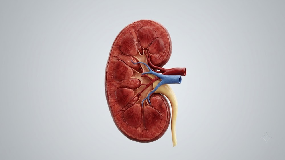
Kidney fibrosis becomes more common with age, with 11% of people of 65 having stage 3 chronic kidney disease. This not only increases mortality risk but also reduces people’s quality of life. Given the link between telomeres and fibrosis in other tissues, it seemed likely that telomere shortening could be associated with kidney fibrosis. Until this study, no link between the two had been shown, in part because of a lack of suitable mouse models.
Bringing in the kidneys
A research team in Spain tackled the question by generating a mouse model that could be used to study kidney fibrosis. They began by studying mice with a mutated form of telomerase reverse transcriptace (TERT). Despite having shortened telomeres, these mice showed no increase in kidney fibrosis compared with wild-type mice. However, with further experiments, the researchers were able to show that something was amiss in the kidneys of these mice.
They accomplished that by dosing the mice with folic acid, a chemical that causes kidney damage. By testing varying doses, they found a folic acid level that caused kidney fibrosis in telomerase mutants but not in wild-type mice. These mice showed all the hallmarks of human kidney disease, making them a potentially valuable model for studying kidney fibrosis and testing potential therapies. The team also developed a second mouse model by deleting the Trf1 gene, which is part of the shelterin complex that protects telomeres. These mutants developed kidney fibrosis without folic acid treatment.
In both mouse models, fibrosis was accompanied by epithelial cells losing their identity and becoming mesenchymal, a process known as epithelial-to-mesenchymal transition (EMT). Consistent with this, the expression of EMT-associated genes increased with fibrosis in both new models. EMT is part of the normal wound healing and regeneration processes, but it’s also active in cancer and fibrotic diseases. These models could thus be invaluable in teasing apart the molecular underpinnings linking EMT and age-related diseases.
Accumulation of short telomeres is a hallmark of aging. Mutations in telomerase or telomere-binding proteins lead to telomere shortening or dysfunction and are at the origin of human pathologies known as ‘telomere syndromes’, which are characterized by loss of the regenerative capacity of tissues and fibrotic pathologies. Here, we generated two mouse models of kidney fibrosis, either by combining telomerase deficiency to induce telomere shortening and a low dose of folic acid, or by conditionally deleting Trf1, a component of the shelterin telomere protective complex, from the kidneys. We find that short telomeres sensitize the kidneys to develop fibrosis in response to folic acid and exacerbate the epithelial-to-mesenchymal transition (EMT) program. Trf1 deletion in kidneys led to fibrosis and EMT activation. Our findings suggest that telomere shortening or dysfunction may contribute to pathological, age-associated renal fibrosis by influencing the EMT program.
Conclusion
While demonstrating the link between telomere length and kidney fibrosis is undoubtedly important, it isn’t particularly surprising. Instead, the true value of this study may lie in the new mouse models that the researchers developed. These offer scientists the tools to investigate how telomere shortening causes fibrosis, digging into the regulation of EMT. Understanding these processes may reveal mechanisms linking aging and telomeres not only with fibrosis but with other conditions such as cancer.

Literature
[1] Saraswait, S, Martínez, P, Graña-Castro, O, and Blasco, MA. Short and dysfunctional telomeres sensitize the kidneys to develop fibrosis (2021), doi: 10.1038/s43587-021-00040-8 [2] Povedano, J. M., Martinez, P., Flores, J. M., Mulero, F. and Blasco, M. A. Mice with pulmonary fibrosis driven by telomere dysfunction. (2015), doi: 10.1016/j.celrep.2015.06.028