A review paper published in Cells has described multiple ways in which the secretion of extracellular vesicles (EVs) changes with aging, leading the authors to propose it as its own hallmark.
Extracellular vesicles and their uses
This review begins with an outline and explanation of extracellular vesicles and their classifications. Cells use extracellular vesicles to signal to one another [1]. Exosomes form from endosomes that are gathered into multivescular bodies, microvesicles are sent directly from the plasma membrane, and apoptotic bodies are the final messages sent by cells undergoing programmed death by apoptosis [2].
EVs readily cross biological barriers, such as the blood-brain barrier, making them appealing therapeutic targets. We have previously published ways in which researchers are attempting to develop EVs to treat tendinopathy and as senotherapeutics. This paper goes into detail on the mechanics of this development, noting that EVs must be purified by their type and cargo and that a single cell can excrete many types, some of which are desirable and some of which are not. In general, EVs are pelleted by centrifuge, with lower speeds being used for larger EVs. Within these sizes, there are multiple subtypes, which can be categorized by their proteomic markers [3].
Critically, EVs can carry RNA [4] and even DNA fragments [5], and the evidence shows that these coding instructions are used to actually code proteins in the recipient cells. EVs also aid in the synthesis of certain molecules, as the enzymes needed to make them are shuttled from cell to cell [6].
Like with every other biological process, EV secretion and uptake changes with aging, and these researchers outline the ways in which it does so.
Extracellular vesicles and senescence
There have been multiple studies linking altered EV secretion to cellular senescence. EVs released by senescent cells exposed to radiation decrease telomerase production in recipient cells, potentially increasing the rate of telomere attrition [7]. Meanwhile, cells that became senescent due to the activation of a cancer-related gene excreted EVs containing a characteristic variety of lipids [8]. Mesenchymal stem cells (MSCs) that are used to develop EV-based therapies can even become senescent, and this is a problem with both replication and EV secretion [9].
In general, senescent cells excrete more EVs than healthy cells do, a finding that has been replicated in fibroblasts [10] and umbilical cord cells [11]. These EVs can induce senescence in the recipient cells, so they can be characterized as part of the senescence-associated secretory phenotype (SASP), the cocktail of factors that senescent cells excrete. However, attempting to measure and compare the size and number of EVs in old and young blood has been an unsuccessful endeavor, and studies attempting to develop such a biomarker are, as a whole, inconclusive.
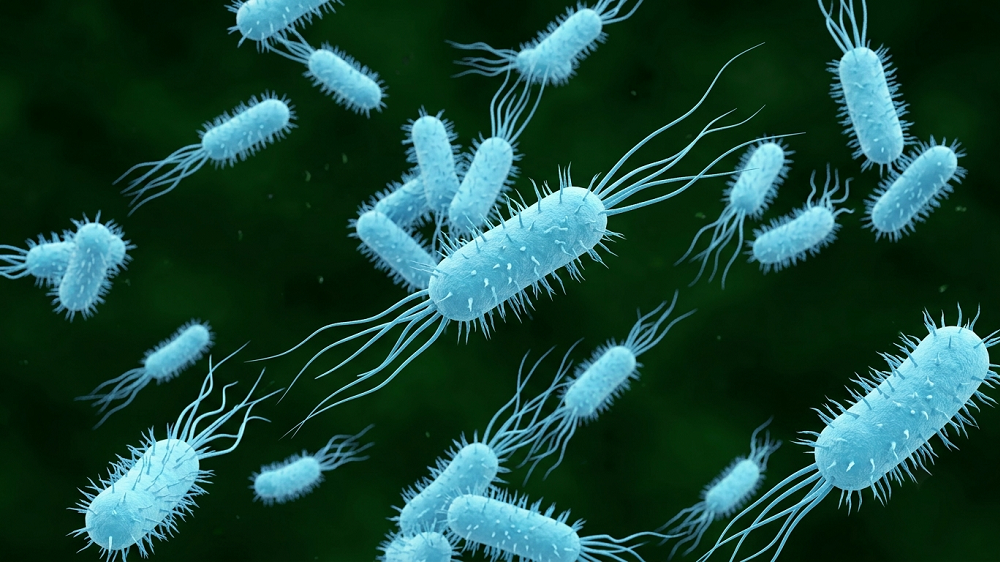
Extracellular vesicles in the gut
Even in the microbiome, EVs change with aging. One species of gut microbiota, found in children but not the elderly, was found to have significantly beneficial effects on the bones of mice [12]. This is true even in the skin, and transplanting EVs from the skin of younger women to older women was shown to help in extracellular matrix maintenance and skin cell proliferation [13].
Conclusion
Of course, whether altered EV secretion is categorized as its own hallmark or as a subtype of altered intercellular communication does not change the underlying biology. It is clear that EVs have a significant impact on many aspects of aging, and they appear to be relatively easier to target than other aspects. While significantly more research is required, it may be the case that EVs represent a low-hanging fruit in gerontology and that they can be used to treat multiple age-related diseases.
Literature
[1] Gurung, S., Perocheau, D., Touramanidou, L., & Baruteau, J. (2021). The exosome journey: From biogenesis to uptake and intracellular signalling. Cell Communication and Signaling, 19(1), 1-19.

[2] Mathieu, M., Martin-Jaular, L., Lavieu, G., & Théry, C. (2019). Specificities of secretion and uptake of exosomes and other extracellular vesicles for cell-to-cell communication. Nature cell biology, 21(1), 9-17.
[3] Kowal, J., Arras, G., Colombo, M., Jouve, M., Morath, J. P., Primdal-Bengtson, B., … & Théry, C. (2016). Proteomic comparison defines novel markers to characterize heterogeneous populations of extracellular vesicle subtypes. Proceedings of the National Academy of Sciences, 113(8), E968-E977.
[4] O’Brien, K., Breyne, K., Ughetto, S., Laurent, L. C., & Breakefield, X. O. (2020). RNA delivery by extracellular vesicles in mammalian cells and its applications. Nature reviews Molecular cell biology, 21(10), 585-606.
[5] Cai, J., Han, Y., Ren, H., Chen, C., He, D., Zhou, L., … & Zeng, C. (2013). Extracellular vesicle-mediated transfer of donor genomic DNA to recipient cells is a novel mechanism for genetic influence between cells. Journal of molecular cell biology, 5(4), 227-238.
[6] Sagini, K., Costanzi, E., Emiliani, C., Buratta, S., & Urbanelli, L. (2018). Extracellular vesicles as conveyors of membrane-derived bioactive lipids in immune system. International journal of molecular sciences, 19(4), 1227.
[7] Al-Mayah, A. H., Bright, S. J., Bowler, D. A., Slijepcevic, P., Goodwin, E., & Kadhim, M. A. (2017). Exosome-mediated telomere instability in human breast epithelial cancer cells after X irradiation. Radiation research, 187(1), 98-106.
[8] Buratta, S., Urbanelli, L., Sagini, K., Giovagnoli, S., Caponi, S., Fioretto, D., … & Emiliani, C. (2017). Extracellular vesicles released by fibroblasts undergoing H-Ras induced senescence show changes in lipid profile. PLoS One, 12(11), e0188840.
[9] Lei, Q., Liu, T., Gao, F., Xie, H., Sun, L. I., Zhao, A., … & Li, Q. (2017). Microvesicles as potential biomarkers for the identification of senescence in human mesenchymal stem cells. Theranostics, 7(10), 2673.
[10] Choi, E. J., Kil, I. S., & Cho, E. G. (2020). Extracellular vesicles derived from senescent fibroblasts attenuate the dermal effect on keratinocyte differentiation. International Journal of Molecular Sciences, 21(3), 1022.
[11] Mensà, E., Guescini, M., Giuliani, A., Bacalini, M. G., Ramini, D., Corleone, G., … & Olivieri, F. (2020). Small extracellular vesicles deliver miR-21 and miR-217 as pro-senescence effectors to endothelial cells. Journal of extracellular vesicles, 9(1), 1725285.
[12] Liu, J. H., Chen, C. Y., Liu, Z. Z., Luo, Z. W., Rao, S. S., Jin, L., … & Xie, H. (2021). Extracellular vesicles from child gut microbiota enter into bone to preserve bone mass and strength. Advanced Science, 8(9), 2004831.
[13] Jo, C. S., Myung, C. H., Yoon, Y. C., Ahn, B. H., Min, J. W., Seo, W. S., … & Hwang, J. S. (2022). The Effect of Lactobacillus plantarum Extracellular Vesicles from Korean Women in Their 20s on Skin Aging. Current Issues in Molecular Biology, 44(2), 526-540.