×

The Blog

Building a Future Free of Age-Related Disease

Rejuvenation Roundup image

Rejuvenation Roundup December 2022

The holiday season is over, the new year is upon us, and we return to our mission of giving us many more years to come. Here’s what’s been done on the rejuvenation front last month.

LEAF News

Team and activities

Lifespan.io Christmas editorial 2022Wishing You a Happy Holiday and a Healthy New Year!: Executive Director Stephanie Dainow and the rest of the Lifespan.io team wished everyone a healthy holiday season at the end of 2022.

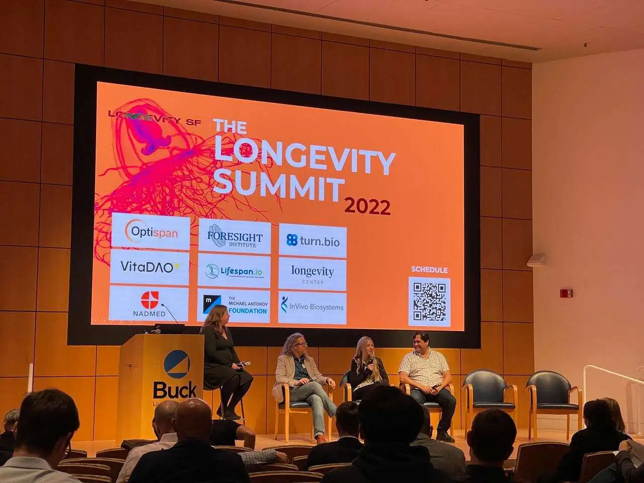
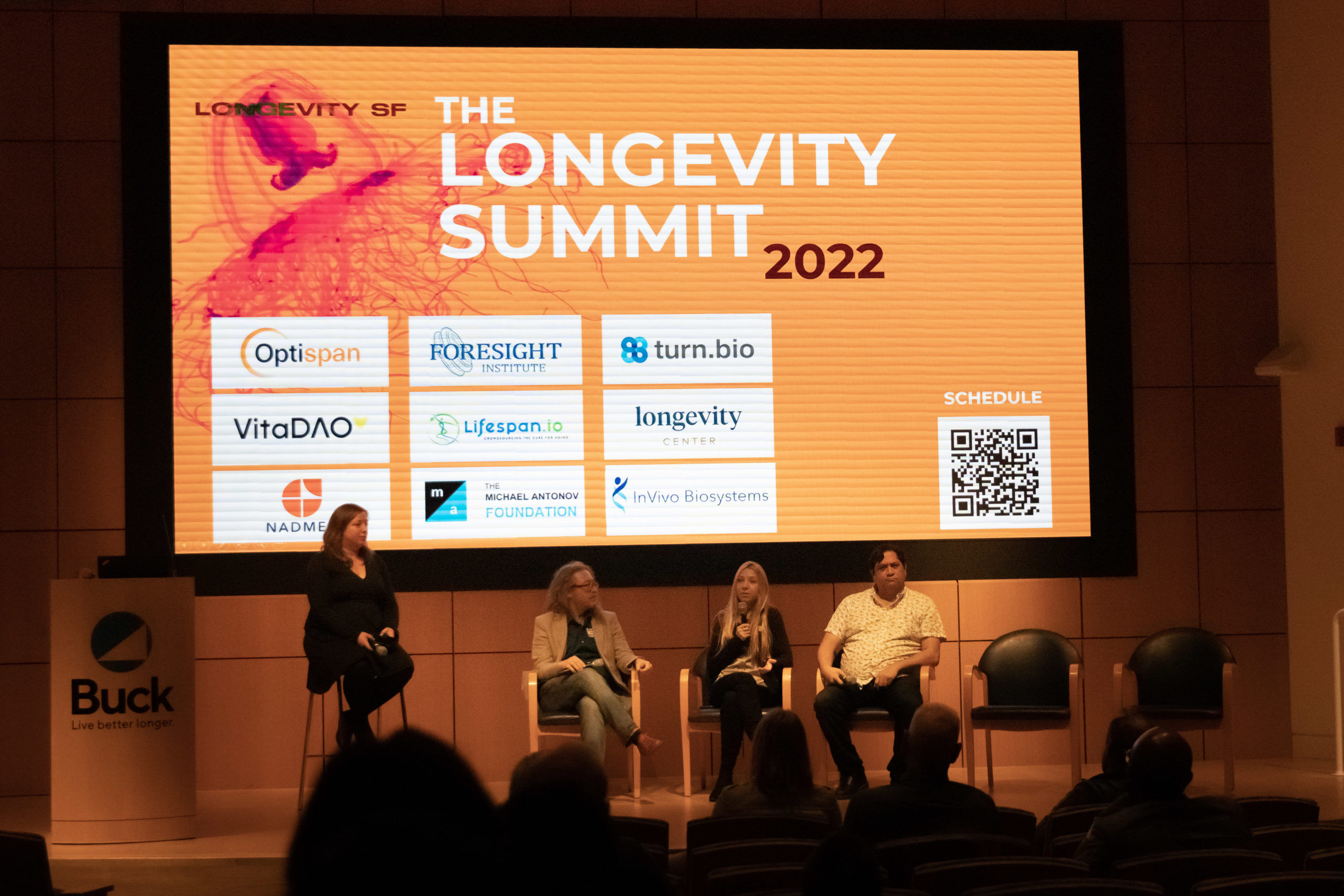
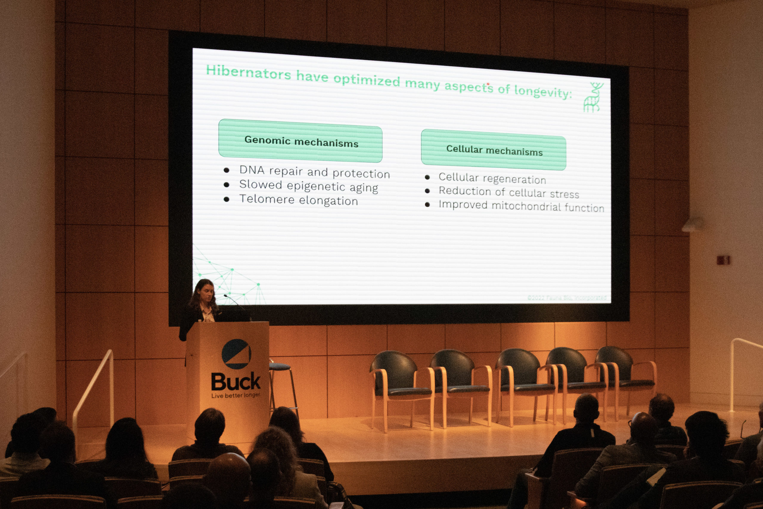
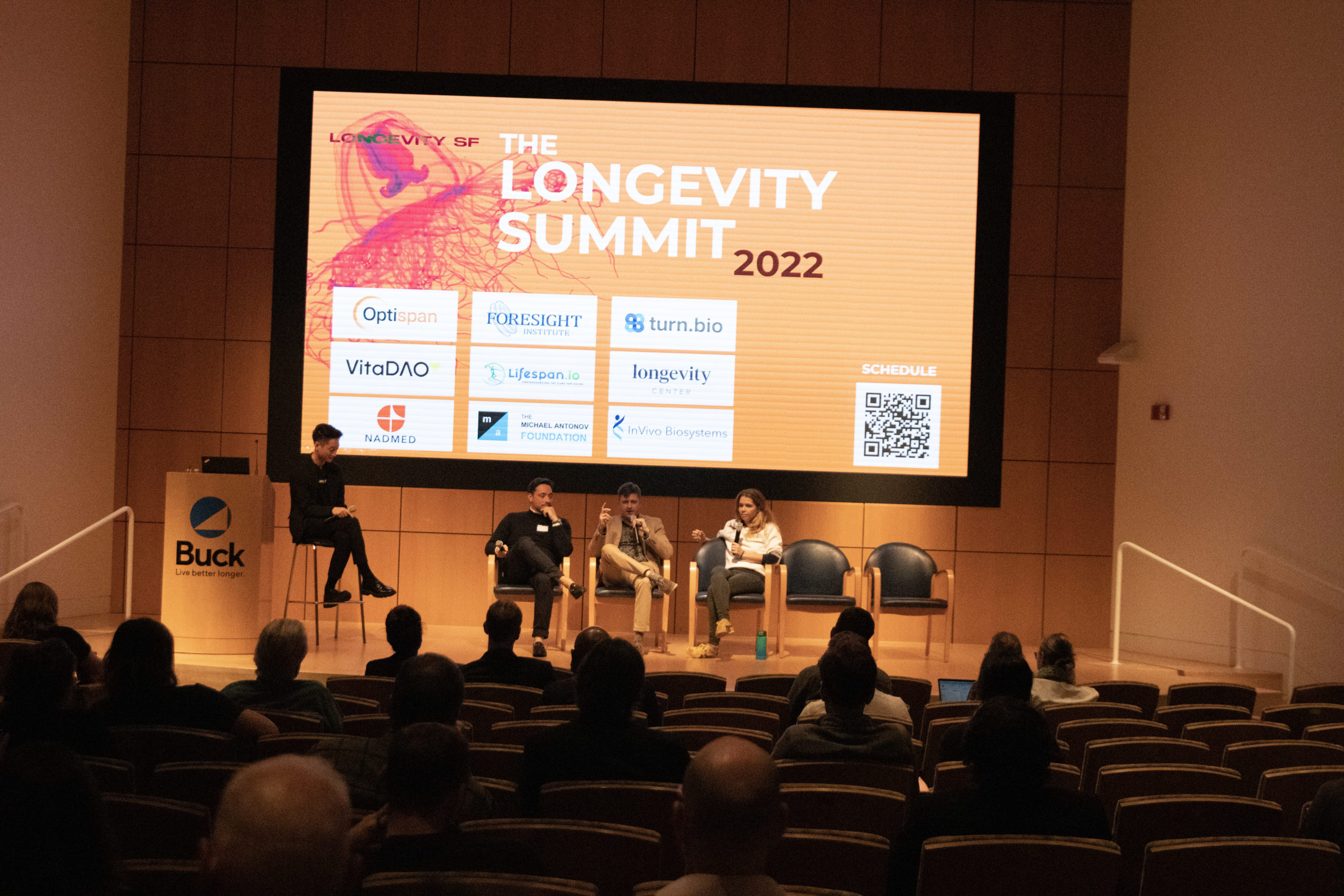
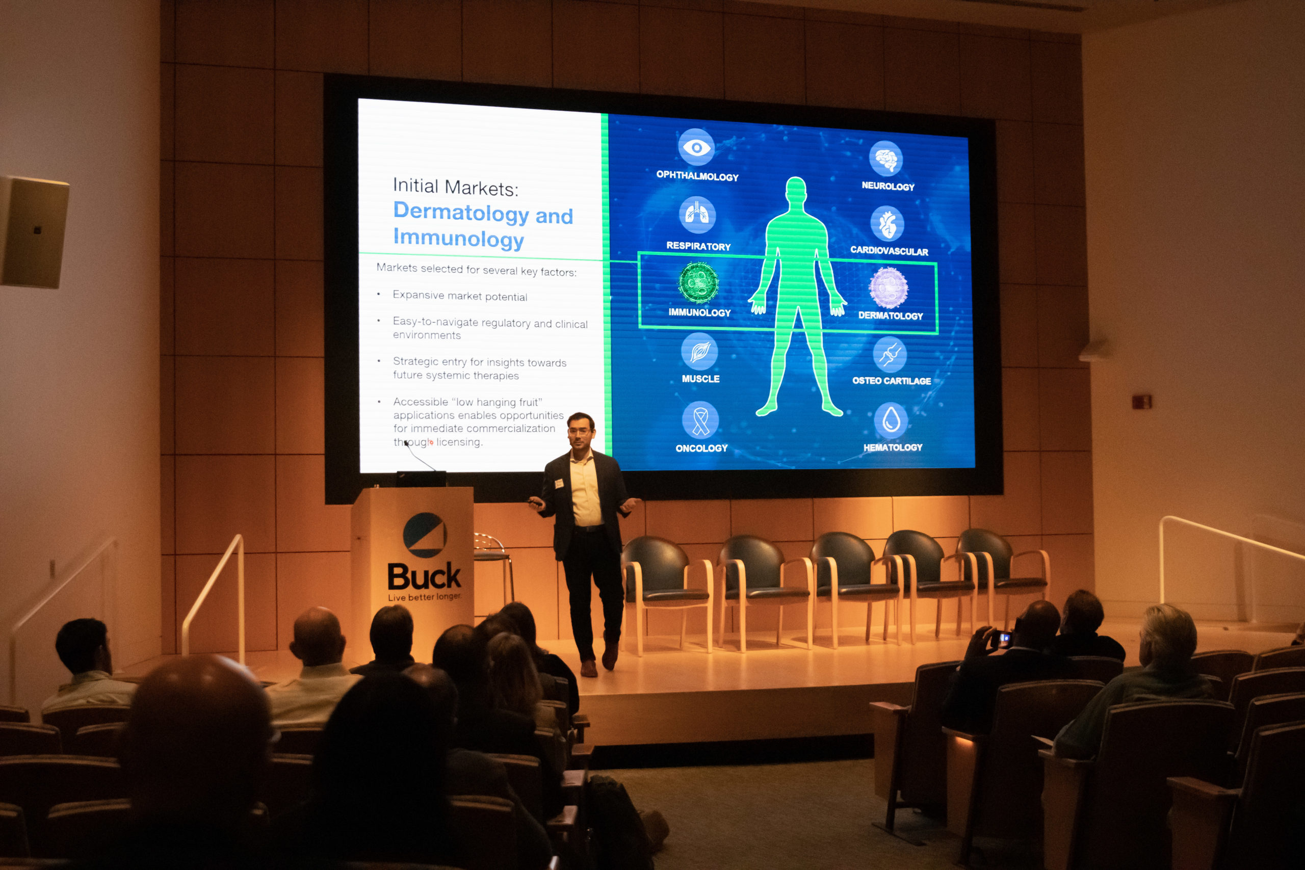
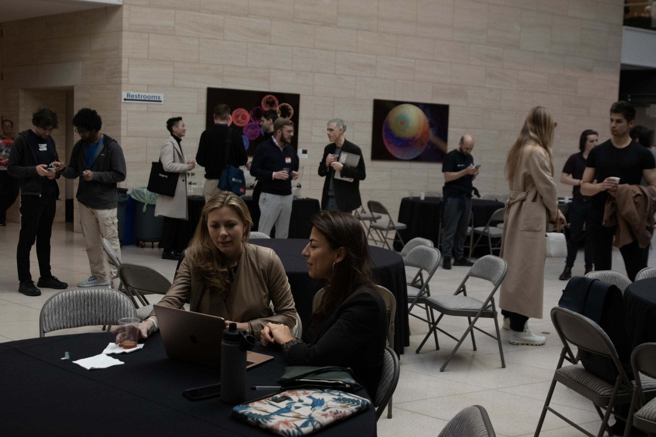
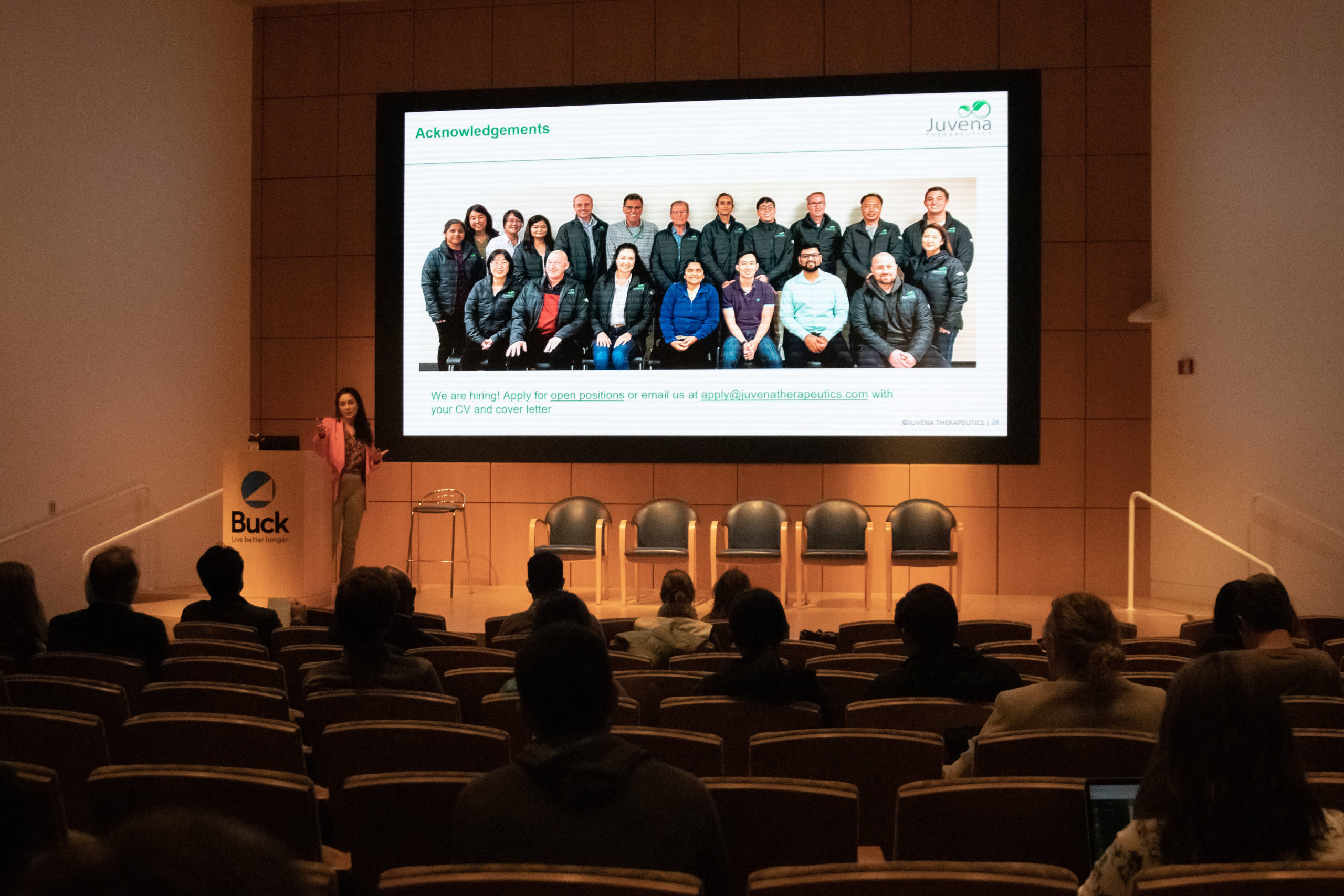
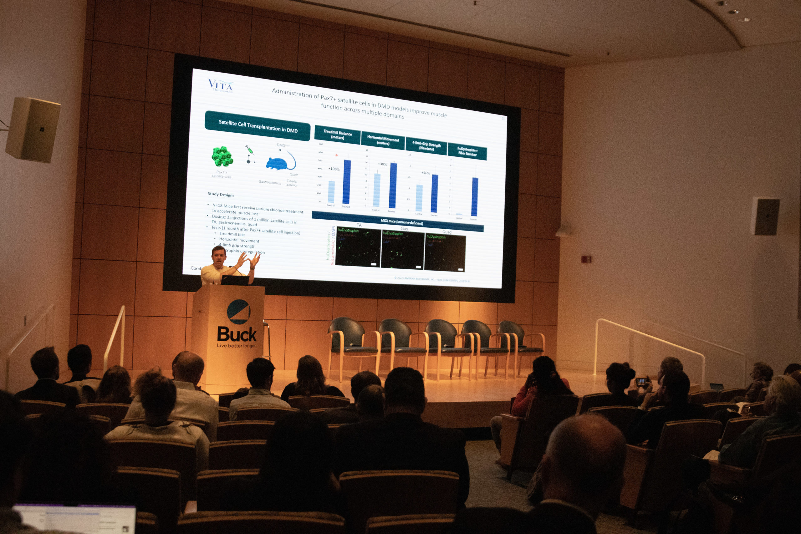
The Science-Packed Longevity Summit at the Buck Institute: The Longevity Summit at the Buck Institute, a two-day geroscience and longevity biotech conference held on December 6-7, was nevertheless densely packed with new research.

Lifespan News

Saving for Longevity: Ryan O’Shea talks about a recent study showing how people’s thoughts about saving money change when confronted with how long they might actually live. Many people retire only to realize that they haven’t saved enough money to be comfortable.

Rapamycin and Egg Cells: Ryan O’Shea discusses a recent study showing that rapamycin improves the potential of egg cells to form embryos. Egg cell viability declines much more rapidly than other aspects of aging accumulate, but there may be ways of improving it.

New Support for Longevity: Foundations that use crypto donations to fund longevity science are the topic of this Lifespan News episode. Both SENS Research Foundation and Lifespan.io have launched end-of-the-year campaigns to support groundbreaking work.

Rejuvenation Roundup Podcast

Ryan O’Shea of Future Grind hosts this month’s podcast, showcasing the events and research discussed here.

Journal Club

For December’s edition of Journal Club, Dr. Oliver Medvedik took a look at a new paper that explores phytocannabinoids in the context of skin aging and rejuvenation.

Advocacy and Analysis

WineholicsDe-Aging Movie Stars Won’t Solve Aging: In the fifth installment of the popular Indiana Jones franchise, Harrison Ford will reportedly be “de-aged” by means of computer graphics, presumably to make him kicking Nazi butt at least somewhat believable. A regular person would react to this piece of showbiz gossip with a chuckle, but for a longevity enthusiast, this raises a bunch of important questions.

The Intersection of Crypto, Gamification, and Longevity: Gamification, the concept of using gaming elements in a non-traditional setting, has long been criticized as at best, a banal pastime, and at worst, a devious manipulation tool employed by unscrupulous companies to acquire profit. However, supporters suggest the opposite: that gamification is a science that utilizes a variety of well-established tools.

Research Roundup

Greek salad bowlPolyphenols Help Reduce Visceral Fat: Pitching two variants of the Mediterranean diet against each other in a randomized controlled trial, scientists have found that the plant-oriented one, which contained more polyphenols, was more effective for weight loss.

Rapamycin, not Dietary Restriction, Fights Infection in Mice: A meta-analysis published in GeroScience has shown that the well-studied drug rapamycin has positive effects on infection in mice, but dietary restriction seems to worsen the problem.

Needle in a haystackFinding Interventions That Truly Impact Aging: Researchers publishing in Nature Communications have determined that interventions that extend lifespan in mice may not have significant effects on the processes of aging. Improvement is not the same as slowing decline.

Intermittent Fasting Protects Mice from Brain Injury: Scientists from Singapore have found that intermittent fasting alleviates damage incurred by chronic cerebral hypoperfusion, the impaired blood flow to the brain that is thought to be a cause of age-related dementia.

SkiingSnowsports for Improved Balance: In a new study published in Journal of Science and Medicine in Sport (JSAMS) Plus, researchers have shown that participating in such sports as snowboarding and skiing might counteract age-associated proprioception decline.

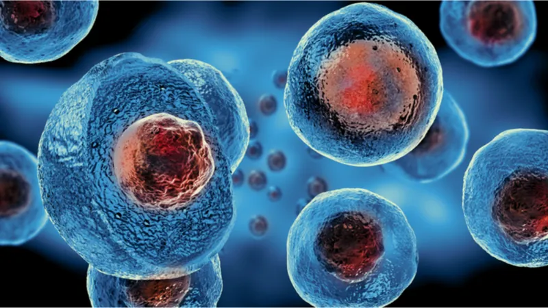
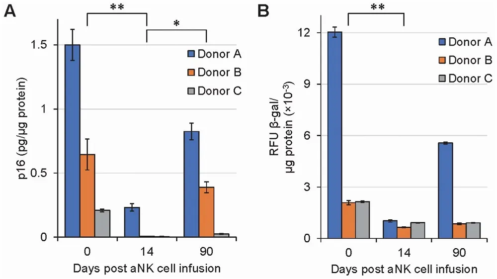
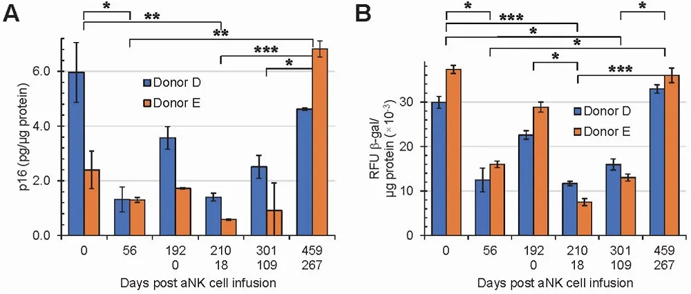
Activated Natural Killer Cells Fight Senescence in Humans: In a study recently published in Biochemistry and Biophysics Reports, researchers have shown that growing natural killer (NK) cells and re-introducing them back into the human bloodstream reduces senescence markers in a wide variety of immune cells.

Destroy cancer
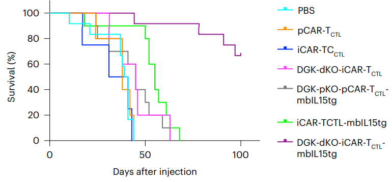
Genetically Enhancing T Cells to Fight Tumors: A team of researchers from multiple Japanese universities has found a way to genetically enhance T cells against solid tumors, as published today in Nature Biomedical Engineering. A focus on signals This highly in-depth paper begins with a discussion of signaling in the response of chimeric antigen receptor (CAR) T cells.

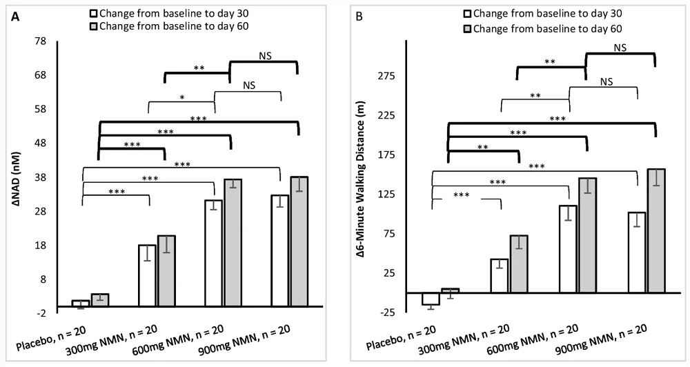
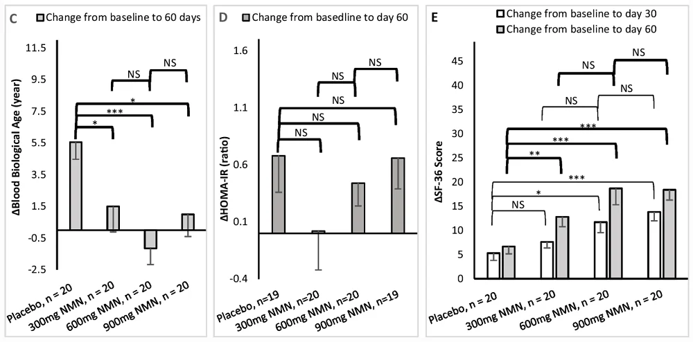
Study Suggests NMN May Improve NAD+ and Walking Speed: A new study suggests that NMN supplementation elevates NAD+ levels and increases walking distance in healthy participants, with 600 mg/day being the optimal dose. This study was based on a randomized, multicenter, double-blind, placebo-controlled clinical trial.

Enlarged cellSize Matters in Cellular Aging: In a new review article published in Frontiers in Cell and Developmental Biology, researchers have suggested adding cellular enlargement to the hallmarks of aging.

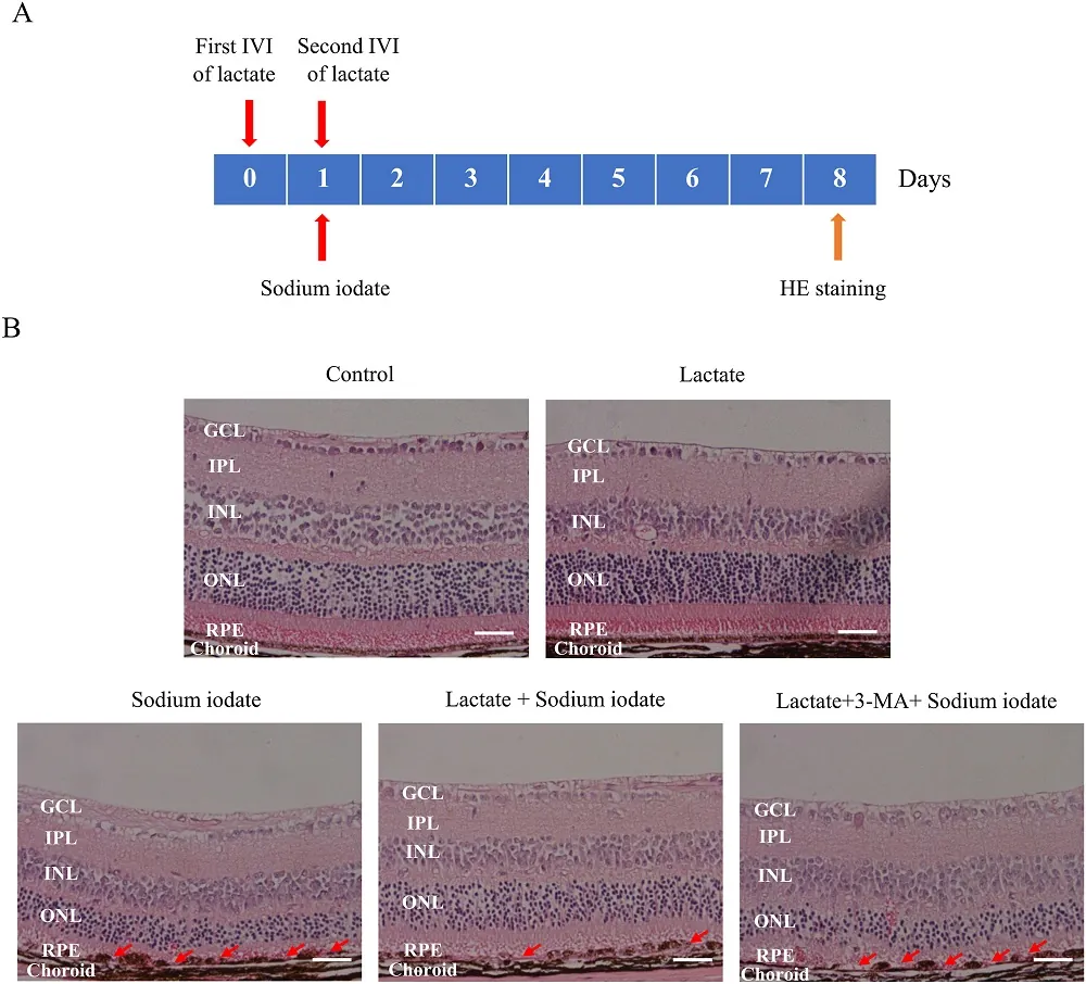
Exploring Autophagy to Fight AMD: Publishing in Free Radical Biology and Medicine, a team of Chinese researchers has investigated the potential role of autophagy in fighting oxidative stress and potentially staving off age-related macular degeneration (AMD).

Wrist wearable deviceShort Bouts of Vigorous Activity May Reduce Mortality Risk: In a study published in Nature Medicine, scientists have shown that short bouts of everyday vigorous physical activity, such as stair climbing, are associated with a considerable reduction in mortality risk, especially in cardiovascular mortality.

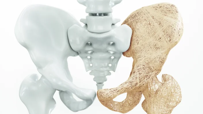
Fighting Osteoporosis Through Cellular Signaling: A paper published in Experimental Gerontology has detailed how a bacterially derived compound may be useful in fighting osteoporosis. This research shows that there may be a way to dig deeper into the root causes of osteoporosis.

RNA strandTranscriptome-Wide Organization Changes in Aging: In a new study published in Nature Aging, researchers have shown that aging is associated with a decreased expression of long transcripts over multiple tissues across several animal species. This study uncovered a conserved molecular feature of age-associated global transcriptome changes.

Clearing Out Senescent Cells Rejuvenates Human Skin: Using a novel senolytic drug, scientists have successfully eliminated senescent cells in human skin transplanted into mice. The treatment led to prolonged skin rejuvenation. It works by inhibiting the enzyme glutaminase, which is essential for the survival of senescent cells.

Heart cells
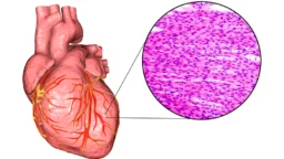
A Connection Between Laminal Dysfunction and Heart Weakness: A paper published in Nature Aging has explained how changes to the lamina contribute to heart weakness in model organisms. Lamin proteins enclose the nucleus in the lamina, the cellular envelope that contains and protects DNA.

Lifespan extension in female mice by early, transient exposure to adult female olfactory cues: These data provide support for the idea that very young mice are susceptible to influences that can have long-lasting effects on health maintenance in later life, and provide a potential example of lifespan extension by olfactory cues in mice.

Inducible Pluripotent Stem Cell-Derived Small Extracellular Vesicles Rejuvenate Senescent Blood–Brain Barrier to Protect against Ischemic Stroke in Aged Mice: In aged stroke mice, iPSC-sEVs significantly mitigated BBB integrity damage, reduced the following infiltration of peripheral leukocytes, and decreased the release of pro-inflammatory factors from the leukocytes, which ultimately inhibited neuronal death and improved neurofunctional recovery.

Ex vivo manufacturing of platelets: beyond the first-in-human clinical trial using autologous iPSC-platelets: This review summarizes current findings on the ex vivo generation of iPSC-PLTs that led to the iPLAT1 study and beyond.

Young plasma transfer recovers decreased sperm counts and restores epigenetics in aged testis: The researchers aimed to show whether blood plasma transfer has an effect on DNA methylation and spermatogenetic cell development.

Senescence atlas reveals an aged-like inflamed niche that blunts muscle regeneration: As senescent cells also accumulate in human muscles, these findings open potential paths for improving muscle repair throughout life.

Targeting anti-apoptotic pathways eliminates senescent melanocytes and leads to nevi regression: These data highlight the important role of redundant anti-apoptotic mechanisms for the survival advantage of senescent melanocytes and the proof-of-concept for a non-invasive combination therapy for nevi removal.

A molecular signature defining exercise adaptation with ageing and in vivo partial reprogramming in skeletal muscle: Considering reduced biological age according to DNA methylome analysis, high-volume exercise training can be classified as an epigenetic reprogramming stimulus.

DNA methylation GrimAge version 2: GrimAge version 2 is an attractive epigenetic biomarker of human mortality and morbidity risk.

Effect of dietary inflammatory potential on the aging acceleration for cardiometabolic disease: A population-based study: In people with CMD, potentially inflammatory diets were associated with age acceleration.

Association between gut microbiota and longevity: a genetic correlation and mendelian randomization study: This study found evidence that gut microbiota is causally associated with longevity, or vice versa, providing novel clues for understanding the roles of gut microbiota in aging development.

Musical and multilingual experience are related to healthy aging: better some than none but even better together: Musical and multilingual experiences are related to healthy aging, especially when combined, which supports the suggestion that a broader spectrum of lifetime experiences relates to cognitive reserve.

Quantifying the benefits of inefficient walking: Monty Python inspired laboratory based experimental study: Adults could achieve 75 minutes of vigorous intensity physical activity per week by walking inefficiently for about 11 min/day.

Gut microbiota of the young ameliorates physical fitness of the aged in mice: These results provide solid evidence that the gut microbiota from the young improves the vitality of the aged.

News Nuggets

BioAge Announces Positive Results Against Muscle Atrophy: BioAge Labs has concluded a Phase 1b study in which human volunteers undergoing 10 days of bed rest were shown to have their related muscle atrophy significantly attenuated by BGE-105.

Turn Biotechnologies Changes Paradigm in Skin Rejuvenation: Turn Biotechnologies has unveiled data at four industry conferences suggesting that a single ERA treatment may be more effective than combination therapies used today. Biomarker analysis demonstrates ERA’s regenerative impact on fibroblast proliferation and collagen VII production.

Coming Up

VitaDAO Longevity Hackathon: On January 20-22, the VitaDAO hackathon, powered by LongHack, will commence. This is a forum for innovators to present ideas and solutions for behavioral health and wellness. Monetary prizes will be awarded. Lifespan.io is an official partner of this event.

We would like to ask you a small favor. We are a non-profit foundation, and unlike some other organizations, we have no shareholders and no products to sell you. All our news and educational content is free for everyone to read, but it does mean that we rely on the help of people like you. Every contribution, no matter if it’s big or small, supports independent journalism and sustains our future.

Lifespan.io Christmas editorial 2022

Wishing You a Happy Holiday and a Healthy New Year!

While our team is taking some well earned rest and relaxation over Christmas (very important for health and longevity) we wanted to share our recent activities with you.

Wishing you a happy holiday!

Stephanie Dainow is the executive director at lifespan.io.Hi, this is Stephanie Dainow, the Executive Director here at Lifespan.io. I would like to take the opportunity to personally wish you all a happy holiday and new year. As you know, our focus is longer, healthier lives with the ones we love, and we hope you’re taking the time to enjoy the holidays and spend time with your loved ones.

I also want to thank those of you who have supported our non-profit foundation this year, it really is greatly appreciated. Your financial support has absolutely enabled our non profit foundation to continue with advocacy initiatives that accelerate efforts to end age-related diseases. Please continue to donate (if you can), and we’re also going to be providing some fascinating metrics about the value of $1 towards advocacy in 2023.

The team will be taking a short break from December 23rd, but we will be back on January 3rd to continue the fight to defeat aging!  In the meantime, we still have tons of evergreen content for you to explore at your leisure. And for those who love Blocko, the host of Life Noggin, check out his latest animated adventures exploring aging research in the content below.

If you want to help drive progress forward, please share our content on social media. The longevity field needs more attention, and effective advocacy requires communities (you) to share information with their networks and educate them about this important cause. Our website provides all the tools you need, and feel free to get in touch if you need help.

See you all in the new year, and have a safe and happy holiday!

SENS Research Foundation video about crosslinks

Life Noggin has teamed up with the SENS Research Foundation to create a series of videos about aging and what science might do about it. This particular video covers crosslinks and how they likely contribute to aging.

Life Noggin is our edutainment channel that explains science and technology in a fun animated pop-sci style. Because we have an audience of 3.26 million subscribers, we can engage the wider science community about all the amazing research happening right now. This is an example of how we use education and edutainment to engage new audiences about aging and rejuvenation research.

We would like to thank SENS Research Foundation for sponsoring this video series. If you are interested in sponsoring your own video on Life Noggin, please feel free to get in touch with us.

Lifespan.io at the Dublin Longevity Summit

Stephanie gave a talk at the Dublin Longevity Summit earlier this year and now the recording has been released. She was also awarded the Rising Star award at this conference for her advocacy and business work in the field.

The Longevity Summit at the Buck

December 6 to 7 saw the Lifespan.io team attending the Longevity Summit at the Buck Institute for Research on Aging. Stephanie Dainow (Executive Director), Arkadi Mazin (Journalist), and Jake Mabey (Marketing) were in attendence.

During the vent, Arkadi took part in a discussion panel talking about the challenges and opportunities present in the field of aging and longevity research.

Arkadi Mazin at the longevity summit 2022

Here is what Arkadi had to say about the panel:

To my knowledge, this was one of the first times when communication and advocacy got some limelight at a aging research conference. The panel I took part in included Allison Duettman, CEO of Foresight Institute, Chris Patil, VP-Media at BioAge Labs, and myself, each of us representing a different facet of the ‘longevity communication network’. We talked about the importance of getting the word out, the challenges in explaining and addressing the concerns people have about increasing human lifespans, and our roles in moving the field forward. 

I stressed the fact that although we at Lifespan.io are passionate about the field’s success, we are also proud to be following strict journalistic standards by providing thorough and impartial coverage. In the end, this responsible approach fosters trust, which is indispensable for getting more people behind the idea of rejuvenation and longevity. 

While scientists are obviously better at science, those of us in the communication and advocacy space might be better in explaining science as well as the philosophical and ethical aspects of life extension. Over the years, we have developed effective communication tools and approaches that our friends in the scientific community can tap into. 

I felt genuine interest in our work coming from the audience, and the feedback me and other participants had received clearly showed that organizing this panel was a great idea and that we need more such interactions in the longevity field.

Thanks to Arkadi for taking part in the panel and representing Lifespan.io. Being a journalist working in this space can often be a challenging but ultimately rewarding experience. Communicating the message that aging is something we can and should do something about is as equally important as the actual research itself.

Because we are a non-profit, we are free from commercial and governmental influence, making us a trusted source for longevity news. As Arkadi mentioned, we have created an ethics code of longevity journalism in order to uphold our trust.

SENS Research Foundation video about stem cell exhaustion

Just in case you wanted more fun videos about aging research, we have teamed up with SENS Research Foundation again! This latest video explores the topic of stem cell exhaustion, one of the reasons we are thought to age.

Once again we wish to thank SENS Research Foundation for sponsoring this video series. If you are interested in sponsoring your own video on Life Noggin, please feel free to get in touch with us.

Announcing the Longevity Cause Fund

Lifespan.io is proud to announce the official launch of the Longevity Cause Fund in partnership with SENS Research Foundation and the Methuselah Foundation, facilitated by Angel Protocol.

Longevity Cause Fund

This is an endowment designed to create sustainable funding for key players in the aging research and advocacy community. 100% of your donation will go directly to our three partner nonprofits fighting the diseases of aging. Half will be used for anti-aging work that is currently underway. The rest will be invested in perpetual endowments that will provide ongoing support for this work — forever. Aging affects us all.

And great news, the first $10k donated is also being fund matched by the Angel Alliance. That means any donation no matter how big or small will be worth even more!

Help us help you to stay healthier for longer by making a donation today!

Heart cells

Changes To The Lamina Contribute To Heart Weakness

A paper just published in Nature Aging has explained how changes to the lamina contribute to heart weakness in model organisms.

A protective enclosure for genetic stability

Lamin proteins enclose the nucleus in the lamina, the cellular envelope that contains and protects DNA. In progeria, a mutation of the gene that codes for the Lamin A protein turns it into progerin instead. This leads to dysmorphia of the nucleus, thus causing epigenetic dysfunction and direct genetic damage [1]. Progeria is a degenerative disease that causes children to age rapidly, and one of its downstream consequences is cardiovascular disease [2].

However, this mutation of Lamin A to progerin also occurs with ordinary aging, and it is associated with heart muscle disease (cardiomyopathy) in this context as well [3]. To further eludicate the relationship between laminal dysfunction and heart disease, the researchers have employed multiple animal models.

The lamina changes with aging in flies

For their first experiments, the researchers chose the familiar Drosophila melanogaster, a species of fruit flies that is commonly used in early-stage aging research. In two distinct wild-type strains, these researchers found that over the flies’ lifespans, the lamina became significantly smaller and more circular in heart muscle cells (cardiomyocytes). This does not occur in skeletal muscle [4] or connective tissue cells [5].

The next experiment determined the roles of specific lamin proteins. In Drosophila, Lamin C is equivalent to human Lamin A, and the researchers found that both Lamin C and Lamin B were decreased with aging in these flies. Therefore, through RNA silencing, they knocked down each of these proteins in the cardiomyocytes of flies.

Flies with the Lamina B knockdown had softer nuclei that did not resemble ordinary aging. However, one-week-old flies with the Lamin C knockdown had cardiomyocytes that resembled those of four-week-old flies. Their hearts were less able to pump blood, and they had significantly reduced survival. Similarly, these flies’ cardiomyocytes had gene expression that was more like that of older flies. Further testing with genetically modified flies confirmed these results.

The researchers also used temperature variations to cause RNA overexpression of Lamin C in adult flies. Flies with this modification had more cell division, more cardiac transcription factor expression, and more heart contraction than their unmodified counterparts.

Of mice and monkeys

The fly findings were recapitulated in other model organisms. Just like in the flies, in cardiomyocytes taken from mice and rhesus monkeys, the lamina were found to be more circular in a way that does not occur in other cells. Gene expression was shown to be similar in aging, with fundamental transcriptional factors being downregulated in aged cardiomyocytes.

Conclusion

These data offer a prospective explanation of a fundamental mechanism behind heart dysfunction in the elderly. Dysfunctional lamina uniquely affect the nuclei of heart cells, and this has a clear connection with a decline in function.

Together, Lamin-mediated misregulation of myogenic transcriptional programs likely has a major impact on mediating heart dysfunction during aging and may precede the development of heart failure.

This research points towards a potential solution. Intervention strategies that increase Lamin A expression in human beings may be of value in preserving genomic stability and gene expression in the heart and thereby extending lifespan. Considerably more work will be required to determine if such an approach is viable.

We would like to ask you a small favor. We are a non-profit foundation, and unlike some other organizations, we have no shareholders and no products to sell you. All our news and educational content is free for everyone to read, but it does mean that we rely on the help of people like you. Every contribution, no matter if it’s big or small, supports independent journalism and sustains our future.

Literature

[1] Goldman, R. D., Shumaker, D. K., Erdos, M. R., Eriksson, M., Goldman, A. E., Gordon, L. B., … & Collins, F. S. (2004). Accumulation of mutant lamin A causes progressive changes in nuclear architecture in Hutchinson–Gilford progeria syndrome. Proceedings of the National Academy of Sciences, 101(24), 8963-8968.

[2] Capell, B. C., Collins, F. S., & Nabel, E. G. (2007). Mechanisms of cardiovascular disease in accelerated aging syndromes. Circulation Research, 101(1), 13-26.

[3] Messner, M., Ghadge, S. K., Goetsch, V., Wimmer, A., Dörler, J., Pölzl, G., & Zaruba, M. M. (2018). Upregulation of the aging related LMNA splice variant progerin in dilated cardiomyopathy. PLoS One, 13(4), e0196739.

[4] Brandt, A., Krohne, G., & Großhans, J. (2008). The farnesylated nuclear proteins KUGELKERN and LAMIN B promote aging-like phenotypes in Drosophila flies. Aging cell, 7(4), 541-551.

[5] Scaffidi, P., & Misteli, T. (2006). Lamin A-dependent nuclear defects in human aging. Science, 312(5776), 1059-1063.

RNA strand

Transcriptome-Wide Organization Changes in Aging

In a new study published in Nature Aging, researchers have shown that aging is associated with a decreased expression of long transcripts over multiple tissues across several animal species [1].

Global changes

It is well known that aging is accompanied by changes in the expression of many genes. Therefore, numerous studies have focused on identifying crucial ‘longevity’ genes and correcting their expression levels in aged animals as a way to potentially rejuvenate them.

Such techniques as real-time quantitative PCR (qPCR or RT-PCR) and RNA sequencing (RNA-seq) are used to compare gene expression levels in different conditions, including aging. More often than not, the research is then reduced to a specific set of genes or pathways for practical reasons.

In this study, the researchers sought to explore aging-associated gene expression changes at a global level instead of evaluating individual genes. They analyzed transcriptomic data for mice and checked if their conclusions held true across several species, including humans.

Length matters

First, the scientists performed RNA-seq on various tissues taken from male mice at 4, 9, 12, 18, and 24 months old. Gene expression changes were inferred by comparing the expression level of each gene at a given age to its expression at 4 months of age in the corresponding tissue.

Next, the researchers applied a machine learning technique to identify which molecular features were associated with the age-related gene expression changes in mice. They show that the length of mature transcripts, i.e. after they’ve been processed and are now ready to be used for protein synthesis, is the most informative feature.

Importantly, with increased age, long transcripts demonstrate a decrease in expression for most tissues. In other words, the abundance of transcripts from long genes compared to short genes changes in old mice. The researchers coined this phenomenon “length-associated transcriptome imbalance”. They confirmed this observation using two other experimental techniques: proteomics and NanoString.

Of mice, men, rats, and killifish

To investigate if the length-associated transcriptome imbalance is a conserved feature across different species, the researchers reverted to previously published studies. They analyzed transcriptomic data from two mouse studies, one rat study, and a killifish study.

Although the amount of available data, e.g. the number of tissues, varied for these species, the results confirmed an age-dependent transcriptome imbalance. For all the organisms considered, approximately 80% of tissues demonstrated an age-related decrease in long transcripts.

Next, the researchers analyzed several sets of single-cell data during mouse aging to investigate if specific cell types or tissues were responsible for the observed transcriptome imbalance. They did not detect any variability and concluded that it was an organism-wide phenomenon.

Finally, the researchers analyzed data available for human tissues. The results were mostly consistent with the previous findings: in both middle-aged (40 to 59) and older adults (60 to 79), a decreased expression of long transcripts was observed. In humans but not mice, one tissue seems particularly prone to this transcriptome imbalance: the brain.

Another set of analyses revealed that the length of both mouse and human genes correlates with their ‘longevity power’. Indeed, the ‘anti-longevity’ genes mostly encode the shortest transcripts, while the longest transcripts are the products of ‘pro-longevity’ genes.

Reversing transcriptome imbalance

The outstanding question, then, is: can or even should anything be done about this age-dependent transcriptome imbalance? Fortunately, the researchers addressed this too. They looked into 11 interventions previously shown to be able to extend mouse lifespan within the Interventions Testing Program of the NIA.

According to the data they analyzed, seven of these anti-aging interventions increase the abundance of long transcripts, including rapamycin, resveratrol, senolytics, and FGF21. On the other hand, metformin and eating every other day were not effective in this respect.

In addition, the researchers highlight that length-associated transcriptome imbalance can be reverted by partial reprogramming, as evidenced by their analysis of a study on retinal ganglion cell rejuvenation [2].

Abstract

Aging is among the most important risk factors for morbidity and mortality. To contribute toward a molecular understanding of aging, we analyzed age-resolved transcriptomic data from multiple studies. Here, we show that transcript length alone explains most transcriptional changes observed with aging in mice and humans. We present three lines of evidence supporting the biological importance of the uncovered transcriptome imbalance. First, in vertebrates the length association primarily displays a lower relative abundance of long transcripts in aging. Second, eight antiaging interventions of the Interventions Testing Program of the National Institute on Aging can counter this length association. Third, we find that in humans and mice the genes with the longest transcripts enrich for genes reported to extend lifespan, whereas those with the shortest transcripts enrich for genes reported to shorten lifespan. Our study opens fundamental questions on aging and the organization of transcriptomes.

Conclusion

This study uncovered a conserved molecular feature of age-associated global transcriptome changes. It is, however, unclear if the transcriptome imbalance is specific to aging. Similarly to cellular enlargement, it could have several molecular origins, including DNA damage and loss of proteostasis. It is nevertheless promising that length-associated transcriptome imbalance is amenable to anti-aging interventions. Future studies should shed light on the role and causes of aging-related transcriptome imbalance.

We would like to ask you a small favor. We are a non-profit foundation, and unlike some other organizations, we have no shareholders and no products to sell you. All our news and educational content is free for everyone to read, but it does mean that we rely on the help of people like you. Every contribution, no matter if it’s big or small, supports independent journalism and sustains our future.

Literature

[1] Stoeger, T. et al. Aging is associated with a systemic length-associated transcriptome imbalance. Nature Aging 1–16 (2022)

[2] Lu, Y. et al. Reprogramming to recover youthful epigenetic information and restore vision. Nature 588, 124–129 (2020)

Hip osteoporosis

Fighting Osteoporosis Through Cellular Signaling

A paper published in Experimental Gerontology has detailed how a bacterially derived compound may be useful in fighting osteoporosis.

Osteoblasts and osteoclasts

Osteoblasts are cells that build bone, and osteoclasts are cells that consume it. These two processes happen consistently and naturally in the human body. However, when osteoclasts consume bone faster than osteoblasts can rebuild it, this leads to the age-related degradation of bone known as osteoporosis [1].

There are two main current treatments for osteoporosis: biphosphonates and the human monoclonal antibody denosumab. While these drugs are the current standard of care, they have rare but serious side effects, including bone death (osteonecrosis) of the jaw and a loss of bodily calcium (hypocalcemia). Denosumab works by inhibiting RANKL, which is a signaling molecule of NF-κB and promotes the growth of osteoclasts (osteoclastogenesis). Therefore, these researchers decided to see if there might be a better method of approaching this target.

Previous research has shown that interferon regulatory factor 8 (IRF-8) inhibits osteoclastogenesis [3] and that suppressing IRF-8 enhances it [4]. This paper’s researchers also noted a recent paper showing that chemicals known as oligosaccharides inhibit osteoclastogenesis [5], although the effects of these chemicals on IRF-8 was not described. With this in mind, the researchers sought to determine if variants of the bacterially derived oligosaccharide glucuronomannan (Gs) and their derivatives (SGs) could be of help against osteoporosis.

A cellular study

The researchers first began their study by conducting tests on viability on RAW264.7 cells, a line of proliferating mouse cells that differentiate into osteoclasts when exposed to RANKL. SGs were found to be too cytotoxic; they killed too many cells and inhibited their proliferation. The larger, more polymerized, Gs were also shown to have this effect, so the researchers focused on only three remaining contenders: G2, G4, and G6.

All three of these prospective drugs were shown to inhibit genes relating to osteoclastogenesis when RAW264.7 cells were also exposed to RANKL, although not all of the genes were equally suppressed. Higher doses were more effective, and G6 was found to be the most effective of the three. As expected, all three drugs delayed the NF-κB signaling pathway.

Most importantly, these compounds were shown to have significant effects on the crucial parts of osteoclastogenesis. IRF-8, which was downregulated by RANKL, was restored, and the RANKL signaling pathway was impaired instead. Formation of the actin ring, a crucial part of osteoclasts, was suppressed. The researchers conclude that these results demonstrate potential effectiveness against osteoclastogenesis and osteoporosis.

Conclusion

This is a cellular study on a mouse line, one of the earliest phases of drug development. Even if this is shown to be effective in living animals, the trial process may discover that oligosaccharides have even worse side effects or are simply not as good as the standard of care. Better methods for fighting osteoporosis may be discovered before this approach proceeds through development.

Still, this research shows that there may be a way to dig deeper into the root causes of osteoporosis, discouraging the overgrowth of bone-eating cells and preventing the associated fractures, weakness, and loss of mobility.

We would like to ask you a small favor. We are a non-profit foundation, and unlike some other organizations, we have no shareholders and no products to sell you. All our news and educational content is free for everyone to read, but it does mean that we rely on the help of people like you. Every contribution, no matter if it’s big or small, supports independent journalism and sustains our future.

Literature

[1] Rodan, G. A., & Martin, T. J. (2000). Therapeutic approaches to bone diseases. Science, 289(5484), 1508-1514.

[2] Gregson, C. L., Armstrong, D. J., Bowden, J., Cooper, C., Edwards, J., Gittoes, N. J., … & Compston, J. (2022). UK clinical guideline for the prevention and treatment of osteoporosis. Archives of osteoporosis, 17(1), 1-46.

[3] Zhao, B., Takami, M., Yamada, A., Wang, X., Koga, T., Hu, X., … & Kamijo, R. (2009). Interferon regulatory factor-8 regulates bone metabolism by suppressing osteoclastogenesis. Nature medicine, 15(9), 1066-1071.

[4] Kodama, J., & Kaito, T. (2020). Osteoclast multinucleation: review of current literature. International journal of molecular sciences, 21(16), 5685.

[5] Wang, S., Feng, W., Liu, J., Wang, X., Zhong, L., Lv, C., … & Mao, Y. (2022). Alginate oligosaccharide alleviates senile osteoporosis via the RANKL–RANK pathway in D-galactose-induced C57BL/6J mice. Chemical Biology & Drug Design, 99(1), 46-55.

Wrist wearable device

Short Bouts of Vigorous Activity May Reduce Mortality Risk

In a study published in Nature Medicine, scientists have shown that short bouts of everyday vigorous physical activity, such as stair climbing, are associated with a considerable reduction in mortality risk, especially in cardiovascular mortality [1].

Everyday activities and health

Exercise is one of the most effective anti-aging interventions available [2], but not everyone has the resources or the willpower to commit to a regular exercise regimen. However, regular exercise is not the only way to put the muscles and cardiovascular system to work. Most people engage in short bursts of activity from time to time, such as climbing a few flights of stairs, running after a bus, carrying heavy bags from the grocery store, and so on. No one had previously compared such everyday actions to health biomarkers, but a new study by an international team of scientists has changed that.

Data from wearable devices

The authors used data from wearable devices stored in UK Biobank, a huge repository of health information that has enabled dozens of scientific papers. Everyday exertions are known as vigorous intermittent lifestyle physical activity (VILPA), which wearable devices can detect. The researchers retrieved data on all-cause, cardiovascular, and cancer mortality for more than 25,000 people (mean age 62) who did not exercise regularly but wore wearables during the 7-year mean follow-up period.

The researchers used 65,000 regular exercisers as controls. This group had slightly higher educational attainment than non-exercisers, higher self-reported health (25.2% reporting being in excellent health versus 13.7% among the non-exercisers), and lower medication use.

Non-exercisers were defined as people who reported no leisure time exercise participation and no more than one recreational walk per week. For the sake of sensitivity analysis, the researchers also analyzed a subset of “perfect non-exercisers” who did not report even taking walks. While for most participants, the general level of physical activity (exerciser or non-exerciser) was only self-reported once, there was a 2000-strong subset of people who had two assessments separated by several years. This subset showed high retention of non-exerciser status; it did not change for 82% of respondents.

A bit of physical activity goes a long way

How much VILPA does the average couch potato accrue? First, virtually all VILPA bouts were no longer than two minutes, and the vast majority were no longer than one minute. The median total VILPA duration was four minutes per day, and the median frequency was three length-standardized bouts per day. At first glance, this is nothing to be proud about.

However, according to the study, this modest amount of activity went a long way in mitigating mortality risks. The effect was dose-dependent, but not perfectly linear, with the bulk of risk reduction associated with small VILPA amounts. Just 3 one-minute bouts of activity a day were associated with 39% less risk of all-cause mortality compared to no VILPA at all. 1.5 bouts a day resulted in 25% less risk, and the effect eventually plateaued at around 11 bouts a day (48% risk reduction).

The effect was much stronger for cardiovascular mortality, in which 1.5 bouts a day resulted in 33%, 3 bouts a day in 41%, and 11 bouts a day in a staggering 65% risk reduction. The analysis was adjusted for multiple covariates, including age, sex, physical activity, smoking, alcohol, sleep duration, fruit and vegetable consumption, education, medication use, and parental history of CVD and cancer. However, the list of covariates did not include BMI, an important factor for both health and physical activity.

Importantly, in terms of health impact, vigorous physical activity of all kinds in regular exercisers was not that dissimilar from VILPA in non-exercisers; according to the researchers, short bouts of vigorous everyday activity can recapitulate a lot of the reduction in mortality risk associated with regular vigorous exercise. The authors note that this is in line with several recent trials that showed improvements in cardiovascular fitness as a result of small amounts of vigorous physical activity [3].

Conclusion

While regular exercise is important, this study shows that even a small amount of vigorous physical activity is associated with a considerable reduction in mortality, especially cardiovascular mortality. When there is not enough everyday activity such as stair climbing, simple exercises like pushups and squats can be relatively easily incorporated into daily routines.

We would like to ask you a small favor. We are a non-profit foundation, and unlike some other organizations, we have no shareholders and no products to sell you. All our news and educational content is free for everyone to read, but it does mean that we rely on the help of people like you. Every contribution, no matter if it’s big or small, supports independent journalism and sustains our future.

Literature

[1] Stamatakis, E., Ahmadi, M. N., Gill, J. M., Thøgersen-Ntoumani, C., Gibala, M. J., Doherty, A., & Hamer, M. (2022). Association of wearable device-measured vigorous intermittent lifestyle physical activity with mortality. Nature Medicine, 1-9.

[2] Duggal, N. A., Pollock, R. D., Lazarus, N. R., Harridge, S., & Lord, J. M. (2018). Major features of immunesenescence, including reduced thymic output, are ameliorated by high levels of physical activity in adulthood. Aging cell, 17(2), e12750.

[3] Allison, M. K., Baglole, J. H., Martin, B. J., MacInnis, M. J., Gurd, B. J., & Gibala, M. J. (2017). Brief intense stair climbing improves cardiorespiratory fitness. Medicine and science in sports and exercise, 49(2), 298-307.

Journal Club

Phytocannabinoids and Skin Rejuvenation

The Journal Club returns for the last time this year at 12:00 Eastern on Tuesday 20th on the Lifespan.io Facebook page. This month, Dr. Oliver Medvedik is taking a look at a new paper that explores phytocannabinoids in the context of skin aging and rejuvenation. Phytocannabinoids are cannabinoids that occur naturally in the cannabis plant and are increasingly being used in a range of beauty products. However, despite their use, the data supporting them is somewhat lacking. This paper attempts to ascertain if they are genuinely useful or not.

Abstract

In light of the increased popularity of phytocannabinoids (pCBs) and their appearance in beauty products without rigorous research on their rejuvenation efficacy, we decided to investigate the potential role of pCBs in skin rejuvenation. Utilizing healthy and stress-induced premature senescent (SIPS) CCD-1064Sk skin fibroblasts, the effects of pCBs on cellular viability, functional activity, metabolic function, and nuclear architecture were tested. Both delta-9-tetrahydrocannabinol (THC) and cannabidiol (CBD) within the range of 0.5 µM to 2.0 µM increased cell growth in a dose-dependent manner while significantly decreasing senescence as measured by beta-galactosidase activity. Utilizing a scratch assay, both THC and CBD (2.0 µM) significantly improved wound healing in both healthy and SIPS fibroblasts. THC and CBD altered nuclear architecture and mRNA levels of cell cycle regulators and genes involved in ECM production. Subsequently, we found ELN, Cyclin D1, PCNA, and BID protein levels altered by SIPS but ameliorated after pCBs exposure in human dermal fibroblasts. Lastly, we compared the efficacy of THC and CBD with common anti-aging nutrient signaling regulators in replicative senescent adult human dermal fibroblasts, CCD-1135Sk. Both THC and CBD were found to improve wound healing better than metformin, rapamycin, and triacetylresveratrol in replicative senescent CCD-1135Sk fibroblasts. Therefore, pCBs can be a valuable source of biologically active substances used in cosmetics, and more studies using clinical trials should be performed to confirm the efficacy of phytocannabinoids.

If you are a Lifespan Hero, you can join us using the following connection details:

Join Zoom Meeting

Meeting ID: 883 1072 6554

Passcode: 239609

Literature

Gerasymchuk, M., Robinson, G. I., Groves, A., Haselhorst, L., Nandakumar, S., Stahl, C., … & Kovalchuk, I. (2022). Phytocannabinoids Stimulate Rejuvenation and Prevent Cellular Senescence in Human Dermal Fibroblasts. Cells, 11(23), 3939.

Exploring Autophagy to Fight AMD

Publishing in Free Radical Biology and Medicine, a team of Chinese researchers has investigated the potential role of autophagy in fighting oxidative stress and potentially staving off age-related macular degeneration (AMD).

AMD, oxidative stress, and autophagy

AMD comes in two major forms. In the nonexudative (dry) form, material builds up behind the retina, while in the exudative (wet) form, abnormal blood vessel growth occurs instead [1]. Wet AMD can be treated with regular injections [2], but dry AMD has no current treatment. Both forms lead to blindness, and AMD is the leading cause of vision loss in the elderly.

Some previous work has shown that oxidative stress plays a major role in the development of AMD [3]. Autophagy, the process of cells consuming their own organelles, declines with aging [4] and is known to serve protective functions against oxidative and other stresses [5]. Therefore, the researchers hypothesized that enhancing autophagy could be an effective strategy for fighting untreatable AMD.

To do this, the researchers turned to lactate, a well-known product of exercise. Lactate has been previously shown to protect against oxidative stress [6], and it protected neurons and reduced inflammation in a mouse model of glaucoma [7].

Effectiveness in cells

For these experiments, the researchers used ARPE-19, a line of human retinal pigment epithelium (RPE) cells, which are the cells most directly affected by AMD and are particularly vulnerable to oxidative stress [8]. To model this stress, they treated these cells with hydrogen peroxide. Interestingly, they found that very small amounts of hydrogen peroxide encourage autophagy and the production of lactate within the cells; to prevent this from altering their experimental data, they chose a higher dose of 600 micromoles, even though that significantly decreased cell viability.

Then, they introduced external sources of lactate. Cells given this lactate had slightly but significantly higher viability along with visible signs of increased autophagy. Introducing a chemical that inhibits autophagy neutralized the effects of the lactate, which is evidence that the autophagy was responsible for the increased viability.

Lactate was also shown to help mitochondria against oxidative stress. Mitochondrial fission and membrane potential were harmed by hydrogen peroxide, and both of these problems were somewhat alleviated by lactate. Lactate pretreatment also significantly reduced the mitochondrial production of reactive oxygen species (ROS); here, introducing another chemical that inhibits autophagy once again neutralized the effects of lactate.

A mouse model showed promise

To validate their cellular work, the researchers turned to mice injected with sodium iodate in a known model of retinal degeneration [9]. With lactate treatment, the number of cells visibly harmed by the sodium iodate, as determined by chemical staining, were substantially decreased. Just like in the cellular experiments, inhibiting autophagy neutralized the effects of the lactate.

Lactate mouse eyes

Conclusion

These results are promising, but this research remains initial and exploratory. The cellular and murine experiments were done with chemical insults rather than genetics. Oxidative stress is unlikely to be the sole factor in the progression of AMD. Further preclinical work, in better models of AMD, will need to be done before this research can progress into human clinical trials.

However, these results may represent a low-hanging fruit, and lactate is a naturally forming chemical that may also have anti-cancer properties. If oxidative stress is found to be a major contributor to AMD and can be successfully ameliorated, it may be possible to help stave off AMD and similar diseases and help people keep their vision for longer.

We would like to ask you a small favor. We are a non-profit foundation, and unlike some other organizations, we have no shareholders and no products to sell you. All our news and educational content is free for everyone to read, but it does mean that we rely on the help of people like you. Every contribution, no matter if it’s big or small, supports independent journalism and sustains our future.

Literature

[1] Spaide, R. F., Jaffe, G. J., Sarraf, D., Freund, K. B., Sadda, S. R., Staurenghi, G., … & Fujimoto, J. (2020). Consensus nomenclature for reporting neovascular age-related macular degeneration data: consensus on neovascular age-related macular degeneration nomenclature study group. Ophthalmology, 127(5), 616-636.

[2] Fogli, S., Del Re, M., Rofi, E., Posarelli, C., Figus, M., & Danesi, R. (2018). Clinical pharmacology of intravitreal anti-VEGF drugs. Eye, 32(6), 1010-1020.

[3] Beatty, S., Koh, H. H., Phil, M., Henson, D., & Boulton, M. (2000). The role of oxidative stress in the pathogenesis of age-related macular degeneration. Survey of ophthalmology, 45(2), 115-134.

[4] Rubinsztein, D. C., Mariño, G., & Kroemer, G. (2011). Autophagy and aging. Cell, 146(5), 682-695.

[5] He, L., Zhang, J., Zhao, J., Ma, N., Kim, S. W., Qiao, S., & Ma, X. (2018). Autophagy: the last defense against cellular nutritional stress. Advances in Nutrition, 9(4), 493-504.

[6] Tauffenberger, A., Fiumelli, H., Almustafa, S., & Magistretti, P. J. (2019). Lactate and pyruvate promote oxidative stress resistance through hormetic ROS signaling. Cell death & disease, 10(9), 1-16.

[7] Harun-Or-Rashid, M., & Inman, D. M. (2018). Reduced AMPK activation and increased HCAR activation drive anti-inflammatory response and neuroprotection in glaucoma. Journal of neuroinflammation, 15(1), 1-15.

[8] Du, J., Yanagida, A., Knight, K., Engel, A. L., Vo, A. H., Jankowski, C., … & Chao, J. R. (2016). Reductive carboxylation is a major metabolic pathway in the retinal pigment epithelium. Proceedings of the National Academy of Sciences, 113(51), 14710-14715.

[9] Kiuchi, K., Yoshizawa, K., Shikata, N., Moriguchi, K., & Tsubura, A. (2002). Morphologic characteristics of retinal degeneration induced by sodium iodate in mice. Current eye research, 25(6), 373-379.

Enlarged cell

Size Matters in Cellular Aging

In a new review article published in Frontiers in Cell and Developmental Biology, researchers have suggested adding cellular enlargement to the hallmarks of aging [1].

Bigger is not always better

Different cell types are known to have different shapes and sizes, which are dictated by their functions. In humans, sperm cells (male gametes) and ova (female gametes) have the smallest and largest diameters, respectively. On the other hand, some neurons are the longest cells: their axons can be over a meter long.

Nevertheless, within a specific cell type, the size variation is negligible. It has been long observed that healthy cells tend to maintain their size and that size changes are characteristic of pathological conditions. Cancer cells are often smaller than normal cells, while senescence leads to cellular enlargement [2].

This can roughly be explained by the different growth and proliferation rates of the two cell types. Cancer cells keep on dividing, so the individual cells don’t grow fast on their own, although this is not universally true. Senescent cells, on the contrary, enter cell cycle arrest: they don’t divide. The production of new cellular material (the biosynthesis rate) exceeds the degradation rate, leading to cellular enlargement.

Cell size is very much dependent on the cell cycle. Often, if a cell experiences an insult such as DNA damage, it halts division to repair the damage. Meanwhile, cell growth continues, leading to cellular enlargement and dysfunction.

In this case, cellular enlargement can be considered a consequence of DNA damage, which leads to cellular dysfunction. The authors, however, argue that cellular enlargement results from various processes other than DNA damage, which, over the course of aging, cause a series of cell cycle arrests.

Of note, there are a number of normal cellular processes that increase the size of cells, such as before division into two daughter cells or during the programmed cell death known as apoptosis.

Cellular enlargement and other hallmarks of aging

In this review, the authors highlight the results of their research on the enlargement of hematopoietic stem cells (HSCs), which give rise to blood cells. They showed that enlarging HSCs by various means makes them dysfunctional. However, the HSCs that stay smaller after such an enlargement-causing treatment are less dysfunctional. Preventing enlargement entirely promotes normal HSC function.

In addition, they note that HSCs enlarge during aging while reducing their size using rapamycin leads to preserved function of aged HSCs. Therefore, the researchers suggest that cellular enlargement could be a new hallmark of aging in addition to the framework of nine hallmarks published in 2013 [3]. Earlier this year, other researchers proposed the inclusion of five more.

These researchers also discussed the interplay between cellular enlargement and some established hallmarks of aging: cellular senescence, genomic instability, telomere attrition, mitochondrial dysfunction, and loss of proteostasis.

Although senescent cells are characterized by enlargement, it is not clear if large HSCs are more prone to become senescent. DNA damage leads to cellular enlargement, while telomere attrition doesn’t seem to play a major role in either enlargement or the aging-associated dysfunction of HSCs. There doesn’t seem to be a clear connection between mitochondrial volume or number and cellular enlargement during aging. Finally, protein synthesis doesn’t seem to be affected by HSC enlargement.

Whether large stem cell size affects other aspects of proteostasis and hallmarks of aging is not yet known.

A detailed analysis

The authors analyzed a large body of literature to explore the association between the enlargement of different cell types and age-related dysfunction. They note that the environment surrounding the cells affects their size. Therefore, by modulating such aspects as the stiffness of the extracellular matrix, it is possible to improve cellular function by altering cellular size.

The researchers also looked into pathology-associated size changes of various cell types, such as adipocytes, red blood cells, neurons, and cardiomyocytes (heart cells). In general, cellular enlargement is indeed an indicator of disease. Interestingly, while neurons mostly shrink with aging, Alzheimer’s disease is characterized by neuron enlargement.

Finally, the authors make an observation that the volume of some cell types is inversely correlated with the lifespan of different animals. However, whether cell size could be used as an indicator of lifespan depends very much on the cell type and the species in question.

Abstract

Years of important research has revealed that cells heavily invest in regulating their size. Nevertheless, it has remained unclear why accurate size control is so important. Our recent study using hematopoietic stem cells (HSCs) in vivo indicates that cellular enlargement is causally associated with aging. Here, we present an overview of these findings and their implications. Furthermore, we performed a broad literature analysis to evaluate the potential of cellular enlargement as a new aging hallmark and to examine its connection to previously described aging hallmarks. Finally, we highlight interesting work presenting a correlation between cell size and age-related diseases. Taken together, we found mounting evidence linking cellular enlargement to aging and age-related diseases. Therefore, we encourage researchers from seemingly unrelated areas to take a fresh look at their data from the perspective of cell size.

Conclusion

This review provides insights into aging-associated cellular enlargement, particularly of hematopoietic stem cells. The authors suggest adding cellular enlargement to the list of the hallmarks of aging because this phenomenon: is observed during normal aging, speeds up aging if worsened, and reverses some aspects of aging if abolished.

However, the authors admit that cellular enlargement does not accompany the aging of all cell types. Moreover, the deviation from the normal cell size is a more precise indicator of pathology. Therefore, it might be premature to coin cellular enlargement as a new hallmark of aging. Nevertheless, analyzing cell size and figuring out how its manipulation affects cellular aging could be an important step for experimental biologists who are attempting to unravel the underlying causes of aging.

We would like to ask you a small favor. We are a non-profit foundation, and unlike some other organizations, we have no shareholders and no products to sell you. All our news and educational content is free for everyone to read, but it does mean that we rely on the help of people like you. Every contribution, no matter if it’s big or small, supports independent journalism and sustains our future.

Literature

[1] Davies, D. M., van den Handel, K., Bharadwaj, S. & Lengefeld, J. Cellular enlargement – A new hallmark of aging? Front Cell Dev Biol 10, 1036602 (2022).

[2] Neurohr, G. E. et al. Excessive Cell Growth Causes Cytoplasm Dilution And Contributes to Senescence. Cell 176, 1083–1097.e18 (2019)

[3] López-Otín, C., Blasco, M. A., Partridge, L., Serrano, M. & Kroemer, G. The hallmarks of aging. Cell 153, 1194–1217 (2013)

Jogging seniors

Study Suggests NMN May Improve NAD+ and Walking Speed

A new study suggests that NMN supplementation elevates NAD+ levels and increases walking distance in healthy participants, with 600 mg/day being the optimal dose [1].

NAD+ and its precursors

While NAD+ can be supplemented via precursors such as NMN, this route has a few roadblocks as well. For instance, studies do not always show that NAD+ precursors increase NAD+ blood levels [2]. Their safety is a bit of a concern as well, since NAD+ can provide energy to cancer cells via glycolysis [3]. However, in general, NAD+ precursors are currently considered safe.

Placebo-controlled study

This study was based on a randomized, multicenter, double-blind, placebo-controlled clinical trial. ‘Multicenter’ means it was conducted in more than one facility, which helps eliminate possible bias due to facility-specific factors. The primary endpoint was NMN’s ability to elevate NAD+ blood levels. As their secondary endpoints, the scientists chose safety and tolerability, physical performance in a walking test, blood biological age, insulin resistance, and overall health via a subjective assessment.

Previous studies were often inconsistent in showing NMN’s health benefits and its ability to affect NAD+ levels in blood. These studies were often sex-biased and/or conducted in people with pre-existing health conditions. This time, the researchers made a point of recruiting healthy men and women aged 40-65 with a wide range of BMI scores. With 80 participants, the sample size was respectable even if not stellar. Three different doses of NMN were investigated: 300, 600, and 900 mg/day.

This study was co-sponsored by the two companies that jointly produce a food-grade NMN product named AbinoNutra™ NMN. However, it was conducted by a respected team of researchers, so there is no reason to doubt its results. It should also be noted here that NMN supplements were recently banned by the FDA in a controversial ruling.

NAD+ levels and fitness affected

NAD+ levels were increased significantly in all study groups compared to placebo and baseline. There was also a significant difference between 300 and 600 mg/day but not between 600 and 900 mg/day. Interestingly, most of the increase in NAD+ levels happened during the first 30 days of the study, while during the second month, the researchers only saw a very mild additional increase.

The six-minute walking distance at baseline was about 300 meters across the groups, which is on the slower side. It significantly improved in the three study groups compared to placebo and baseline. Here, too, the difference between 300 and 600 mg/day was large, but the difference between 600 and 900 mg/day was virtually nonexistent. The gains in walking distance were substantial, with both 600 and 900 mg/day groups adding about 150 meters: a 50% increase.

The participants were not required to perform any regular physical activity during the experiment, and the tests were done just three times: at baseline, after the first month, and after the second month. If there was any habituation to the walking test, it should have been noticeable in the placebo group. However, in this group, no increase in walking distance occurred. This means that the 50% increase occurred due to NMN supplementation. It is possible that participants on NMN began feeling more invigorated early into the study period, and increased their physical activity accordingly, which led to better results measured in the clinic.

NMN human study 1

Asterisks above the bars designate different levels of statistical significance, while NS stands for ‘non-significant’.

Puzzling biological age results 

The researchers measured blood biological age using Aging.AI 3.0, which was developed by Alexander Zhavoronkov’s company InSilico. Unlike epigenetic clocks, Aging.AI has yet to see widespread use in scientific studies. According to this metric, in the placebo group, biological age increased by 5.5 years on average from baseline (during the mere two months of the trial’s duration), while in all the study groups, there was barely any change at all. These results look hardly intuitive or interpretable. Unfortunately, the researchers did not use epigenetic clocks that could have provided a benchmark.

For insulin resistance measurements, the researchers used the HOMA-IR (Homeostatic Model Assessment for Insulin Resistance) test. All of those results were statistically insignificant.

The participants were also asked to complete a 36-question health and quality of life questionnaire (SF-36). The scores mostly followed the same dynamic as NAD+ levels and walking distance: there was some increase in the 300 mg/day group compared to placebo (statistically significant at 60 days), and a much more pronounced increase in both the 600 mg/day and 900 mg/day groups. However, there was no significant difference between those two groups.

NMN human study ages

Conclusion

This interesting trial addresses some design problems found in previous studies, establishing 600 mg/day of NMN as a preferred dose that seems to significantly affect NAD+ levels and physical performance. As in previous NMN trials, no safety problems were reported. However, it does not mean that either efficacy of safety of NMN supplementation have been proven beyond any doubt; therefore, there should be more studies with larger sample sizes, different endpoints, and longer follow-up periods.

We would like to ask you a small favor. We are a non-profit foundation, and unlike some other organizations, we have no shareholders and no products to sell you. All our news and educational content is free for everyone to read, but it does mean that we rely on the help of people like you. Every contribution, no matter if it’s big or small, supports independent journalism and sustains our future.

Literature

[1] Yi, L., Maier, A. B., Tao, R., Lin, Z., Vaidya, A., Pendse, S., … & Kumbhar, V. (2022). The efficacy and safety of ß-nicotinamide mononucleotide (NMN) supplementation in healthy middle-aged adults: a randomized, multicenter, double-blind, placebo-controlled, parallel-group, dose-dependent clinical trial. GeroScience, 1-15.

[2] Huang, D. (2022). A Multicentre, Randomized, Double Blind, Parallel Design, Placebo Controlled Study to Evaluate the Efficacy and Safety of Uthever (NMN Supplement), an Orally Administered Supplementation in Middle Aged and Older Adults. Frontiers in Aging, 26.

[3] Yaku, K., Okabe, K., Hikosaka, K., & Nakagawa, T. (2018). NAD metabolism in cancer therapeutics. Frontiers in oncology, 8, 622.

Destroy cancer

Genetically Enhancing T Cells to Fight Tumors

A team of researchers from multiple Japanese universities has found a way to genetically enhance T cells against solid tumors, as published today in Nature Biomedical Engineering.

A focus on signals

This highly in-depth paper begins with a discussion of signaling in the response of chimeric antigen receptor (CAR) T cells, as good signals are vital for their effectiveness [1]. Three specific biochemical signals are mentioned: a primary signal that spurs the downstream activation of T cells, a co-stimulatory signal that sits on the surface of T cells to enhance the primary signal, and a cytokine signal that promotes the activity and survival of T cells.

Previous work has dedifferentiated T cells into induced pluripotent stem cells (iPSCs), which then redifferentiate back into rejuvenated T cells [2]. However, these cells have been imperfect, particularly with regards to signaling. One experiment used added signals to enhance the effectiveness of such cells [3], but these researchers note that this approach is much stronger against blood-borne cancers than solid tumors.

Thorough enhancement

Seeking to improve cellular signaling from the inside out, the researchers performed three modifications to their cells. The first was to select a CAR differentiation line that did not send exhaustion signals, promoting the formation of T cells. For the second, the researchers used the well-known CRISPR/Cas-9 technique to inhibit the cells’ response to counteracting signals that might be found within the tumor. For their final modification, the team enhanced the production of the cytokine IL-15.

The researchers tested their modifications every step of the way. The first modification, applying this CAR to these T cells, made the cells much more effective than similar cells without it, but this modification could also be applied to primary T cells, which were much more robust and effective than these iPSC cells.

The second modification improved the cells further. The cells’ metabolic fitness was improved, they survived for longer, and they fought tumors more effectively. Simply put, they became better at being T cells. These genetically modified cells were nearly as effective against solid tumors as the primary CAR-T cells.

The third modification changed the game.

Modified CAR-T Survival

While this suite of modifications could not be effectively applied to primary T cells, the iPSCs took it very well, significantly outperforming the other groups in every respect and dramatically improving survival against a model of ovarian cancer. Further testing showed that it was nearly as effective against a model of liver cancer. Tumors, while they still grew somewhat, were much slower to grow when these triply modified cells were present.

Conclusion

While proven to work only in mice, this approach represents a significant breakthrough for immunotherapy. It is easy to envision that this team, or other teams, may discover even more alterations that improve iPSC-derived T cells even further against cancer. Because these modifications begin with a single cloned cell, they can be applied with 100% accuracy, sidestepping a problem common to gene therapy.

However, as the researchers note, this approach is not without its potential drawbacks. The modifications to these cells primarily worked by making them considerably more aggressive, which might enhance negative immune reactions in human trials. This paper recommends suicide genes and other modifications to prevent immunorejection and make such cells safe for general use against cancerous tumors in human beings.

We would like to ask you a small favor. We are a non-profit foundation, and unlike some other organizations, we have no shareholders and no products to sell you. All our news and educational content is free for everyone to read, but it does mean that we rely on the help of people like you. Every contribution, no matter if it’s big or small, supports independent journalism and sustains our future.

Literature

[1] Klebanoff, C. A., Gattinoni, L., Palmer, D. C., Muranski, P., Ji, Y., Hinrichs, C. S., … & Restifo, N. P. (2011). Determinants of Successful CD8+ T-Cell Adoptive Immunotherapy for Large Established Tumors in Mice. Clinical Cancer Research, 17(16), 5343-5352.

[2] Nishimura, T., Kaneko, S., Kawana-Tachikawa, A., Tajima, Y., Goto, H., Zhu, D., … & Nakauchi, H. (2013). Generation of rejuvenated antigen-specific T cells by reprogramming to pluripotency and redifferentiation. Cell stem cell, 12(1), 114-126.

[3] Iriguchi, S., Yasui, Y., Kawai, Y., Arima, S., Kunitomo, M., Sato, T., … & Kaneko, S. (2021). A clinically applicable and scalable method to regenerate T-cells from iPSCs for off-the-shelf T-cell immunotherapy. Nature communications, 12(1), 1-15.

Clearing Out Senescent Cells Rejuvenates Human Skin

Using a novel senolytic drug, scientists have successfully eliminated senescent cells in human skin transplanted into mice. The treatment led to prolonged skin rejuvenation [1].

Senescence and senolytics

Senescent cells, also known as “zombie cells”, are cells that have stopped proliferating after being subjected to any of several types of stress (replicative, chemical, radiational, etc.) but evade clearance by the immune system. Such cells secrete the senescence-associated secretory phenotype (SASP), a cocktail of mostly harmful molecules that harm neighboring cells, driving more of them towards senescence and promoting inflammation. Cellular senescence is one of the hallmarks of aging.

Clearing away senescent cells with senolytics is a popular emerging strategy in the longevity field, but creating effective therapies have proved tricky. However, attempts continue, as evidenced by this new study coming from Japan.

Clearing out senescent fibroblasts in vitro

The researchers experimented with a recently discovered senolytic, which has a long chemical name shortened to BPTES. It works by inhibiting the enzyme glutaminase, which is essential for the survival of senescent cells [2]. Their goal was to see whether BPTES can effectively target senescent human skin cells in vitro and in vivo and whether clearing out those cells can lead to actual skin rejuvenation.

For their in vitro experiments, the researchers used human fibroblasts. Skin is thought to be one of the organs most affected by cellular senescence, with 15-60% of fibroblasts in the skin of aging mice being senescent [3]. Senescent cells are not always harmful. They play a role in wound healing [4], which is obviously important when it comes to skin, but with age, the “dark side” of senescent cells prevails.

The researchers induced senescence in the fibroblasts by three different method: replication, radiation, and treatment with doxorubicin, a chemotherapy drug. Such thoroughness is required, because senescence phenotypes differ significantly. The researchers then confirmed that some of the fibroblasts became senescent.

Here again, three different popular markers were used: senescence-associated ß-galactosidase (SA-ß-gal), p16, and p21. For additional robustness, senescence was also confirmed using morphological analysis, as senescent cells are generally larger and flatter than healthy cells. Another assay confirmed that proliferation levels in the culture had dropped as expected.

BPTES was used in different doses to eliminate the senescent fraction of the fibroblasts. The effect on the senescent cells was dose dependent. The highest dose decreased senescent cell viability almost to zero while barely affecting the viability of non-senescent cells, which shows both high efficacy and high specificity.

Rejuvenating human skin… in mice

To investigate the effect of BPTES on actual aged human skin, the researchers transplanted patches of it into naked mice. Prior to the transplantation, the samples were stained for senescence markers, which showed an abundance of senescent cells. The researchers then treated the transplants with BPTES and followed up for one month.

The treatment drastically reduced the number of cells that tested positive for SA-ß-gal, p16, and p21. Importantly, the levels of cellular senescence remained low even after a month of follow-up, indicating a protracted effect. The researchers also measured the proliferation marker Ki67 and saw a very significant increase in the rate of cell division following the treatment. BPTES also attenuated the levels of several SASP molecules that were elevated in the skin prior to transplantation (metalloproteinases and the inflammatory cytokines IL-1a, IL6, and TNFα).

Collagen density, which determines skin elasticity and decreases with age, was significantly increased by the treatment, as shown by histological analysis. The mRNA expression of Col1A1, the gene that produces type I collagen, was more than twice as high in the treated transplants than in controls. Moreover, collagen levels remained high during the month-long follow-up. This suggests that senolysis might be a viable strategy for long-term skin rejuvenation.

Skin Senolytics

Conclusion

In this study, the researchers were able to cause an impressive reduction of cellular senescence in human skin both in vitro and in vivo. The treatment had a protracted rejuvenative effect on aged human skin samples transplanted into mice. It is easy to see how this can lead to actual human trials in the near future.

We would like to ask you a small favor. We are a non-profit foundation, and unlike some other organizations, we have no shareholders and no products to sell you. All our news and educational content is free for everyone to read, but it does mean that we rely on the help of people like you. Every contribution, no matter if it’s big or small, supports independent journalism and sustains our future.

Literature

[1] Takaya, K., Ishii, T., Asou, T., & Kishi, K. (2022). Glutaminase inhibitors rejuvenate human skin via clearance of senescent cells: a study using a mouse/human chimeric model. Aging, 14.

[2] Johmura, Y., Yamanaka, T., Omori, S., Wang, T. W., Sugiura, Y., Matsumoto, M., … & Nakanishi, M. (2021). Senolysis by glutaminolysis inhibition ameliorates various age-associated disorders. Science, 371(6526), 265-270.

[3] Wang, C., Jurk, D., Maddick, M., Nelson, G., Martin-Ruiz, C., & Von Zglinicki, T. (2009). DNA damage response and cellular senescence in tissues of aging mice. Aging cell, 8(3), 311-323.

[4] Wilkinson, H. N., & Hardman, M. J. (2020). Senescence in wound repair: emerging strategies to target chronic healing wounds. Frontiers in cell and developmental biology, 8, 773.

Cascading Waterfall

Activated Natural Killer Cells Used To Clear Senescent Cells

In a study recently published in Biochemistry and Biophysics Reports, researchers have shown that growing natural killer (NK) cells and re-introducing them back into the human bloodstream reduces senescence markers in a wide variety of immune cells.

Peripheral blood mononuclear cells

This research focuses on peripheral blood mononuclear cells (PBMCs), a category that includes T cells, NK cells, B cells, and other immune cells that constantly send signals to one another [1]. Senescence in these cells drives senescence and other aspects of aging in other organs [2]. Normally, NK cells keep the senescent cell population under control [3], a task that goes unaccomplished if they, themselves, become senescent. While the idea of using NK cells to fight senescence has been approached, previous studies in this area were restricted to mouse models and in vitro experiments, on which we have previously reported.

A protocol has already been developed for extracting NK cells from people, propagating and activating them, and then returning them to their donors [4]. These cells are known as autologous NK (aNK) cells. Therefore, these researchers took the logical next step: with a total of five volunteers, they set out to determine whether or not aNK cells are a viable approach for affecting senescence in human beings.

Improving effectiveness

The first thing the researchers did was determine if they were correctly isolating and expanding the correct portion of cells. They found that after harvesting PBMC cells and applying their activation technique, an average of 91% of the resulting cells were aNK cells. This number varied between donors; while the sample size is too small to determine statistical significance in this respect, the 70-year-old donor’s PBMCs resulted in the fewest aNK cells compared to the other donors, who were all in middle age.

The researchers then determined if the technique they were using to activate and propagate the cells was effective against natural targets. The activated cells were considerably more effective than their unaffected counterparts, destroying larger percentages of cancer cells and senescent cells at lower doses in vitro.

Three of the volunteers, including the 70-year-old, received one billion aNK cells and were monitored for 30 days. The senescence markers p16 and ß-gal both declined dramatically in all three of these individuals’ PBMCs after two weeks; the 70-year-old’s returned to approximately half of its initial high value after 30 days. One of the younger donors, who has inflammatory bowel disease, had many inflammatory markers substantially downregulated by this treatment.

Effectiveness of aNK cells

The final two volunteers underwent a much longer-term experiment, with two billion aNK cells injected at two separate times: once at the beginning and once after 192 days. The effects were similar; the senescence markers were substantially decreased at the beginning of the experiment, rose over time, and were reduced once more by the second infusion of aNK cells.

Effectiveness of aNK cells 2

No adverse effects were detected in any of these volunteers.

Conclusion

While there were only a small handful of participants, this study is an eye-opening proof of concept and suggests that aNK cells can be valuable in at least temporarily ameliorating systemic inflammation and potentially delaying other aspects of aging. It may one day be possible to affect the cells that give rise to aNK cells, restoring their effectiveness and providing a powerful, long-term weapon against inflammaging. That day might be a long way off, however, so a biotechnology company might determine that guiding an aNK cell-based therapy through clinical trials is a wise investment.

We would like to ask you a small favor. We are a non-profit foundation, and unlike some other organizations, we have no shareholders and no products to sell you. All our news and educational content is free for everyone to read, but it does mean that we rely on the help of people like you. Every contribution, no matter if it’s big or small, supports independent journalism and sustains our future.

Literature

[1] Grievink, H. W., Luisman, T., Kluft, C., Moerland, M., & Malone, K. E. (2016). Comparison of three isolation techniques for human peripheral blood mononuclear cells: cell recovery and viability, population composition, and cell functionality. Biopreservation and biobanking, 14(5), 410-415.

[2] Yousefzadeh, M. J., Flores, R. R., Zhu, Y. I., Schmiechen, Z. C., Brooks, R. W., Trussoni, C. E., … & Niedernhofer, L. J. (2021). An aged immune system drives senescence and ageing of solid organs. Nature, 594(7861), 100-105.

[3] Sagiv, A., Burton, D. G., Moshayev, Z., Vadai, E., Wensveen, F., Ben-Dor, S., … & Krizhanovsky, V. (2016). NKG2D ligands mediate immunosurveillance of senescent cells. Aging (Albany NY), 8(2), 328.

[4] Kundu, S., Gurney, M., & O’Dwyer, M. (2021). Generating natural killer cells for adoptive transfer: expanding horizons. Cytotherapy, 23(7), 559-566.

Gamification

The Intersection of Crypto, Gamification, and Longevity

Gamification, the concept of using gaming elements in a non-traditional setting, has long been criticized as at best, a banal pastime, and at worst, a devious manipulation tool employed by unscrupulous companies to acquire profit. However, supporters suggest the opposite: that gamification is a science that utilizes a variety of well-established tools and can have a beneficial impact if employed for the right reasons.

Having recently found its usage in numerous industries, such as online learning, human resources training, and even improving health outcomes, gamification could find its next use case in longevity. According to Keith Comito of Lifespan.io in an interview with The Enjin Room Podcast, there is potential for “incentivizing you to take care of yourself.”

What is gamification?

Gamification has acquired numerous definitions over the years; however, one of the most succinct is from Brian Burke from his book Gamify: How Gamification Motivates People to Do Extraordinary Things, describing it as “the use of game mechanics and experience design to digitally engage and motivate people to achieve their goals.” Burke is a research VP at Gartner, and he suggests that gamification includes three key elements—game mechanics, experience design, digital engagement—designed to motivate people to change or alter their behaviors.

This links in concepts from psychology, behavioral science, and pedagogy:

  • Accomplishment: Players are rewarded for doing a specific action in the form of badges, positive messages, monetary rewards, new unlocked levels, etc. This potentially triggers the well-known dopamine reward pathway in the brain.
  • Empowerment: By achieving milestones or leveling up, players feel empowered with a sense of achievement. This encourages them to continue.
  • Social influence: By utilizing tactics such as group quests and multiplayer modes, a social element is added to the game. Social networking features can also be included.
  • Ownership: Elements such as avatars and collectibles play into players’ senses of ownership and uniqueness within a gaming environment. This helps them take responsibility for their actions in that environment.
  • Scarcity: Prizes or achievements that are limited in number or are considered ‘rare’ encourage players to acquire them.
  • Unpredictability: While repetitive actions can build results, they can also become boring, causing players to become disinterested. Different mechanics, such as quests and storytelling, can promote player interest.
  • Avoidance—the sunk cost bias is one wherein a person continues an action even when it becomes unprofitable due to time, finances or other resources invested. The game works for positive stimuli, a player is unwilling to risk progress made so far and give up.

How is gamification used now?

In an industry estimated to reach a value of $30.5 billion by 2025, gamification is becoming increasingly employed across a variety of industries and spheres, with an equally vast range of outcomes, such as:

  • Online learning: Platforms such as Coursera and DuoLingo utilize gamification practices to encourage their users to continue to engage with educational material. This often includes videos, story-telling, and quizzes that catch and keep the attention of their users.
  • Corporate training: The pharmaceutical giant AstraZeneca implemented a voluntary gamified training program to update their agents on a new drug. The program, called “Go to Jupiter”, reportedly achieved 97% engagement rates among employees.
  • Health outcomes: Over 10 years ago, the health insurance company Aetna partnered with MindBloom to introduce a gamified experience to their clients in order to boost their health outcomes.

How can gamification be integrated into longevity?

Organizations such as Humanity are already taking steps into the intersection of technology and human health.

For example, this company’s proprietary app utilizes wearables to track biomarkers and behavioral tracking to deliver users scores that are based on their rates of aging compared to their biological ages. Like many gamified projects, it requires users to meet specific goals, such as getting a certain amount of exercise per day and consuming proper nutrition, and social features, such as comparisons to average scores and that of friends.

CureDAO is another platform using gamification methodologies to improve human health outcomes. Still in its initial stages, this DAO employs a tokenomics system to encourage users to engage in healthy behaviors. It also supports collaboration by rewarding cooperation. However, behind the platform, a wider game is at play, seeking to utilize data to “discover how millions of factors like foods, drugs, and supplements affect human health.” The platform draws on the power of all this data, employs open-source technology, and seeks to uncover how different factors affect human health and how they can be modified.

These platforms highlight interesting concepts of how gamification strategies can be engaged to deliver real-life outcomes within the longevity field. Further solutions may seek to employ other blockchain-related technologies, such as:

  • Tokenomics systems: The goal is to create a reward system that allows human health outcomes to be connected to digital economics. For example, rewarding behavior that improves health or offering rewards for data sharing. This could utilize such technologies as Pay-to-Earn or Play-and-Earn.
  • Virtual identity: This draws from the sense of ownership, as a digital persona (avatar) adapts or evolves alongside the actual human engaging with the system.
  • Social networking: In a blockchain-based network, participants constantly interact with one another. This can also add a competitive element as participants could compete with friends/community to meet goals and objectives.

Are there any drawbacks?

Although gamification technology has great potential for promoting longevity, it is not without its drawbacks. Like the majority of technology, questions arise as to how data will be managed and which specific data is private and public. The inherent risks of data security, such as fraud and identity theft risks, mean that organizations deciding to engage in gamification technology for longevity purposes need to pay additional attention to how data is stored and whether it meets international security standards.

In addition to this, existing challenges within the blockchain sphere, such as voting protocol fairness issues, mean that to manage equity, mechanisms must be addressed at early project stages to avoid further challenges down the line.

Summary

Gamification is a long-established and considerably effective strategy in many spheres. Its use in the longevity field is still in its early stages, with some projects recently emerging, such as CureDAO and the Humanity app.

We would like to ask you a small favor. We are a non-profit foundation, and unlike some other organizations, we have no shareholders and no products to sell you. All our news and educational content is free for everyone to read, but it does mean that we rely on the help of people like you. Every contribution, no matter if it’s big or small, supports independent journalism and sustains our future.
Skiing

Snowsports for Improved Balance

In a new study published in Journal of Science and Medicine in Sport (JSAMS) Plus, researchers have shown that participating in such sports as snowboarding and skiing might counteract age-associated proprioception decline [1].

Ankles keep you balanced

It is no secret that exercise is one of the key components of lifelong health, which was even shown to slow down cancer progression in a recent study. However, the recommended intensity, frequency, and prevalence of different training modalities for various groups of people are still debated.

In addition to strength and cardio, physical activity guidelines usually include regular balance training. Indeed, keeping balance is an important skill, especially for older adults who have higher risks of falling and breaking bones due to their age-related fragility.

Balance is tightly connected with proprioception, the awareness of body position. The brain needs to integrate sensory information coming from various parts of the body via mechanoreceptors to control balance.

Of all the body parts, ankle proprioception is a critically important component of balance control, as evidenced by postural instability in individuals who suffer from neuromuscular disorders that affect distal leg muscles [2].

This proprioceptive decline is also observed in older adults. On the other hand, professional sports such as gymnastics, soccer, and badminton promote ankle proprioception and could potentially contract the age-associated lack of balance control.

In this study, the researchers noted that while snowsports are limited by environmental conditions, they are more popular activities among the general public than team-based sports. This study sought to explore whether snowsports are effective in protecting against age-related proprioception decline.

Snowsports improve proprioception

142 advanced and elite skiers and snowboarders between 12 and 70 years of age (average of 25.5) participated in this study. Their ankle proprioception was assessed using an Active Movement Extent Discrimination Assessment (AMEDA) device. This apparatus consists of a platform housing a fixed and a movable plate that rotates around its central axis.

During the test, the participants place their feet on each plate, which is randomly tilted to one of five positions. The participants are asked to identify the position of the tilted foot while looking straight ahead. The proprioception score is calculated based on how accurately the participant recognizes the foot position over 50 trials.

The researchers determined that there was no age-related proprioception decline among the participants, as they found no difference between the under-19, 19-39, 40-59, and 60+ age groups.

Interestingly, prior ankle injury did not affect proprioception as measured by AMEDA, as both participant groups with and without unstable ankles did equally well on the test. In addition, there was no difference between the male and female participants or among the participants involved in different snowsports (skiing only, snowboarding only, the two snowsports combined).

These results show that regular snowsport participation such as skiing and snowboarding, that occur in dynamic and changing environments, may i) protect against age-related proprioception decline, and ii) provide a rehabilitative effect for CAI. Thus, snowsport participation may be beneficial for active healthy aging and fall prevention.

Conclusion

This enlightening study demonstrates that engaging in snowsports could be an effective way to contract age-associated proprioception deficits. Although additional studies are required to confirm that these activities are superior to other sports in sustaining balance control in old age, sliding across unpredictable environments could be a determinant of their beneficial effect on ankle stability. In addition, the ankle rehabilitative effect of snowsports discovered in this study could be instrumental in developing successful recovery programs for people with ankle injuries.

We would like to ask you a small favor. We are a non-profit foundation, and unlike some other organizations, we have no shareholders and no products to sell you. All our news and educational content is free for everyone to read, but it does mean that we rely on the help of people like you. Every contribution, no matter if it’s big or small, supports independent journalism and sustains our future.

Literature

[1] Tracey J. D., et al. Age-related proprioceptive decline is not seen in lifelong skiing and snowboarding participants: Lessons for balanced active healthy ageing. JSAMS Plus, 2772-6967 (2022). https://doi.org/10.1016/j.jsampl.2022.100010.

[2] Lencioni T., et al. Postural stabilization and balance assessment in Charcot–Marie–Tooth 1A subjects. Gait & Posture, 481-486 (2014). https://doi.org/10.1016/j.gaitpost.2014.07.006.