×

The Blog

Building a Future Free of Age-Related Disease

ARDA Therapeutics

A New Startup Is Going After Rogue Cells

In a blog post, the founder and CEO of Arda Therapeutics, Adam Freund, explained his company’s intriguing strategy along with why it is imperative to counter aging.

From Calico to Arda

It’s not every day that the longevity field sees a new player with a fresh message joining the ranks. On January 25th, Dr. Freund announced the arrival of Arda Therapeutics in a blog post. After completing his Ph.D. in molecular and cell biology at Berkeley, Dr. Freund was briefly a postdoctoral fellow at the Buck Institute under Judith Campisi, and then at Stanford, working on projects that focused on aging and cancer biology. Importantly, before founding Arda, Dr. Freund spent almost seven years as a principal investigator at Calico. Freund spoke about his work at Calico at last year’s Longevity Therapeutics conference.

According to Freund, Arda Therapeutics is backed by some well-known venture capital funds, including Andreessen Horowitz and The Longevity Fund. The company is also actively hiring, so we do not yet know who will join Freund on the scientific team.

More than senolytics

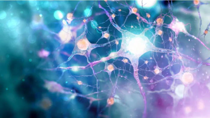
Arda proposes an interesting strategy based on eliminating harmful cells. This is already being done for cancer and senescent cells, but Freund makes an argument for a much wider approach based on the fact that many diseases are caused by dysregulation of a relatively small subset/subtype of cells. As an example, he mentions overgrowth of stromal cells that can lead to fibrosis and overactivation of certain immune cell types that causes chronic inflammation [1].

The traditional approach is to bring those cells back to normal with drugs, but this can be very challenging because of the immense complexity and interconnectedness of cellular processes, many of which are still unknown to us.

Rather that trying to fix misbehaving cells, it might be easier to simply eliminate them. Scientists know a great deal about the mechanisms of cellular death and how to trigger them. At times, the body can repopulate the niche, replacing the dead cells with healthy ones.

For this approach to succeed, such cells must be precisely identified. This is not an easy task, but recent advances in bioscience and AI can provide a roadmap. In this regard, Freund mentions single-cell sequencing that allows researchers to build much more detailed cell maps based on subtle differences between cellular subtypes. Interestingly, from the few papers that have been published by Calico, we know that its scientists use large-scale single-cell sequencing [2].

In cancer, trying to eliminate harmful cells involves harsh treatments that exert a heavy toll on the patient’s health and quality of life. According to Freund, the problem is that cancer cells proliferate uncontrollably, which is not the case with other harmful cells. As a result, to eradicate cancer, every single cancer cell must be killed, while to alleviate other diseases, it might be enough to simply lower the burden of “bad cells”. We would add that due to their fast proliferation, cancer cells continuously evolve to evade threats, which makes them an even more formidable adversary.

In his blog post, Freund is being remarkably honest about the challenges that lie ahead. For instance, even when a harmful subtype of cells is identified, it might be tricky to devise a precise targeting and delivery method. Yet, recent breakthroughs in this field, such as lipid nanoparticles [3], inspire hope.

Unabashedly anti-aging

Freund does not shy away from admitting that his company will target age-related diseases and aging itself, as he explains his beef with aging in a passionate and eloquent way:

For as long as I have been a scientist, I have been driven to understand aging. Frankly, I have trouble understanding why it’s not a more common obsession. After all, it’s going to kill you… At Arda, we hypothesize that there are shared biological mechanisms that affect multiple aspects of age-related deterioration, and that targeting those mechanisms is a promising path to extending healthy lifespan. More specifically, we suspect that many aspects of aging are driven by the hyper-activation of particular cell types, and that if we can remove those cells, we will delay (and possibly reverse) tissue deterioration. If we are even marginally successful, we will push back the greatest killer that has ever existed, improve quality of life at older ages, and give everyone more time with the people they love. We strive to not just add years to life, but life to years.

We at Lifespan.io couldn’t agree more. Unfortunately, Freund’s attitude is in sharp contrast with some other companies, such as Altos Labs, that are trying to position themselves as strictly non-longevity, even though cellular reprogramming, the main avenue that Altos Labs will be pursuing, is more longevity-oriented than clearing out rogue cells.

We would like to ask you a small favor. We are a non-profit foundation, and unlike some other organizations, we have no shareholders and no products to sell you. All our news and educational content is free for everyone to read, but it does mean that we rely on the help of people like you. Every contribution, no matter if it’s big or small, supports independent journalism and sustains our future.

Literature

[1] Thameem Dheen, S., Kaur, C., & Ling, E. A. (2007). Microglial activation and its implications in the brain diseases. Current medicinal chemistry, 14(11), 1189-1197.

[2] Roux, A., Zhang, C., Paw, J., Zavala-Solorio, J., Vijay, T., Kolumam, G., … & Kimmel, J. C. (2021). Partial reprogramming restores youthful gene expression through transient suppression of cell identity. bioRxiv.

[3] Li, Y., Su, Z., Zhao, W., Zhang, X., Momin, N., Zhang, C., … & Weiss, R. (2020). Multifunctional oncolytic nanoparticles deliver self-replicating IL-12 RNA to eliminate established tumors and prime systemic immunity. Nature Cancer, 1(9), 882-893.

Rat maze

Senolytics Restore Cognitive Ability in Aged Rats

Polish researchers publishing in Aging have discovered that the well-known senolytic combination of dasatinib and quercetin improves the cognitive capabilities of older rats.

Understanding neuroplasticity

While new neurons can be formed in the adult hippocampus through neurogenesis [1], learning and behavioral changes caused by changes in the prefrontal cortex are not the result of this process. Instead, neurons in the prefrontal cortex form protrusions called dendritic spines, which then go on to become synapses between other neurons.

Rather than the mass neuronal death seen in Alzheimer’s disease, it is this fundamental ability to form new connections that declines with “normal” aging [2]. The downstream effects of this gradual loss of ability affect practically every aspect of cognitive function, including reasoning, memory, psychomotor abilities, and your ability to remember and understand the contents of this paragraph along with my ability to write it.

As neuroplasticity is directly related to changes in gene expression, it has been shown to be strongly affected by epigenetics, specifically the methylation of histone H3 [3]. The researchers hypothesized that this change in gene expression is linked to the accumulation of senescent cells, specifically due to the senescence-associated secretory phenotype (SASP).

In order to test their hypothesis, the researchers administered the well-known senolytic combination of dasatinib and quercetin (D+Q) to rats.

A series of positive effects

In this experiment, rats were trained to avoid a specific place through small electric shocks. As expected, older rats performed worse on this task than younger rats, which learned to avoid the specific place quickly. However, rats that had performed poorly on this task performed very similarly to younger rats after an administration of D+Q, showing that their short-term memory and skill learning abilities were significantly improved, and they retained this improvement even five weeks after administration of D+Q had ceased. D+Q administration had no apparent effect on younger rats.

The researchers then examined the effects of the SASP on inflammatory cytokine levels. While each individual cytokine was not strongly affected, rats given D+Q had a broad decrease in nearly all of the cytokines tested, including interleukins and interferon-γ. The anti-inflammatory cytokine IL-10, which actually increases with aging, was further increased with D+Q in both young and old animals.

In the next step in this experiment, the researchers carefully examined the dendritic spines of the animals, focusing on the hippocampus. The basal dendrites were unaffected; however, the apical dendritic spines of aged animals given D+Q were shown to be thicker and substantially longer than those of their untreated counterparts, corresponding to their increased ability to retain memory and knowledge.

Finally, the researchers took a look at H3 histone methylation, where the differences were stark and clear. Correlating with cognitive decline, the methlylation of the H3K9me3 site increases with aging, while that of H3K27me3 decreases; D+Q treatment halved H3K9me3 while significantly increasing H3K27me3.

Conclusion

The role of inflammaging and the SASP on cognitive decline has remained an open question, and this study has significantly helped to close it. While senolytics have not been shown to affect diseases such as Alzheimer’s and are unlikely to be the last word in “normal” cognitive decline, this study shows that they have a significant effect on the ability of older animals to perform basic cognitive tasks, right down to the neuronal level.

As always, human trials are required to see whether or not these or any other senolytics have positive effects on the human brain and whether or not targeting inflammaging is a viable method of allowing older people to retain their cognitive abilities.

We would like to ask you a small favor. We are a non-profit foundation, and unlike some other organizations, we have no shareholders and no products to sell you. All our news and educational content is free for everyone to read, but it does mean that we rely on the help of people like you. Every contribution, no matter if it’s big or small, supports independent journalism and sustains our future.

Literature

[1] Fares, J., Bou Diab, Z., Nabha, S., & Fares, Y. (2019). Neurogenesis in the adult hippocampus: history, regulation, and prospective roles. International Journal of Neuroscience, 129(6), 598-611.

[2] Bloss, E. B., Janssen, W. G., Ohm, D. T., Yuk, F. J., Wadsworth, S., Saardi, K. M., … & Morrison, J. H. (2011). Evidence for reduced experience-dependent dendritic spine plasticity in the aging prefrontal cortex. Journal of Neuroscience, 31(21), 7831-7839.

[3] Parkel, S., Lopez-Atalaya, J. P., & Barco, A. (2013). Histone H3 lysine methylation in cognition and intellectual disability disorders. Learning & Memory, 20(10), 570-579.

Fibrous foods

Dietary Fiber and Probiotics Affect Melanoma Immunotherapy

A study published in Science by Dr. Jennifer A. Wargo and her colleagues spanning multiple centers recently examined if dietary fiber and commercially available probiotics affect immunotherapy response in cancer patients [1]. They hypothesized that bacteria from two probiotic species would be associated with a response to this therapy.

The human patients

By using stool samples, the researchers examined the gut microbiomes of 438 patients with melanoma. Most of the patients were receiving systemic therapy for metastatic melanoma (n=321) and were classified as responders or non-responders based on Response Evaluation Criteria in Solid Tumors (RECIST).

Of these patients, 87% were receiving immune checkpoint blockade (ICB) immunotherapy. This type of therapy uses monoclonal antibody-based therapies to block the desired interaction at immune checkpoints. For instance, a common treatment used to prevent T lymphocytes from attacking other cells is an immune therapy called anti-programmed cell death 1 (anti-PD-1). Most of the patients in this study on ICB immunotherapy were also on anti-PD-1 immunotherapy. Data was collected on dietary habits and probiotic supplements used within the past month.

No significant differences with probiotic use in late-stage melanoma patients

49 out of 158 of the late-stage melanoma patients were on a probiotic supplement in the past month. The authors noted that the patients on the probiotics had lower BMIs, reported higher consumption of vegetables and legumes, and were less likely to be on statins than patients who were not taking a probiotic supplement. When looking at survival rates in patients treated with ICB immunotherapy, there were no significant differences between patients who took a probiotic supplement and patients who did not.

The effects of probiotics on larger tumors in mice

Germ-free mice received donor stool from responders as determined by RECIST. These mice were then given one of two types of commonly purchased probiotic strains, Bifidobacterium longum and Lactobacillus rhamnosus, as well as treatment with immunotherapy to treat the tumors. Compared to the control group, the mice on either of the probiotics had significantly larger tumors. The research found similar results when they did the same experiment in non-germ-free mice and specific pathogen-free mice that had melanoma tumors.

Probiotics vs. sterile water in mice

The researchers then examined mice fed either of two probiotic strains alongside a control group fed sterile water. The treated mice had reduced levels of two immune cell types that play important roles in helping to fight cancer: interferon-γ and CD8+ T cells. The treated groups also had reduced cytotoxic t-cells in tumor tissues, which are immune cells that aid in helping fight tumor cells. The authors cite research that has shown similar and opposite findings, and follow with:

Together, these studies support the need for more careful investigations of the effects of current commercially available probiotic formulations on immunity and cancer immunotherapy response.

Fiber slowed tumor growth in mice and humans

Among mice receiving an anti-PD-1 therapy, mice on a fiber-rich diet showed delayed tumor growth compared with mice on a fiber-poor diet. The researchers also profiled the gut microbiomes of the mice, and there were significant differences in the fiber-rich and fiber-poor diets. Immune cell analysis revealed that the mice fed a high-fiber diet had higher CD45+ T cells in their tumors and higher T cell activation and interferon responses.

To assess the effects of dietary fiber intake on ICB immunotherapy response in humans, the researchers chose the National Cancer Institute Dietary Screener Questionnaire. Even though a fiber intake of 20 grams per day was considered sufficient, only 30% of people on ICB immunotherapy, 37 out of 128, reached this level.

Additional analysis showed that higher fiber intake was highly correlated with vegetables, fruit, whole grains, and, to a lesser extent, calcium intake. People who ingested less than 20 grams per day were more likely to be classified as obese and to be on blood pressure-reducing medication.

Patients who consumed sufficient fiber demonstrated improved survival rates. After adjustment for clinical parameters, every 5-gram increase in fiber intake corresponded with a 30% lower risk of cancer progression or death. In this study, similar results were found when assessing dietary fiber intake in relation to ICB immunotherapy response. People who consumed sufficient fiber and did not take probiotic supplements had significantly greater survival rates. The authors follow the human diet data results with:

Ongoing dietary intervention studies in the setting of ICB are critical for establishing whether a targeted and achievable diet change at the initiation of ICB can safely and effectively improve outcomes (NCT04645680).

Conclusion

While the researchers stressed the need for further research and independent validation, this study shows that dietary fiber and probiotic use are associated with different outcomes in humans and mice on ICB immunotherapy.

This study highlights an overarching point and a valuable message for clinicians and patients: this type of research comes with a need for additional careful investigations with interdisciplinary and multi-center collaboration. Specific supplements may need to be taken with caution within the context of specific scenarios, such as ICB immunotherapy.

The authors concluded with:

These notable (and perhaps unexpected) findings from studies in this observational patient cohort are corroborated by parallel studies in preclinical models with preliminary mechanistic insights. In light of these collective results, dietary habits and probiotic supplement use should be considered in patients receiving ICB and in efforts to modulate the gut microbiota. These factors should be more thoughtfully evaluated in strategies to improve cancer outcomes.

We would like to ask you a small favor. We are a non-profit foundation, and unlike some other organizations, we have no shareholders and no products to sell you. All our news and educational content is free for everyone to read, but it does mean that we rely on the help of people like you. Every contribution, no matter if it’s big or small, supports independent journalism and sustains our future.

Literature

[1] Spencer, C. N., McQuade, J. L., Gopalakrishnan, V., McCulloch, J. A., Vetizou, M., Cogdill, A. P., Khan, M., Zhang, X., White, M. G., Peterson, C. B., Wong, M. C., Morad, G., Rodgers, T., Badger, J. H., Helmink, B. A., Andrews, M. C., Rodrigues, R. R., Morgun, A., Kim, Y. S., Roszik, J., … Wargo, J. A. (2021). Dietary fiber and probiotics influence the gut microbiome and melanoma immunotherapy response. Science (New York, N.Y.), 374(6575), 1632–1640. https://doi.org/10.1126/science.aaz7015

Balanced stones

Meta-Analysis Discovers Goldilocks Zone for IGF-1

A new meta-analysis from Aging Cell has shown that both high and low levels of IGF-1 are related to mortality risk [1].

Insulin-like growth factor 1 (IGF-1)

IGF-1 is one of the body’s major metabolic hormones. It can have effects similar to insulin, and it mediates the effects of growth hormone. IGF-1 has growth-promoting effects in almost every tissue, inhibiting apoptosis and increasing the synthesis of proteins and DNA [2]. High expression of IGF-1 has been shown to reduce lifespan and healthspan in worms, mice, and flies [3-5]. In fact, the expression of very low levels of IGF-1 has increased mouse lifespan by as much as 40% [6].

Serum levels of IGF-1 are commonly measured in the clinic. This has provided researchers with a wealth of data on the hormone, but studies of IGF-1 levels in humans have shown mixed results. High IGF-1 is associated with an increased risk of several cancers [7]. However, low levels of IGF-1 are associated with diseases such as cardiovascular disease, diabetes, and frailty [8]. Many studies have linked both high and low levels of IGF-1 to mortality, while others have found no relationship [9].

Because of these conflicting results, it is unclear whether there is any value in measuring IGF-1 to predict remaining lifespan. However, an international collaboration of researchers recently conducted a meta-analysis of these findings to determine if an ideal range exists for IGF-1 to predict all-cause mortality [1].

IGF-1 shows a U-shaped relationship with mortality

19 studies that included a total of 30,876 participants were included in the meta-analysis. The studies were conducted from 2007 to 2018 and included populations from the United States, Japan, China, and various European nations.

First, the authors looked at high versus low IGF-1 and mortality, similar to the methodology of previous studies. In this analysis, no difference in risk of death was found between the high and low levels of IGF-1 groups.

Rather than only grouping participants into “high” and “low” levels of IGF-1, 9 of the studies included detailed enough information to conduct a dose-response meta-analysis. This analysis showed a U-shaped response, with both the lowest levels and highest levels of IGF-1 being associated with higher mortality.

The researchers then analyzed their dataset using an intermediate group that approximately aligned with the lowest mortality from the dose-response analysis (120-160 ng/mL). With low, intermediate, and high IGF-1 groupings, mortality risk still did not differ between high and low IGF-1 levels. However, the intermediate group had statistically significant lower mortality risks than both the high and the low groups. These findings did not change when the intermediate range was expanded to 100-180 ng/mL IGF-1 or when excluding studies that did not include participants older than 70.

Lastly, researchers used the Third National Health and Nutrition Examination Survey (NHANES III, 1988–1994) to see which dietary differences were correlated with IGF-1. Higher consumption of proteins, carbohydrates, and 13 different vitamins and minerals were associated with higher IGF-1 levels, as were eggs, milk, cheese, yogurt, and butter. These findings were similar to previous studies and confirm that IGF-1 levels can be modified by dietary changes.

In conclusion, by analyzing and comparing different ranges of IGF-1 in 30,876 subjects, we find that both high and low levels of IGF-1 increase mortality risk, and for the first time, we identify a specific mid-range being associated with the lowest mortality (120–160 ng/ml). Using the NHANES III survey, we show an association between high intake of animal proteins, carbohydrates, and milk-based products and IGF-1 levels. These results can point to diagnostic, nutritional, and pharmacological strategies to optimize IGF-1 levels and help reduce mortality.

Conclusion

IGF-1 has been of great interest to longevity researchers due to its key role in metabolism, influence on healthspan and lifespan in model organisms, and association with mortality in humans. Its association with mortality has been scrutinized in recent years with the publication of several seemingly conflicting results. This study appears to explain these contradictory findings by identifying the relationship between IGF-1 and mortality as U-shaped rather than linear.

As always, the conclusions that can be drawn from these sorts of studies are limited. It cannot be said whether low and high IGF-1 contributed to mortality in these participants or if it was simply correlated. Additionally, it is well established in the scientific literature that studies with nonsignificant results disproportionately go unpublished. While the authors did not detect any publication bias in their statistical analysis, meta-analyses can only be as good as the studies that go into them.

Ultimately, these results do well to highlight the complexity of aging biology. It is not always enough to simply lower the concentration of a protein that is problematic at high levels or raise one that is problematic at low levels. If the concentration is raised or lowered too far, it can become problematic once again. If the level of IGF-1 is a contributor to aging (as opposed to simply the result of aging), this study highlights for the first time an ideal “healthy” range and includes several dietary measures that may be used to modulate its concentration in the body.

We would like to ask you a small favor. We are a non-profit foundation, and unlike some other organizations, we have no shareholders and no products to sell you. All our news and educational content is free for everyone to read, but it does mean that we rely on the help of people like you. Every contribution, no matter if it’s big or small, supports independent journalism and sustains our future.

Literature

[1] Rahmani, J. et al. Association between IGF-1 levels ranges and all-cause mortality: A meta-analysis. Aging Cell (2022). https://doi.org/10.1111/acel.13540

[2] Clemmons, D.R. Metabolic actions of insulin-like growth factor-I in normal physiology and diabetes. Endocrinology and Metabolism Clinics of North America (2012). https://doi.org/10.1016%2Fj.ecl.2012.04.017

[3] Fontana, L., Partridge, L., & Longo, V.D. Extending healthy lifespan—From yeast to humans. Science (2010). https://doi.org/10.1126/scien ce.1172539

[4] Kenyon, C. A pathway that links reproductive status to lifespan in Caenorhabditis elegans. Annals of the New York Academy of Sciences (2010). https://doi.org/10.1111/j.1749-6632.2010.05640.x

[5] Podshivalova, K., Kerr, R. A., & Kenyon, C. How a mutation that slows aging can also disproportionately extend end-of-life decrepitude. Cell Reports (2017). https://doi.org/10.1016/j.celrep.2017.03.062

[6] Bartke, A., Sun, L.Y., & Longo, V. Somatotropic signaling: Trade-offs between growth, reproductive development, and longevity. Physiological Reviews (2013). https://doi.org/10.1152/physr ev.00006.2012

[7] Renehan, A.G. et al. Insulin-like growth factor (IGF)-I, IGF binding protein-3, and cancer risk: Systematic review and meta-regression analysis. The Lancet (2004). https://doi.org/10.1016/s0140-6736(04)16044-3

[8] Higashi, Y. et al. IGF-1, oxidative stress and atheroprotection. Trends in Endocrinology & Metabolism (2010). https://doi.org/10.1016/j.tem.2009.12.005

[9] Burgers, A.M.G. et al. Meta-analysis and dose-response metaregression: Circulating insulin-like growth factor I (IGF-I) and mortality. The Journal of Clinical Endocrinology & Metabolism (2011). https://doi.org/10.1210/jc.2011-1377

DNA background

Cellular Reprogramming Rejuvenates Multiple Organs in Mice

In a preprint showcased in bioRxiv, scientists have shown that a single cycle of partial cellular reprogramming can reverse multiple age-related changes in mice.

Starting from a blank slate

As was first shown in 2006 by a group led by Shinya Yamanaka, introduction of four transcription factors (proteins that influence gene expression) can make differentiated cells revert to a pluripotent, stem-cell-like state [1]. These four Yamanaka factors are abbreviated as OSKM. Since then, other combinations of reprogramming factors have been suggested, but the four basic Yamanaka factors still attract most of the interest.

On their way back to pluripotency, cells become rejuvenated, in much the same way that an aged egg cell gives rise to a brand-new organism. Scientists have been trying for years to use Yamanaka factors to rejuvenate somatic cells but without making them lose their cellular identity, as this can lead to cancer and the formation of teratomas, ghastly tumors consisting of cells of multiple types [2]. This so-called partial reprogramming is one of the most coveted goals in geroscience, pursued by both Google’s biotech company Calico and the newborn Jeff Bezos-backed Altos Labs.

What can one cycle do?

Partial reprogramming via several cycles of OSKM induction has been attempted with encouraging results, such as a significant lifespan extension in progeric mice [3]. In this new pre-print paper, a group of researchers studied the effect of a single such cycle, using several techniques. First, they analyzed methylation patterns of DNA that strongly correlate with aging, which is the basis of methylation clocks that measure biological age. Second, they studied the transcriptome – how a cycle of reprogramming affects gene transcription. Finally, they measured the levels of several metabolites in the blood serum.

This research, which is still waiting to be peer reviewed, was done on a strain of genetically modified mice with additional copies of OSKM genes that can be transiently turned on by the drug doxycycline. The researchers treated 55-week-old mice (a solid middle age for these rodents) with a relatively low dose of doxycycline for one week and then followed them up for another four weeks. As controls, they used age-matched mice that had not undergone doxycycline treatment along with young mice.

Interestingly, in these genetically modified mice, OSKM genes react to doxycycline differently in various tissues. The pancreas is the most responsive organ, with OSKM expression rising sharply with the commencement of the treatment. Indeed, the results of the treatment were also most pronounced in the pancreas, but even there, it did not lead to irreversible morphological changes such as teratoma formation.

Rather than using one of the established methylation clocks, the researchers first compared the methylation patterns of old and young mice. Clear differences were detected, with the methylation profiles of old OSKM-treated mice falling between those of young and old untreated animals. 36% of the age-related methylation changes identified in the study were reversed by a single cycle of OSKM induction.

OSKM against the hallmarks of aging

The scientists then analyzed the transcriptomes of young versus old mice and identified differentially expressed genes. Yet again, old OSKM-treated mice landed between young and old controls, indicating partial rejuvenation. Most genes upregulated by OSKM in old mice were those whose expression declined with age, while genes downregulated by OSKM were those upregulated by aging.

The researchers took a closer look at some sets of genes firmly associated with aging processes, such as mTOR signaling. As expected, this gene-set was upregulated in old versus young control mice. Conversely, the gene-set that governs DNA replication was downregulated with age. OSKM treatment reverted many of those changes back to the youthful phenotype. The scientists concluded that OSKM treatment resulted in a “positive reconfiguration of the transcriptome against key hallmarks of aging”.

Spleen, liver, and blood

As mentioned above, pancreas is especially receptive to OSKM induction, but the scientists observed similar, if less conspicuous, patterns in two other organs: the spleen and liver. They also found that OSKM treatment had reversed some age-related changes in the levels of some blood metabolites, such as those involved in collagen production.

Some changes induced by the OSKM treatment were noticeable at its start, while others only began to appear after the treatment ended. Overall, about half of the changes disappeared after 2 weeks. This tells us that some initial effects of transient cellular reprogramming are truly transient and that future treatments based on cellular reprogramming will probably consist of multiple cycles.

Conclusion

If these findings withstand peer review, they will contribute a lot to our understanding of partial cellular reprogramming using Yamanaka factors. As the rapid evolution of biotechnologies makes it easier and cheaper to perform multi-omic analysis, we will be seeing more papers addressing multiple aspects of aging, such as methylation, gene expression, and metabolism.

We would like to ask you a small favor. We are a non-profit foundation, and unlike some other organizations, we have no shareholders and no products to sell you. All our news and educational content is free for everyone to read, but it does mean that we rely on the help of people like you. Every contribution, no matter if it’s big or small, supports independent journalism and sustains our future.

Literature

[1] Takahashi, K., & Yamanaka, S. (2006). Induction of pluripotent stem cells from mouse embryonic and adult fibroblast cultures by defined factors. cell, 126(4), 663-676.

[2] Abad, M., Mosteiro, L., Pantoja, C., Cañamero, M., Rayon, T., Ors, I., … & Serrano, M. (2013). Reprogramming in vivo produces teratomas and iPS cells with totipotency features. Nature, 502(7471), 340-345.

[3] Ocampo, A., Reddy, P., Martinez-Redondo, P., Platero-Luengo, A., Hatanaka, F., Hishida, T., … & Belmonte, J. C. I. (2016). In vivo amelioration of age-associated hallmarks by partial reprogramming. Cell, 167(7), 1719-1733.

Turning back the clock

A Current Overview of Rejuvenation Techniques

Vadim Gladyshev and his team have published a review in Aging Cell about the current techniques and innovations in rejuvenation biotechnology, providing an overview of how these researchers see the state of the field.

The reversibility of aging

The reviewers begin by discussing the connection between the perceived irreversibility of aging and the lack of regeneration of many tissues in adult humans. They then contrast this with species in which this regeneration does occur, such as the limbs of certain salamanders [1] and in the heart tissue of newborn mice [2].

The paper then mentions a couple of methods by which this rejuvenation might be achieved. These are epigenetic reprogramming, which the researchers explain has been used to regenerate nerve axons after eye injury [3], and drug-based interventions, such as Greg Fahy’s approach towards reversing thymic involution [4].

The necessity of biomarkers

Critically, the researchers do not make the mistake of categorizing aging as a single variable that is shifted up or down. Instead, they list three broad metrics by which age reversal may be categorized: molecular damage, cellular functionality, and physiological improvement. They then go on to state that functional interventions that can be classified as truly reversing aging will reverse these phenotypes at multiple levels.

However, in order to identify which approaches actually show promise in effecting real-world age reversal, consistent sets of measurements must be used. The researchers note that physiological markers are often subjective and useful only in older animals, and mice must be sacrificed at multiple time points in order for their organs to be analyzed thoroughly.

Measurements based on molecular changes, such as epigenetics, however, do not share this problem. The researchers dive into the field of -omics, pointing out that the epigenome, transcriptome, and immunome can all be analyzed. They list multiple notable clocks, including PhenoAge and the vaunted GrimAge, and point out a large body of research showing that induced pluripotent stem cells (iPSCs) are biologically younger according to these clocks.

Systemic molecules and tissue transplantation

Ever since the Conboy research team discovered the true importance of the systemic environment on cellular behavior back in 2005, discovering that cells change their behavior due to its influence [5], many researchers have looked further into the potential benefits of replacing or removing certain molecules in the bloodstream.

While much research has focused on the benefits of introducing young blood into older animals, such as by connecting old and young animals together via heterochronic parabiosis or by introducing plasma fractions into older animals [6], other research has discovered that there are contaminants in old blood that contribute to aging and that simple dilution is enough to cause positive changes [7]. This is what Dr. Gladyshev classifies as the “deleteriome”: the toxic compounds and by-products that stimulate biological aging [8].

The researchers also discuss the transplanation of tissues, such as a study in which introducing youthful bone marrow into aged animals increased their lifespan by 12% [9] along with a brain tissue study showing that embryonic neurons could be integrated into the brains of adult animals [10].

Reprogramming and iPSCs

The researchers also thoroughly discuss the effects of cellular reprogramming and iPSCs, topics with which much of the rejuvenation community is very familiar. They cite a very long list of research studies showing that iPSCs are substantially epigenetically younger than their original dermal fibroblasts, with some clocks even reporting negative ages.

Along with the study in axonal regeneration in repairing eye injuries [3], the researchers cite a potentially very promising study in which human muscle cells were exposed to an mRNA treatment that caused them to become rejuvenated, according to multiple metrics, while still remaining functional muscle cells [11].

Naturally, the researchers also reiterate the main caveat with transient expression of the Yamanaka factors in the rejuvenation of somatic cells: if such factors are expressed for too long, such a treatment could rejuvenate cells all the way back to pluripotency, destroying their ability to function.

Embryonic development

This paper points out that there must be a minimum point of the age of cells, and as this occurs soon after the fusion of sperm and egg cells, there must be some sort of rejuvenative effect that turns these aged germ cells into a young embryo. Epigenetic clocks were employed in order to discover its existence and its effects [12].

This review returns to the concept of damage dilution, suggesting that the various byproducts of aging that are present in the oocyte are diluted through the multiple divisions of a newly formed embryo, thereby reducing their harmful effects.

Conclusion

Obviously, this paper does not purport to discuss the entirety of the rejuvenation biotechnology space, and it makes no mention of genomics nor does it discuss the extracellular matrix. While senescent cells are mentioned, their effects on the systemic environment through the senescence-associated secretory phenotype (SASP) are not, which some readers might see as a glaring oversight. However, Dr. Gladyshev and his team provide an enlightening perspective towards the current state of the rejuvenation field.

We would like to ask you a small favor. We are a non-profit foundation, and unlike some other organizations, we have no shareholders and no products to sell you. All our news and educational content is free for everyone to read, but it does mean that we rely on the help of people like you. Every contribution, no matter if it’s big or small, supports independent journalism and sustains our future.
Literature

[1] Haas, B. J., & Whited, J. L. (2017). Advances in decoding axolotl limb regeneration. Trends in Genetics, 33(8), 553-565.

[2] Bryant, D. M., O’Meara, C. C., Ho, N. N., Gannon, J., Cai, L., & Lee, R. T. (2015). A systematic analysis of neonatal mouse heart regeneration after apical resection. Journal of molecular and cellular cardiology, 79, 315-318.

[3] Lu, Y., Brommer, B., Tian, X., Krishnan, A., Meer, M., Wang, C., … & Sinclair, D. A. (2020). Reprogramming to recover youthful epigenetic information and restore vision. Nature, 588(7836), 124-129.

[4] Fahy, G. M., Brooke, R. T., Watson, J. P., Good, Z., Vasanawala, S. S., Maecker, H., … & Horvath, S. (2019). Reversal of epigenetic aging and immunosenescent trends in humans. Aging cell, 18(6), e13028.

[5] Conboy, I. M., Conboy, M. J., Wagers, A. J., Girma, E. R., Weissman, I. L., & Rando, T. A. (2005). Rejuvenation of aged progenitor cells by exposure to a young systemic environment. Nature, 433(7027), 760-764.

[6] Horvath, S., Singh, K., Raj, K., Khairnar, S., Sanghavi, A., Shrivastava, A., … & Katcher, H. L. (2020). Reversing age: dual species measurement of epigenetic age with a single clock. bioRxiv.

[7] Mehdipour, M., Skinner, C., Wong, N., Lieb, M., Liu, C., Etienne, J., … & Conboy, I. M. (2020). Rejuvenation of three germ layers tissues by exchanging old blood plasma with saline-albumin. Aging (Albany NY), 12(10), 8790.

[8] Gladyshev, V. N. (2016). Aging: progressive decline in fitness due to the rising deleteriome adjusted by genetic, environmental, and stochastic processes. Aging cell, 15(4), 594-602.

[9] Guderyon, M. J., Chen, C., Bhattacharjee, A., Ge, G., Fernandez, R. A., Gelfond, J. A., … & Li, S. (2020). Mobilization-based transplantation of young-donor hematopoietic stem cells extends lifespan in mice. Aging cell, 19(3), e13110.

[10] Falkner, S., Grade, S., Dimou, L., Conzelmann, K. K., Bonhoeffer, T., Götz, M., & Hübener, M. (2016). Transplanted embryonic neurons integrate into adult neocortical circuits. Nature, 539(7628), 248-253.

[11] Sarkar, T. J., Quarta, M., Mukherjee, S., Colville, A., Paine, P., Doan, L., … & Sebastiano, V. (2020). Transient non-integrative expression of nuclear reprogramming factors promotes multifaceted amelioration of aging in human cells. Nature communications, 11(1), 1-12.

[12] Kerepesi, C., Zhang, B., Lee, S. G., Trapp, A., & Gladyshev, V. N. (2021). Epigenetic clocks reveal a rejuvenation event during embryogenesis followed by aging. bioRxiv.

Objecting to Rejuvenation is Objecting to Medicine

Some people are uneasy about the idea of extending human healthy lifespan by using rejuvenation technologies that target the aging process directly. Even people who accept the possibility that such therapies can be developed are not always convinced that developing them is a good idea.

Interestingly enough there are only a few arguments that most people use. These arguments can actually be easily adapted to make a case against the medicine that already exists, which the vast majority of people on the planet currently benefit from—and the consensus is virtually universal that people who do not yet benefit from it should be given this opportunity as soon as possible.

Meanwhile, back in medieval Europe…

The question is: would people who accept these arguments as valid objections to rejuvenation accept them also as valid objections against “normal” medicine? For example, how many present-day people would agree with what these two people from medieval Europe are talking about?

A – Did you hear about John’s son?

B – Yes, he came down with a fever and never recovered. What a tragedy.

A – Indeed. He and his wife had lost three other children to a fever before.

B – Oh, that’s terrible. Did they try to ask for a doctor’s help?

A – They couldn’t afford it for the other children, but when a fourth one became ill, they were so desperate about it that they did all they could to find the money. Anyway, not even the doctor could save the child’s life, even with all the leeches and poultices at his disposal.

B – Of course, I know nothing about medicine, but sometimes I think doctors don’t either. Their practices are a bit… scary, and as far as I have heard, most people they treat die anyway.

A – That may be, but doctors still have the best wisdom and techniques, at least for those who can afford them.

B – Who knows, maybe one day, doctors will actually know how to cure us for real. It could be as simple as drinking a potion or eating some sort of biscuit containing specific medicinal herbs, and in a few days, you’re back on your feet, no matter the disease.

A – That seems like fantasy to me. Doctors have existed for centuries, and they never managed to perform such miracles. If this were at all possible with knowledge and technique alone, wouldn’t one of them have managed to do so by now? Besides, perhaps it is for the best to leave things the way they are; doctors have gone far enough into God’s domain, and I don’t even want to imagine what would happen if they went even farther.

B – That is true. Surely, there must be a reason for all the diseases that plague us. Common folks are more affected, true, but they also take nobles on occasion. It’s difficult to say if this is because commoners sin more than nobles and that this is God’s way of punishing them or because they are more pious and God wants to call them to Himself sooner, but it is obvious that the will of Providence is at play.

A – Exactly. But I think there is more than this to it. Maybe the reason why diseases exist is to make our lives less miserable. Maybe they are blessings in disguise.

B – I don’t understand. They do cause a lot of suffering, not only to the diseased but also their families.

A – That is true, but how much more suffering would they endure if they went on living, especially among us commoners? It might explain why diseases affect common people more than the nobility. They live better lives, so it makes sense for them to live longer and enjoy it; but what about us? Our lives are harder and deprived of all the comforts and luxuries that rich people can afford. Is it worth living longer for us?

B – You speak truth, and I also think that if, one day, doctors will really be able to cure everyone of certain ailments, this will only make poor people’s lives worse. Very few people can afford the services of doctors even though they aren’t of much use; imagine how expensive it would be if they actually could cure you! Rich people would be healthy, and the rest of us would simply have to die knowing that they could be saved if only they had the money.

A – You are right, it is definitely better if there is no cure for anyone rather than a cure that is only for some. But, still, I dream of a day when medicine eventually becomes cheaper, or maybe the commoners won’t be so poor.

B – A day when even the likes of you and me could live in a fairly comfortable house, with our basic necessities covered, without having to work so hard every day to bring just a little food to the table, and while being able to afford the services of a doctor whenever we need one? You dream of Heaven on Earth, friend; it won’t happen until Judgment Day.

A – We won’t be able to achieve this ourselves, even centuries from now?

B – Again, it hasn’t happened until now, I don’t see why it should happen later. Even if it did, the consequences would be even more dire. It’s hard enough as it is to produce enough food for everyone, and if doctors could cure all diseases and everyone was able to afford these cures, there would be far too many mouths to feed. Therefore, in His infinite wisdom, the good God has decided that some of us must fall prey to disease.

A – I see your point, but in such a world where doctors can treat all ailments with their own knowledge, maybe we would be able to produce more food with less work, so that hundreds of millions, maybe even billions, could eat every day, while farming would not be as laborious.

B – You sure have a wild imagination! And how could that be accomplished, pray tell?

A – Perhaps there might be more machines that do work in place of animals, faster and better. Possibly even in place of people.

B – Machines that work the fields without a person maneuvering them? Walking water mills? Clockwork horses? Oh! How about a sewing machine to go with our spinning wheel? My wife would love such a thing, if it could ever exist.

A – We have some machines for some tasks. Why could we not have more?

B – Because they could never work, that’s why. I sure hope you’re never going to talk such nonsense with others, because not everyone has my sense of humor.

A – Maybe you are right. It was a bit of a stretch; windmills and water mills must sit where they are, after all. Diseases may be a necessary evil, as well. I’ve seen people who survived ailments like the one that killed John’s son, and as they grew older, their lives became more and more miserable. Old age was killing them more slowly and with far more cruelty than fever or plague. A poor old man dies on the street if he has no family to care for him or if his family cannot afford it. I would rather die the way John’s son did, surrounded by my loved ones, than as a crippled old man begging under a bridge.

B – Now you’re talking sense, and this is probably one of the most compelling reasons why we should leave diseases alone. Again, maybe it makes sense for the royalty to live that long, because they will not end up dying like old beggars, but for the rest of us, that would be a curse.

A – True. Besides, I suppose that at some point, one would get tired of living and would rather go. I guess this must be why even people who don’t die early in life eventually die of old age; even if you are part of the upper class, what can you possibly look forward to after you’ve seen your children and grandchildren grow up? Even if you know how to read and have a taste for music and the theatre, there are only so many books and so many composers and playwrights.

B – Precisely.

A – Yes, while being able to cure diseases might appear to be a good thing at first, when you think about it, you realize that it would not be.

B – Indeed, and this is what we must always remind ourselves of when disease does strike and sorrow makes us lose our objectivity.

Now back to the present

The arguments presented by our two friends from the medieval era are fundamentally the same ones that a lot of people bring up when they try to rationalize and justify the diseases of old age, saying that the defeat of aging might, at first, appear to be a good thing, but would actually not be that good after all.

However, given the knowledge we have today, it is very easy to counter their arguments; in any event, not too many people would agree that the conversation above would have made a good case against vaccines and modern medicine, which have brought infectious diseases under strict control and save countless lives that would otherwise be lost on a daily basis.

Just like the arguments in the conversation above would not be a valid reason to give up on the medicine we are used to, they are not a reason to give up on the medicine of the future—the rejuvenation biotechnologies that may one day prevent and reverse age-related diseases. Claiming otherwise is nothing but a double standard.

We would like to ask you a small favor. We are a non-profit foundation, and unlike some other organizations, we have no shareholders and no products to sell you. All our news and educational content is free for everyone to read, but it does mean that we rely on the help of people like you. Every contribution, no matter if it’s big or small, supports independent journalism and sustains our future.

Neurons

Telomerase Alleviates Alzheimer’s Symptoms not via Telomeres

Scientists publishing in Nature Aging have shown that telomerase reverse transcriptase slows neurodegeneration but not due to its telomere-building activity [1].

Chromosomes’ bodyguards

The word “telomerase” rings a huge bell for people who are familiar with geroscience. The primary function of this ribonucleoprotein complex (it consists of both protein and RNA) is to repair telomeres. Telomerase replenishes telomeres to some extent, adding new nucleotides in a process that is catalyzed by a subunit of telomerase called TERT (telomerase reverse transcriptase). When telomeres get too short, the cell stops dividing. Telomere length is associated with aging [2], but this story is not about telomeres.

TERT versus AD

Scientists have noticed that higher TERT levels in neurons are associated with better protection from Alzheimer’s disease (AD) [3]. TERT expression also declines in Alzheimer’s disease, but neurons do not divide, so this effect cannot be attributed to TERT rebuilding telomeres. This is interesting but not very surprising, since rather than performing one job in the cell, proteins usually have many others. TERT has long been known to have both “canonical” (telomere-building) and “non-canonical” activities.

The authors of this new paper studied the role of TERT in neurons using murine and human models of Alzheimer’s disease (AD). In accordance with previous studies, in genetically engineered AD-prone mice, TERT levels were significantly lower than in sex- and age-matched controls.

However, when the researchers introduced an artificial TERT-producing gene to maintain healthy levels of TERT in the brain even as the mice aged and developed AD, this slowed disease progression and accumulation of amyloid beta (Aß), the hallmark of Alzheimer’s. In conditional mutants – mice that had their artificial TERT gene switched on in the middle of AD progression – TERT activation also led to a striking decline in Aß accumulation in the hippocampus.

TERT induction had several other benefits: it alleviated AD-associated neuroinflammation, resulted in decreased expression of amyloid precursor protein (APP), increased neuronal health, and, finally, led to a significant improvement in cognitive function.

Since it is much harder to experiment on humans than on mice, the researchers made do with studying human neurons in vitro. They derived neurons from human induced pluripotent stem cells (iPSCs) and designed them to produce abnormal quantities of APP, thus creating a cellular model of AD. The researchers then introduced TERT-carrying viral vectors into the neurons, with results similar to what had been observed in murine neurons. Interestingly, TERT induction in human neurons not only decreased APP protein levels but also triggered the activation of the well-known anti-aging gene SIRT1 as well as of several genes related to synaptic plasticity and inflammation suppression.

Definitely not the telomeres

Though it was rather obvious that telomeres had no role here, the researchers spent some time establishing this as a fact. In some of the experiments, they used a catalytically inactive TERT mutant – that is, a protein that is very similar to TERT except that it does not facilitate telomere extension. In these experiments, mutant TERT exerted the same effects as normal TERT.

The scientists hypothesize that this non-catalytic neuroprotective activity by TERT might be the reason why neurons maintain a certain level of TERT expression despite being post-mitotic (not dividing anymore). The researchers also note that, according to previous studies, TERT reactivation improves function of other postmitotic cells such as cardiomyocytes and hepatocytes – apparently, without telomere replenishment being involved [4].

In the experiments, TERT had wide-ranging effects on gene networks related to AD and neuronal health. Since TERT is known to be able to influence gene expression, the researchers hypothesize that this is how it works in neurons as well, though the mechanism of action is not entirely clear.

Conclusion

This paper highlights a lesser-known side of TERT that does not involve telomeres. According to the authors, while TERT research has focused primarily on its canonical functions, “there is a growing appreciation that TERT also functions in postmitotic tissues via modulation of gene expression”. Studying the non-canonical activities of TERT and other proteins can broaden our understanding of molecular biology and might lead to breakthroughs in geroscience.

We would like to ask you a small favor. We are a non-profit foundation, and unlike some other organizations, we have no shareholders and no products to sell you. All our news and educational content is free for everyone to read, but it does mean that we rely on the help of people like you. Every contribution, no matter if it’s big or small, supports independent journalism and sustains our future.

Literature

[1] Shim, H. S., Horner, J. W., Wu, C. J., Li, J., Lan, Z. D., Jiang, S., … & DePinho, R. A. (2021). Telomerase reverse transcriptase preserves neuron survival and cognition in Alzheimer’s disease models. Nature Aging, 1-13.

[2] Vaiserman, A., & Krasnienkov, D. (2021). Telomere length as a marker of biological age: state-of-the-art, open issues, and future perspectives. Frontiers in Genetics, 11, 1816.

[3] Spilsbury, A., Miwa, S., Attems, J., & Saretzki, G. (2015). The role of telomerase protein TERT in Alzheimer’s disease and in tau-related pathology in vitro. Journal of Neuroscience, 35(4), 1659-1674.

[4] Sahin, E., Colla, S., Liesa, M., Moslehi, J., Müller, F. L., Guo, M., … & DePinho, R. A. (2011). Telomere dysfunction induces metabolic and mitochondrial compromise. Nature, 470(7334), 359-365.

Journal Club January 2022 – CAR T cells produced in vivo to treat cardiac injury

Hosted by Dr. Oliver Medvedik, Journal Club returns on Tuesday 25th January at 12:00 Eastern and will be broadcast live on the Lifespan.io Facebook page. The topic for this month is the paper ‘CAR T cells produced in vivo to treat cardiac injury‘ which looks at how researchers created CAR T cells to target scar tissue in the heart. Being able to remove fibrotic tissue from the heart could help the heart to heal properly following an injury.

Making CAR T cells in vivo

Cardiac fibrosis is the stiffening and scarring of heart tissue and can be fatal. Rurik et al. designed an immunotherapy strategy to generate transient chimeric antigen receptor (CAR) T cells that can recognize the fibrotic cells in the heart (see the Perspective by Gao and Chen). By injecting CD5-targeted lipid nanoparticles containing the messenger RNA (mRNA) instructions needed to reprogram T lymphocytes, the researchers were able to generate therapeutic CAR T cells entirely inside the body. Analysis of a mouse model of heart disease revealed that the approach was successful in reducing fibrosis and restoring cardiac function. The ability to produce CAR T cells in vivo using modified mRNA may have a number of therapeutic applications. —PNK

Lifespan Heroes can join us live on the call using the Zoom connection info below.

Join Zoom Meeting https://lifespan-io.zoom.us/j/84575150529?pwd=cEJLOGg0dG1hdWluUnY0bGNEdGlyZz09

Meeting ID: 845 7515 0529 Passcode: 099277

Literature

Rurik, J. G., Tombácz, I., Yadegari, A., Méndez Fernández, P. O., Shewale, S. V., Li, L., … & Epstein, J. A. (2022). CAR T cells produced in vivo to treat cardiac injury. Science375(6576), 91-96.

CAR T Cells Produced in Vivo to Treat Cardiac Injury

Hosted by Dr. Oliver Medvedik, Journal Club returns on Tuesday 25th January at 12:00 Eastern and will be broadcast live on the Lifespan.io Facebook page. The topic for this month is the paper ‘CAR T cells produced in vivo to treat cardiac injury‘, which looks at how researchers created CAR T cells to target scar tissue in the heart. Being able to remove fibrotic tissue from the heart could help the heart to heal properly following an injury.

Making CAR T cells in vivo

Cardiac fibrosis is the stiffening and scarring of heart tissue and can be fatal. Rurik et al. designed an immunotherapy strategy to generate transient chimeric antigen receptor (CAR) T cells that can recognize the fibrotic cells in the heart (see the Perspective by Gao and Chen). By injecting CD5-targeted lipid nanoparticles containing the messenger RNA (mRNA) instructions needed to reprogram T lymphocytes, the researchers were able to generate therapeutic CAR T cells entirely inside the body. Analysis of a mouse model of heart disease revealed that the approach was successful in reducing fibrosis and restoring cardiac function. The ability to produce CAR T cells in vivo using modified mRNA may have a number of therapeutic applications. —PNK

Literature

Rurik, J. G., Tombácz, I., Yadegari, A., Méndez Fernández, P. O., Shewale, S. V., Li, L., … & Epstein, J. A. (2022). CAR T cells produced in vivo to treat cardiac injury. Science375(6576), 91-96.

ALEC is a lifespan comparison tool for researchers.

An Open Access Research Tool for Lifespan Comparisons

Dr. Leon Peshkin talks us through a new open access research tool he has helped to develop. The Animal Life Expectancy Comparisons Research Tool (ALEC) is free for researchers to use now.

If a drug increases lifespan in mice by 15%, should I take it?

The idea of intervention testing in a model organism is simple: while keeping all conditions equal, test whether animals subjected to an intervention show a statistically significant difference in lifespan compared to a control group. An intervention could be a particular drug, caloric restriction, a diet of different nutrients (e.g. more protein), a change in feeding regime, etc.

The aim of such experiments is not to directly search for an elixir of life but to use previously characterized molecules as exploratory tools in order to find out which biological processes control healthy lifespan.

The key aspect of these tests is treating and observing the studied animals for a substantial part, if not the entirety, of their respective lifespans. A single instance of a dog living to 30 years or a mouse living for 5 would, in itself, be sensational. When lifespan changes slightly, say an extra 15%, we need data on hundreds of animals. But 15% in comparison to what?

Controlling the controls

During my compulsory service in the Russian army, I briefly picked up the habit of smoking. Smokers were excused for frequent breaks, while the rest had to keep doing hazardous tasks, such as fixing sewage pipes, moving heavy equipment, and applying toxic paint. My peers got injuries and chronic respiratory illnesses.

In retrospect, a brief episode of chain smoking would appear to have had a substantial benefit within our “cohort”. Baselines matter! Reporting life extension in sick mutants, or animals kept in suboptimal conditions, has very different implications from a study involving a healthy control group.

Lifespan curve
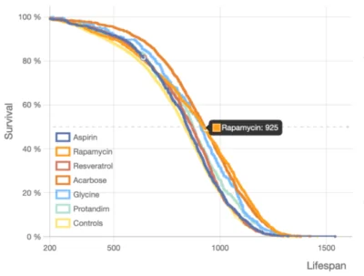
Take a close look at these so-called lifespan curves, each of which shows a surviving fraction of a cohort as a function of time, always going from 100% to zero. The precise shape of such a curve and slight differences between multiple curves tell us a lot about an intervention and its interaction with an organism.

This plot compares effects of several drugs from an Intervention Testing Program (ITP) in UM-HET3 mice [1]. It is meant to illustrate that the notion of “statistically significant” is not at all trivial. On one hand, rapamycin gave about 15% extra lifespan compared to controls. However, the same ITP data shows a 15% median lifespan difference between control male mice held at two different facilities in otherwise identical conditions.

Cross-checking the literature, we see that there are 20 papers on rapamycin in mice [3,4]. One shows a median lifespan increase of 8%, while another shows a 37% increase in the same strain/dose (128 ppm). Getting to the bottom of such differences would be crucial e.g. for Longevica, whose business is based on the outcome of another large intervention testing project: 1000+ compounds in B6C3F1/J mice over 3 years, tested in 15 mice per compound.

Longevica is looking to complement its own data with all the drugs ever tested in mice, creating an open research platform. In a separate effort, GeneAge and OpenGenes have been compiling data on all genetic interventions affecting lifespan.

Remarkable conservation of processes

A remarkable conservation across a wide range of species, from molecular- to organ-level processes, enables testing interventions in one species and generalizing the results to others. Such universality has indeed been observed: caloric restriction and rapamycin affect lifespans across species, from apes to flies. Alas, there are no perfect species – some are prohibitively expensive to use at scale, some live too long, and some only react to unreasonably high doses.

We have to do what we can; expensive animals are used scarcely, producing small (possibly not statistically significant) cohorts, while worms are subjected to extreme dosages when drugs crash out of solution. Our hope is that a big picture will come through when we combine reports of interventions in one species and closely related interventions in related species. Thousands of results of intervention testing for lifespan extension have been published to date [1-2].

When and where is such an integration supposed to occur? Agglomerating all the knowledge in the entirety of the relevant literature would be a superhuman feat. Perhaps artificial intelligence could help, but AI starts with computer-readable data, and current publishing practices are a long way from forcing scientists to express their premises and findings in a framework of constrained ontologies and logical predicates.

Someone has to marshal the sources and distill the literature into a uniform representation while working out a proper database schema. Early attempts [3-5] leave a lot to be desired. On top of the inherent discordance of the literature, the scope and quality of extracted information is very limited.

The same drug (caffeine), tested in the same strain of worms and at the same dose (7.5 mM), decreased lifespan by 12% in one study and increased it by 20% in another. It turns out that a drastically different temperature influences metabolism, but this was not reflected in the baseline data. Baselines matter!

A system focused on baseline data

All this underscores a need for a system that contains detailed baseline data on model organisms and control conditions in order to allow us to contrast the controls used in a given paper to others reporting on the same strain and to verify that the lifespan follows the same distribution.

Rich data on control groups’ lifespans will provide answers. What are the longest and shortest living strains of mice? How does temperature affect the lifespan of worms? Which husbandry conditions produce the longest lifespan in certain species?

To address this need for a data repository and an open research platform, Dr. Leon Peshkin conceived ALEC – Animal Life Expectancy Comparisons – a lifespan data browser whose application depends on the data it is populated with.

The most challenging and expensive part of the project will be data extraction and curation. A pilot site is already populated with a substantial seed dataset kindly provided by the ITP [1]; it is hosted by Lifespan.io and available for interactive exploration, and there are multiple illustrations of what can be uncovered within ALEC.

This pilot effort, spearheaded by Leon Peshkin and Olga Spiridonova, is focused mainly on lifespan in controls across species and strains. Longevica and OpenGenes can now develop GitHub-style branches of the ALEC platform.

We would like to ask you a small favor. We are a non-profit foundation, and unlike some other organizations, we have no shareholders and no products to sell you. All our news and educational content is free for everyone to read, but it does mean that we rely on the help of people like you. Every contribution, no matter if it’s big or small, supports independent journalism and sustains our future.

Literature

[1] ITP https://www.nia.nih.gov/research/dab/interventions-testing-program-itp

[2] ITP in worms https://citp.squarespace.com

[3] https://genomics.senescence.info/drugs

[4] Barardo, D., et al, de Magalhaes, J.P. (2017) “The DrugAge database of ageing-related drugs.” Aging Cell 16(3):594-597

[5] http://geroprotectors.org

Old man eye

Heat Shock Protein Fights Senescence in Macular Degeneration

Researchers publishing in Experimental Eye Research have discovered that the peptide aB-crystallin can reduce the cellular senescence that affects the progression of age-related macular degeneration (AMD).

What is αB-crystallin?

αB-crystallin is a well-known heat shock protein, a family of proteins that is used in temperature regulation. This protein has been previously demonstrated by this research team to have positive effects on retinal pigment epithelium (RPE) cells, including its benefits for mitochondria [1], its positive effects on oxidative stress [2], and its ability to prevent cellular death [3].

With this study, the researchers pinpointed another effect of this protein, one that is related to those three and many more: cellular senescence. In order to manage its levels, they used a chaperone peptide, a collection of amino acids that is generally smaller than a protein, called mini Cry.

Cellular senescence seems to be negated by mini Cry

For this study, the researchers induced senescence in RPE cells by giving them a precise dose of the antibiotic doxorubicin. As expected, the cells were shown to have a senescent phenotype, as measured by SA-β-gal, p16, and p21 accumulation, which are common biomarkers of cellular senescence. Co-administration of mini Cry decreased these levels nearly to those of a control group that was never administered doxorubicin at all.

These results were confirmed by using mini Cry on cells that were driven senescent by oxidative stress, which was induced in this study through hydrogen peroxide. The effects of mini Cry on peroxide-treated cells were nearly identical to its effects on doxorubicin-treated cells.

The researchers also returned to the mitochondria that they studied previously, noting that mitochondrial biogenesis, the process by which mitochondria are created, is actually increased in cellular senescence. However, this is reduced back to, or below, untreated control group levels with the administration of mini Cry.

The mitochondria’s ability to perform their basic function, provide energy from glucose and oxygen through glycolysis, is also affected; glycolysis increases with cellular senescence as well, and mini Cry reduced it, also to levels below that of the control group in some cases.

Finally, the researchers chose to examine one of the most well-known and dreaded aspects of cellular senescence: the SASP. The inflammatory mediators and interleukins that are associated with the SASP, including TNF-α, IL-6, and IL-8, were decreased to or below control group levels with mini Cry.

The researchers examined a mouse model to briefly examine the effects of senescence on RPE cells, where they found evidence for its association with AMD and subretinal fibrosis, but they did not test the effects of mini Cry on it.

Conclusion

These researchers’ continued efforts and examinations of the biology of RPE cells has yielded interesting findings that might possibly be useful for the treatment of AMD. If mini Cry can be dosed and tested in animals and human beings, it may provide an amelioration of this common and often blinding disease.

However, possibly the most interesting finding from a life extension viewpoint is that the researchers have been measuring many different positive effects of αB-crystallin of over the years, all of which seem to hinge upon a single upstream cause. There is no such thing as a complete panacea, but if more research can be directed into treatments that can possibly provide broad and positive effects against multiple aspects of aging, such treatments could potentially ameliorate multiple “different” conditions at once.

We would like to ask you a small favor. We are a non-profit foundation, and unlike some other organizations, we have no shareholders and no products to sell you. All our news and educational content is free for everyone to read, but it does mean that we rely on the help of people like you. Every contribution, no matter if it’s big or small, supports independent journalism and sustains our future.

Literature

[1] Sreekumar, P. G., Wang, M., Spee, C., Sadda, S. R., & Kannan, R. (2020). Transporter-Mediated Mitochondrial GSH Depletion Leading to Mitochondrial Dysfunction and Rescue with aB Crystallin Peptide in RPE Cells. Antioxidants, 9(5), 411.

[2] Sreekumar, P. G., Li, Z., Wang, W., Spee, C., Hinton, D. R., Kannan, R., & MacKay, J. A. (2018). Intra-vitreal aB crystallin fused to elastin-like polypeptide provides neuroprotection in a mouse model of age-related macular degeneration. Journal of Controlled Release, 283, 94-104.

[3] Sreekumar, P. G., Chothe, P., Sharma, K. K., Baid, R., Kompella, U., Spee, C., … & Hinton, D. R. (2013). Antiapoptotic properties of a-crystallin–derived peptide chaperones and characterization of their uptake transporters in human RPE cells. Investigative ophthalmology & visual science, 54(4), 2787-2798.

Launch Button

Altos Labs’ $3 Billion Launch to Reverse Cellular Aging

In September 2021, we reported that Altos Labs was about to launch an ambitious project to develop partial cellular reprogramming to the point where it could be safely used in humans to reverse cellular aging. That launch has now officially started, and Altos Labs is shaping up to be a real contender in the battle against aging and age-related diseases.

Altos Labs has considerable backing behind it

Among the backers of Altos Labs are Russian-born billionaire Yuri Milner along with Jeff Bezos, who is the world’s richest person and the former CEO of Amazon who stepped down last July.

Altos Labs has also attracted significant scientific talent. Shinya Yamanaka, one of the men who started it all, is in the scientific leadership. Juan Carlos Izpisua Belmonte, the first researcher to demonstrate that Yamanaka factors could be used in living animals to make them younger again, is also on the team.

Nobel Prize winner Jennifer Doudna, one of the researchers involved in the discovery of the gene editing tool CRISPR-Cas9, is on the board. We also recently learned that Morgan Levine has also joined the Altos team and brings her expertise of epigenetic clocks with her.

Cellular aging can be reversed

Sounds interesting, but what is Altos Labs doing, and what is its approach to targeting aging?

In a nutshell, as we age, our cells change their gene expression patterns and move from a profile that is pro-youth to one that is pro-aging. This change was always assumed to be a one-way-street until 2006, when Drs. Takahashi and Yamanaka showed that it was possible to reprogram mouse cells using just four transcription factors: Oct4, Sox2, Klf4, and c-Myc (OSKM). These genes became known as the Yamanaka factors.

It was discovered that these Yamanaka factors could reprogram adult cells back to an embryonic state called pluripotency, a flexible state in which the cell behaves like an embryonic stem cell and can become any other cell type in the body.

It was also shown that reprogramming these cells back to pluripotency caused them to behave like young cells again. Telomere length, mitochondrial function, and oxidative stress levels were reset to those of younger cells.

The problem was that exposing cells to the Yamanaka factors totally reset the cell type, which is a problem if a heart cell forgets it’s a heart cell while part of the organ! Thankfully, it was soon discovered that exposing cells to the Yamanaka factors for just long enough was enough to reset their cellular age without making them forget what type of cells they were. This was the birth of partial cellular reprogramming.

Now the race is on to bring partial cellular reprogramming to people, but the big question is: can it be made safe for human use? Altos is gearing up to find out and has the funds and the people to do it.

Official launch press release

SAN FRANCISCO, Jan. 19, 2022 /PRNewswire/ — Altos Labs™ (Altos™) launched today as a new biotechnology company dedicated to unraveling the deep biology of cellular rejuvenation programming. Altos’ mission is to restore cell health and resilience to reverse disease, injury, and the disabilities that can occur throughout life. The company launches with a community of leading scientists, clinicians, and leaders from both academia and industry working together towards this common mission.

Altos Labs

The Altos executive team will be composed of Hal Barron, MD (incoming CEO), Rick Klausner, MD (Chief Scientist and Founder), Hans Bishop (President and Founder), and Ann Lee-Karlon, PhD (Chief Operating Officer). Hal Barron is currently President of R&D and Chief Scientific Officer at GSK and will join Altos as CEO and Board co-chair effective August 1, 2022. Klausner was former director of the National Cancer Institute and entrepreneur, Bishop was former CEO of GRAIL and Juno Therapeutics, and Lee-Karlon was former Senior Vice President at Genentech.

Altos will be initially based in the US in the San Francisco Bay Area and San Diego, and in the UK in Cambridge. The company will also have significant collaborations in Japan. Set within these geographies, activity will be organized across the Institutes of Science and the Institute of Medicine. The Altos Institutes of Science will pursue deep scientific questions and integrate their findings into one collaborative research effort. The Altos Institute of Medicine will capture knowledge generated about cell health and programming to develop transformative medicines.

The three Altos Institutes of Science will be led by Juan Carlos Izpisua Belmonte, PhD, Wolf Reik, MD, and Peter Walter, PhD. Thore Graepel, PhD, will serve as global head of computational science, artificial intelligence, and machine learning. Prior to joining Altos, Izpisua Belmonte was professor and chair at the Salk Institute, Reik was director of the Babraham Institute and is an honorary professor at the University of Cambridge, and Walter was professor at the University of California, San Francisco (UCSF) and investigator at the Howard Hughes Medical Institute. Graepel previously served as research lead at Google DeepMind and professor at University College London. Within the Institutes of Science, an extraordinary group of Principal Investigators (PIs) will collaboratively pursue the many aspects of cell health and programming.

The Altos Board of Directors and advisors include Nobel Laureates and scientific leaders. The Board will be co-chaired by Rick Klausner, Hans Bishop, and Hal Barron (current director and incoming co-chair) and includes the following Board directors: Frances Arnold, PhD (Linus Pauling Professor of Chemical Engineering, Bioengineering and Biochemistry at the California Institute of Technology and Nobel Laureate), Hal Barron, MD (Chief Scientific Officer and President, R&D, of GSK), Jennifer Doudna (Li Ka Shing Chancellor’s Chair and Professor of Chemistry and Molecular and Cell Biology at the University of California, Berkeley, President of the Innovative Genomics Institute, and Nobel Laureate), Maria Leptin, PhD (President of the European Research Council), Robert Nelsen (Co-founder and Managing Director of ARCH Venture Partners), Rajiv Shah, MD (President of the Rockefeller Foundation), and David Baltimore, PhD (President Emeritus and Judge Shirley Hufstedler Professor of Biology at the California Institute of Technology and Nobel Laureate), as lead independent director. Shinya Yamanaka, MD, PhD (Director of the Center for iPS Cell Research and Application at Kyoto University and Nobel Laureate), will serve as senior scientific advisor to Altos without remuneration, overseeing research activities in Japan.

“I am deeply honored to have been offered this once in a lifetime opportunity to lead such a unique company with a transformative mission to reverse disease,” said Hal Barron. “It’s clear from work by Shinya Yamanaka, and many others since his initial discoveries, that cells have the ability to rejuvenate, resetting their epigenetic clocks and erasing damage from a myriad of stressors. These insights, combined with major advances in a number of transformative technologies, inspired Altos to reimagine medical treatments where reversing disease for patients of any age is possible.”

Altos is designed to integrate the best features of academia and industry — from academia the freedom to pursue the most challenging problems in biology, and from industry the focus on a shared mission, ability to foster deep collaborations, and the passion and commitment to transform science into medicines.

“Altos seeks to decipher the pathways of cellular rejuvenation programming to create a completely new approach to medicine, one based on the emerging concepts of cellular health,” said Rick Klausner. “Remarkable work over the last few years beginning to quantify cellular health and the mechanisms behind that, coupled with the ability to effectively and safely reprogram cells and tissues via rejuvenation pathways, opens this new vista into the medicine of the future. Altos begins with many of the leading scientists who are creating this new science. Together, we are building a company where many of the world’s best scientists can collaborate internally and externally and develop their research with the speed, mission, and focus of private enterprise. Our success will depend upon a culture of intense collaboration, enthusiasm, and openness.”

Will this be another Calico?

Some people in our community may think that Altos could turn out to be another Google Calico, with lots of hype and little to show in real terms years later. This could, of course, be true, but it is at least equally likely that this could also be an important step forward for our field. Altos has the backing, it has the people, and it has chosen an approach that has huge potential in the context of changing how we age.

We cannot, of course, predict the future, and a lot of this depends on the successful and safe translation of partial cellular reprogramming to people. There is no doubt that something that is so potentially transformative, and could have such a significant impact on aging, is going to need years of testing and refinement.

While enthusiasm for partial cellular reprogramming is currently high, make no mistake: this is something for the long haul. It is our view that we are a good decade or perhaps more away from partial cellular reprogramming reaching people. This is due to the complexity of the biology involved, the clinical trial process, and the inevitable setbacks on the road to getting it to work safely.

We wish Altos the best of luck, and we will be following this company’s progress in the coming years. Meanwhile, while we wait for this and other technologies to arrive, we should all strive to stay as healthy and active as possible.

We would like to ask you a small favor. We are a non-profit foundation, and unlike some other organizations, we have no shareholders and no products to sell you. All our news and educational content is free for everyone to read, but it does mean that we rely on the help of people like you. Every contribution, no matter if it’s big or small, supports independent journalism and sustains our future.
Hearing Loss

Epigenetic Age Acceleration Is Linked to Hearing Loss

A study published in Frontiers in Aging Neuroscience examined the relationship between epigenetic age acceleration and hearing [1].

Since 1958, a team of researchers from the National Institute on Aging Intramural Research program has been conducting the Baltimore Longitudinal Study of Aging (BLSA). The BLSA study continuously enrolls volunteers free of chronic conditions from a variety of age groups and conducts visits every 1 to 4 years, depending on age. Participants younger than 60 were seen every 4 years, participants between 60 and 79 were seen every 2 years, and participants who were at least 80 were seen every year.

Major revisions were made in 2003 to the BLSA study to add phenotypic measurements and molecular biomarkers. Due to the longitudinal nature of the study, changes in technology have occurred during the study effort, and extensive efforts have been made over time to control for these changes in analysis according to Dr. Luigi Ferruci and colleagues [2].

Epigenetic aging and hearing loss in a longitudinal study

236 individuals from the BLSA study were enrolled in this secondary analysis study. DNA was extracted from blood samples and CpG methylation status was determined from 485,577 CpG sites. Hearing was tested by trained technicians in a soundproof booth with an audiometer device.

Epigenetic age acceleration was measured with the GrimAge [3], Intrinsic Epigenetic Age Acceleration (IEAA) Horvath [4], Hannum [5], Phenoage [6], and Dunedin Pace of Aging (DunedinPACE) [7] clocks.

GrimAge and DunedinPACE had the strongest association with hearing

After adjusting for age, sex, race, and time, hearing was statistically associated with the GrimAge and DunedinPACE clocks. Both the direction and magnitude of the associations continued to be consistent after adjustment for congestive heart failure, hypertension, peripheral arterial disease, and smoking history.

An additional subanalysis was done on 197 participants, as some of the epigenetic clocks were limited to people who were at least 60 years old. In this older subset of participants, similar to the prior result, the GrimAge clock and the DunedinPACE clock were statistically associated with hearing. The researchers then ran an additional analysis to adjust for the variable of smoking cigarettes history. Like the prior two results, the GrimAge clock and the DunedinPACE clock were statistically associated with hearing. When the variable changed from the better hearing ear to the worse hearing ear, the results remained consistent.

Conclusion

This study was the first to examine the relationship between hearing loss via audiometric measurement and epigenetic clocks. The authors note some possible confounding factors and potential inaccuracies in the study, make it clear that future studies are needed, and finish as follows:

In conclusion, our findings demonstrate that not all epigenetic clocks were strongly correlated with hearing. Only those epigenetic clocks established using many cardiovascular measurements with longitudinal information were associated with hearing. Future research is needed to study the potential subclinical cardiovascular causes of hearing and to investigate the relationship between DNA methylation and hearing longitudinally.

We would like to ask you a small favor. We are a non-profit foundation, and unlike some other organizations, we have no shareholders and no products to sell you. All our news and educational content is free for everyone to read, but it does mean that we rely on the help of people like you. Every contribution, no matter if it’s big or small, supports independent journalism and sustains our future.

Literature

[1] Kuo, P. L., Moore, A. Z., Lin, F. R., & Ferrucci, L. (2021). Epigenetic Age Acceleration and Hearing: Observations From the Baltimore Longitudinal Study of Aging. Frontiers in aging neuroscience, 13, 790926. https://doi.org/10.3389/fnagi.2021.790926

[2] Kuo, P. L., Schrack, J. A., Shardell, M. D., Levine, M., Moore, A. Z., An, Y., Elango, P., Karikkineth, A., Tanaka, T., de Cabo, R., Zukley, L. M., AlGhatrif, M., Chia, C. W., Simonsick, E. M., Egan, J. M., Resnick, S. M., & Ferrucci, L. (2020). A roadmap to build a phenotypic metric of ageing: insights from the Baltimore Longitudinal Study of Aging. Journal of internal medicine, 287(4), 373–394. https://doi.org/10.1111/joim.13024

[3] Lu, A. T., Quach, A., Wilson, J. G., Reiner, A. P., Aviv, A., Raj, K., Hou, L., Baccarelli, A. A., Li, Y., Stewart, J. D., Whitsel, E. A., Assimes, T. L., Ferrucci, L., & Horvath, S. (2019). DNA methylation GrimAge strongly predicts lifespan and healthspan. Aging, 11(2), 303–327. https://doi.org/10.18632/aging.101684

[4] Horvath S. (2013). DNA methylation age of human tissues and cell types. Genome biology, 14(10), R115. https://doi.org/10.1186/gb-2013-14-10-r115

[5] Hannum, G., Guinney, J., Zhao, L., Zhang, L., Hughes, G., Sadda, S., Klotzle, B., Bibikova, M., Fan, J. B., Gao, Y., Deconde, R., Chen, M., Rajapakse, I., Friend, S., Ideker, T., & Zhang, K. (2013). Genome-wide methylation profiles reveal quantitative views of human aging rates. Molecular cell, 49(2), 359–367. https://doi.org/10.1016/j.molcel.2012.10.016

[6] Levine, M. E., Lu, A. T., Quach, A., Chen, B. H., Assimes, T. L., Bandinelli, S., Hou, L., Baccarelli, A. A., Stewart, J. D., Li, Y., Whitsel, E. A., Wilson, J. G., Reiner, A. P., Aviv, A., Lohman, K., Liu, Y., Ferrucci, L., & Horvath, S. (2018). An epigenetic biomarker of aging for lifespan and healthspan. Aging, 10(4), 573–591. https://doi.org/10.18632/aging.101414

[7] Belsky, D. W., Caspi, A., Arseneault, L., Baccarelli, A., Corcoran, D. L., Gao, X., Hannon, E., Harrington, H. L., Rasmussen, L. J., Houts, R., Huffman, K., Kraus, W. E., Kwon, D., Mill, J., Pieper, C. F., Prinz, J. A., Poulton, R., Schwartz, J., Sugden, K., Vokonas, P., … Moffitt, T. E. (2020). Quantification of the pace of biological aging in humans through a blood test, the DunedinPoAm DNA methylation algorithm. eLife, 9, e54870. https://doi.org/10.7554/eLife.54870

Rat bones

Quercetin Improves Bone Regeneration in Older Rats

A new study in ACS Applied Materials and Interfaces used a combined geroscience and tissue engineering approach to regenerate bone in aged rats [1].

Replacement or rejuvenation?

Too often, tissue engineering and longevity therapeutics are viewed as competing strategies – two different paths to potentially combat aging. In tissue engineering, cells and biomaterials are used to form tissue that replaces aged or diseased tissues. Meanwhile, longevity therapeutics include drugs that target one or more aging pathways in order to rejuvenate existing aged or diseased tissue. When viewed in competition, the question becomes “Which strategy deserves our precious time, attention, and funding?”

In reality, these two fields are quite complementary. Current tissue engineering strategies rely heavily on the body’s innate ability to heal and self-regenerate – which declines considerably with age. Additionally, while evidence is mounting that various drugs can modulate aging, at least in animal models, their effects have been far from total reversal of disease. Eventually, tissue damage becomes too extensive to ever return to a healthy state. If drugs can improve the aged body’s regenerative potential, it would follow that tissue engineering approaches would be more successful in older adults if used in combination with those treatments.

While the two fields have been relatively separate, progressing at their own rates in parallel, the first studies that utilize both approaches are now beginning to be conducted. Most recently, scientists at Sichuan University have used the senolytic quercetin and a TG-18 hydrogel to regenerate a bone defect in aged rats [1].

Finding the best senolytic

Many senolytic drugs have shown cell type-specific effects. To focus their study on bone regeneration, the researchers used bone marrow-derived mesenchymal stem cells (BMSCs) from rats. Cellular senescence was induced in these cells in vitro by either hydrogen peroxide or d-galactose treatment. Both senescent and non-senescent cells were treated with 10 different known senolytics (dasatinib, quercetin, navitoclax, A-1331852, A-1155463, ABT-737, fisetin, geldanamycin, 17-AAG, and 17-DMAG) at a concentration of 10 µmol.

Each drug except navitoclax significantly reduced the ratio of senescent BMSCs (as measured by SA-ß-gal staining) after hydrogen peroxide or d-galactose treatment. Dasatinib, quercetin, geldanamycin, 17-AAG, and 17-DMAG notably reduced this ratio to a greater extent than the other drugs. However, only fisetin and quercetin showed no toxicity to non-senescent cells, with dasatinib, geldanamycin, 17-AAG, and 17-DMAG especially showing notable toxicity. Because of these results, the researchers chose to move forward with quercetin for the remainder of their experiments.

Quercetin was then optimized for concentration. In these dose optimization studies, the researchers investigated the induction of senescence and effectiveness of quercetin in their BMSCs beyond just SA-ß-gal, confirming their results with the DNA damage marker γH2AX, the SASP markers IL-1ß and IL-6, and the cell cycle regulators p16, p21, and p53. A 20 µmol concentration showed the greatest effectiveness at reducing senescence without toxicity.

At this concentration, quercetin treatment also improved the proliferation and osteogenic differentiation of the rat BMSCs exposed to hydrogen peroxide but not of non-senescent control cells. This suggested that eliminating senescent cells may be able to improve bone regeneration.

A drug release platform that responds to senescent cells

In order to deliver the drug directly to the site of injury and to release it more rapidly in the presence of senescent cells, a triglycerol monostearate (TG-18) hydrogel was utilized for this study.

Senescent cells are known to release matrix metalloproteinases (MMPs) as part of their SASP. In this study, elevated levels of MMPs were observed in the in vitro senescent BMSCs and in the bone tissue of aged rats in vivo. TG-18 is a hydrogel that can encapsulate drugs like quercetin and is disassembled by MMP enzymes. Therefore, the researchers hypothesized the quercetin would be released more rapidly in the presence of senescent cells.

A maximum loading concentration of quercetin was determined to be approximately 3% by weight. When the TG-18 hydrogel was immersed in cell culture media conditioned by senescent cells, quercetin was released more rapidly compared to media from control cells. Additionally, TG-18 degraded more rapidly in the skull defects created in old rats compared to young rats.

Bone regeneration was greater with the combination treatment

To investigate its ability to facilitate bone regeneration, TG-18 loaded with 0.2% quercetin, 2% quercetin, or no quercetin was implanted into aged rats. The hydrogel was also supplemented with 2% hydroxyapatite in all groups to further facilitate bone regeneration.

Bone defects were surgically created in either the femur or skull and filled with the TG-18 hydrogel. At 3 months, for both femur and skull defects, senescence was decreased with quercetin treatment as measured by p16, γH2AX, and MMP expression. Bone formation was also greater with quercetin treatment as measured by microCT, Masson trichrome staining, and OCN and OPN expression. For each of these measures, the 2% quercetin showed slightly better, although not statistically significant, results relative to the 0.2% group.

In this study, we screened out quercetin as the suitable senolytic drug for clearing senescent rBMSCs. According to the secretion of MMPs in senescent rBMSCs, a senescence-responsive hydrogel loading quercetin was prepared to eliminate the senescent rBMSCs in the bone defects (Scheme 1). In vivo, bone repair assay confirmed that the senescence-responsive hydrogel efficiently eliminated local senescent cells and promoted the repair of bone defects in aged rats. This work presents a promising strategy for local removal of the senescent rBMSCs to promote bone regeneration in aged individuals.

Conclusion

Many previous studies have shown similar success at regenerating bone in rats. However, these studies have almost always been conducted in younger rats, which regenerate quite well on their own. Future studies would do well to follow in the footsteps of this one, as these treatments will ultimately be used primarily in older adults.

We know that senescence is critical for wound healing, but in older organisms, the senescent response is typically prolonged and interferes with the healing process [2,3]. This study provides compelling evidence that senolytics can, in fact, be beneficial for bone regeneration in older organisms. With TG-18, the local delivery and controlled release of quercetin initiated by the presence of senescent cells is also an exciting strategy moving forward.

Notably, however, the defects treated in this study were very small. Larger defects are more difficult to heal, as fibrotic scar tissue infiltrates the area more quickly than bone can form. Whether this strategy would be successful in humans or in larger defects cannot be claimed from these results. Although, there are also many improvements to this strategy, such as the inclusion of other biomaterials, cells, and/or growth factors, that future studies could use when targeting the regeneration of larger bone defects.

We would like to ask you a small favor. We are a non-profit foundation, and unlike some other organizations, we have no shareholders and no products to sell you. All our news and educational content is free for everyone to read, but it does mean that we rely on the help of people like you. Every contribution, no matter if it’s big or small, supports independent journalism and sustains our future.

Literature

[1] Xing, X. et al. Local elimination of senescent cells promotes bone defect repair during aging. ACS Appl Mater Interfaces (2022). https://doi.org/10.1021/acsami.1c22138

[2] Demaria, M. et al. An essential role for senescent cells in optimal wound healing through secretion of PDGF-AA. Developmental Cell (2014). https://doi.org/10.1016/j.devcel.2014.11.012

[3] da Silva, P.F.L. et al. The bystander effect contributes to the accumulation of senescent cells in vivo. Aging Cell (2019). https://doi.org/10.1111/acel.12848