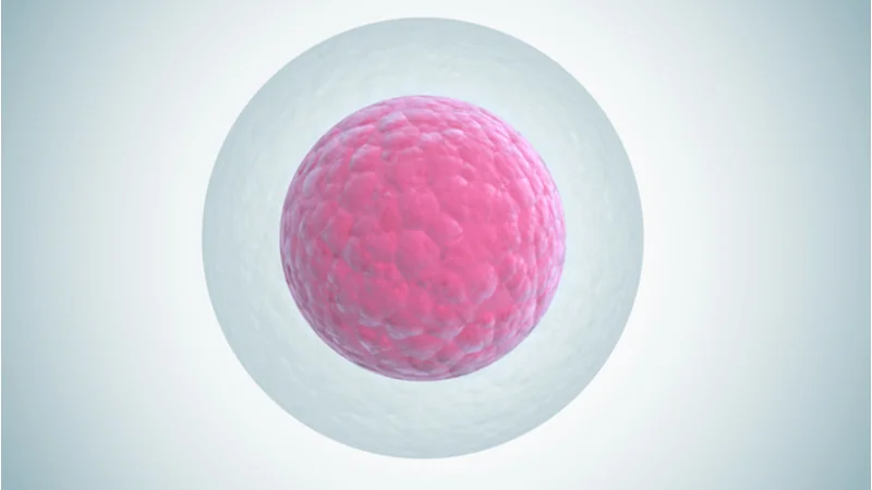
A group of researchers has documented in Aging that Epitalon, a synthetic peptide made of four amino acids, slows the aging of egg cells (oocytes) after ovulation.
Aging makes fertilization difficult
Oocyte aging is well-known to lead to fertilization deficiencies and a host of other problems that are visible in the cells themselves, including abnormalities in the fundamental parts of chromosomes [1] and the deterioration of cortical granules, which are organelles necessary to stop an egg from being fertilized by multiple sperm [2].
Assisted reproductive technology is often used as a way to fight back against age-related fertility problems; however, as it involves oocytes being cultured outside the body, it can introduce aging in other ways, as oocytes that are not quickly fertilized after ovulation begin to accumulate reactive oxygen species (ROS) and become senescent [3].
Therefore, the researchers sought an agent that could stave off these negative effects. After reviewing the literature, they chose Epitalon, a short peptide that is only four amino acids long. Epitalon is based on epithalamin, a nervous system regulator that is secreted by the pineal gland and has been shown to decrease ROS in Drosophila fruit flies [4].
Epitalon reduces ROS and benefits morphology
It only takes 24 hours in culture for egg cells to start showing notable effects of aging. ROS, as shown by fluorescence staining, is nearly absent in freshly ovulated egg cells, but in that time, the effects are dramatically visible. Epitalon was shown to benefit these cells on an interesting dose-response curve: 0.1 millimole (mM) of Epitalon reduced ROS nearly to the level of the control group, but 1 mM of Epitalon performed significantly worse in this respect, and 2 mM had an even weaker effect.
The researchers then looked at morphology, focusing on fragmentation before and after fertilization with sperm. As before, 0.1 mM of Epitalon was the most effective dose, and aged cells treated with this dose had approximately half of the fragmentation of untreated aged cells. Higher doses showed less positive effects.
Chromosomes, organelles, and mitochondria
The researchers then looked at the effects of Epitalon on cortical granules and spindles, which are fundamental structures responsible for chromosomal structure during cellular division. While the effects were not as strong as in other areas, Epitalon was shown to significantly decrease abnormalities in both of these areas.
This peptide was also found to improve mitochondrial function in these cells. Fluorescence staining found that Epitalon restored the mitochondrial membranes of treated cells to the level of the control group, and mtDNA copy number, another marker of mitochondrial function, was partially restored.
DNA damage was also measured by fluorescence: the DNA repair molecule γ-H2AX, which is produced in response to this damage, was dramatically increased in aged cells but partially decreased by Epitalon. Early cellular death due to apoptosis was similarly decreased.
Conclusion
While this paper has shown a potentially interesting tool for keeping oocytes viable for use in assisted reproductive technology, it did not go as far as to bring mouse pups to term. Therefore, we still do not know whether it has side effects in developing embryos. A more thorough assessment in mice, and perhaps other mammals, is needed alongside a test of Epitalon on the egg cells of human women.
However, if it is found to be effective, Epitalon may become part of a strategy for maximizing the viability of embryos conceived through in vitro fertilization while minimizing the risk of genomic damage, which can lead to miscarriages or potentially crippling birth defects.
Literature
[1] Jones, K. T. (2008). Meiosis in oocytes: predisposition to aneuploidy and its increased incidence with age. Human reproduction update, 14(2), 143-158.
[2] Ducibella, T., Duffy, P., Reindollar, R., & Su, B. (1990). Changes in the distribution of mouse oocyte cortical granules and ability to undergo the cortical reaction during gonadotropin-stimulated meiotic maturation and aging in vivo. Biology of Reproduction, 43(5), 870-876.
[3] Moghadam, A. R. E., Moghadam, M. T., Hemadi, M., & Saki, G. (2022). Oocyte quality and aging. JBRA Assisted Reproduction, 26(1), 105.
[4] Khavinson, V. K., Myl’nikov, S. V., Oparina, T. I., & Arutyunyan, A. V. (2001). Effects of peptides on generation of reactive oxygen species in subcellular fractions of Drosophila melanogaster. Bulletin of Experimental Biology and Medicine, 132(1), 682-685.


