Researchers publishing in Nature Aging have discovered how Alzheimer’s-related protein aggregates are also related to senescent cells and osteoporosis.
Beyond the brain

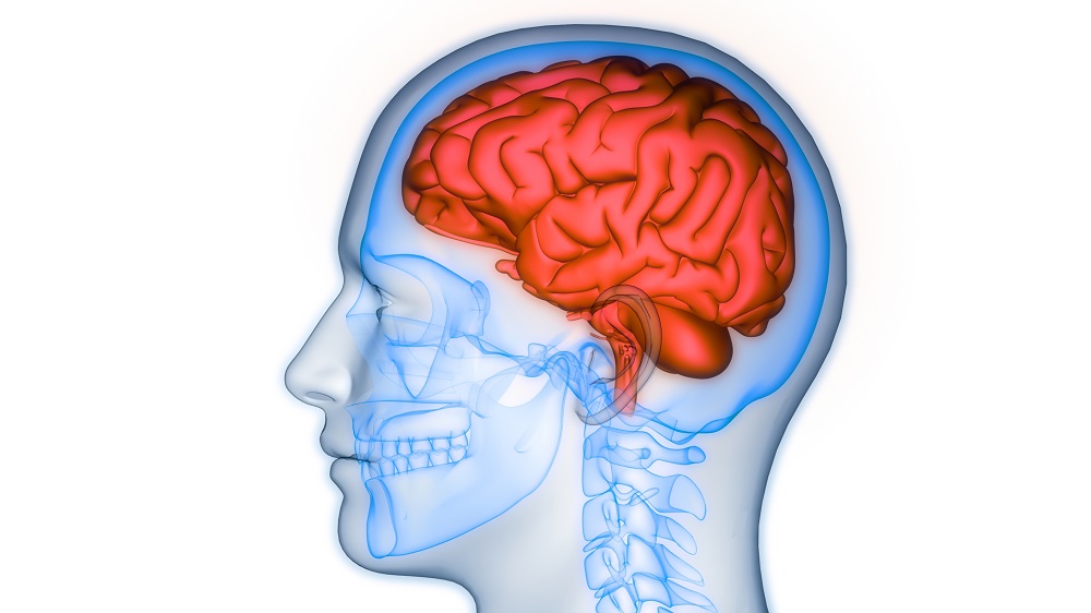
The amyloid tangles that result from a loss of proteostasis are very well-known in the context of Alzheimer’s and other neurodegenerative diseases. However, amyloid fibrils can appear in many other organs, including the liver, kidneys, and heart [1]. When this occurs throughout the body, it is known as systemic amyloidosis, a potentially fatal condition [2]. Like other proteostasis diseases, this can be caused by rare genetic disorders but is most often linked to aging [3].
Interestingly, the incidence of proteostasis diseases seems to be linked between entirely different organs. Compared to an average person of the same age, someone with Alzheimer’s is more likely to have been suffering from bone loss well before Alzheimer’s was diagnosed [4]. In Alzheimer’s model mice, the rate of bone loss is above that of wild-type mice [5].
Despite some work being done in intervertebral and related tissues [6], the causes and consequences of amyloid deposition in bone have been the subject of very little previous work. These researchers sought to fill that gap, discovering a relationship between these amyloids and senescent cells.
Nerve amyloids affecting bone tissue
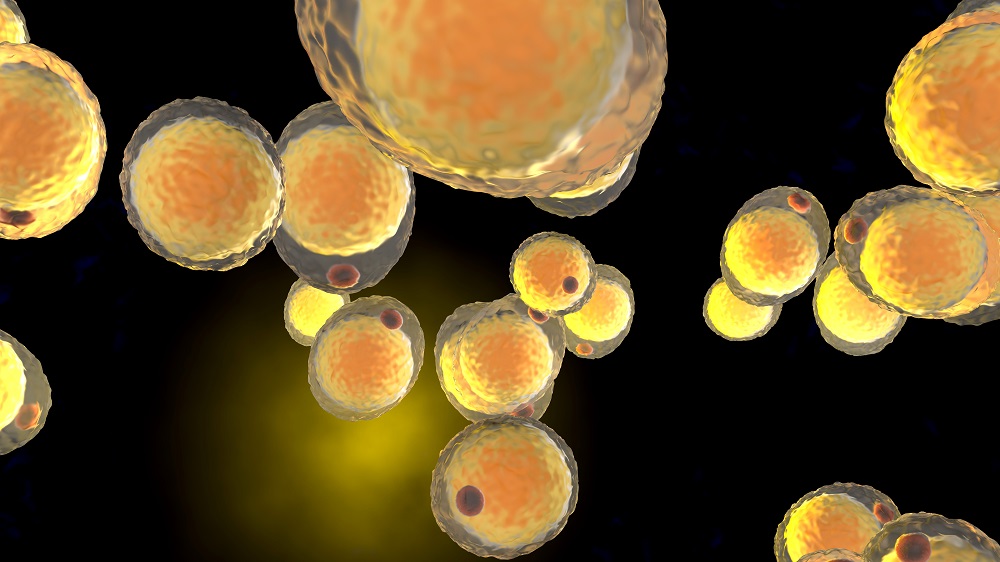
These researchers reared two variants of Alzheimer’s model mice and compared them to wild-type mice at 9 months of age. While the two variants did not share the exact same metrics, both types had prematurely aged bones. There were significant indicators of osteoporosis, including thinner and less dense bones, and they had fat deposits throughout their bones that wild-type mice did not.
The researchers found antibodies against amyloid beta (Aβ) in the bones of the Alzheimer’s mice. Their data suggested that these amyloids may have originated from nervous tissue, which implies that Alzheimer’s itself causes some of this premature bone aging. However, older wild-type mice, which do not get Alzheimer’s, also had Aβ deposits. In both cases, these Aβ deposits formed rings around fat cells in the bone marrow, which prompted the researchers to surmise that the fat cells were stabilizing them.
These fat cells were found to have substantial markers of cellular senescence in Alzheimer’s mice, including the well-known p16, p21, and SA-β-gal. p19, a regulator of the relationship between p21 and the tumor suppressor p53, was also upregulated, as was the DNA damage marker γH2AX. The relationship between CEBPα, which drives the formation of fat cells in bone tissue, and p19 was found to be a crucial part of this accelerated senescence.
Further experiments found that it was these senescent cells that were causing the bone loss. The researchers transplanted these fat cells from Alzheimer’s model mice into 4-month-old wild-type mice alongside a control group that had transplants from other wild-type mice. The senescent fat cells derived from the Alzheimer’s mice secreted signals (the SASP) that led to significant bone loss, and removing these cells with the senolytic combination of dasatinib and quercetin ameliorated some of the damage.
A SASP factor can cause amyloid aggregation
An antibody array found that the main factor involved in this bone loss was SAP/PTX2, which was found to be largely localized to the senescent fat cells. Administering either the dasatinib and quercetin combination or ruxolitinib, a compound that inhibits the SASP, to Alzheimer’s mice was sufficient to reduce the level of SAP to that of the wild-type mice. Importantly, SAP was found to be directly related to amyloid formation itself; introducing SAP to unaggregated amyloid beta peptides caused them to aggregate.

The researchers then tested another compound, CPHPC, which directly targets SAP. This compound was found to aid against both Aβ deposition and bone loss. Osteoclasts, cells that are responsible for destroying bone, were significantly less prevalent in the CPHPC-treated Alzheimer’s mice.
This direct relationship between a SASP factor and Aβ deposition is surprising and suggests new potential therapies. While this approach does not affect the production of amyloids within cells, a SASP factor that causes these amyloids to aggregate is a clear target. However, this approach has not yet been tested in human beings, it is not clear if other amyloids are involved, and it has yet to be determined if senolytics, senomorphics, or compounds such as CPHPC may be effective against amyloid-related osteoporosis.
Literature
[1] Gertz, M. A., Comenzo, R., Falk, R. H., Fermand, J. P., Hazenberg, B. P., Hawkins, P. N., … & Grateau, G. (2005). Definition of organ involvement and treatment response in immunoglobulin light chain amyloidosis (AL): a consensus opinion from the 10th International Symposium on Amyloid and Amyloidosis. American journal of hematology, 79(4), 319-328.
[2] Gertz, M. A., & Dispenzieri, A. (2020). Systemic amyloidosis recognition, prognosis, and therapy: a systematic review. Jama, 324(1), 79-89.
[3] Hipp, M. S., Kasturi, P., & Hartl, F. U. (2019). The proteostasis network and its decline in ageing. Nature reviews Molecular cell biology, 20(7), 421-435.
[4] Tan, Z. S., Seshadri, S., Beiser, A., Zhang, Y., Felson, D., Hannan, M. T., … & Kiel, D. P. (2005). Bone mineral density and the risk of Alzheimer disease. Archives of neurology, 62(1), 107-111.
[5] Dengler-Crish, C. M., Ball, H. C., Lin, L., Novak, K. M., & Cooper, L. N. (2018). Evidence of Wnt/β-catenin alterations in brain and bone of a tauopathy mouse model of Alzheimer’s disease. Neurobiology of aging, 67, 148-158.
[6] Mihara, S., Kawai, S., Gondo, T., & Ishihara, T. (1994). Intervertebral disc amyloidosis: histochemical, immunohistochemical and ultrastructural observations. Histopathology, 25(5), 415-420.