×

The Blog

Building a Future Free of Age-Related Disease

Food clock

Caloric Restriction Might Slow Down Human Aging

In a paper published in Nature Aging, researchers have shown that caloric restriction modestly slows down the pace of aging in healthy young people as measured by one of the DNA methylation clocks [1].

CALERIE design

A plethora of animal data has shown beneficial effects of caloric restriction for health and longevity. Human trials, albeit not that numerous, also suggest rejuvenation-promoting properties of eating less.

Comprehensive Assessment of Long-term Effects of Reducing Intake of Energy (CALERIE) was a two-phase trial designed to explore the effect of caloric restriction in healthy people. Phase 2 was a randomized controlled trial conducted in three centers over a two-year period.

The participants were men aged 21-50 years and premenopausal women aged 21-47 years who had normal BMIs or were slightly overweight. They were assigned to either a caloric restriction group (25% less than what they normally consume) or a control group (regular diet).

117 and 71 people in the experimental and control groups, respectively, completed the trial. Interestingly, the participants in the caloric restriction group were first provided with three meals for a month so that they could get used to the portion sizes.

The participants’ adherence to the caloric restriction diet was then assessed by their weight loss. Based on previous studies, scientists know how much weight the participants are expected to lose with a 25% reduction in caloric intake. Therefore, they can just compare the participants’ actual weight loss with a predicted weight loss trajectory.

The many clocks

In this publication, the researchers focused on DNA methylation (DNAm) data obtained from the participants of the CALERIE phase 2 trial. Epigenetic clocks use genome-wide methylation patterns to estimate biological age and predict mortality risks.

After the development of the first-generation Horvath clock [2], the second generation was built to match DNA methylation patterns to some clinical parameters, such as glucose and C-reactive protein levels. This includes PhenoAge [3] and GrimAge [4], the latter of which is highly predictive of mortality. The researchers of this study primarily used the principal component versions of these two clocks, which have greater accuracy.

Other clocks were developed by assessing epigenetic changes across multiple time points. The one used in this study is called DunedinPACE. This epigenetic clock was fitted to 19 biomarkers, including waist-to-hip ratio and cardiorespiratory fitness, and it shows high test-retest reliability [5].

The (not so impressive?) results

The PhenoAge and GrimAge clocks did not show any difference between the caloric restriction and control groups. However, DunedinPACE demonstrated a reduction in the aging pace of the participants on the restrictive diet.

An important shortcoming the researchers faced was that, on average, participants in the caloric restriction group could only achieve an approximately 12% reduction in consumed calories, not the prescribed 25%.

The scientists then tested if the pace of aging was slower in people who managed to achieve a higher caloric reduction. Indeed, the effect was better in people who achieved more than 10% caloric restriction than people who managed less than 10% as measured by DunedinPACE.

The authors admit that while only one out of three aging clocks showed a difference between caloric restriction and regular diet groups, this might be explained by the different methods on which the clocks are based. DunedinPACE could be a more sensitive measure, and the effect of caloric restriction on aging that it detected is in line with previous CALERIE results [6].

Abstract excerpt

Here we report the results of a post hoc analysis of the influence of CR on DNAm measures of aging in blood samples from the Comprehensive Assessment of Long-term Effects of Reducing Intake of Energy (CALERIE) trial, a randomized controlled trial in which n = 220 adults without obesity were randomized to 25% CR or ad libitum control diet for 2 yr. We found that CALERIE intervention slowed the pace of aging, as measured by the DunedinPACE DNAm algorithm, but did not lead to significant changes in biological age estimates measured by various DNAm clocks including PhenoAge and GrimAge.

Conclusion

This study adds to the growing body of evidence showing that caloric restriction has a beneficial effect on longevity. However, it also raises a number of questions and confirms some reservations. For example, it is unclear if caloric restriction works for people with lower BMIs. In this study, the participants were either on the higher end of normal BMI or overweight.

This study has also shown that achieving significant caloric restriction is not feasible for most people. Moreover, the effect of caloric restriction seems to depend on which measures are chosen to estimate it and varies from person to person.

We would like to ask you a small favor. We are a non-profit foundation, and unlike some other organizations, we have no shareholders and no products to sell you. All our news and educational content is free for everyone to read, but it does mean that we rely on the help of people like you. Every contribution, no matter if it’s big or small, supports independent journalism and sustains our future.

Literature

[1] ​​Waziry R, Ryan CP, Corcoran DL, Huffman KM, Kobor MS, Kothari M et al. Effect of long-term caloric restriction on DNA methylation measures of biological aging in healthy adults from the CALERIE trial. Nature Aging (2023): 1–10.

[2] Horvath S. DNA methylation age of human tissues and cell types. Genome Biol (2013); 14: R115.

[3] Levine, Morgan E et al. “An epigenetic biomarker of aging for lifespan and healthspan.” Aging vol. 10,4 (2018): 573-591.

[4] Lu, Ake T et al. “DNA methylation GrimAge strongly predicts lifespan and healthspan.” Aging vol. 11,2 (2019): 303-327.

[5] Belsky DW, Caspi A, Corcoran DL, Sugden K, Poulton R, Arseneault L et al. DunedinPACE, a DNA methylation biomarker of the pace of aging. Elife (2022): 11.

[6] ​​Spadaro O, Youm Y, Shchukina I, Ryu S, Sidorov S, Ravussin A et al. Caloric restriction in humans reveals immunometabolic regulators of health span. Science 2022; 375: 671–677.

Protein muscle

High Protein Intake Associated With Sarcopenia

Researchers publishing in Age and Ageing have found that, rather than being protective, an increase in dietary protein is associated with an increased chance of sarcopenia.

Sarcopenia is a well-known disorder that occurs with aging. People with sarcopenia lack adequate muscular function, leading to frailty, a higher risk of falls, and a functional decline in daily living activities that leads to a decreased quality of life [1].

Previous research into the relationship between protein consumption and sarcopenia has noted the phenomenon of anabolic resistance, in which muscle protein is more difficult to synthesize in older adults [2]. Therefore, the European Society for Clinical Nutrition and Metabolism (ESPEN) recommends that older adults consume 1 to 1.3 grams of protein per kilogram of body weight a day [3].

Muscle strength, muscle mass, and sarcopenia

This study used data from the TwinsUK cohort, a registry of almost 15,000 fraternal and identical twins and triplets [4]. This study chose community-dwelling older adults that had detailed muscular data available; this was a subgroup of 3,302. There were no exclusionary factors.

As is usual for this kind of study, there were a substantial number of confounding factors, many of which were related to one another. While aging is, of course, the primary association, education, BMI, and income were all found to have associations with muscle strength, muscle mass, and sarcopenia.

There was no significant association between muscle strength and protein intake one way or another. However, protein intake below the ESPEN recommendations was, to the researchers’ surprise, significantly correlated with a reduced incidence of low muscle mass and sarcopenia. Similarly, protein intake in excess of the ESPEN recommendations was correlated with a greater risk of low muscle mass and sarcopenia.

These results were confirmed when twins were examined against one another. Shared twin factors, such as genetics and early life history, did not meaningfully change the statistical relationship between protein intake and sarcopenia. Other multivariable analysis models also confirmed these findings.

Analysis

The researchers considered multiple potential reasons behind these findings. One of them is the idea that the causality might be reversed: that people who suffer from sarcopenia may be consuming more protein in an effort to treat the condition. The researchers find this to be unlikely, as sarcopenia is seldom diagnosed [5].

They also note that dietary studies that did not focus on protein, but rather on higher consumption of fruits and vegetables, showed an association with higher grip strength [6]. Diets that are high in protein might also be high in inflammatory or other negative factors that promote sarcopenia, so it might be the source of protein that is the root cause of these results.

Conclusion

While this study was biased towards healthy volunteers, it offers highly contrary evidence to the conventional wisdom surrounding protein and sarcopenia, and it may result in re-evaluations of ESPEN and other dietary health guidelines. This is an association study that does not prove causation, but it makes it clear that simply eating more protein is not likely to protect anyone against developing sarcopenia. Exercise may be somewhat effective in fighting back against this disease, but more fundamental biological interventions are likely to be required to prevent it for good.

We would like to ask you a small favor. We are a non-profit foundation, and unlike some other organizations, we have no shareholders and no products to sell you. All our news and educational content is free for everyone to read, but it does mean that we rely on the help of people like you. Every contribution, no matter if it’s big or small, supports independent journalism and sustains our future.

Literature

[1] Cruz-Jentoft, A. J., Bahat, G., Bauer, J., Boirie, Y., Bruyère, O., Cederholm, T., … & Zamboni, M. (2019). Sarcopenia: revised European consensus on definition and diagnosis. Age and ageing, 48(1), 16-31.

[2] Welch, A. A. (2014). Nutritional influences on age-related skeletal muscle loss. Proceedings of the Nutrition Society, 73(1), 16-33.

[3] Deutz, N. E., Bauer, J. M., Barazzoni, R., Biolo, G., Boirie, Y., Bosy-Westphal, A., … & Calder, P. C. (2014). Protein intake and exercise for optimal muscle function with aging: recommendations from the ESPEN Expert Group. Clinical nutrition, 33(6), 929-936.

[4] Verdi, S., Abbasian, G., Bowyer, R. C., Lachance, G., Yarand, D., Christofidou, P., … & Steves, C. J. (2019). TwinsUK: the UK adult twin registry update. Twin Research and Human Genetics, 22(6), 523-529.

[5] Avgerinou, C. (2020). Sarcopenia: why it matters in general practice. British Journal of General Practice, 70(693), 200-201.

[6] Robinson, S. M., Jameson, K. A., Batelaan, S. F., Martin, H. J., Syddall, H. E., Dennison, E. M., … & Hertfordshire Cohort Study Group. (2008). Diet and its relationship with grip strength in community‐dwelling older men and women: the Hertfordshire cohort study. Journal of the American Geriatrics Society, 56(1), 84-90.

Lifespan.io Editorial

Our Organization Grows to Support Rejuvenation Research

Spring is not far away here in the Northern Hemisphere, and with it, the signs of life and new growth are appearing. Much like the change of seasons, our organization is also growing and preparing to continue the fight against age-related diseases.

Matt Kaeberlein joins our SAB

Matt Kaeberlein

We are delighted to announce that Dr. Matt Kaeberlein has joined our Scientific Advisory Board.

He is the Chief Science Officer at Optispan Geroscience and a Professor of Laboratory Medicine and Pathology at the University of Washington (UW) School of Medicine, with adjunct appointments in Genome Sciences and Oral Health Sciences.

Matt is an important figure in our field, helping to drive progress while keeping things grounded and scientific. Our field has a history of overpromising and under delivering and Matt helps to keep the hype in check and present a balanced and realistic view of the aging and rejuvenation space.

We are looking forward to working with him in the future and he had this to say about joining the scientific advisory board.

I am pleased to join the scientific advisory board. Lifespan.io fills an important need in the field of aging research, providing unbiased journalism, high-quality educational materials, and effective advocacy for aging research. I look forward to working closely with the team at Lifespan.io to advance progress toward our shared goal to solve the biggest problem in human history.

Stephanie Dainow at Rejuvenation Startup Summit 2022

Our friends over at the Forever Healthy Foundation recently published a video from their Rejuvenation Startup Summit 2022 featuring Stephanie Dainow, Executive Director of Lifespan.io.

During this talk, Stephanie focused on how to make the longevity movement mainstream. The presentation covered how to bring more funding and attention to the field and how Lifespan.io is helping to challenge widely held biases about life extension and helping to pave the way for rejuvenation technologies to be accepted and adopted by mainstream audiences.

New interactive maps upgrade educational articles

As part of making our website’s educational knowledge hubs even better, we are starting to add interactive data visualizations to some of them. These highly interactive maps and charts allow you to dive into the data and better understand certain topics.

The first educational topic that has had a revamp is ‘What is the Average Life Expectancy Worldwide?’. We added a brand new interactive chart that shows the averages of lifespan and healthspan for most countries in the world. If you are curious to find out how long you might live based on your country, you can find that out here.

The second topic we have added interactive charts to is ‘What is the Average Life Expectancy in America?’. The first interactive map we included here shows all US counties or states and the life expectancy associated with each. This second interactive map shows life expectancy and healthy life expectancy in the US.

There are lots of ways to interact with all three of these visualizations, so if you are at all curious about life expectancy and healthspan based on where you live, check it out. Our knowledge hubs are a great resource for people interested in longevity and the biology of aging that will always be 100% free.

Save the date for Ending Age-Related Diseases 2023

On August 10-12, 2023, we will be holding our sixth annual Ending Age-Related Diseases conference to bring entrepreneurs and investors, pharma and biotech companies, researchers, and government organizations together.

Fostering the exchange of information and encouraging cooperation between the various parts of the industry is a large goal for our non-profit organization.

Solving aging will need a coordinated industry working together to turn aging research into prescribable medicine. Lifespan.io is really helping to move things forward and is the focus of our conference.

This will be a hybrid event – both virtual and physical – taking place in New York City and online.

We will be announcing speakers, opening ticket sales, and publishing a program in the coming months. If you would like to stay informed about the conference and ticket offers, register for our free newsletter today!

Support Lifespan.io when you use email

GatedThere’s a new way to support Lifespan.io while reducing the amount of spam and nonsense emails you get!

Most of us get too many emails, cluttering our inbox and wasting hours of our time.

We have teamed up with Gated, a platform that asks people you don’t know to donate to charity in order to contact you. With Gated, anyone trying to get in touch with you is encouraged to pay $2 (more if they like) to send you an email.

This helps to cut down on the spam and timewasters bothering you and supports LIfespan.io to continue our important work. Sign up today, and help us to solve aging, one email at a time!

The Rejuvenation Roadmap is expanding

As part of our ongoing mission to educate people about aging and rejuvenation research, we are currently increasing the range of resources we offer on our website. The Rejuvenation Roadmap is a curated database that tracks the various initiatives in the field and shows their progress in a way that is quick and easy.

We have been busy adding many new companies and drug candidates to the roadmap, and we have added a brand new Biomarkers of Aging section as well.

Our commitment to making this curated database even bigger and better will continue this year; check back often to see all the new companies and drugs we are adding every month.

Support us with the Longevity Cause Fund

Spring Editorial Banner

The Longevity Cause Fund is a cryptocurrency fundraiser in partnership with SENS Research Foundation and the Methuselah Foundation, facilitated by Angel Protocol.

All proceeds from the fundraiser will go to three non-profit foundations in support of aging research and advocacy.

This program is designed to create sustainable funding for key players in the aging research and advocacy community. From the total amount, half will be used for anti-aging work that is currently underway. The remainder will be invested in perpetual endowments to provide ongoing support for this work — forever.

Aging affects us all, but there is some good news!  The first $10k donated is being fund-matched by the Angel Alliance. That means any donation, no matter how big or small, will be worth even more!  Help us help you to stay healthier for longer by making a donation today!
We would like to ask you a small favor. We are a non-profit foundation, and unlike some other organizations, we have no shareholders and no products to sell you. All our news and educational content is free for everyone to read, but it does mean that we rely on the help of people like you. Every contribution, no matter if it’s big or small, supports independent journalism and sustains our future.
A4LI Logo

Announcing the Formation of the Longevity Science Caucus

The Alliance for Longevity Initiatives (A4LI) would like to applaud the efforts of Congressman Gus Bilirakis (R-FL) and Congressman Paul Tonko (D-NY) for co-chairing and launching the newly formed bipartisan Congressional Caucus for Longevity Science.

The “Longevity Science Caucus” aims to increase funding for aging and longevity biotechnology, streamline regulations, and promote initiatives to increase the healthy average lifespan of Americans. This is a major step towards ensuring that the latest advancements in aging and longevity research are made accessible to all.

A4LI is proud to have led this historic effort, and we are confident that the Longevity Science Caucus will make a significant impact on the future of health and wellness for all Americans. We look forward to working closely with the members of this caucus to achieve its goals. We hope that you share our excitement for this new era in which the pursuit of a longer, healthier lifespan is a national priority. We would appreciate your support in spreading the word about this important announcement by sharing our posts on Twitter, LinkedIn, and Facebook. The links to our posts are provided below:

For more information about the Longevity Science Caucus, check out the Longevity Science Caucus press release.

We would like to ask you a small favor. We are a non-profit foundation, and unlike some other organizations, we have no shareholders and no products to sell you. All our news and educational content is free for everyone to read, but it does mean that we rely on the help of people like you. Every contribution, no matter if it’s big or small, supports independent journalism and sustains our future.
Midday Exercise Might Be Better In Preventing Mortality

Midday Exercise Might Be Better In Preventing Mortality

Scientists have found that the time of day when you exercise might be an important factor in lowering all-cause and cardiovascular mortality [1].

Exercise is good, but what about timing?

The value of physical activity for health and longevity is unquestionable [2], even if some doubts remain about its optimal composition and quantity. However, little is known about the effect of daily exercise timing.

This new study published in Nature Communications is one of many to utilize the trove of health information accumulated in UK Biobank, a huge open repository of health data. In addition to more conventional stuff, such as bloodwork and dietary patterns, UK Biobank also contains data on about 100,000 UK residents who had agreed to wear sophisticated accelerometers constantly for 7 days. This data enables researchers to precisely determine exercise intensity and timing. It also covers physical activity that is not part of structured exercise, such as carrying groceries or walking stairs, which is something that most related studies are missing.

Physical activity is associated with a massive reduction in mortality

The researchers divided the participants into four groups by prevalent timing of MVPA (moderate-to-vigorous physical activity): morning, evening, midday-afternoon, and mixed. The mean follow-up period was seven years.

First, the data confirmed yet again that physical activity is associated with massively reduced mortality. Interestingly, and largely in line with other recent studies, the association increased rapidly between 0 and 150 minutes of MVPA per week and plateaued at around 200 minutes per week. This was true for all three types of mortality considered: all-cause, cardiovascular, and cancer. For cardiovascular mortality, a massive four-fold reduction was observed. The effect was weaker, though still remarkable, for cancer mortality.

Exercise timing graph

Midday beats mornings and evenings

The main result, however, was that people who had the lion’s share of their MVPA either in the morning or in the evening seemed to enjoy a smaller reduction in all-cause and cardiovascular mortality than those who exercised mostly during midday-afternoon or mixed hours. Even in models adjusted for numerous potentially confounding variables, such as age, sex, ethnicity, socioeconomic status, education level, diet, smoking, alcohol intake, sleep quality, and total MVPA volumes, the midday-afternoon and mixed hours groups showed 28% and 26% reduction in cardiovascular mortality, respectively, compared to the morning group. The evening group results were largely similar to those of the morning group. The difference for all-cause mortality was significant as well but not for cancer mortality.

These associations held even after numerous sensitivity analyses and were more pronounced for older people, males, people with existing cardiovascular conditions, and less physically active people.

What are the mechanisms?

Speculating about possible mechanisms behind their results, the researchers mention the circadian differences in cardiometabolic reaction to physical exercise that were uncovered by previous studies. For example, one study showed faster recovery of systolic blood pressure after exercise in the late afternoon than in the early morning [3]. This would be consistent with the stronger effect shown in people with existing cardiovascular conditions. Another study found that post-meal walking improves glucose control more effectively than morning/afternoon walking does [4].

Like all populational studies, this one cannot establish causation and is prone to contamination by numerous potentially confounding variables. It also has design limitations, such as the short, one-week duration of accelerometer wearing. This might be enough to capture weekly activity patterns but not long-term changes in physical activity. On the other hand, the study implements robust safeguards and presents convincing results that should be explored further.

To our knowledge, this large cohort study provides the first evidence that MVPA is associated with lower risks of all-cause, CVD, and cancer mortality regardless of the time of day. Another interesting finding was that the midday-afternoon and mixed MVPA timing groups, as compared to the morning group, showed substantially decreased all-cause and CVD mortality risks, but not cancer mortality. The associations between MVPA timing and mortality risk were independent of sociodemographic factors, lifestyle, comorbidities, sleep duration, sleep midpoint, and total MVPA volume. These findings were robust to multiple testing corrections and sensitivity analyses. In addition, the observed protective effects of MVPA timing were more pronounced among the elderly, males, less active individuals, or those with preexisting CVDs.

Conclusion

While the effects of daily timing of sleep and feeding are drawing most of the attention, the timing of physical activity is not as well-researched. The results of this study suggest that when we exercise might be just as important as how we exercise. However, more research is needed to confirm this effect and uncover the mechanisms behind it.

We would like to ask you a small favor. We are a non-profit foundation, and unlike some other organizations, we have no shareholders and no products to sell you. All our news and educational content is free for everyone to read, but it does mean that we rely on the help of people like you. Every contribution, no matter if it’s big or small, supports independent journalism and sustains our future.

Literature

[1] Feng, H., Yang, L., Liang, Y. Y., Ai, S., Liu, Y., Liu, Y., … & Zhang, J. (2023). Associations of timing of physical activity with all-cause and cause-specific mortality in a prospective cohort study. Nature Communications, 14(1), 1-10.

[2] DiPietro, L. (2001). Physical activity in aging: changes in patterns and their relationship to health and function. The Journals of Gerontology Series A: Biological Sciences and Medical Sciences, 56(suppl_2), 13-22.

[3] Qian, J., Scheer, F. A., Hu, K., & Shea, S. A. (2020). The circadian system modulates the rate of recovery of systolic blood pressure after exercise in humans. Sleep, 43(4), zsz253.

[4] DiPietro, L., Gribok, A., Stevens, M. S., Hamm, L. F., & Rumpler, W. (2013). Three 15-min bouts of moderate postmeal walking significantly improves 24-h glycemic control in older people at risk for impaired glucose tolerance. Diabetes care, 36(10), 3262-3268.

The Journal Club is a monthly livestream hosted by Dr. Oliver Medvedik which covers the latest aging research papers.

Journal Club February 2023

The Journal Club, hosted by Dr. Oliver Medvedik, returns to the Lifespan.io Facebook page at 12:00 on Tuesday, February 28th. This month, we are covering a recent paper that showed a method of restoring youthful collagen production in aged skin [1]. If you would like some background, check out our article ‘New Way to Help Aging Cells Produce Collagen‘, which covers this paper.

Abstract

The success of messenger RNA therapeutics largely depends on the availability of delivery systems that enable the safe, effective and stable translation of genetic material into functional proteins. Here we show that extracellular vesicles (EVs) produced via cellular nanoporation from human dermal fibroblasts, and encapsulating mRNA encoding for extracellular-matrix α1 type-I collagen (COL1A1) induced the formation of collagen-protein grafts and reduced wrinkle formation in the collagen-depleted dermal tissue of mice with photoaged skin. We also show that the intradermal delivery of the mRNA-loaded EVs via a microneedle array led to the prolonged and more uniform synthesis and replacement of collagen in the dermis of the animals. The intradermal delivery of EV-based COL1A1 mRNA may make for an effective protein-replacement therapy for the treatment of photoaged skin.

Join us for the livestream

https://lifespan-io.zoom.us/j/89506164162?pwd=b2gzMGhyTWtSb2dJTjhwdzBIS0VZUT09

Meeting ID: 895 0616 4162

Passcode: 483416

Literature

[1] You, Y., Tian, Y., Yang, Z., Shi, J., Kwak, K. J., Tong, Y., … & Lee, A. S. (2023). Intradermally delivered mRNA-encapsulating extracellular vesicles for collagen-replacement therapy. Nature Biomedical Engineering, 1-14.

Sinclair Why We Age

Reviewing David Sinclair’s First Lifespan Book

David Sinclair, PhD, esteemed molecular geneticist at Harvard, believes that aging is a disease that should be attacked at its roots. Furthermore, he believes if we are to make efficient and sensible use our medical resources, we must move away from the “whack-a-mole” approach to treating individual age-related diseases. In his first book, Lifespan: Why We Age – and Why We Don’t Have To, Dr. Sinclair expounds on his Information Theory of Aging.

The politics of aging research

Dr. Sinclair asserts that the failure to define aging as a disease has resulted in the poor allocation of research dollars that could further enhance our understanding of aging biology and the development of treatments for aging. Chasing down cures for individual diseases, such as cancer, cardiovascular disease, and Alzheimer’s, demonstrates that we have surrendered to the idea that aging can’t be treated as a disease, despite aging being our greatest danger. Dr. Sinclair points out that stopping the progression of one disease is futile when it is simply replaced by another. While lifespans have increased with this approach, healthspans haven’t.

Aging theories of the past

Historically, aging theories premised on free radical damage and the like cite natural selection and slowly accumulating DNA damage as the primary drivers of aging. However, if DNA damage was as significant as once believed, cloning would not be possible because cloning depends on intact DNA to occur.

Evidence has led researchers to shift away from the free radical theory of aging towards the Hallmarks of Aging model. Sinclair argues that these ultimately arise due to a loss of information. In the Hallmarks, this is defined as epigenetic alterations, which occurs when methylation of cellular DNA drifts in predictable ways that establish the aging phenotype.

The Information Theory of Aging

The Information Theory of Aging posits that the loss of information in the epigenome is the universal cause of aging. Sinclair’s theory emerged from decades of research focused on the expression of longevity genes and their associated longevity factors.

According to the information theory of aging, DNA is a form of digital information that remains sufficiently intact to reset cells to a youthful phenotype. What changes is the placement of methyl groups in regions of DNA that block gene expression. This is a form of analog information. The regulation of gene expression using methyl groups is what defines the epigenome. Sinclair argues that these methylation changes are what cause the emergence of the hallmarks of aging.

Longevity genes, when expressed, produce longevity factors: proteins and related compounds that are associated with increased longevity. These factors include sirtuins, FOX proteins, NAD+, and target of rapamycin (TOR).

Sirtuins are a family of seven proteins that regulate inflammation, energy metabolism, and DNA repair. They function in the presence of NAD, and suboptimal sirtuin expression can occur if NAD+ is not produced in sufficient amounts. NAD+ production tends to decrease with increasing age. This coenzyme helps turn macronutrients like carbohydrate, fat, and protein into the energy necessary for driving cellular processes.

TOR regulates growth and metabolism. It provides a distress signal when DNA repair is needed and plays a vital roles in autophagy. Autophagy, which means “self-eating”, is a metabolic process by which expired proteins and other cellular constituents are degraded and used for energy or recycled.

AMPK (AMP activated protein kinase) activates glucose and fatty acid metabolism when cellular energy reserves get low.

Longevity genes are conserved, meaning they can be found in nearly every organism that exists because they appeared very early in the evolution of life and are essential for enabling life to endure stressful environmental changes.

What we can do to express our longevity genes

Dr. Sinclair holds that integrating daily practices such as fasting, cold exposure, and sauna use can activate these genes and extend our healthspan. He states that maintaining optimal activity of these genes and their products is the key to maximizing the natural longevity of any species. Therefore, he offers a number of evidence-based strategies that include dietary adjustments, exercise, cold exposure, and avoidance of pollutants such as cigarette smoke.

The dietary approach advocates eating less, fasting intermittently, reducing protein intake, and consuming a diet rich in vegetables, which contain phytonutrients that can positively affect methylation patterns and mTOR.

Sinclair points out that people who exercise regularly have larger telomeres (non-coding regions at the ends of DNA that erode with somatic cell division). Exercise also shifts cells into longevity mode by raising NAD levels, which activates and supports survival networks. He also recommends high-intensity interval training.

Having covered what we can do to maximize longevity, Sinclair then shifts his focus to medicines that extend human life: rapamycin, metformin, resveratrol, and NAD boosters.

Rapamycin has been shown to extend lifespan in mice by 9-14%; however, this comes at the expense of lowered immune response.

Metformin, a diabetes drug, may also promote longevity. It appears to offer some protection from cancer in 25 out of 26 clinical studies. Additionally, metformin mimics certain aspects of caloric restriction.

Resveratrol, a naturally occurring molecule found in the skins of red grapes and certain berries, appears to benefit heart health and up to a 20% extension in lifespan in mice.

NAD boosters, such as NR and NMN, appear to increase lifespan in animal studies.

Critique

The Information Theory of Aging is premised on the notion that age-related changes in the body are primarily a function of epigenetic drift, however, Sinclair ignores certain factors, such as advanced glycation end products (AGEs) and the accumulation of lysosomal aggregates. Such factors are likely to continually undermine tissue elasticity and extensibility, drive inflammation, and disrupt vital processes such as autophagy.

What the future may hold

Several drugs and emerging technologies may extend healthspan. The development of personalized medicine, DNA screening, and a wide array of biotrackers may enable individuals to preemptively guard their health.

Additionally, senolytics could rid the body of senescent cells, which evidence suggests could delay the onset of many of the inflammaging-related diseases that arise during middle age. Further, the development of partial reprogramming protocols that use Yamanaka factors to reset DNA methylation patterns to a more youthful phenotype is rapidly progressing.

Finally, Dr. Sinclair offers a well-rounded finish by addressing the impacts of global life extension and the corresponding population increase that would be expected. In doing so, he cites a number of potentially dystopian scenarios, including indefinite service among dubious politicians, the effects of excessive consumption and waste, and equitable distribution of cutting-edge treatments.

On the positive side, he notes the tremendous savings that are possible in shifting perspectives from individual disease research to aging as a disease. Dr. Sinclair believes that although a long-lived population presents several socioeconomic and environmental challenges, human ingenuity will overcome them.

We would like to ask you a small favor. We are a non-profit foundation, and unlike some other organizations, we have no shareholders and no products to sell you. All our news and educational content is free for everyone to read, but it does mean that we rely on the help of people like you. Every contribution, no matter if it’s big or small, supports independent journalism and sustains our future.
Lumbar spine

High Cholesterol Associated With Lower Bone Mineral Density

A team of researchers has described an association between reduced bone density in the lumbar spine and high total cholesterol in a paper published in Aging.

Previous studies were conflicting

In various populations, conflicting evidence has been found regarding a relationship between cholesterol and bone density. One study found that high HDL cholesterol was associated with higher bone density in post-menopausal women [1], another study found that there was no such relationship in South Korean women [2], but more studies [3, 4] found that there was a negative relationship in larger populations.

Making firm conclusions with a broad population

In an attempt to settle the issue with broad data, these researchers chose the large-scale National Health and Nutrition Examination Survey (NHANES). This data had been collected between 1999 and 2006. In an effort to avoid conflicting factors, the researchers specifically chose participants who were at least 60 years old and had neither cardiovascular disease nor cancer. There were a total of 3,290 people included, and approximately half of them were over the age of 70.

While there was only approximately a 6% difference, the high number of people involved in this study showed a very significant association between high total cholesterol and low bone density in the lumbar spine. There were several other factors associated with high total cholesterol. On average, people with low total cholesterol had slightly higher incomes, more blood urea nitrogen and uric acid, less protein in the blood, more serum calcium, and, interestingly, slightly higher BMIs.

Some of these variables had other associations. People with low protein concentrations had a somewhat nonlinear relationship between high total cholesterol and low bone density, and there was a much stronger relationship in people who had low serum calcium. In general, people with lower BMIs had less dense bones at all cholesterol levels.

Conclusion

While the researchers offer a few hypotheses for a potential causal connection, such as an increase in bone turnover, this is an association study that does not prove any sort of causal relationship. It is possible that other factors are the cause of both higher total cholesterol and less bone density. However, it does provide physicians with potential areas of interest when looking at blood biomarkers, and it also suggests potential study directions to researchers.

We would like to ask you a small favor. We are a non-profit foundation, and unlike some other organizations, we have no shareholders and no products to sell you. All our news and educational content is free for everyone to read, but it does mean that we rely on the help of people like you. Every contribution, no matter if it’s big or small, supports independent journalism and sustains our future.

Literature

[1] Zolfaroli, I., Ortiz, E., García-Pérez, M. Á., Hidalgo-Mora, J. J., Tarín, J. J., & Cano, A. (2021). Positive association of high-density lipoprotein cholesterol with lumbar and femoral neck bone mineral density in postmenopausal women. Maturitas, 147, 41-46.

[2] Cui, L. H., Shin, M. H., Chung, E. K., Lee, Y. H., Kweon, S. S., Park, K. S., & Choi, J. S. (2005). Association between bone mineral densities and serum lipid profiles of pre-and post-menopausal rural women in South Korea. Osteoporosis International, 16, 1975-1981.

[3] Tang, Y., Wang, S., Yi, Q., Xia, Y., & Geng, B. (2021). High-density lipoprotein cholesterol is negatively correlated with bone mineral density and has potential predictive value for bone loss. Lipids in health and disease, 20(1), 1-17.

[4] Makovey, J., Chen, J. S., Hayward, C., Williams, F. M., & Sambrook, P. N. (2009). Association between serum cholesterol and bone mineral density. Bone, 44(2), 208-213.

Arm muscle

Nicotinamide Riboside Improves Muscle Energy in Trial

Scientists have shown that long-term treatment with a popular NAD+ precursor can raise NAD+ levels in blood and muscle and increase mitochondria content and function [1].

Twin-based design

NAD+ is an important coenzyme that performs multiple functions, including mitochondrial energy production. NAD+ levels decrease with aging, which has been shown to underlie other age-related diseases. Supplementing NAD+ has shown various health benefits in animal models and humans.

Of all NAD+ precursors, nicotinamide riboside (NR) and nicotinamide mononucleotide (NMN) are considered the most promising and are extensively studied. Previous short-term studies of NR have reported improved physical performance, blood pressure, and several other markers [2,3].

This new clinical trial, coming from Finland and published in Science Advances, utilizes an interesting twin-based design. The researchers recruited 24 pairs of monozygotic twins, 20 of of which had substantially different BMIs (BMI-discordant, a rare occurrence in identical twins), and 4 of which had similar BMIs (BMI-concordant).

The first 20-pair cohort received the same escalating doses of NR for a period of five months. This allowed the researchers to observe the effects of NR supplementation in genetically identical humans with different BMI levels. In the second mini-cohort of BMI-concordant twins, one of the twins was used as a randomized placebo-treated control. The pairs were aged 33 to 41, with an average age of about 40, and men and women were almost equally represented. The daily NR dose was gradually increased from 250 mg to 1000 mg over the first month of the study.

On average, both twins in the BMI-discordant pairs were above the obesity threshold (mean BMI of 27.4 for the leaner twin and 32.8 for the heavier twin). BMI-concordant pairs were obese as well, with a mean BMI of around 32.

More NAD+, more fat

The primary outcome was mitochondrial biogenesis in muscle and white adipose tissue (WAT), but the researchers also analyzed body composition and various metabolic markers. The levels of NAD+ and its metabolites did not differ significantly at baseline in the BMI-discordant pairs. The effect of NR supplementation was significant and largely similar for both twins: the treatment increased whole-blood NAD+ levels 2.3-fold. NR also promoted NAD+ biosynthesis in muscle and WAT.

Interestingly, over the course of the study, both body mass and fat percentage increased significantly (3 kg on average) in BMI-discordant pairs, despite food diaries and questionnaires not showing any changes in food consumption or physical activity. Lean body mass and liver fat contents remained the same. There were no significant changes in adipocyte characteristics, but the expression of PPARγ, an important transcription factor controlling adipogenesis, was upregulated by the treatment. Those changes were largely similar between heavier and leaner twins.

Changes in fat mass coincided with a decrease in insulin sensitivity, although glucose homeostasis markers remained within normal ranges. Markers of cardiovascular health, including blood lipids, blood pressure, and pulse rate, remained unchanged.

Increased mitochondrial biogenesis and stem cell differentiation

On the bright side, NR treatment seemed to significantly improve mitochondrial biogenesis in muscle, according to electronic microscopy and mitochondrial DNA count. The expression of several factors regulating mitochondrial biogenesis, including SIRT1, increased as well. However, most of those changes were absent in WAT.

Since NR had been shown to increase the number and function of satellite cells (muscle stem cells) in mice [4], the researchers were interested to know whether the same holds for humans. Experiments showed reduced stemness and increased differentiation, suggesting that NR treatment activates stem cells and promotes their differentiation and fusion into existing muscle fibers, which would be consistent with unaffected muscle mass.

In BMI-concordant twins, NR’s effects on body composition and metabolic health did not significantly differ from placebo, probably due to the small sample size. In this study, NR supplementation was well tolerated with limited side effects.

Conclusion

According to the researchers, their study “provides the first evidence that long-term NR supplementation increases muscle and WAT NAD+ biosynthesis in humans regardless of BMI” and shows the value of long-term NAD+ supplementation studies. While some of the results are hard to interpret, this study generally suggests that NR treatment can substantially raise NAD+ levels in muscle and boost satellite cell differentiation without increasing muscle mass. However, fat mass increase and reduced insulin sensitivity are notable concerns.

We would like to ask you a small favor. We are a non-profit foundation, and unlike some other organizations, we have no shareholders and no products to sell you. All our news and educational content is free for everyone to read, but it does mean that we rely on the help of people like you. Every contribution, no matter if it’s big or small, supports independent journalism and sustains our future.

Literature

[1] Lapatto, H. A., Kuusela, M., Heikkinen, A., Muniandy, M., van der Kolk, B. W., Gopalakrishnan, S., … & Pirinen, E. (2023). Nicotinamide riboside improves muscle mitochondrial biogenesis, satellite cell differentiation, and gut microbiota in a twin study. Science Advances, 9(2), eadd5163.

[2] Elhassan, Y. S., Kluckova, K., Fletcher, R. S., Schmidt, M. S., Garten, A., Doig, C. L., … & Lavery, G. G. (2019). Nicotinamide riboside augments the aged human skeletal muscle NAD+ metabolome and induces transcriptomic and anti-inflammatory signatures. Cell reports, 28(7), 1717-1728.

[3] Martens, C. R., Denman, B. A., Mazzo, M. R., Armstrong, M. L., Reisdorph, N., McQueen, M. B., … & Seals, D. R. (2018). Chronic nicotinamide riboside supplementation is well-tolerated and elevates NAD+ in healthy middle-aged and older adults. Nature communications, 9(1), 1286.

[4] Zhang, H., Ryu, D., Wu, Y., Gariani, K., Wang, X., Luan, P., … & Auwerx, J. (2016). NAD+ repletion improves mitochondrial and stem cell function and enhances life span in mice. Science, 352(6292), 1436-1443.

Brain construction

Human Organoids for Brain Regeneration

Researchers publishing in Cell Stem Cell have used organoids derived from human cells to regrow the brain tissue of rats.

The need to repair permanent damage

While human beings do generate new neurons (neurogenesis) [1], this ability is very limited, in both region and amount. Brain injuries [2] and strokes [3] are well-known sources of permanent damage, but, of course, age-related diseases can do the same thing.

Previous research has shown that rodent fetal brain tissue can be grafted into the rodent brain [4], with the transplanted neurons able to take up their responsibilities [5]. However, this approach is unethical and infeasible for human patients. Fortunately, brain tissue derived from induced pluripotent stem cells (iPSCs) has been shown to form organoids that mimic the properties of human brain regions [6].

While some previous research has explored the idea of organoid transplantation [7], this research team holds that such work did not go into sufficient depth. Building upon that work, this team built a biological system that reports engraftment in detail, seeking to analyze just how well such an approach takes hold in the visual cortices of rats.

Human cells in rat brains

The researchers conducted a series of experiments using organoids created from multiple cell lines of human-derived iPSCs, which were modified to express fluorescent proteins for better visualization of engraftment. Parts of the the visual cortices of rats were removed, and the tissue was replaced with these organoids. Immunosuppressants were used to minimize the immune response.

Approximately four-fifths of the organoids successfully took root in the animals, which were studied at one, two, and three months after the transplantation surgery. There appeared to be no gradual failure of these organoids over this time frame.

As expected, the grafts matured over time, transitioning away from stem cells to fully functional neurons. However, even at three months, there were still numerous stem cells present in these organoids. Fortunately, fully pluripotent cells, which might have signified tumors, were not found among them. There was also no evidence that any host neurons migrated into these grafts.

The engrafted organoids appeared to work. Synaptic connections were demonstratably formed between the organoid and the rest of the brain, and information processing was confirmed to occur within the organoid. The organoids were found to have significantly more connections than comparable tissue; the researchers suspect that this represents a youthful state, as substantial neural pruning, which represents brain maturity, had not yet occurred when these measurements were taken. In other aspects of neural activity, the organoids appeared to work nearly identically to the rest of the brain.

Problems with inflammation and surgery

One month after the transplantation, there were increased numbers of astrocytes and other glial cells in the grafted areas compared to injury-only control groups. However, this situation seemed to stabilize over time. CD68+ microglia, which represent macrophages and inflammation, were slightly more present in the grafted groups. This shows ongoing inflammation, which the researchers believe is due to the immunosuppressants having incomplete effects, as has been shown in other studies [8].

Unfortunately, the testing procedure was not completely safe. Out of a total of 46 animals tested across the various experiments, eight died before the examinations took place; three of those deaths were directly linked to complications of the surgery. As these experiments involved both injury and transplantation, it is impossible to say what part of the surgery was the true cause of death.

Conclusion

While this is still a rat experiment, the use of human cells makes these results very promising for survivors of brain injury and stroke. However, the ongoing need for immunosuppressants, a common issue in human organ transplants, makes this procedure potentially dangerous.

To develop this approach for use in human beings, it may be possible to use autologous, patient-derived cells or mass-produced allogeneic cells that do not stimulate the immune system. The researchers also highlighted the need for proper maintenance of structure and blood vessel perfusion (vascularization) to maintain organoid function and health. Further experiments and human clinical trials will determine if such methods can be used to restore cognitive ability and quality of life to people who have experienced brain damage, whether that damage occurred slowly or quickly.

We would like to ask you a small favor. We are a non-profit foundation, and unlike some other organizations, we have no shareholders and no products to sell you. All our news and educational content is free for everyone to read, but it does mean that we rely on the help of people like you. Every contribution, no matter if it’s big or small, supports independent journalism and sustains our future.

Literature

[1] Kempermann, G., Gage, F. H., Aigner, L., Song, H., Curtis, M. A., Thuret, S., … & Frisén, J. (2018). Human adult neurogenesis: evidence and remaining questions. Cell stem cell, 23(1), 25-30.

[2] Thurman, D. J., Alverson, C., Dunn, K. A., Guerrero, J., & Sniezek, J. E. (1999). Traumatic brain injury in the United States: a public health perspective. The Journal of head trauma rehabilitation, 14(6), 602-615.

[3] Hankey, G. J., Jamrozik, K., Broadhurst, R. J., Forbes, S., & Anderson, C. S. (2002). Long-term disability after first-ever stroke and related prognostic factors in the Perth Community Stroke Study, 1989–1990. Stroke, 33(4), 1034-1040.

[4] Santos-Torres, J., Heredia, M., Riolobos, A. S., Jiménez-Díaz, L., Gómez-Bautista, V., de la Fuente, A., … & Yajeya, J. (2009). Electrophysiological and synaptic characterization of transplanted neurons in adult rat motor cortex. Journal of neurotrauma, 26(9), 1593-1607.

[5] Girman, S. V., & Golovina, I. L. (1990). Electrophysiological properties of embryonic neocortex transplants replacing the primary visual cortex of adult rats. Brain Research, 523(1), 78-86.

[6] Qian, X., Su, Y., Adam, C. D., Deutschmann, A. U., Pather, S. R., Goldberg, E. M., … & Ming, G. L. (2020). Sliced human cortical organoids for modeling distinct cortical layer formation. Cell Stem Cell, 26(5), 766-781.

[7] Mansour, A. A., Gonçalves, J. T., Bloyd, C. W., Li, H., Fernandes, S., Quang, D., … & Gage, F. H. (2018). An in vivo model of functional and vascularized human brain organoids. Nature biotechnology, 36(5), 432-441.

[8] Espuny-Camacho, I., Michelsen, K. A., Gall, D., Linaro, D., Hasche, A., Bonnefont, J., … & Vanderhaeghen, P. (2013). Pyramidal neurons derived from human pluripotent stem cells integrate efficiently into mouse brain circuits in vivo. Neuron, 77(3), 440-456.

Pink Hourglass

Young Ovarian Tissue Rejuvenates Old Mice

In a paper published in Frontiers in Endocrinology, researchers have shown that transplanted young ovarian tissue improves the immune function of old recipient mice even if they are hormone-depleted [1].

Not just a reproductive matter

Although women generally live longer than men, women spend almost half of their lives in poorer health due to reproductive decline, which starts when women are in their 30s and culminates in menopause, i.e. ovarian failure, at roughly 50 years of age.

Menopause is associated with increased frailty and risks of developing various diseases, including osteoporosis and cardiovascular diseases. Moreover, premature ovarian failure, prior to 40 years of age, increases mortality rates.

Given that early menopause predicts shorter life expectancy [2], therapeutics aimed at slowing down or reversing reproductive aging are urgently needed. Fortunately, biotech startups eager to tackle this issue are emerging.

Currently, hormone replacement therapy, which consists mostly of estrogen, is the main menopause management option, but the outcomes depend greatly on several factors, including when it is initiated [3]. It seems that the reduced hormone levels observed in menopause are just half of the story.

Indeed, targeting the hallmarks of aging instead of manipulating sex hormone levels has already proved to be effective for reversing ovarian aging in mice. Similarly, in this paper, the researchers show that the physiological benefits observed in post-reproductive mice that receive young ovary transplants are estradiol-independent.

Immunity and ovaries

The researchers focused on the association between immunosenescence, particularly the decline of naïve T-cells, and reproductive failure in mice.

With aging, the number of naïve T-cells, which respond to new pathogens, declines, while the number of memory T-cells increases. As a result, the immune system cannot respond adequately to novel bacteria and viruses.

There is some experimental evidence regarding a connection between ovaries and immune function in mice: surgically removing ovaries leads to pathological changes in glands that are crucial for the normal functioning of the immune system.

In this paper, the researchers summarized their findings on transplanting ovaries from young, 60-day-old mice to old, post-reproductive mice. They show that the treatment reversed naïve T-cell decline and improved several health parameters. Interestingly, the treatment did not affect the thymus, suggesting that other beneficial factors are at play.

An unexpected turn

The assumption was that the transplanted young ovaries rejuvenate old mice because of their ability to increase estrogen levels. To confirm this, the researchers depleted young ovaries of hormones and then transplanted those ovaries into the old mice.

Surprisingly, the recipient mice demonstrated even better recovery of naïve T-cells compared to old mice that received fully functional young ovaries. Moreover, a similar effect was observed when the researchers transplanted only somatic cells isolated from young ovaries into the ovaries of post-reproductive mice.

The researchers then measured estradiol levels in mice transplanted with young ovaries, mice transplanted with hormone-depleted young ovaries, mice transplanted with isolated young ovarian somatic cells, and age-matched controls. Estradiol levels were low in all of these groups.

Therefore, exposing post-reproductive mice to young ovarian tissue is enough to improve their T-cell function. This suggests that the restoration of immune function depends on ovarian-dependent factors other than estradiol levels.

Abstract excerpt

Apparently, the influence of ovarian function on immune function may be an indirect effect, likely moderated by other physiological functions. Estradiol is significantly reduced in post-reproductive females, but was not increased in post-reproductive females that received new ovaries, suggesting an estradiol-independent, but ovarian-dependent influence on immune function. Further evidence for an estradiol-independent influence includes the restoration of immune function through the transplantation of young ovaries depleted of follicles and through the injection of isolated ovarian somatic cells into the senescent ovaries of old mice.

Conclusion

This research, although done in mice, provides a hint as to why hormone replacement therapy alone is not enough to rejuvenate the reproductive system of females. It opens up a venue to explore estradiol-independent factors that could be effective at postponing or reversing reproductive failure first in animal models, and, finally, in women.

We would like to ask you a small favor. We are a non-profit foundation, and unlike some other organizations, we have no shareholders and no products to sell you. All our news and educational content is free for everyone to read, but it does mean that we rely on the help of people like you. Every contribution, no matter if it’s big or small, supports independent journalism and sustains our future.

Literature

[1] ​​King TL, Bryner BS, Underwood KB, Walters MR, Zimmerman SM, Johnson NK et al. Estradiol-independent restoration of T-cell function in post-reproductive females. Front Endocrinol 2023; 14: 1066356.

[2] Asllanaj E, Bano A, Glisic M, Jaspers L, Ikram MA, Laven JSE et al. Age at natural menopause and life expectancy with and without type 2 diabetes. Menopause 2019; 26: 387–394.

[3] Flores VA, Pal L, Manson JE. Hormone Therapy in Menopause: Concepts, Controversies, and Approach to Treatment. Endocr Rev 2021; 42: 720–752.

Pure lithium

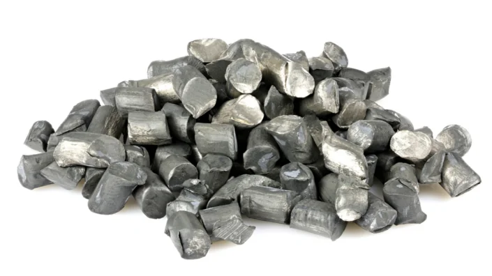
Lithium Is Linked to Lower Mortality

In an epidemiological study published in Aging, scientists have shown that patients who take lithium have much lower all-cause mortality than patients on other psychotropic drugs [1].

Is lithium a longevity drug?

Lithium has long been prescribed for certain psychiatric conditions, mainly bipolar disorder. With time, evidence began to accumulate that lithium might have other benefits for health and longevity; this is similar to what happened with metformin, which is an anti-diabetes drug.

Lithium is thought to work by promoting autophagy and stress resistance via inhibition of glycogen synthase kinase-3 beta (GSK-3β). However, because GSK-3β has multiple functions related to many pathologies [2], scientists are just beginning to unravel lithium’s full potential.

Lithium has been shown to extend lifespan in simple animal models, such as drosophila, even when administered later in life [3]. Human epidemiological studies also showed a slight bump in life expectancy in regions with trace amounts of lithium in drinking water [4]. Of course, epidemiological studies cannot establish causation and are notoriously hard to interpret.

Lithium vs other psychotropic drugs

In this new study, epidemiological as well, the researchers used data from UK Biobank – a huge repository of medical information on almost half a million of British citizens – to see if taking lithium as a drug was associated with lower all-cause mortality. They analyzed a cohort of patients with F3X mood disorders, so called because they are in the International Classification of Diseases (ICD-10) as F30 to F39. According to the study, those diagnoses significantly shorten lifespan by increasing the risk of death from suicide, respiratory diseases, mental disorders, COVID-19, and other proximate causes..

Patients who took lithium were compared to patients who took psychotropic drugs for at least three consecutive months. While the two groups were largely similar, they differed from each other as well as from the general population. For instance, patients who were prescribed lithium normally took the drug longer than patients who were prescribed other antipsychotic medications and had more consecutive F3X episodes. Such heterogeneity makes analyzing the data somewhat harder.

To account for as many confounding variables as possible, the scientists used a tool called propensity score matching (PSM), which theoretically “allows to analyze an observational non-randomized study so that it mimics a randomized controlled trial in a post-hoc fashion”. The researchers controlled for several comorbidities, including diabetes and cancer, and traits, such as sleep quality, smoking, physical activity, alcohol intake, and body mass index (BMI).

A massive decrease in mortality

Despite UK Biobank’s huge size, the resulting cohorts were not that big – just 276 lithium users and 552 users of other psychotropic drugs. However, the trend was clear: lithium significantly extended lifespan. Lithium users had 3.6 times lower chances of dying at a given age compared to other antipsychotic drug users. Importantly, the effect was sex-independent, unlike with some known geroprotective drugs. The causes of death in lithium users and other drug users did not differ significantly, implying that lithium might affect underlying mechanisms of aging and not just mechanisms behind one or two specific diseases.

While the data looks very encouraging, it should be interpreted cautiously. First, the study was populational and had a small sample size. Second, patients on psychotropic drugs generally have more health problems and higher mortality than the rest of the population, so it is not clear how effective lithium would be in extending lifespan in otherwise healthy people. The researchers themselves note that “an alternative interpretation for these findings is that lithium in F3X-diagnosed patients has fewer lifespan-decreasing effects than other medications used to treat F3X diagnoses, and therefore it protects its users from side effects which may lead to premature death”. Finally, lithium treatment requires a lot of caution and supervision, since lithium levels must be kept inside a narrow range.

This study is the first to evaluate the effects of lithium on lifespan in a large aging observational cohort. We have employed a state-of-the-art statistical algorithm, namely propensity score matching, to take advantage of the large amount of observational data in the UK Biobank and simulate a randomized control trial setting to evaluate whether lithium is a geroprotective agent in humans. We have unveiled that lithium at therapeutic concentrations is not only effective in ameliorating mental health, but it is also able to decrease mortality in individuals affected by F3X diagnoses, a demographics that would normally be more fragile and shorter-lived compared to healthy individuals, as also shown in this study.

Conclusion

The results of this study (a 3.6-fold decrease in mortality) certainly look promising and will fuel the growing interest in lithium as a potential geroprotector. However, more research is needed. For instance, while there is data suggesting that metformin users, who are usually diabetic, have fewer chances of dying than even their healthy peers, we don’t have such data for lithium yet.

We would like to ask you a small favor. We are a non-profit foundation, and unlike some other organizations, we have no shareholders and no products to sell you. All our news and educational content is free for everyone to read, but it does mean that we rely on the help of people like you. Every contribution, no matter if it’s big or small, supports independent journalism and sustains our future.

Literature

[1] Araldi, E., Jutzeler, C. R., & Ristow, M. (2023). Lithium treatment extends human lifespan: findings from the UK Biobank. Aging15.

[2] Mancinelli, R., Carpino, G., Petrungaro, S., Mammola, C. L., Tomaipitinca, L., Filippini, A., … & Giampietri, C. (2017). Multifaceted roles of GSK-3 in cancer and autophagy-related diseases. Oxidative medicine and cellular longevity2017.

[3] Castillo-Quan, J. I., Li, L., Kinghorn, K. J., Ivanov, D. K., Tain, L. S., Slack, C., … & Partridge, L. (2016). Lithium promotes longevity through GSK3/NRF2-dependent hormesis. Cell reports15(3), 638-650.

[4] Zarse, K., Terao, T., Tian, J., Iwata, N., Ishii, N., & Ristow, M. (2011). Low-dose lithium uptake promotes longevity in humans and metazoans. European journal of nutrition, 50, 387-389.

Enlarged heart

The Link Between NAD+ and Heart Failure

A paper published today in Nature Cardiovascular Research has explained a relationship between dilated cardiac myopathy (DCM), epigenetic dysregulation, NAD+, and the epigenetic regulatory molecule Kdm8.

A gradual decline of ability

DCM occurs when the left ventricle of the heart becomes enlarged and loses its ability to pump enough blood. The heart muscle gradually thins, causing additional mechanical stress, and this leads to cell death [1] and fibrosis [2]. This makes it the primary reason for heart transplantation [3], as this condition slowly leads to heart failure [4].

The mitochondria of the heart muscle cells (cardiomyocytes) are negatively affected by this condition as well, and their ability to process food and oxygen declines; the powerhouses of the cell lose their power [5]. The resulting depletion of ATP is a key component of DCM’s severity [6]. Previous studies have shown that introducing NAD+ to mice afflicted by this condition restores their heart function [7].

Histone methyltransferases and demethylases, which regulate epigenetics, have been reported to have significant impacts on cardiac function [8]. This research focuses on lysine demethylase 8 (Kdm8), without which mouse embryos die of heart failure before they are born [9]. Previous experiments, however, have not examined the effects of this regulator on living mouse hearts.

Mice with reduced Kdm8 develop DCM

To test their hypothesis, the researchers developed a mouse crossbreed with the KDM8 gene heavily downregulated, most notably in the cardiomyocytes. These mice had relatively normal hearts at 2 months of age but began to show signs of DCM at 4 and 6 months. Mice with two copies of the mutation lived shorter lives (median 7.3 months) than mice with one copy (median 9.3 months), and their deaths were due to heart failure. Mice with no copies of the gene, but which had a different mutation due to the experimental procedure, lived for 27.3 months on average.

As expected, genes related to mitochondrial oxidation were heavily downregulated in the mutants compared to the control group. Mice with one copy of the mutation had more mitochondria, but they were much smaller. Further examination of gene methylation showed that the Kdm8-depleted cardiomyocytes had widespread dysregulation in many other aspects as well, most notably NAD+ metabolism.

Multiple aspects of NAD+, including both its creation and consumption, were heavily downregulated with the loss of Kdm8. This included both NAMPT and the well-known sirtuins, which have been associated with longevity. At 6 months of age, available ATP was roughly half that of controls.

These results proved the researchers’ basic hypothesis: that Kdm8 is a critical component of mitochondrial energy use in cardiomyocytes, which strongly affects heart function.

Conclusion

While this study was not conducted from the point of view of aging research, it touches upon a critical aspect. It has long been known that sirtuins and NAD+ metabolism decline with aging, which is part of deregulated nutrient sensing and heavily related to mitochondrial dysfunction. This in-depth paper describes a specific way in which this depletion appears to be related to epigenetic alterations as well. While potential treatments were not explored in this study, it may be that Kdm8 is a worthwhile target for restoring metabolic ability to the aging heart.

Literature

[1] Aharinejad, S., Andrukhova, O., Lucas, T., Zuckermann, A., Wieselthaler, G., Wolner, E., & Grimm, M. (2008). Programmed cell death in idiopathic dilated cardiomyopathy is mediated by suppression of the apoptosis inhibitor Apollon. The Annals of thoracic surgery, 86(1), 109-114.

[2] Agapitos, E., Kavantzas, N., Nanas, J., Margari, Z., Bakouris, M., Kassis, K., … & Davaris, P. (1996). The myocardial fibrosis in patients with dilated cardiomyopathy. The application of image analysis in the myocardial biopsies. General & diagnostic pathology, 141(5-6), 305-311.

[3] Manolio, T. A., Baughman, K. L., Rodeheffer, R., Pearson, T. A., Bristow, J. D., Michels, V. V., … & Harlan, W. R. (1992). Prevalence and etiology of idiopathic dilated cardiomyopathy (summary of a National Heart, Lung, and Blood Institute workshop). The American journal of cardiology, 69(17), 1458-1466.

[4] McNally, E. M., & Mestroni, L. (2017). Dilated cardiomyopathy: genetic determinants and mechanisms. Circulation research, 121(7), 731-748.

[5] Dávila-Román, V. G., Vedala, G., Herrero, P., De Las Fuentes, L., Rogers, J. G., Kelly, D. P., & Gropler, R. J. (2002). Altered myocardial fatty acid and glucose metabolism in idiopathic dilated cardiomyopathy. Journal of the American College of Cardiology, 40(2), 271-277.

[6] Lopaschuk, G. D., Karwi, Q. G., Tian, R., Wende, A. R., & Abel, E. D. (2021). Cardiac energy metabolism in heart failure. Circulation research, 128(10), 1487-1513.

[7] Diguet, N., Trammell, S. A., Tannous, C., Deloux, R., Piquereau, J., Mougenot, N., … & Mericskay, M. (2018). Nicotinamide riboside preserves cardiac function in a mouse model of dilated cardiomyopathy. Circulation, 137(21), 2256-2273.

[8] Davis, K., Azarcon, P., Hickenlooper, S., Bia, R., Horiuchi, E., Szulik, M. W., & Franklin, S. (2021). The role of demethylases in cardiac development and disease. Journal of Molecular and Cellular Cardiology, 158, 89-100.

[9] Ishimura, A., Minehata, K. I., Terashima, M., Kondoh, G., Hara, T., & Suzuki, T. (2012). Jmjd5, an H3K36me2 histone demethylase, modulates embryonic cell proliferation through the regulation of Cdkn1a expression. Development, 139(4), 749-759.

Victory cup

Meet hack-age, Winners of the January 2023 LongHack

On January 22nd, hack-age won the 2023 LongHack competition, which carried a grand prize of 5,500 VITA and 3,500 USD. Lifespan.io caught up with the team behind the winning project to learn more about their idea and their experience competing in the hackathon.

What is LongHack?

LongHack is a hackathon organized by VitaDAO and its partners with the aim of accelerating longevity research and increasing awareness of the field. The January event was the second-ever hackathon held, and it brought together hundreds of interested participants. This was narrowed down to ten impressive teams: AGen-iNET, AdStella Therapeutics, BELLEROPHON, Chiranjivi, DETECT-ALL, GeneWeavers, Gigachads, Grey Matter, hack-age, and PharmaPros.

Out of these ten, four teams, including hack-age, took home a prize in honor of their work. 5,500 VITA and 3,500 USDC went to hack-age, 2,500 VITA and 1,500 USDC went to AdStella, 1,000 VITA and 1,000 EUR went to AGen-iNET, and 1,000 VITA went to GeneWeavers.

Who is the team behind the first-prize winning entry?

The hack-age team is comprised of four individuals: Dauken Seitkali, a data scientist and founder of AgzaThera, a drug discovery biotech; Mikolaj Rzuczkowski, On Deck biotech longevity fellow and medical doctor; Roman Litvinov, Senior Researcher; and Rakhan Aimbetov, LabDAO lead, analyst at Longaevus Technologies, and founder of hack-age.

What does hack-age do and hope to achieve?

The group describes itself as a goal-driven association of passionate individuals unified by a vision: rejuvenation of the extracellular matrix.

Accumulation of advanced glycation end-products (AGEs) during a person’s lifetime contributes to the development of age-related conditions and health issues. Their removal and/or prevention of formation, could be a promising therapeutic modality that could consequently reduce or mitigate the physiological deterioration connected with aging.

The goal of hack-age is to utilize machine learning (ML) and artificial intelligence (AI) technologies, specifically in silico screens and modeling tools, to create and develop such therapies through to the clinical/pre-clinical stage.

Simple sugars, such as glucose, and toxic sugar-derived metabolites react with and harm proteins and DNA in our bodies. This is especially evident during diabetes when there’s elevated blood glucose levels due to reduced capacity of our cells to take it up, leading to accelerated damage.

This damage eventually compounds and brings about various pathologies, i.e., diabetic complications. The enzyme called glyoxalase I (Glo1) removes one such very reactive and noxious chemical, methylglyoxal, but during diabetes its activity is overridden by too much influx of sugars and derived metabolites.

Our solution is to enhance the natural capacity of Glo1 by means of directed evolution, or getting desired traits by molecular biology techniques, and subsequent delivery of instructions to make the engineered enzyme into the cells of people suffering from diabetes.

Thanks for speaking with us. You must be excited about your victory. So, could you tell us how you first heard about LongHack and what inspired you to enter?

Rakhan: I first learned about the VitaDAO Longevity Hackathon as one of the projects in a 2022 Gitcoin grants round. When the hackathon dates were made known, I decided I should be there – alone or with a team. Fortunately, I had people in my network whom I wanted to see at my side during the contest. Since glycation is my main scientific interest, I assembled a team of folks I knew were into glycation too. Basically, we were on the same page from the get-go.

Dauken: I’ve heard about LongHack from my teammate Rakhan. I was inspired by hackathon tracks and experts.

Roman: My inspiration is the exceptional complexity of the problem of aging. Events like the hackathon stimulate those parts of the brain that generate new ideas, and sometimes these ideas can even be realized.

Mikolaj: I originally heard about it through an announcement on On Deck Longevity Biotech the year of its inauguration. I joined this year because my team was involved.

Was this your first hackathon experience?

Rakhan: Yes, it was. The main thing that drove me was curiosity – I wanted to experience what being in a hackathon feels like.

Dauken: No, but it was my first hackathon that was held entirely online.

Roman: The very first. In my life.

Mikolaj: No, my second. I had participated in a GPT-3 hackathon a year or two prior.

Tell us about your LongHack experience. How was it? Did you experience any challenges? Did you get much sleep?

Rakhan: It was very natural! We got together, we discussed, we interacted with mentors, we ideated, strategized, built, and executed. The biggest challenge was to focus on one idea and sacrifice things that are dear to our hearts but distract from the project’s core. And we peeled and carved until the message was crisp and super simple.

Dauken: The LongHack events and sessions were well organized, mentors were available to help with any issues. The most challenging was fitting the time zone difference, in our region it was night-time.

Roman: It all began as an opportunity to entertain myself in my work-free time, but turned into hack-age, a company that eventually will focus its efforts on the development of unique anti-aging biotech (and pharmaceutical) products. Sleep is the most needful thing in order to be able to generate ideas. Sometimes sleep becomes the sole pleasure in life since all other time is dedicated to hard work.

Mikolaj: It was a great and intense experience. I’m fortunate to have worked with such dedicated and motivated individuals, who were able to work together and integrate mentors’ advice into our workflow.

How did you come up with the idea for the project, and what was the most interesting part of the process?

Rakhan: We reasoned that our thing must be broad in scope, generalizable and scalable, and easy to build at the same time. So, we brainstormed and made a longlist of pathways, targets, and a contingency plan too.

By the end of the ideation weekend, we conceptualized the software platform and narrowed down our list to one use case to showcase the platform’s capabilities. In effect, our project was oriented towards a SaaS business model initially.

During the main hackathon part, though, we pivoted 180 degrees to offer an actual therapy candidate that can be made using the platform we built as our main product. This was really exciting!

Personally, the most interesting part was making our idea and presentation slightly better with every iteration until they’re tight and shiny and we’re happy with them. So, when I presented, I was 100% sure that what we have is good.

Dauken: Our team was formed a few months before the hackathon started and we had some ideas for a longevity project. Then on ideation weekend, we came up with a final idea for the project that best fits our interest and meets LongHack requirements. The most interesting part was decision making on the project strategy.

Roman: In the ideation process, communication is crucial. When you explain your project to mentors, something clicks in your head and an idea pops up. And yes, an important role of a mentor is to be an idea acceptor and taster. I would love to especially thank Helena [Helena Slama, LongHack mentor and Chief Medical Advisor to IHCHI – Institute for Human Centred Health Innovation] who conducted the “grandma test” for newly born ideas. That’s exactly how the idea to name our vector-delivered deglycating enzyme the “vaccine against aging” came to be. Helena’s lessons hold a special place in my memory.

Mikolaj: I had joined later into the project’s inception, though I had been interested in the involvement of the ECM in aging ever since reading Fedinstev’s and Moskalev’s review on stochastic non enzymatic modification of long-lived macromolecules. I was excited to join the team, as they all had interest in directed evolution, which I’ve been curious about as a mechanism to develop aging therapeutics for some time.

Now that you’ve won, what’s your next steps?

Rakhan: We’ve come up with a ton of good ideas. Now we will execute on them and test for viability.

Dauken: It is just the first baby steps of our big journey. Our next steps are to finish in the silico part and move to laboratory experiments.

Roman: The next step is the launch of hack-age as a company and further combat for mitigation of the manifestations of age in the matrix (now in practice).

Mikolaj: We’re excited to continue developing our pipeline and working together with LabDAO to achieve our goals.

Will you enter any more hackathons?

Rakhan: Definitely!

Dauken: More likely to enter accelerators as our project is going to be more mature, I believe.

Roman: You can come up with only one thing in your life, but this thing better be significant. That’s why too frequent participation in hackathons might lose the essence of a hackathon as a stage for idea generation. For the next event, I would rather suggest inviting the previous contest winners so that they can tell if they managed to turn their idea into a viable project. However, if thoughts require warm-up, participation to satisfy this need is a good reason for showing up.

Mikolaj: Looking forward to it!

What advice would you give to someone thinking of joining a hackathon / LongHack?

Rakhan: Do your homework and show up with a plan. Have a core team that knows each other and worked together on something in the past, so that you don’t lose time on getting to know each other’s strengths and are less likely to run into personality incompatibility issues. And foremostly, set your ego aside and aim to win big — as a team!

Dauken: Be flexible to change your mind or to look at your idea from other angles.

Roman: Cultivate interest and love for what you do, and you will think about it constantly. Then ideas will visit often, so often that you will have to write them down. Afterwards, their realization will become a question of time and circumstance.

Mikolaj: Definitely give it a go. Even if you aren’t able to work on something that interests you in your first hackathon, you’ll be able to use that experience for subsequent ones (my time in the GPT3 hackathon was spent helping with someone else’s interests, but my time in LongHack was right in line with my personal interest).

Thanks for your time, Rakhan, Dauken, Roman, and Mikolaj. We wish you all the best in your next steps, and congratulations on your victory.

Summary

Ten selected teams worked from January 20th to 22nd, competing in the LongHack weekend to solve an issue related to longevity science. Four teams took home a prize, with hack-age taking first place and winning 5,500 VITA and 3,500 USDC for their entry based on an AI/ML solution for AGEs.

We would like to ask you a small favor. We are a non-profit foundation, and unlike some other organizations, we have no shareholders and no products to sell you. All our news and educational content is free for everyone to read, but it does mean that we rely on the help of people like you. Every contribution, no matter if it’s big or small, supports independent journalism and sustains our future.
Tendon inflammation

Extracellular Vesicles for Tendon Healing

Researchers publishing in Journal of Nanobiotechnology have found that extracellular vesicles derived from young cells are effective in treating a model of a common tendon disease.

Tendinopathy and stem cells

Swelling, weakness, tenderness, and tendon pain during activities are the characteristic symptoms of chronic tendinopathy [1], a condition that is exacerbated by aging and obesity into degeneration and rupture [2]. This condition is notoriously difficult to treat, and even surgery is poorly effective [3].

Previous research has investigated potential treatments that involve tendon stem/progenitor cells (TSPCs) and mesenchymal stem cells (MSCs) [4]. Adipose-derived MSCs (ADMSCs) have a higher yield and quality than other stem cell sources, and they have previously been used to treat tendinopathy in a rat model [5].

One of the core parts of MSC effectiveness involves extracellular vesicles (EVs), which have been explored in treating multiple diseases, including tendinopathy [6]. However, donor EVs are strongly affected by the donor’s health, including aging [7].

NAMPT, a core component of NAD+ synthesis, declines with age [8] and is contained in EVs. Hypothesizing that NAMPT is an important part of tendinopathy recovery through EV administration, the researchers designed an experiment that compares the EVs of young and old animals.

Graphical abstract

EVs and NAMPT

Comparing MSCs in animals and cells

For the first part of their experiment, the researchers compared MSCs from 22-month-old mice to 2-month-old mice. As expected, the older mice had significantly more senescent MSCs, as measured by the well-known senescence biomarkers p16INK4A, p21, and SA-β-gal, along with significantly decreased NAD+ and NAMPT.

This decrease in NAMPT was also found to apply to these MSCs’ EVs; EVs derived from old MSCs had less than half the NAMPT of their younger counterparts. Interestingly, old MSCs generated more than twice as many EVs as young MSCs did, sacrificing quality for quantity, which the researchers attribute to inflammatory stress.

Similar effects were also found in macrophages. Administering IL-1β to macrophages caused signs of cellular senescence, which were ameliorated by young-derived EVs. These EVs also encouraged macrophages to take up the M2 (healing) phenotype, as opposed to the M1 (inflammatory) phenotype, and they decreased inflammatory signals given off by macrophages.

After inducing tendinopathy in mice through collagenase I, the EVs’ effects on the disorder were examined. EVs derived from older animals had a minimal effect on biomarkers, including matrix metalloproteinase and collagen levels. Microscopic examination revealed similar results, with tendon fibrosis ameliorated only by young-derived EVs.

In a cellular study, the resarchers simulated tendinopathy by exposing tenocytes, which are fibroblasts found only in the tendons, to IL-1β. The affected cells experienced death by apoptosis at much higher rates, but this was almost entirely ameliorated by young-derived EVs. Old-derived EVs had a negligible effect.

These results were recapitulated in an examination of gene expression. Tendinopathy causes pathological remodeling of the extracellular matrix, but this too was ameliorated by young-derived EVs. Levels of collagen-related gene expression were also restored.

The connection with NAMPT and NAD+

Finally, the researchers examined the cells’ biochemistry to determine what effects the NAMPT-rich EVs were having on tenocytes exposed to IL-1β. An NAD+ signaling pathway that was substantially downregulated by IL-1β was largely restored to previous values. The mitochondria were found to be significantly and positively affected, including such metrics as oxygen respiration, energy production, and membrane integrity. Once again, only the young-derived EVs were successful in these respects.

Conclusion

The researchers describe their approach as “one stone, two birds”, emphasizing the fact that young EVs can both directly restore tenocytes and discourage macrophages from making things worse with inflammation. If clinical trials prove that these effects occur in human beings as well as mouse and cellular models, chronic tendinopathy may become a more easily treatable condition along with other age-related diseases.

We would like to ask you a small favor. We are a non-profit foundation, and unlike some other organizations, we have no shareholders and no products to sell you. All our news and educational content is free for everyone to read, but it does mean that we rely on the help of people like you. Every contribution, no matter if it’s big or small, supports independent journalism and sustains our future.

Literature

[1] Millar, N. L., Silbernagel, K. G., Thorborg, K., Kirwan, P. D., Galatz, L. M., Abrams, G. D., … & Rodeo, S. A. (2021). Tendinopathy. Nature reviews Disease primers, 7(1), 1.

[2] Gomez-Florit, M., Labrador-Rached, C. J., Domingues, R. M., & Gomes, M. E. (2022). The tendon microenvironment: engineered in vitro models to study cellular crosstalk. Advanced Drug Delivery Reviews, 114299.

[3] Dan, M., Phillips, A., Johnston, R. V., & Harris, I. A. (2019). Surgery for patellar tendinopathy (jumper’s knee). Cochrane Database of Systematic Reviews, (9).

[4] Schneider, M., Angele, P., Järvinen, T. A., & Docheva, D. (2018). Rescue plan for Achilles: Therapeutics steering the fate and functions of stem cells in tendon wound healing. Advanced drug delivery reviews, 129, 352-375.

[5] Oshita, T., Tobita, M., Tajima, S., & Mizuno, H. (2016). Adipose-derived stem cells improve collagenase-induced tendinopathy in a rat model. The American Journal of Sports Medicine, 44(8), 1983-1989.

[6] Chen, S. H., Chen, Z. Y., Lin, Y. H., Chen, S. H., Chou, P. Y., Kao, H. K., & Lin, F. H. (2021). Extracellular vesicles of adipose-derived stem cells promote the healing of traumatized Achilles tendons. International Journal of Molecular Sciences, 22(22), 12373.

[7] Hooten, N. N., Byappanahalli, A. M., Vannoy, M., Omoniyi, V., & Evans, M. K. (2022). Influences of age, race, and sex on extracellular vesicle characteristics. Theranostics, 12(9), 4459.

[8] Yoshida, M., Satoh, A., Lin, J. B., Mills, K. F., Sasaki, Y. O., Rensing, N., … & Imai, S. I. (2019). Extracellular vesicle-contained eNAMPT delays aging and extends lifespan in mice. Cell metabolism, 30(2), 329-342.