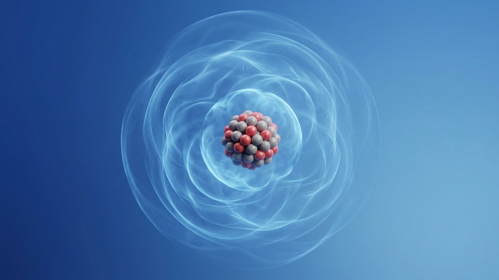
Human Organoids for Brain Regeneration
- Permanent brain damage may finally have an upcoming solution.

Researchers publishing in Cell Stem Cell have used organoids derived from human cells to regrow the brain tissue of rats.
The need to repair permanent damage
While human beings do generate new neurons (neurogenesis) [1], this ability is very limited, in both region and amount. Brain injuries [2] and strokes [3] are well-known sources of permanent damage, but, of course, age-related diseases can do the same thing.
Previous research has shown that rodent fetal brain tissue can be grafted into the rodent brain [4], with the transplanted neurons able to take up their responsibilities [5]. However, this approach is unethical and infeasible for human patients. Fortunately, brain tissue derived from induced pluripotent stem cells (iPSCs) has been shown to form organoids that mimic the properties of human brain regions [6].
While some previous research has explored the idea of organoid transplantation [7], this research team holds that such work did not go into sufficient depth. Building upon that work, this team built a biological system that reports engraftment in detail, seeking to analyze just how well such an approach takes hold in the visual cortices of rats.
Human cells in rat brains
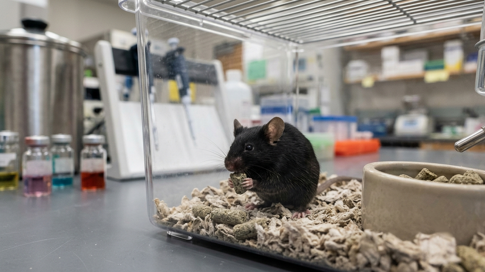
The researchers conducted a series of experiments using organoids created from multiple cell lines of human-derived iPSCs, which were modified to express fluorescent proteins for better visualization of engraftment. Parts of the the visual cortices of rats were removed, and the tissue was replaced with these organoids. Immunosuppressants were used to minimize the immune response.
Approximately four-fifths of the organoids successfully took root in the animals, which were studied at one, two, and three months after the transplantation surgery. There appeared to be no gradual failure of these organoids over this time frame.
As expected, the grafts matured over time, transitioning away from stem cells to fully functional neurons. However, even at three months, there were still numerous stem cells present in these organoids. Fortunately, fully pluripotent cells, which might have signified tumors, were not found among them. There was also no evidence that any host neurons migrated into these grafts.
The engrafted organoids appeared to work. Synaptic connections were demonstratably formed between the organoid and the rest of the brain, and information processing was confirmed to occur within the organoid. The organoids were found to have significantly more connections than comparable tissue; the researchers suspect that this represents a youthful state, as substantial neural pruning, which represents brain maturity, had not yet occurred when these measurements were taken. In other aspects of neural activity, the organoids appeared to work nearly identically to the rest of the brain.
Problems with inflammation and surgery
One month after the transplantation, there were increased numbers of astrocytes and other glial cells in the grafted areas compared to injury-only control groups. However, this situation seemed to stabilize over time. CD68+ microglia, which represent macrophages and inflammation, were slightly more present in the grafted groups. This shows ongoing inflammation, which the researchers believe is due to the immunosuppressants having incomplete effects, as has been shown in other studies [8].
Unfortunately, the testing procedure was not completely safe. Out of a total of 46 animals tested across the various experiments, eight died before the examinations took place; three of those deaths were directly linked to complications of the surgery. As these experiments involved both injury and transplantation, it is impossible to say what part of the surgery was the true cause of death.
Conclusion
While this is still a rat experiment, the use of human cells makes these results very promising for survivors of brain injury and stroke. However, the ongoing need for immunosuppressants, a common issue in human organ transplants, makes this procedure potentially dangerous.
To develop this approach for use in human beings, it may be possible to use autologous, patient-derived cells or mass-produced allogeneic cells that do not stimulate the immune system. The researchers also highlighted the need for proper maintenance of structure and blood vessel perfusion (vascularization) to maintain organoid function and health. Further experiments and human clinical trials will determine if such methods can be used to restore cognitive ability and quality of life to people who have experienced brain damage, whether that damage occurred slowly or quickly.
Literature
[1] Kempermann, G., Gage, F. H., Aigner, L., Song, H., Curtis, M. A., Thuret, S., … & Frisén, J. (2018). Human adult neurogenesis: evidence and remaining questions. Cell stem cell, 23(1), 25-30.
[2] Thurman, D. J., Alverson, C., Dunn, K. A., Guerrero, J., & Sniezek, J. E. (1999). Traumatic brain injury in the United States: a public health perspective. The Journal of head trauma rehabilitation, 14(6), 602-615.
[3] Hankey, G. J., Jamrozik, K., Broadhurst, R. J., Forbes, S., & Anderson, C. S. (2002). Long-term disability after first-ever stroke and related prognostic factors in the Perth Community Stroke Study, 1989–1990. Stroke, 33(4), 1034-1040.
[4] Santos-Torres, J., Heredia, M., Riolobos, A. S., Jiménez-Díaz, L., Gómez-Bautista, V., de la Fuente, A., … & Yajeya, J. (2009). Electrophysiological and synaptic characterization of transplanted neurons in adult rat motor cortex. Journal of neurotrauma, 26(9), 1593-1607.
[5] Girman, S. V., & Golovina, I. L. (1990). Electrophysiological properties of embryonic neocortex transplants replacing the primary visual cortex of adult rats. Brain Research, 523(1), 78-86.
[6] Qian, X., Su, Y., Adam, C. D., Deutschmann, A. U., Pather, S. R., Goldberg, E. M., … & Ming, G. L. (2020). Sliced human cortical organoids for modeling distinct cortical layer formation. Cell Stem Cell, 26(5), 766-781.
[7] Mansour, A. A., Gonçalves, J. T., Bloyd, C. W., Li, H., Fernandes, S., Quang, D., … & Gage, F. H. (2018). An in vivo model of functional and vascularized human brain organoids. Nature biotechnology, 36(5), 432-441.
[8] Espuny-Camacho, I., Michelsen, K. A., Gall, D., Linaro, D., Hasche, A., Bonnefont, J., … & Vanderhaeghen, P. (2013). Pyramidal neurons derived from human pluripotent stem cells integrate efficiently into mouse brain circuits in vivo. Neuron, 77(3), 440-456.