×

The Blog

Building a Future Free of Age-Related Disease

Muscle factor

Promoting Muscle Regeneration With an Immune Factor

A paper published today in Nature Aging has shown that a macrophage-regulating factor has a significant impact on muscle regeneration.

Healing slows with aging

The researchers note two key reasons for the decline of regenerative abilities with aging. The first is stem cell exhaustion, the gradual depletion of stem cells and ensuing lack of functional cells. The second, which this paper focuses on, is changes to the surrounding environment, the effects of which are the subject of well-known parabiosis experiments.

However, instead of experimenting with young blood as a whole, the researchers focus on just one of its factors: mesencephalic astrocyte-derived neurotrophic factor (MANF) [1], which declines with aging and has been shown to have positive effects on the retina [2] and other tissues [3].

A factor necessary for regeneration

The researchers created a population of mice that stopped producing MANF upon exposure to a hormone therapy. When the quadriceps of these mice were injured and MANF blocked, these mice were found to have substantially weaker regeneration, with fewer new muscle fibers, fewer muscle stem cells, and many more dead fibers remaining in the tissue.

This is explained by the cellular populations in the tissue. Mice without MANF2 were found to have roughly the same amounts of pro-inflammatory macrophages but far fewer macrophages associated with regeneration. By blocking MANF at different times, the researchers showed that this particular effect occurs while the animal is injured; temporarily blocking MANF before an injury did not have an effect.

Further experiments in cellular culture, using cells from wholly MANF-deficient mice, showed that MANF is essential to macrophages changing their phenotypes towards regeneration and away from inflammation. MANF was also found to be valuable for lysosomal degradation of foreign proteins, and macrophages without it were unable to properly respond to necrotic debris, which is a logical explanation for the dead fibers remaining in MANF-deficient mice.

Restoring age-related MANF decline

This study confirmed that old (22-24 months) mice have substantially fewer healing-oriented macrophages than young (2-6 months) mice. Gene expression analysis showed that their macrophages had some things in common with young, MANF-deficient macrophages, particularly the problems with lysosomal degradation, but the aged cells also had other problems with cellular movement structures.

The researchers performed their final and most crucial experiment, delivering recombinant MANF to aged mice. This treatment worked; macrophages were restored to youthful populations, necrotic fibers were more thoroughly cleared, and new muscle fibers were created in greater quantities.

MANF therapy at 4 μg i.m. resulted in a complete rescue of the repair-associated myeloid response.

Conclusion

While this is a mouse study, MANF appears to play the same basic biological role in human beings. Further work, development, and clinical trials will be necessary to determine if MANF-based treatments can restore health and function to old, damaged muscle tissue.

We would like to ask you a small favor. We are a non-profit foundation, and unlike some other organizations, we have no shareholders and no products to sell you. All our news and educational content is free for everyone to read, but it does mean that we rely on the help of people like you. Every contribution, no matter if it’s big or small, supports independent journalism and sustains our future.

Literature

[1] Sousa-Victor, P., Neves, J., Cedron-Craft, W., Ventura, P. B., Liao, C. Y., Riley, R. R., … & Jasper, H. (2019). MANF regulates metabolic and immune homeostasis in ageing and protects against liver damage. Nature metabolism, 1(2), 276-290.

[2] Neves, J., Zhu, J., Sousa-Victor, P., Konjikusic, M., Riley, R., Chew, S., … & Lamba, D. A. (2016). Immune modulation by MANF promotes tissue repair and regenerative success in the retina. Science, 353(6294), aaf3646.

[3] JOntti, M., & Harvey, B. K. (2020). Trophic activities of endoplasmic reticulum proteins CDNF and MANF. Cell and Tissue Research, 382(1), 83-101.

The Journal Club is a monthly livestream hosted by Dr. Oliver Medvedik which covers the latest aging research papers.

Journal Club March 2023 + Zoom Connection Details

The Journal Club returns on Tuesday 28th March at 12:00 PM Eastern time to the Lifespan.io Facebook channel. This month Dr. Oliver Medvedik will be exploring a recent study which looked at the effects of prolonged fasting on human macrophages and how metabolites from that fasting increased median lifespan in Caenorhabditis elegans.

Abstract

Periodic prolonged fasting (PF) extends lifespan in model organisms and ameliorates multiple disease states both clinically and experimentally owing, in part, to its ability to modulate the immune system. However, the relationship between metabolic factors, immunity, and longevity during PF remains poorly characterized especially in humans.

This study aimed to observe the effects of PF in human subjects on the clinical and experimental markers of metabolic and immune health and uncover underlying plasma-borne factors that may be responsible for these effects.

Join the livestream

Lifespan Heroes get access to the livestream using the connection details below:

Join Zoom Meeting https://lifespan-io.zoom.us/j/88373018752?pwd=RC9KcEZKOU1YMDFOTlpGaEt6ZzRWZz09 Meeting ID: 883 7301 8752 Passcode: 374339

Literature

Rhodes, C. H., Zhu, C., Agus, J., Tang, X., Li, Q., Engebrecht, J., & Zivkovic, A. M. (2022). Human fasting modulates macrophage function and upregulates multiple bioactive metabolites that extend lifespan in Caenorhabditis elegans: a pilot clinical studyThe American Journal of Clinical Nutrition.

Running seniors

Long-Term Resistance Exercise Increases Autophagy

In a new systemic review published in Autophagy Reports, researchers have demonstrated that exercise plays a role in regulating autophagy, depending on its type [1].

Manipulating autophagy 

Disabled autophagy was recently acknowledged as a hallmark of aging. Indeed, boosting the ability of cells to get rid of unnecessary and damaged organelles and molecules is considered a promising anti-aging strategy.

As shown in animal and some human studies, autophagy activation can be achieved with fasting and exercise, therapeutic interventions such as rapamycin and lithium, and even ultrasound treatment.

At the same time, just like with any other molecular process, autophagy manipulation should be fine-tuned in a context-specific manner to achieve beneficial results. Hyperactivation of autophagy might be toxic in some situations, such as for DNA damage repair [2].

Exercise is believed to activate autophagy, particularly mitophagy, the removal of damaged mitochondria. This conclusion mostly comes from animal studies, whereas human data on exercise-induced autophagy is less consistent.

In this systematic review, the researchers analyzed the results of 26 studies assessing the autophagic response to exercise in humans. The studies measured various autophagy markers in skeletal muscles, peripheral blood mononuclear cells (PBMCs), or both in participants engaging in various exercise forms and intensities.

Autophagy response to exercise

First, the researchers showed that the levels of two autophagy markers, microtubule-associated proteins 1A/1B light chain 3B (LC3-II) and sequestosome 1 (SQSTM1), were consistent with reduced autophagy following an acute bout of resistance exercise and increased autophagy following long-term resistance exercise.

The researchers did not detect any change in LC3-II following either acute or long-term (over several weeks) moderate- and vigorous-intensity endurance exercise. Only long-term vigorous-intensity endurance exercise was accompanied by altered levels of SQSTM1.

Next, the researchers found a tissue-specific autophagy response to exercise. Exercise was associated with lowered LC3-II and increased SQSTM1 levels in skeletal muscle, which corresponds to attenuated autophagy. This effect was largely due to resistance exercise. The opposite effect, induced autophagy, was observed in PBMCs in response to exercise. This suggests distinct autophagy regulation mechanisms in different tissues.

Finally, the researchers showed that several other autophagic and mitophagic markers, such as BNIP3, Beclin-1, and ATG12, were increased in the analyzed studies, including those that involve endurance exercise. However, because it was not accompanied by changes in LC3-II and SQSTM1 levels, the effect of endurance training on autophagy remains unclear.

Abstract excerpt

Our findings demonstrate that physical exercise probably regulates autophagy in an exercise modality- and tissue-dependent manner in humans, although further investigation is needed. Customized exercise prescriptions should be aimed for when implementing exercise to regulate autophagy in humans.

Conclusion

This study demonstrated that long-term resistance training might be the best exercise for increasing autophagy in humans. The beneficial health effects of other exercise types, such as endurance training, might be via mechanisms other than activated autophagy.

The limitations of this systematic review include autophagy measurements limited to western blot analysis of some protein markers, analysis of only two tissues, and studies conducted mostly on young and middle-aged healthy participants.

We would like to ask you a small favor. We are a non-profit foundation, and unlike some other organizations, we have no shareholders and no products to sell you. All our news and educational content is free for everyone to read, but it does mean that we rely on the help of people like you. Every contribution, no matter if it’s big or small, supports independent journalism and sustains our future.

Literature

[1] Chen X-K, Zheng C, Siu PM-F, Sun F-H, Wong SH-S, Ma AC-H. Does Exercise Regulate Autophagy in Humans? A Systematic Review and Meta-Analysis. Autophagy Reports 2023; 2: 2190202.

[2] Guo C, Zhao Y. Autophagy and DNA damage repair. Genome Instability & Disease 2020; 1: 172–183.

Mediterranean Diet Might Lower Risk of Dementia

Mediterranean Diet Might Lower Risk of Dementia

In a large-scale observational study, British scientists have shown that high levels of adherence to the Mediterranean diet might substantially lower the risk of dementia [1].

A healthy diet

The Mediterranean diet, which is based on ingredients such as olive oil, vegetables, legumes, fish, and whole grains, is considered one of the healthiest in the world. While most of the evidence comes from observational studies that have well-known limitations, there is also a handful of randomized clinical trials [2]. The Mediterranean diet has been linked to decreased risk of cardiovascular and all-cause mortality [3], improved cognitive function [4], and other health benefits, but data on its effect on dementia has been sparce and inconclusive.

Power of the study

This study, like many other recent studies, was based on data from UK Biobank, a huge repository of health information on some half a million of British citizen. Compared to previous studies, it had several strengths, including its size. The researchers assembled a cohort of about 60 thousand older adults with 882 cases of dementia recorded during a 9-year average follow-up period, which allowed for considerable statistical power.

A known problem with such studies is that they often make do with a single questionnaire to ascertain dietary patterns. In this cohort, however, participants not only filed in a detailed 24-hour assessment of their diets online, but most did this more than once, which allowed the researchers to gauge adherence.

The researchers employed two similar scores widely used to measure adherence to the Mediterranean diet: MEDAS and PYRAMID. Moreover, they improved the existing MEDAS scoring system that awards a binary score for 14 food types (you either meet the requirement or not) by making it continuous. The authors also adjusted for numerous potential confounding variables, including age, socioeconomic status, educational attainment, BMI, physical activity, smoking, and even sleep duration, and conducted several sensitivity analyses.

More adherence is associated with more benefits

In the fully adjusted model, high adherence to the Mediterranean diet according to the continuous MEDAS score was associated with a 23% decrease in the risk of all-cause dementia. This result was highly statistically significant. High adherence according to the PYRAMID score was associated with a smaller 14% risk decrease and nearly reached the significance threshold. Medium adherence did not seem to provide a lot of protection. A sensitivity analysis showed the negative correlation between Mediterranean diet and fatal cases of dementia to be the strongest.

Med diet graph

Few other studies have considered the relationship between diet and genetic risk for dementia. Those that did mostly limited it to the APOE genotype, which is known to be strongly correlated with Alzheimer’s risk. In this study, the researchers developed a polygenic score that combined information from multiple weighted risk alleles.

Importantly, no significant interaction between adherence to the Mediterranean diet and a polygenic risk for dementia was detected. In other words, the Mediterranean diet seemed to help lower the risk of dementia even in people who were genetically predisposed to it. However, this association did not hold in some scenarios, which is why the authors of the study call for more research.

Limited demographic scope

The researchers also note an important limitation inherent to UK Biobank: it mostly contains data on people who self-reported their ethnic backgrounds as British, Irish, or otherwise European, which may limit the applicability of the results to other populations. The scientists were also unable to reliably ascertain consumption of one of the most important components of the Mediterranean diet, olive oil, as the questionnaire only asked participants whether they used olive oil for cooking. It is possible that with this data available, the correlation between the Mediterranean diet and risk of dementia would have been even more robust.

Dr Oliver Shannon of Newcastle University, one of the study’s lead authors, notes in a blog post that currently, our options for treating dementia are very limited, which makes prevention especially important. “Much more research is needed to identify the best diet that people could follow to try and reduce their risk of dementia”, Shannon admits. “However, the findings from our study contribute towards a growing body of evidence to suggest that following a more Mediterranean-like diet could be an effective way to help ‘oil your cogs’ and reduce your risk of developing dementia”.

Conclusion

This study, while observational and hence insufficient to establish causation, provides further evidence of the Mediterranean diet’s connection to lifespan and healthspan.

We would like to ask you a small favor. We are a non-profit foundation, and unlike some other organizations, we have no shareholders and no products to sell you. All our news and educational content is free for everyone to read, but it does mean that we rely on the help of people like you. Every contribution, no matter if it’s big or small, supports independent journalism and sustains our future.

Literature

[1] Shannon, O. M., Ranson, J. M., Gregory, S., Macpherson, H., Milte, C., Lentjes, M., … & Stevenson, E. (2023). Mediterranean diet adherence is associated with lower dementia risk, independent of genetic predisposition: findings from the UK Biobank prospective cohort study. BMC medicine, 21(1), 1-13.

[2] Valls-Pedret, C., Sala-Vila, A., Serra-Mir, M., Corella, D., De la Torre, R., Martínez-González, M. Á., … & Ros, E. (2015). Mediterranean diet and age-related cognitive decline: a randomized clinical trial. JAMA internal medicine, 175(7), 1094-1103.

[3] Hidalgo-Liberona, N., Meroño, T., Zamora-Ros, R., Rabassa, M., Semba, R., Tanaka, T., … & Cherubini, A. (2021). Adherence to the Mediterranean diet assessed by a novel dietary biomarker score and mortality in older adults: the InCHIANTI cohort study. BMC medicine, 19(1), 1-13.

[4] Tsivgoulis, G., Judd, S., Letter, A. J., Alexandrov, A. V., Howard, G., Nahab, F., … & Wadley, V. G. (2013). Adherence to a Mediterranean diet and risk of incident cognitive impairment. Neurology, 80(18), 1684-1692.

Elderly lifting

Vital Muscle Enzyme Declines With Aging

Research published in Nature Metabolism has described PCYT2, an enzyme essential for muscle function, and how it declines with aging.

A necessary lipid synthesizer in muscle

Human beings need PCYT2 as part of the Kennedy pathway to synthesize two critical components of the phosopholipid membrane that surrounds cells [1]. People with genetic deficiencies of PCYT2 have stunted growth, severe motor function disorders, and muscle weakness [2], and people with similar deficiencies in the Kennedy pathway have similar symptoms [3].

The researchers recapitulated similar findings in zebrafish and mice. Without PCYT2, both of these species were considerably smaller than normal. Vital lipids, including long-chain fatty acids, were substantially depleted in the leg muscles of young PCYT2-deficient mice, which also had substantially thinner fibers. Further examination showed that the cellular membranes of their muscle cells were looser, weaker, and softer than those of normal mice.

As expected, mice with this condition did not age well. These mice frequently had spinal problems, and muscle wasting and atrophy, along with the associated low bone density, were common afflictions.

Interestingly, PCYT2 only seems to be necessary for muscle tissue. Mice that were genetically engineered specifically to lack PCYT2 in fat tissues seemed to be totally unaffected at any age.

A potentially recoverable decline with aging

In both mice and people, PCYT2 declines with age, as shown by tissue taken from ordinary, wild-type mice and human volunteers. 45- to 62-year-old people have substantially less PCYT2 than 20- to 30-year-old people. The researchers surmise that this may be a big part of the reason why resistance training becomes less beneficial with age [4].

The researchers then tested whether gene therapy could have an impact. Injecting middle-aged, pre-sarcopenia mice with an adeno-associated virus (AAV) to deliver PCYT2 to muscle tissue, they found that the affected mice had significantly higher grip strength, muscle fiber size, and muscular mitochondrial function than the control group of aged mice, although these values were not quite restored to the levels of young mice.

PCYT2 AAV

Conclusion

This research provides some evidence for, but does not prove, the possibility that PCYT2 deficiency with aging contributes to sarcopenia. However, as the authors state, upregulating this vital compound could be a potential treatment for muscle frailty with age. If clinical trials confirm this research, an effective gene therapy or mRNA-based treatment for frailty may be on the horizon.

Literature

[1] Gibellini, F., & Smith, T. K. (2010). The Kennedy pathway—de novo synthesis of phosphatidylethanolamine and phosphatidylcholine. IUBMB life, 62(6), 414-428.

[2] Vaz, F. M., McDermott, J. H., Alders, M., Wortmann, S. B., Kölker, S., Pras-Raves, M. L., … & Banka, S. (2019). Mutations in PCYT2 disrupt etherlipid biosynthesis and cause a complex hereditary spastic paraplegia. Brain, 142(11), 3382-3397.

[3] Ahmed, M. Y., Al-Khayat, A., Al-Murshedi, F., Al-Futaisi, A., Chioza, B. A., Pedro Fernandez-Murray, J., … & Crosby, A. H. (2017). A mutation of EPT1 (SELENOI) underlies a new disorder of Kennedy pathway phospholipid biosynthesis. Brain, 140(3), 547-554.

[4] Gault, M. L., & Willems, M. E. (2013). Aging, functional capacity and eccentric exercise training. Aging and disease, 4(6), 351.

Global obesity

The Human Cost of Metabolic Diseases

A new paper published in Cell Metabolism has shown the growing influence of metabolic diseases in an aging population.

Related diseases studied together

Metabolic diseases include hypertension, type 2 diabetes, hyperlipidemia, obesity, and non-alcoholic fatty liver disease [1]. These diseases, representing fundamental problems with how the human body uses energy, are heavily linked to systemic inflammation [2].

The authors note that previous studies relating to these metabolic diseases have measured each of them independently. This paper looks at these diseases collectively, from a public health perspective, treating these non-communicable diseases with the same gravity as a pandemic.

Metabolic diseases

Prevalence and mortality

This study, which was based on 2019 data, estimated that there were a full 43.8 million cases of type 2 diabetes, 18.5 million cases of hypertension, and a full 1.2 billion cases of non-alcoholic fatty liver disease: roughly a seventh of the world’s population. The other two diseases could not be accurately measured.

Between 2000 and 2019, when controlling for age, the per-capita death rate has been either holding steady or, in the case of hyperlipidemia, on a downtrend. Of course, with an aging and growing population, the absolute number of people dying from these diseases has been growing rapidly. Globally, obesity was the main contributor to these metabolically related deaths; however, this varied greatly by countries, with poorer countries being less obese and more prone to diabetes-related mortality after standardization for age.

Non-alcoholic fatty liver disease substantially increased, even after adjustment for age, particularly in males. The prevalence of this metabolic disease increased almost a full percentage point every year from 2000 to 2019, a situation that the authors describe as “alarming”. Fortunately, the total amount of suffering and death, as measured by disability-adjusted life years (DALYs) and mortality, seemed to decrease for people who have this disease. The authors hypothesize that this might be due to increased diagnosis of this disease, even when its symptoms are not particularly acute.

Recommendations

As this is a policy-oriented study for global health, the authors spend time discussing what might be done. They hold that the current environment for most people is obesogenic, tending towards diets and practices that lead to obesity, and they list genetics and modifiable risk factors such as smoking and sedentary lifestyles. They also note that type 2 diabetes medications that protect against cardiovascular events might reduce the death and suffering caused by such events [3].

Conclusion

The elephant in the room is, of course, aging. The authors specifically tune their analysis to exclude the effects of aging and differentially aged populations for exactly this reason; aging has a drastic effect on everything they are attempting to measure.

However, the unspoken assumption, on both an individual and population level, is that it is not modifiable. Therefore, treatments that focus on the age-related root causes of metabolic diseases are not mentioned, and the policy recommendations given are those that are well known but very hard to implement on a wide scale: diet, exercise, and other lifestyle-related habits.

As this paper points out, the total death and suffering caused by metabolic diseases is enormous: more than ten million people died of such diseases in 2019 alone. If policymakers are going to do something about them, they would be well-advised to look for interventions that strike them at their root.

We would like to ask you a small favor. We are a non-profit foundation, and unlike some other organizations, we have no shareholders and no products to sell you. All our news and educational content is free for everyone to read, but it does mean that we rely on the help of people like you. Every contribution, no matter if it’s big or small, supports independent journalism and sustains our future.

Literature

[1] Ng, C. H., Wong, Z. Y., Chew, N. W., Chan, K. E., Xiao, J., Sayed, N., … & Muthiah, M. (2022). Hypertension is prevalent in non-alcoholic fatty liver disease and increases all-cause and cardiovascular mortality. Frontiers in Cardiovascular Medicine, 9.

[2] Sethi, J. K., & Hotamisligil, G. S. (2021). Metabolic Messengers: tumour necrosis factor. Nature metabolism, 3(10), 1302-1312.

[3] Chew, N. W., Ng, C. H., Muthiah, M. D., & Sanyal, A. J. (2022). Comprehensive review and updates on holistic approach towards non-alcoholic fatty liver disease management with cardiovascular disease. Current Atherosclerosis Reports, 24(7), 515-532.

Capitol Hill

Rep. Bilirakis on the Longevity Science Caucus

We in the longevity field have received powerful allies on Capitol Hill with the creation of the bipartisan Congressional Caucus for Longevity Science. We had the opportunity to ask questions of one of its co-chairs.

Longevity is bipartisan

The fight against aging must become one of humanity’s main priorities if we want to see meaningful progress on a global scale. This requires recruiting allies among politicians and other decision makers.

Recently, a major step in that direction was made. Reps. Gus Bilirakis (R-FL) and Paul Tonko (D-NY) announced the formation of the Congressional Caucus for Longevity Science. According to the press release, the caucus “aims to educate Members about the growing field of aging and longevity biotechnology, and promote initiatives aimed at increasing the healthy average lifespan of all Americans.”

The two co-chairs are joined by three other founding members: Dan Crenshaw (R-TX), Michael Burgess (R-TX), and Anna Eshoo (D-CA). In a time when political divisions run deep, both Bilirakis and Tonko stressed and praised the bipartisan nature of this endeavor in their comments.

Policy, budget, research

A congressional caucus is a force to be reckoned with. For instance, it can promote legislation that would improve the FDA’s stance on longevity drug trials, influence budget allocation, and use the vast analytical resources available to Congress to further investigate the economic and societal benefits of healthspan and lifespan extension.

The Alliance for Longevity Initiatives (A4LI), a lobbying group founded in 2021, helped facilitate the formation of the caucus. You can read our interview with A4LI’s founder and CEO Dylan Livingston here. The president of Lifespan.io, Keith Comito, is an A4LI board member.

We contacted Rep. Bilirakis and asked him a few questions about the caucus.

Rep Bilirakis

How can politicians help longevity research?

It is important for policymakers to stay up to date on longevity research, as we have a unique platform that allows us the chance to highlight the development that is occurring in this space. Additionally, with my role in Congress, I welcome the opportunity to shape forward-focused policy with a strong public-private partnership that allows innovation to flourish.

What was your motivation for founding the caucus?

I helped form this caucus to learn more about the growing field of aging and longevity biotechnology and look forward to the opportunity to promote initiatives that work towards and encourage the healthy average lifespan of all Americans. Truly, I want all Floridians and all Americans to live their fullest, healthiest lives in a way in which health care costs don’t break the bank for them as they enjoy their retirement years.

What would be your answer to the various misconceptions about life extension, such as that it will lead to overpopulation and increased healthcare costs, or that longevity therapies will only be available to the rich?

Increasing life expectancy and promoting positive health outcomes is an extremely important priority, and we formed the caucus so that we can address such misconceptions. The goal is not just for people to live longer, but also for people to live longer, healthier and more enriching lives. Proactive and preventative health measures will undoubtedly help reduce healthcare costs, which is a win-win. Many business leaders and economists have expressed concern about the declining US birthrate and its implications for the US workforce.

The US birthrate has declined by almost 20% since 2007. With fewer babies being born to replace our aging population, it is a reasonable concern. Therefore, the work the Caucus is doing to ensure our aging population stays as healthy as possible for as long as possible, could potentially have a positive impact on the economy as it could help prevent workforce interruptions.

What economic impact would an increase in healthy longevity have?

We anticipate that healthy longevity will lend itself to a full and productive workforce, which will be wonderful for the United States economy. Additionally, through improved disease prevention and maintenance, the healthcare system would realize potential cost savings.

Do you have concrete actions planned for the Caucus?

We look forward to growing membership and pursuing educational opportunities for Members of Congress.

Are you in touch with prominent longevity scientists and other leaders in the field?

Yes, we are very excited to work with a multitude of stakeholders, including prominent longevity scientists. It is a fast-growing field, and I certainly welcome the opportunity to stay informed on the work that is being done.

What is your positive message to Americans about healthy longevity?

It is important that Americans feel empowered to take ownership of their health and that they have access to resources that allow them to live full, healthy, long lives.

We would like to ask you a small favor. We are a non-profit foundation, and unlike some other organizations, we have no shareholders and no products to sell you. All our news and educational content is free for everyone to read, but it does mean that we rely on the help of people like you. Every contribution, no matter if it’s big or small, supports independent journalism and sustains our future.
Sleep apnea

Sleep Apnea and Inflammatory Biomarkers of Tooth Decay

A study published in Heliyon has explained a relationship between sleep apnea and an increase of inflammatory factors in the mouth that are connected to the gum disease peridontitis.

LLP Biostarks
Check out Biostarks to learn more about their test kits.

Connected dangers

This paper begins with a discussion of peridontitis, which is caused by infection with anaerobic organisms [1] that form pockets in the gums, leading to tooth loss. Peridontitis is associated with multiple diseases, some of which are directly connected to aging, including cardiovascular disease [2], arthritis [3], and diabetes [4].

In this study, the researchers focus particularly on obstructive sleep apnea, a disorder that is characterized by a collapse of the airway during sleep, resulting in reduced airflow and impeded breathing and linked to respiratory diseases [5].

It should be unsurprising that the common thread linking all these various disorders is inflammation, and multiple research papers have linked sleep apnea to increased inflammatory biomarkers [6]. Here, the researchers identify those biomarkers individually.

Five different factors

The researchers focused on five different biomarkers: the four interleukins IL-1β, IL-6, IL-17A, and IL-33, along with the well-studied inflammation-linked cytokine TNF-α. While this study was relatively small, with a total of only 90 people, it had significant results even in the most basic measurements: people with severe sleep apnea were statistically likely to have severe peridontitis.

While all of these biomarkers seemed to have correlations, only IL-6 reached the level of statistical significance between groups. Interestingly, IL-6 seemed to be richer in the gingival crevicular fluid taken from the gums of people with sleep apnea, while peridontitis was associated with richer IL-6 concentrations in the saliva. The presence of Candida yeast in the mouth was found to increase cytokines as well.

This chart shows a percentile-based analysis of the concentrations of inflammatory cytokines in each group and subgroup. Inflammatory tendencies

Conclusion

Does sleep apnea cause peridontitis, or does peridontitis cause sleep apnea? While this is an association study that does not prove causation, this study bolsters previous research showing that that the dry mouth [7] and lack of oxygen caused by sleep apnea increase peridontitis-related inflammation [8]. While some research is conflicting, the authors believe that the association is bidirectional and that systemic inflammation is a contributor to both conditions.

Whatever the causal relationship is, this study shows the value of biomarkers in understanding disease, particularly if they are taken from different sources in the same person. Peridontitis is relatively easy to detect in the dentist’s office, but biomarkers of inflammation can better show the true extent of inflammation. While sleep apnea is notably difficult to treat, anyone with this condition is particularly encouraged to maintain good oral health to minimize the risk of bacterial infection, subsequent inflammation, and inflammation-related, age-related diseases.

We would like to ask you a small favor. We are a non-profit foundation, and unlike some other organizations, we have no shareholders and no products to sell you. All our news and educational content is free for everyone to read, but it does mean that we rely on the help of people like you. Every contribution, no matter if it’s big or small, supports independent journalism and sustains our future.

Literature

[1] Cortelli, S. C., Cortelli, J. R., Romeiro, R. L., Costa, F. O., Aquino, D. R., Orzechowski, P. R., … & Duarte, P. M. (2013). Frequency of periodontal pathogens in equivalent peri-implant and periodontal clinical statuses. Archives of oral biology, 58(1), 67-74.

[2] Bui, F. Q., Almeida-da-Silva, C. L. C., Huynh, B., Trinh, A., Liu, J., Woodward, J., … & Ojcius, D. M. (2019). Association between periodontal pathogens and systemic disease. Biomedical journal, 42(1), 27-35.

[3] Xiao, F., Li, C., Lin, Y., Peng, Z., Xu, X., Wen, Y., … & Zhang, P. (2021). Increased risk of periodontitis occurrence in patients with rheumatoid arthritis and its association with the levels of IL-1β and TNF-α in gingival crevicular fluid. Ann. Palliat. Med, 10, 9078-9087.

[4] Arregoces, F. E., Uriza, C. L., Porras, J. V., Camargo, M. B. F., & Morales, A. R. (2014). Relation between ultra-sensitive C-reactive protein, diabetes and periodontal disease in patients with and without myocardial infarction. Arquivos Brasileiros de Endocrinologia & Metabologia, 58, 362-368.

[5] Locke, B. W., Lee, J. J., & Sundar, K. M. (2022). OSA and chronic respiratory disease: mechanisms and epidemiology. International Journal of Environmental Research and Public Health, 19(9), 5473.

[6] Shrivastava, A., & Chand, P. (2021). Molecular determinants of obstructive sleep apnea. Sleep Medicine, 80, 105-112.

[7] Gamsiz‐Isik, H., Kiyan, E., Bingol, Z., Baser, U., Ademoglu, E., & Yalcin, F. (2017). Does obstructive sleep apnea increase the risk for periodontal disease? a case‐control study. Journal of periodontology, 88(5), 443-449.

[8] Karatas, O., Balci Yuce, H., Tulu, F., Taskan, M. M., Gevrek, F., & Toker, H. (2020). Evaluation of apoptosis and hypoxia‐related factors in gingival tissues of smoker and non‐smoker periodontitis patients. Journal of Periodontal Research, 55(3), 392-399.

Cholesterol

Inflammation Strongly Predicts Mortality After Statin Use

Analyzing data from three clinical trials, scientists have shown that excessive inflammation is a much stronger predictor of cardiovascular and all-cause mortality in patients on statins than excessive LDL cholesterol levels. These findings can upend the current standard of care for such patients [1].

The two types of risk

Statins, drugs that lower the levels of low-density lipoprotein (LDL), aka “bad cholesterol”, are doing a great job in reducing cardiovascular mortality [2]. However, in some people, LDL levels remain higher than the norm despite statin therapy, which elevates cardiovascular risk. Patients on statins are also more vulnerable if their inflammation levels are high. The former condition is known as residual cholesterol risk (RCR), and the latter as residual inflammatory risk (RIR) [3].

What was not known until now is each one’s exact contribution to mortality, which can be crucial in a choice of adjunctive cardiovascular therapy. According to a new paper published in the Lancet, RIR is the much more important factor.

Three different trials

The researchers analyzed data from three statin clinical trials – PROMINENT, REDUCE-IT, and STRENGTH. Participants were divided into four groups – those with neither RCR nor RIR, those with RCR only, those with RIR only, and those with both. This analysis was adjusted for multiple potential confounders, including age, gender, smoking status, BMI, blood pressure, and previous history of cardiovascular disease.

Blood levels of LDL-cholesterol and high-sensitivity C-reactive protein (hs-CRP), a measure of inflammation, were grouped into quartiles, with quartile limits being largely similar across all three studies. One of the studies included only people with diabetes, while in two others, diabetes patients constituted a majority. The mean BMI of all three studies’ participants was about 32.

CRP is the key

In all three trials, baseline CRP proved to be a significant predictor of mortality. The average fully adjusted hazard ratio for the highest vs the lowest CRP quartile was 1.31 for major cardiovascular events, 2.68 for cardiovascular mortality, and 2.42 for all-cause mortality. A hazard ratio measures how much more likely an event is to happen; an HR of 2.68 indicates an almost three-fold increase in likelihood.

The contribution of higher LDL cholesterol was much more modest. In combined data, it was statistically significant only for cardiovascular mortality (HR = 1.27) and all-cause mortality (HR = 1.16) at the highest quartile. Conversely, for CRP, the combined trial data was highly significant in all scenarios, except for cardiovascular events for the second quartile.

The danger of inflammation

According to Paul Ridker, MD, a preventive cardiologist at Brigham and Women’s Hospital and the study’s corresponding author, “the new data should be a wake-up call for preventive cardiologists and their patients.”

“Virtually all patients with or at risk for atherosclerotic disease are appropriately treated with aggressive statin therapy”, Ridker said. “Yet, in our study of patients already taking a statin, hsCRP – a measure of residual inflammatory risk – was a more powerful determinant of having a future heart attack or dying from cardiovascular disease than was LDL – cholesterol – a measure of residual cholesterol risk. The data are a powerful demonstration that to beat heart disease, we need to lower both cholesterol and inflammation, not just cholesterol alone.”

While several clinical trials have found that inhibiting inflammation reduces cardiovascular risks, the uptake of anti-inflammatory therapy in daily practice has been slow. One particular anti-inflammatory drug the paper mentions is colchicine. Although two large, randomized trials showed that colchicine reduces cardiovascular event rates at least on par with much more expensive cholesterol-lowering medications [4], it is still not widely used in this context.

Conclusion

This study constitutes an important addition to previous research that has highlighted inflammation as a major factor in atherosclerosis. The results strongly suggest that in high-risk patients who already receive statin therapy, cardiologists should strive to reduce excessive inflammation as opposed to excessive LDL levels. Apart from the obvious implications for the current standard of care in atherosclerosis, the study also adds to our growing understanding of how inflammation impacts various aspects of aging.

We would like to ask you a small favor. We are a non-profit foundation, and unlike some other organizations, we have no shareholders and no products to sell you. All our news and educational content is free for everyone to read, but it does mean that we rely on the help of people like you. Every contribution, no matter if it’s big or small, supports independent journalism and sustains our future.

Literature

[1] Ridker, P. M., Bhatt, D. L., Pradhan, A. D., Glynn, R. J., MacFadyen, J. G., & Nissen, S. E. (2023). Inflammation and cholesterol as predictors of cardiovascular events among patients receiving statin therapy: a collaborative analysis of three randomised trials. The Lancet.

[2] Bots, S. H., Onland-Moret, N. C., Jancev, M., Hollander, M., Tulevski, I. I., Hofstra, L., … & den Ruijter, H. M. (2022). Statins are associated with a large reduction in all-cause mortality in women from a cardiac outpatient population. Open Heart, 9(1), e001900.

[3] Kalkman, D. N., Aquino, M., Claessen, B. E., Baber, U., Guedeney, P., Sorrentino, S., … & Mehran, R. (2018). Residual inflammatory risk and the impact on clinical outcomes in patients after percutaneous coronary interventions. European heart journal, 39(46), 4101-4108.

[4] Fiolet, A. T., Opstal, T. S., Mosterd, A., Eikelboom, J. W., Jolly, S. S., Keech, A. C., … & Cornel, J. H. (2021). Efficacy and safety of low-dose colchicine in patients with coronary disease: a systematic review and meta-analysis of randomized trials. European heart journal, 42(28), 2765-2775.

Fat mouse closeup

Excessive Fat, Not Sugar, Leads to Obesity in Mice

In a new study published in Endocrinology and Metabolism, researchers have shown that fat rather than sugar is the macronutrient that drives obesity and other detrimental metabolic changes if it constitutes a large proportion of dietary calories [1].

Obesity, diet, and the microbiome

Obesity is associated with a dysregulation of several molecular pathways, which lead to chronic low-grade inflammation and accelerated aging. A connection between almost all age-associated diseases and obesity has been shown, although the exact mechanisms are often not known.

There are hot debates regarding what foods lead to obesity and how much impact they have, and this has spurred the development of various diets that promise great weight loss results. While the link between excessive food consumption and weight gain seems obvious, which dietary components are to blame has remained unclear.

In addition to inflammation, age-related microbiome alterations have recently been proposed as an additional hallmark of aging. Obesity is associated with altered microbiome composition [2], although the connection might be bidirectional.

The composition of the microbiome is partly shaped by the foods consumed. Therefore, this research focuses on understanding which food components promote gut health or cause detrimental metabolic changes leading to obesity. In this study, the researchers explored the link between the excessive consumption of different macronutrients, obesity, and gut microbiota in male C57BL/6J (Black 6) mice, an inbred strain that is commonly used in research.

The fattening

The researchers divided 50 mice into four groups: 20 mice on a low-sugar (7% calories) and low-fat (10% calories) control diet, 10 mice on a high-sugar diet (17% calories) diet, 10 mice on a high-fat (45% calories) and high-sugar (17% calories) diet, and 10 mice on a high-fat diet (60% calories).

After following their respective diets for 8 weeks, mice on the high-fat diet gained the most body weight and fat mass. These mice also showed the highest glucose intolerance among the four groups and developed insulin resistance, as did the group following a high-fat and high-sugar diet.

Next, the researchers dissected and analyzed the liver, brown and white adipose tissues, muscles, and hypothalami of the mice. They did not detect any liver inflammation in any of the groups, suggesting that this organ was not profoundly affected by the diets. However, they detected increased triglyceride levels in all the hypercaloric diets compared to the control group.

In addition, the researchers showed increased levels of some inflammatory markers in white adipose tissue surrounding the internal organs of mice fed either a high-fat diet or a high-fat and high-sugar diet. Adipocyte size was significantly bigger in mice on the high-fat diet.

The researchers also showed differential expression of several inflammatory markers in the muscle tissue and hypothalamus between mice fed high-sugar, high-fat, and high-sugar + high-fat diets. These results suggest that excessive consumption of either of these macronutrients leads to different metabolic changes

The gut

In the last set of experiments, the researchers analyzed the microbial composition and the microbial gene expression in the guts of the four groups of mice. They showed that dietary fat content was the major contributor explaining the variations between both the bacterial composition and gene expression of the groups. This means that fat intake determined the composition of the mice’s microbiomes to a large extent. Sugar was also a contributing factor, at least to the microbial gene expression variation.

Abstract excerpt

HF diet-fed mice showed the highest body weight and fat mass gains and displayed the most impaired glucose and insulin profiles. HS, HF/HS, and HF diets differently affected hepatic cholesterol content and mRNA expression of several markers associated with immune cells, inflammation, oxidative and ER stress in several organs/tissues. In addition, HF diet feeding resulted in a decreased microbial load at the end of the experiment. When analyzing the gut microbiota composition, we found that HS, HF/HS, and HF diets induced specific changes in the abundance of certain bacterial taxa. This was not associated with a specific change in systemic inflammatory markers, but HS mice exhibited higher FGF21 plasma levels compared with HF diet-fed mice. Taken together, our results highlight that dietary intake of different macronutrients distinctively impacts the development of an obese/diabetic state and the regulation of metabolic inflammation in specific organs. We propose that these differences are not only obesity-driven but that changes in the gut microbiota composition may play a key role in this context.

Conclusion

This study showed that excessive fat consumption is a major contributor to obesity, inflammation, and microbiotal changes in mice. While it is not clear if the same effect should be expected in humans, it seems reasonable to avoid a diet that is skewed towards heavy fat consumption.

We would like to ask you a small favor. We are a non-profit foundation, and unlike some other organizations, we have no shareholders and no products to sell you. All our news and educational content is free for everyone to read, but it does mean that we rely on the help of people like you. Every contribution, no matter if it’s big or small, supports independent journalism and sustains our future.

Literature

[1] Suriano F, Vieira-Silva S, Falony G, de Wouters d’Oplinter A, Paone P, Delzenne NM et al. Fat and not sugar as the determining factor for gut microbiota changes, obesity, and related metabolic disorders in mice. Am J Physiol Endocrinol Metab 2023; 324: E85–E96.

[2] Muscogiuri G, Cantone E, Cassarano S, Tuccinardi D, Barrea L, Savastano S et al. Gut microbiota: a new path to treat obesity. Int J Obes Suppl 2019; 9: 10–19.

Fitness watch

Epigenetic Biomarker for Measuring Aging Through Fitness

A new biomarker for measuring biological aging based on physical fitness has been published in Aging, and it has been found to be useful in predicting health issues.

LLP Biostarks
Check out Biostarks to learn more about their test kits.

A clock with a different purpose

Epigenetic clocks are most notable for their direct relationship to biological age, but some have been trained on metrics such as mortality risk (GrimAge) [1] and the rate of aging (DunedinPACE) [2]. As would be expected, these aging biomarkers have been shown to be modifiable by biological factors. Obesity is related to epigenetic aging [3], and there are quantifiable differences in epigenetic markers between athletes and other people [4].

Since directly testing for fitness parameters often a time-consuming and difficult endeavor [5], this study’s authors decided to use such epigenetic differences to their advantage. Instead of seeing these differences as side effects or interesting data points, these researchers decided to create a clock with them, one that uses fitness as a basis for biological age.

Four metrics of fitness

To develop this clock, the researchers collected data from a new Budapest study along with the Baltimore Longitudinal Study on Aging and the Offspring cohort of the well-known Framingham Heart Study. They also conducted a validation analysis using six entirely different datasets, including the well-known CALERIE study. Some metrics were substituted in validation cohorts that did not include them.

Separate clocks were built on four metrics: gait speed, grip strength, a lung measurement of  forced expiratory volume in one second (FEV1), and VO2max, a key measurement of cardiovascular fitness. Combining these clocks together with GrimAge, the researchers created male and female versions of a unified clock, DNAmFitAge, and its acceleration-oriented counterpart, FitAgeAcceleration.

Validating each of these DNA metrics yielded results that were shown to be significantly, but only slightly, correlated in most of the cohorts. Most notably, results from the CALERIE trial were uncorrelated in this respect, which the researchers hypothesize is due to that trial’s stringent enrollment requirements. While these biomarkers were built around fitness, and are correlated with fitness, it appears that they cannot accurately measure the fitness of completely healthy people.

These particular biomarkers were also notable predictors of mortality risk. On average, having a handgrip strength one kilogram greater than peers of the same age and sex meant a 5% decrease in all-cause mortality risk. Gait speed and FEV were found to be good predictors of diabetes and comorbidities.

DNAmFitAge correlated fairly well with chronological age, with an r of 0.77. In some age-restricted cohorts, this correlation was much weaker, but over larger and broader datasets such as CALERIE, the correlation was stronger. Male bodybuilders were found to be an average of 2.74 years biologically younger according to DNAmFitAge, but there were too few female bodybuilders in the study to measure properly.

Conclusion

While this set of biomarkers isn’t perfect, this approach has its strengths. The researchers hold that FitAgeAcceleration is suitable as a supplement, rather than a replacement, for the corresponding GrimAge acceleration clock. They also note that this clock can serve as a strong motivator for people to become more physically active and so help preserve their health.

We would like to ask you a small favor. We are a non-profit foundation, and unlike some other organizations, we have no shareholders and no products to sell you. All our news and educational content is free for everyone to read, but it does mean that we rely on the help of people like you. Every contribution, no matter if it’s big or small, supports independent journalism and sustains our future.

Literature

[1] Lu, A. T., Quach, A., Wilson, J. G., Reiner, A. P., Aviv, A., Raj, K., … & Horvath, S. (2019). DNA methylation GrimAge strongly predicts lifespan and healthspan. Aging (albany NY), 11(2), 303.

[2] Belsky, D. W., Caspi, A., Corcoran, D. L., Sugden, K., Poulton, R., Arseneault, L., … & Moffitt, T. E. (2022). DunedinPACE, a DNA methylation biomarker of the pace of aging. Elife, 11, e73420.

[3] Horvath, S., Erhart, W., Brosch, M., Ammerpohl, O., von Schönfels, W., Ahrens, M., … & Hampe, J. (2014). Obesity accelerates epigenetic aging of human liver. Proceedings of the National Academy of Sciences, 111(43), 15538-15543.

[4] Spólnicka, M., Pośpiech, E., Adamczyk, J. G., Freire-Aradas, A., Pepłońska, B., Zbieć-Piekarska, R., … & Branicki, W. (2018). Modified aging of elite athletes revealed by analysis of epigenetic age markers. Aging (Albany NY), 10(2), 241.

[5] Huggett, D. L., Connelly, D. M., & Overend, T. J. (2005). Maximal aerobic capacity testing of older adults: a critical review. The Journals of Gerontology Series A: Biological Sciences and Medical Sciences, 60(1), 57-66.

George Church Interview

Prof. George Church on Cellular Reprogramming and Longevity

Professor of Genetics at Harvard Medical School, a veteran geroscientist, and a serial entrepreneur, George Church hardly needs an introduction. While we are always happy to discuss the present and future of geroscience with him, this interview focuses on the two gene therapy papers that he recently co-authored, which drew a lot of attention due to their spectacular and surprising results. In this interview, Prof. Church interprets these results and gives his opinions on a range of longevity-related topics, such as cellular reprogramming and supplements.

Two recent papers that you were involved in have caused quite a buzz in the longevity community. Let’s start with the telomerase and follistatin paper. My first question is, why didn’t you try their combination?

That’s a good question, especially given that we were using this huge vector. The whole point of cytomegalovirus is that it can package a lot more. I think it’s just that these experiments are not cheap, including the mice. What is distinctive about both papers is that they have a Kaplan-Meier plot, and those are more expensive than just showing some reversal of physiological decay or of an age-related disease. Both groups are thinking about combinations, they just haven’t done all of them yet.

Do you have a prediction about the possible effect of a telomerase and follistatin combination?

Those are fairly independent pathways. There are arguably ten major pathways of aging, and I think you need to get all ten pathways if you want to impact longevity beyond a certain point. It’s been estimated that doing just one of those ten pathways might get you two years of life extension in humans and maybe a few months in mice. But, if you develop something that impacts multiple pathways, some combination drug, that will be very interesting to see when we get to that.

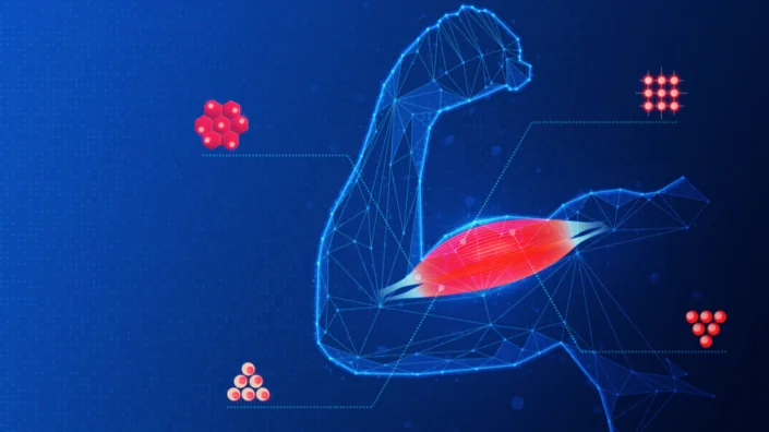
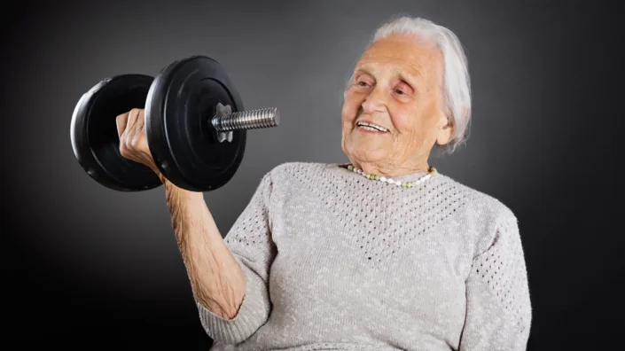
Follistatin, in particular, is a rather unusual choice of an anti-aging treatment, but it did deliver fantastic results. Do we have a mechanistic explanation for that?

I think the original explanation is that cachexia is muscle wasting that accompanies a lot of aging. You could argue that’s just a late-stage thing, and to do real aging reversal you’d want to get to the earlier stages, or you could argue that this is one of the ten pathways, and you want to have that part of a combination, but the answer is that it’s still not clear why it works in itself.

Cachexia accompanies cancer, the main cause of death in lab mice. Could this hint at an answer?

It’s one of the mechanisms that cancer can cause death by, but there are others. Cancer can cause death by making it hard to breathe. It can screw up almost any physiological process, depending on where it starts and where it metastasizes, but muscle wasting can be a product of fairly normal aging without cancer as well. Seems like it’s a nice tool to have in your toolbelt, but I admit it is a little surprising that it works by itself.

Telomerase is also an important factor in cancer. Do you think telomerase treatments can have adverse effects in humans that we don’t know about yet?

First, there are effects that we do know about. If you give a long steady dose, it will increase the probability of tumorigenesis. It also sets you up for additional genetic and epigenetic changes that can make an even more serious cancer. But, if you give intermittent doses, it’s like with Yamanaka factors in the second study: if you give too much, cells could go back too far in time. So, I think it’s all about moderation.

The other thing that was done in germline experiments, or earlier animal models, is to use specific safeguards against cancer so that when you then introduce the telomerase, you can express it at higher levels and for longer periods of time. For instance, messing around with the p16 and p53 pathways, i.e., having extra copies of tumor suppressors, can protect you against certain forms of cancer and allow you to use things that would otherwise put you at risk.

The results of this paper were spectacular, and it got us all very excited, but the sample size was small. Are there any plans for a bigger follow-up study?

It’s all about resources. Both those companies are scrappy, young, underfunded, but they make up for it in creativity and delivery. Those are the only two gene therapy papers that I know of that show significant Kaplan-Meier plots. And I really like the gene therapy approach that they share, because you can target alleles, splice isoforms, and gene family members (paralogs). This is something that’s very hard to do with small molecules, but very easy to do with either protein or gene therapies.

The slight advantage of gene therapies over protein therapies is that you have a few ways of rigging the specificity. You can have specificity of the vector, specificity of the nucleic acid, and finally, specificity of the protein. With proteins, you only have the latter.

The only thing I don’t like about gene therapies is the cost, but one positive side of the COVID dilemma was that we tested five different vaccines that were formulated in the form of a gene therapy on billions of people and got the price down to as low as 2 dollars for one of those five. It’s a whole new ballgame now if you have a large market, and I think pandemics and aging are the largest.

Let’s move to the OSK paper that came out of Rejuvenate Bio. What is the importance of it? Was it the simultaneous delivery of three factors in one vector or something else?

We had already done three genes at once at my lab and Rejuvenate Bio. Those were different from the OSK Yamanaka factors. The latter are transcription factors that need to be delivered to the nucleus, while the other set that we had, TGF beta receptor 2 in soluble form, Klotho, and FGF21, could be soluble, secreted, hence they could go into the interstitial, intercellular matrix, or they could go out into the blood. In a certain sense, it requires less delivery, or less ubiquitous delivery of the nucleic acid, because then the proteins do some of the delivery.

That’s the fundamental difference between those two. The significance of using the OSK is that this was the best example that we had of rejuvenation, as opposed to just fighting the symptoms of some age-related disease, making your muscles hurt less or something. This was truly rejuvenating ancient cells all the way back to embryo.

So, if you can control that, dial it up and down, it has some promise. This encouraged several groups, including Altos Labs, which I worked with briefly (many of my alumni are core members of Altos Labs). It is also one of the things that encouraged our collaboration with David Sinclair.

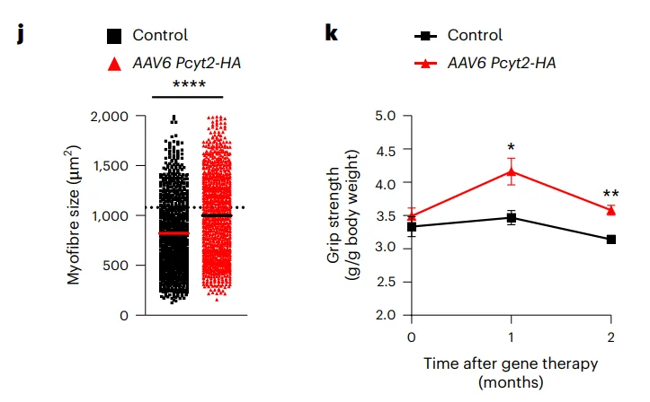
But, except for Rejuvenate Bio, so far, all of those are fairly distantly related to longevity. They might cure something like a crushed optic nerve, but damage to the eye is not aging, certainly not longevity. Now, this wasn’t merely the Kaplan-Meier plot where you could see extended life. The injection occurred at the age of 124 weeks. For a mouse, it’s very late in life. Half of the mice in the cohort are already dead at that point.

That was indeed a very interesting design. I think those were the oldest mice in any study that I’m aware of. What was the rationale behind that? What were you trying to prove?

The logic behind this was that if you do it as late as possible and show that it still has an effect, that’s very encouraging. It broadens the market, that’s one way of thinking about it, but it also tells you that you are really getting a reversal as opposed to delay, and that’s a fundamental difference. Many drugs that are considered cures are reversing something, but people somehow put reversing aging  in a special category, like breaking the sound barrier. However, just like breaking the sound barrier was easy enough to do once we figured it out, reversal of fundamental epigenetic programs that are a natural part of development and aging starts looking easier than it originally did.

Do we understand how cellular reprogramming improves health and longevity?

There have been two major camps in aging since long ago. One says that aging happens due to damage, to proteins, lipids, RNA, and DNA, and that you have to go in there with your repair kit and fix it as a therapist. The other camp says that it’s all epigenetic, and that if you convince the cell that it’s young, it will get its own toolkit out and start repairing as much as it can. Some things are beyond repair. If you delete all copies of a tumor suppressor, that’s not something a young cell can repair. But most things are fixable with epigenetics – at least, that’s how the second hypothesis goes.

I believe in a hybrid model. I think most of the work can be done epigenetically. A surprising amount of it can be done via the bloodstream, but probably not all of it. Then, there’s a residual amount that you can fix with the Yamanaka factors and another residual amount that you can fix by restoring genes.

Since we do the epigenetic reprogramming by adding in genes, it’s not that fundamental a difference between adding in genes that will go into the blood, adding genes that will reprogram the nucleus, and adding genes that are missing, like tumor suppressors. In a certain sense, they are all addressable by multiplex gene therapy. That’s why being able to either use multiple rounds of dosing or to have bigger vectors will become increasingly important.

Given the rising popularity of partial reprogramming, what is its overall place in the longevity landscape?

I think there are subtle but important differences between anti-aging drugs and drugs that improve biomarkers in the way that statins improve cholesterol. That doesn’t mean such drugs increase longevity, just that they improve this one biochemical. It could actually hurt you; for instance, it could improve cardiovascular chances for some subset of the population, but for another subset, it could hasten muscle pain.

So, affecting biomarkers is one thing. Reversing diseases of aging is different. You could do it just by addressing that particular disease, or you could do it more broadly, affecting multiple diseases. You might get FDA approval for one of them, but it’s actually affecting multiple ones, and maybe acting preventatively. Say, there might be a cure for muscle wasting that helps prevent a variety of diseases.

Finally, you’re really at the core of aging  when you reprogram shared elements – with good feedback systems that already exist in the body or with feedback systems that you introduce as part of the therapy.

I understand that the combination therapy with the three soluble factors you mentioned earlier is already in clinical trials in dogs. Do you have any preliminary results?

The thing about clinical trials is that you ideally minimize talking about them before they’re done. This is a second animal trial. We did it in mice already, and it looked good. With the dogs, it’s not just an animal trial, it’s also a product, a veterinary product. I understand that some of the pet owners involved have said good things, but, of course, you have to be very cautious with anecdotal evidence. There could be wishful thinking, placebo effect involved. These are randomized clinical trials, so you really won’t know until you break blinding at the end.

But the nice thing about veterinary clinical trials is that they’re typically much faster than human clinical trials. COVID-19 trials were an exception, 12 months, but most human clinical trials last about 10 years, and most veterinary trials are more like 18 months. So, we’ll know soon.

Still, the endgame is delivering those therapies to humans, and you will need a human trial anyway.

We could have gone straight from mice to humans, but we decided to create a veterinary product. People spend a fair amount of money on end of life in dogs, they even clone dogs, but of course, a clone is not what you want. So, it’s valuable to have two or more animal preclinical trials before you go into clinical trials. Even though we’re looking into specific diseases, for instance, the mitral valve disease in spaniels, it’s not limited to that. We hope that it will hit many different diseases. We’ve tested it in about five or six different diseases already, in mice and dogs.

To move this into humans, you use the same genes. You might use a human version of them, just like you might switch from mouse to dog genes, but other than that, mode of delivery. You might also change the vector. We use AAV vectors, which is the oldest approved vector and one of the most popular ones. However, dogs have natural immunity to most of the popular AAV vectors.

That’s clearly solvable. It’s even solvable for redosing. One of the advantages of certain gene therapies is that you don’t have to redose. You might have a once-and-done, lifetime expression, but a couple of my companies are working on improving the AAV delivery. Shape Therapeutics and Dyno Therapeutics are both making quite a progress on this. So, you can make radically different capsids that have radically different tissue tropisms and immune evasion. You basically make viruses that don’t exist in the wild or in previous pharmaceuticals, and you can probably keep generating those things for quite a while.

First, with dogs, you can get a lot of feedback on how to maximize your chances of success. Second, it’s a product, so you can make a profit on the first product to pay for the development of the second product. Human clinical trials are expensive. You could do it by investment, but that would result in dilution of the original founder’s stake in it.

Also, dogs are just a particularly good intermediate between mice and humans, because they live in a human environment, they have some compatible behavioral traits and eating habits. You can identify personality changes, subtle behavioral changes that you don’t identify in mice, because you don’t cohabit with them like you do with dogs.

None of this means that we couldn’t go directly to humans, it’s just that failures in human trials can put a damper on the whole field. I hope that all people in the field do their homework and don’t rush trials, because they can screw it up for us. We’re trying not to screw it up for them, and, hopefully, that will be reciprocated.

By the way, you do have the reputation of being a serial entrepreneur. How are your numerous startups doing? Are you still bullish about longevity biotech?

I co-founded 46 companies. I’m on the scientific advisory board of a few more. Seven of them are related to aging reversal and longevity.  They all involve pre-clinical and clinical trials. Some are on nutritional supplements, which you can sell without FDA approval, but nevertheless, they’re doing trials, and that’s a strong preference for my involvement in a company: they have to be willing and planning to do, and also have the resources to do, clinical trials, even if not legally required.

I think the whole field is very healthy economically and scientifically. We have passed through multiple “valleys of death”. We’re now in the solid science phase, and this field is going to be very impactful, maybe more impactful than any other pharmaceuticals in history, including even antibiotics, because our very ability to fight off diseases is age-related. Almost every single form of human morbidity and mortality has an age-related component to it. If you want to have a pleiotropic effect on many different diseases, this is the way to go.

So, it seems like a good choice. It also has a good chance of being cost-effective enough. I’m not promising this on behalf of any of my companies, but since manufacturers of COVID vaccines can get gene therapy to be as cheap as two dollars a dose, others can do that as well, and that’s important to me from the standpoint of equitable distribution of technology. It’s not about “can we develop a billion-dollar drug?” Rather, it’s “can we distribute it to everyone who needs it?” And the same way that we got the price of DNA sequencing down seven orders of magnitude, 20-million-fold, I think we need to at least consider that for every new medical technology.

Speaking of supplement companies, you are involved with two of them, NOVOS and Elysium.

Yes, and I was referring to them when I said they’re willing to do clinical trials even though they are not required to do so.

This is commendable, but they are already selling supplements. Do you think it’s not too early to be marketing anti-aging stuff to people, considering the state of the research?

I think we need to be very cautious about supplements that do not go through clinical trials and also about what trials they went through. If you do a clinical trial on ALS or some other degenerative disease, then that’s all you’re addressing. You’re not addressing the broader population. And, you have to be cautious, because some supplements have been shown to be detrimental if taken for prolonged periods in particular individuals that may have a pharmacological predisposition.

For example, some encourage cancer. And that’s true for all things you put in your body. Just because your grandparents got away with it doesn’t mean that it’s going to work, especially as we now have a more complex environment with a lot of other toxins.

Also, because we’re staying employed later and later in life, we need to reevaluate the things that were generally recognized as safe. So, I’m very enthusiastic about testing and retesting things that have been accepted without formal clinical trials. There’s a lot of placebo effects, long-term effects, and wishful thinking that we need to put to the test. I hope those two companies continue along that pathway.

So, you think they’re being prudent enough for you to associate your name with them?

I’m a scientific advisor. We shouldn’t ignore companies that need advice, otherwise, they will be operating without advice. As long as they’re doing clinical trials, they’ve got my attention.

Which directions in geroscience are you personally excited about at the moment?

I think we really need to hit all ten pathways and think about their interactions. In my opinion, the easiest way to go from a hypothesis or a pile of molecular data to a therapy is via gene therapy. In a couple of weeks, you can essentially go from a new research article to a therapy and get that therapy into mice.

So, I’m particularly excited about those directions. We don’t know yet whether it should be cell-autonomous or something that spreads through the blood, probably both. I’m excited about the possibility that this could be low-cost for equitable distribution. All those things are clustered around this multi-drug therapy, which I think is fairly well-validated in other cases: multi-drug antibiotics, multivalent antivirals, cancer, some other diseases as well.

So, you think we’ve accumulated enough knowledge to start moving in the direction of combination anti-aging treatments?

Yes, except you have to test them all in vitro and in animal models, in the exact formulation that you want to use, even if the ingredients were already tested individually. Sometimes with gene therapy, you can’t test the very exact molecular form, because there’s a difference between the animal form and the human form. But, other than that, everything should be as close as possible.

In one of your recent talks, you said something, and I hope I’m not misinterpreting, about cell therapies being superior to gene therapies.

It’s not either or. Cells can deliver genes and proteins. The advantage of cell therapy in principle is that you can debug the gene therapy ex vivo and then deliver it. For example, if you apply gene therapy in vivo to a billion cells, you’ve got a billion chances for genetic mischief, but with cell therapies, especially clonal cell therapies, you can debug it and make sure this is genetically and epigenetically what you want, that all cells are basically identical.

The downside of it is that many cells just don’t deliver very well. For example, neurons are highly connected. Maybe you can deliver a few neuronal stem cells, including into parts of the brain that normally don’t have neuronal stem cells, but you also want to keep most of your connections intact and not replace them with some generic neuronal progenitor. The bottom line is that there’s room for a lot of different strategies and innovations.

We would like to ask you a small favor. We are a non-profit foundation, and unlike some other organizations, we have no shareholders and no products to sell you. All our news and educational content is free for everyone to read, but it does mean that we rely on the help of people like you. Every contribution, no matter if it’s big or small, supports independent journalism and sustains our future.
Damaged DNA

Mitochondria, DNA, and Oxidative Stress

A paper published in Experimental Gerontology has provided a fresh and detailed look at the effects of oxidative stress on longevity.

Revisiting an old theory

The free radical theory of aging, which purports that reactive oxygen species (ROS) are the core driver of aging, was developed all the way back in 1956 [1] and expanded upon in 1972 with a focus on the mitochondria [2]. While this limited theory has been superseded by more comprehensive and detailed models of aging, experiments have repeatedly confirmed that ROS do, in fact, drive mitochondrial damage [3], which is linked to a great many aspects of aging [4].

This review paper explores modern research into this old theory, elucidating the relationship between mitochondrial ROS and the longevity of different species.

Not consumption but production

Previous experiments relating to oxidative stress have focused on antioxidants, substances that are intended to reduce the amounts of ROS. Unfortunately, experimental evidence found that feeding antioxidants to animals did not have any beneficial impact in most cases, regardless of how they were administered [5]. In fact, organisms with more naturally occurring antioxidants in their bodies age more rapidly on average [6].

Instead, mitochondrial ROS production seems to be the key factor. The reviewers cite a multitude of studies showing that organisms that produce more free radicals in the mitochondria age more quickly. They even contend that this represents evidence for this aspect of aging being evolutionarily programmed: free radical leak, a key component of ROS production, varies by species, is strongly correlated with longevity, and is not well connected with body size. Pigeons, for example, naturally live much longer than rats and have considerably less ROS despite their high oxygen use [7]. Bats also have the same advantages [8].

A focus on mitochondrial DNA fragments

The researchers then turn their focus to the physical mechanisms involved. While the researchers note one study showing that oxidative stress leads to direct mitochondrial DNA damage, which is strongly associated with reduced longevity [9], along with another study showing that deleted mitochondrial DNA is associated with accelerated aging [10], the researchers hold that these deletions, in and of themselves, are insufficient to be substantial direct causes of aging.

Instead, they note that mitochondrial DNA fragments can accumulate in nuclear DNA in a process called numtogenesis [11], which these researchers hypothesize is driving a significant part of aging. This hypothesis is supported by evidence showing that rapamycin decreases nuclear mitochondrial DNA accumulation at the same rate it increases longevity [12]. Mitochondrial DNA can even circulate through the body, contributing to systemic inflammaging [13].

An amino acid as a target

The researchers posit that methionine reduction, which has been shown to increase longevity in mice [14], has such beneficial effects because methionine and its metabolites are connected to increased ROS production [15] along with multiple distinctly toxic effects within cells. They also suggest that methionine is a major part of the reason why high-protein diets are linked to oxidative stress and decreased longevity [16].

Conclusion

While these reviewers’ conclusions of the roles of ROS and mitochondrial DNA in aging may seem surprising, they are supported by experiments. Therefore, it is worthwhile to attempt to develop interventions that directly target ROS generation at its root or remove free-floating mitochondrial DNA from cells and tissues. If such interventions can be shown to work in small mammals, it may be very much worth it to test them in clinical trials in order to demonstrate whether or not they can reduce aging biomarkers and lengthen the lifespans of people.

We would like to ask you a small favor. We are a non-profit foundation, and unlike some other organizations, we have no shareholders and no products to sell you. All our news and educational content is free for everyone to read, but it does mean that we rely on the help of people like you. Every contribution, no matter if it’s big or small, supports independent journalism and sustains our future.

Literature

[1] Harman, D. (2002). Aging: a theory based on free radical and radiation chemistry. Science of Aging Knowledge Environment, 2002(37), cp14-cp14.

[2] Harman, D. (1972). The biologic clock: the mitochondria?. Journal of the American Geriatrics Society, 20(4), 145-147.

[3] Halliwell, B., & Gutteridge, J. M. (2015). Free radicals in biology and medicine. Oxford university press, USA.

[4] Berry, B. J., & Kaeberlein, M. (2021). An energetics perspective on geroscience: mitochondrial protonmotive force and aging. Geroscience, 43(4), 1591-1604.

[5] Barja, G. (2004). Aging in vertebrates, and the effect of caloric restriction: a mitochondrial free radical production–DNA damage mechanism?. Biological Reviews, 79(2), 235-251.

[6] Perez-Campo, R., Lopez-Torres, M., Cadenas, S., Rojas, C., & Barja, G. (1998). The rate of free radical production as a determinant of the rate of aging: evidence from the comparative approach. Journal of Comparative Physiology B, 168, 149-158.

[7] Barja, G., Cadenas, S., Rojas, C., Perez-Campo, R., & Lopez-Torres, M. (1994). Low mitochondrial free radical production per unit O2 consumption can explain the simultaneous presence of high longevity and high aerobic metabolic rate in birds. Free radical research, 21(5), 317-327.

[8] Brunet-Rossinni, A. K., & Austad, S. N. (2004). Ageing studies on bats: a review. Biogerontology, 5, 211-222.

[9] Barja, G., & Herrero, A. (2000). Oxidative damage to mitochondrial DNA is inversely related to maximum life span in the heart and brain of mammals. The FASEB Journal, 14(2), 312-318.

[10] Trifunovic, A., Wredenberg, A., Falkenberg, M., Spelbrink, J. N., Rovio, A. T., Bruder, C. E., … & Larsson, N. G. (2004). Premature ageing in mice expressing defective mitochondrial DNA polymerase. Nature, 429(6990), 417-423.

[11] Singh, K. K., Choudhury, A. R., & Tiwari, H. K. (2017, December). Numtogenesis as a mechanism for development of cancer. In Seminars in cancer biology (Vol. 47, pp. 101-109). Academic Press.

[12] Martínez-Cisuelo, V., Gómez, J., García-Junceda, I., Naudí, A., Cabré, R., Mota-Martorell, N., … & Barja, G. (2016). Rapamycin reverses age-related increases in mitochondrial ROS production at complex I, oxidative stress, accumulation of mtDNA fragments inside nuclear DNA, and lipofuscin level, and increases autophagy, in the liver of middle-aged mice. Experimental gerontology, 83, 130-138.

[13] Picca, A., Lezza, A. M. S., Leeuwenburgh, C., Pesce, V., Calvani, R., Bossola, M., … & Marzetti, E. (2018). Circulating mitochondrial DNA at the crossroads of mitochondrial dysfunction and inflammation during aging and muscle wasting disorders. Rejuvenation Research, 21(4), 350-359.

[14] Fang, H., Stone, K. P., Wanders, D., Forney, L. A., & Gettys, T. W. (2022). The origins, evolution, and future of dietary methionine restriction. Annual Review of Nutrition, 42, 201-226.

[15] Gomez, J., Sanchez-Roman, I., Gomez, A., Sanchez, C., Suarez, H., Lopez-Torres, M., & Barja, G. (2011). Methionine and homocysteine modulate the rate of ROS generation of isolated mitochondria in vitro. Journal of bioenergetics and biomembranes, 43, 377-386.

[16] Żebrowska, E., Maciejczyk, M., Żendzian-Piotrowska, M., Zalewska, A., & Chabowski, A. (2019). High protein diet induces oxidative stress in rat cerebral cortex and hypothalamus. International journal of molecular sciences, 20(7), 1547.

Depression

New Treatment Alleviates Depression Symptoms in Mice

Scientists have shown that the protein GDF11 can reverse depression-like symptoms in naturally aged mice and in a mouse model of depression. Depressed humans have lower GDF11 as well [1].

Depression and aging

Severe mental disorders, including depression, have been linked to significantly shorter lifespans [2]. According to one study, depressed people “have a higher incidence of various diseases of aging, such as cardiovascular and cerebrovascular diseases, metabolic syndrome, and dementia.” [3] This makes depression an important target of longevity research.

However, treating depression is difficult. Many prescription antidepressants are addictive, have serious side effects, and don’t always work. In fact, the whole theory behind serotonin-increasing antidepressants was called into question by a recent study [4], although its findings were probably taken out of proportion.

In this new study, published in Nature Aging, the researchers investigated the protein called growth differentiation factor 11 (GDF11), a member of the transforming growth factor beta (TGF-β) superfamily, as a potential antidepressant.

GDF11 plays an important role in the development of the central nervous system and regulates neurogenesis in adults. Depression has been linked to a decrease in the volume of the hippocampus, the main locus where adult neurogenesis occurs. Previous research has shown that GDF11 supplementation can increase hippocampal neurogenesis in aged mice [5].

Smarter and happier?

In the study, naturally aged mice showed a significant increase in depression-like symptoms compared to young controls. For instance, their grooming frequency dropped almost threefold. The tail suspension test (in which a mouse is briefly suspended by its tail) showed that old mice remained immobile for longer, which indicates a diminished will to escape.

The intruder avoidance test showed that older mice were more apathetic and less inclined to chase intruders out of their territory. Finally, the researchers tested for the inability to experience pleasure (anhedonia), finding that aged mice were less attracted to a sucrose solution. GDF11 treatment restored all but one of those parameters to youthful levels.

GDF11 Depression 1

As expected, older mice also experienced a decline in cognitive abilities. In a test designed specifically to assess hippocampus-dependent spatial memory, the novel object location test, aged mice spent much less time investigating the novel location. This cognitive decline was reversed to youthful levels by the treatment. However, the treatment failed to improve anxiety and physical performance in aged mice.

Increased neurogenesis confirmed

The researchers then investigated the mice’s brains. Analysis of neurogenesis markers revealed a 53% increase in immature neuroblasts in the subgranular zone (SGZ), the part of the hippocampus heavily populated by neuronal stem cells, indicating improved neurogenesis.

The treatment also decreased levels of cellular senescence in SGZ almost to the levels observed in young controls, as measured by senescence-associated β-galactosidase. Two other markers of senescence, p16 and p19, which are elevated in aged mice, were also significantly reduced by GDF11. In treated mice, several autophagy-related proteins were upregulated, with FoxO3a being upregulated even compared to young controls.

In cultured hippocampal neurons, GDF11 caused a significant increase in autophagy and neuronal activity. To investigate whether the increase in neuronal activity had been caused by autophagy, the researchers knocked down the autophagy-related protein Beclin1, which halved the GDF11-induced effect. The same results were obtained by blocking autophagy with the antibiotic Bafilomycin A1.

Of depressed mice and men

The researchers also experimented with young mice in which depression-like behavior was induced by corticosterone (a well-established murine model of depression). Just like with naturally aged mice, GDF11 alleviated most depression-like symptoms. For instance, in the well-known open field test, where avoidance of the central zone signals decreased activity and exploration, GDF11-treated mice performed on par with healthy controls.

GDF11 Depression 2

Finally, the researchers recruited 57 young adults with MDD (major depressive disorder) and found that GDF11 levels in their blood were significantly lower than in age-matched controls. In a larger cohort of 759 young adults (103 with an ongoing depressive episode and 656 controls) aged between 21 and 32 years old, the difference was even more pronounced, reinforcing the link between GDF11 and depression.

Conclusion

The link between depression and aging calls for a serious investigation. This study suggests that the relationship between them might be mediated by decreased hippocampal neurogenesis, which can be attenuated by systemic administration of GDF11. Lower levels of GDF11 in depressed young adults lend some support to this hypothesis.

We would like to ask you a small favor. We are a non-profit foundation, and unlike some other organizations, we have no shareholders and no products to sell you. All our news and educational content is free for everyone to read, but it does mean that we rely on the help of people like you. Every contribution, no matter if it’s big or small, supports independent journalism and sustains our future.

Literature

[1] Moigneu, C., Abdellaoui, S., Ramos-Brossier, M., Pfaffenseller, B., Wollenhaupt-Aguiar, B., de Azevedo Cardoso, T., … & Katsimpardi, L. (2023). Systemic GDF11 attenuates depression-like phenotype in aged mice via stimulation of neuronal autophagy. Nature Aging, 1-16.

[2] Fiorillo, A., & Sartorius, N. (2021). Mortality gap and physical comorbidity of people with severe mental disorders: the public health scandal. Annals of General Psychiatry, 20(1), 1-5.

[3] Wolkowitz, O. M., Epel, E. S., Reus, V. I., & Mellon, S. H. (2010). Depression gets old fast: do stress and depression accelerate cell aging?. Depression and anxiety, 27(4), 327-338.

[4] Moncrieff, J., Cooper, R. E., Stockmann, T., Amendola, S., Hengartner, M. P., & Horowitz, M. A. (2022). The serotonin theory of depression: a systematic umbrella review of the evidence. Molecular psychiatry, 1-14.

[5] Ozek, C., Krolewski, R. C., Buchanan, S. M., & Rubin, L. L. (2018). Growth Differentiation Factor 11 treatment leads to neuronal and vascular improvements in the hippocampus of aged mice. Scientific reports, 8(1), 1-13.

Blood DNA

Young Blood Alters Gene Expression in Old Brain Cells

Research published today in Nature Aging has shown that heterochronic parabiosis, the circulatory joining of young and old organisms, has rejuvenative effects on the gene expression of multiple types of brain cells in mice.

Parabiosis affects a wide variety of factors at once

Heterochronic parabiosis is a well-known aging intervention in mouse studies, and some studies have shown that factors in young blood improve brain health [1] while other studies have shown that factors in old blood cause harm to this organ [2]. While some work has focused on the changes to the proteins in blood (proteome) [3], these researchers note that the proteome is only part of the equation: exosomes, lipids, and other nonprotein factors are also likely to be having an effect.

Here, the researchers are not attempting to discover which of these myriad factors is having what effects. Instead, this paper has analyzed, at the gene transcription level of single cells, what effects heterochronic parabiosis has on multiple types of brain cells.

Largely expected results

Like with other parabiosis research, 3- to 4-month-old mice had their circulatory systems joined to 20- to 22-month-old mice, with same-age mice joined the same way as control groups. Individual, unjoined mice were also used as controls. After filtration and selection, over 100,000 cells that consisted of 75 different types were analyzed, and the researchers found that parabiosis did not cause them to lose their cellular identities.

This paper’s findings were in concordance with previously published work on parabiosis showing multiple beneficial and rejuvenative effects on gene expression. The researchers found that 700 of the over 20,000 genes they analyzed had their expression changed with aging, 442 gene expressions were changed in old animals by youthful blood, and 155 gene expressions were changed in young animals by old blood. Beneficial effects on gene expression networks, downregulation of senescence, and the improvement of intercellular communication in a way that appears to encourage the formation of new brain cells (neurogenesis) were also noted.

This paper highlights the strong, youth-promoting effects of parabiosis on the endothelial cells that make up the blood-brain barrier, with the suggestion that this may be due to these cells’ direct and constant contact with relatively large volumes of blood. This observation supports the previous finding that heterochronic parabiosis substantially improves brain vasculature [1].

However, some of the findings were less enthusiastic. The gene expressions that were common to aging and rejuvenation were different between cells, and parabiosis only had a limited effect on gene expression. The researchers also made the counterintuitive finding that heterochronic parabiosis may work through pathways that are not recognized to be part of aging.

Conclusion

Interspersed throughout its detailed data explaining the many various genes and pathways upregulated and downregulated with parabiosis and aging, this paper provides three key takeaways:

These data are again consistent with the notion that parabiosis is likely to act in part by regulating processes important to vascular structure and health.

These data suggested that heterochronic parabiosis changes the metabolic profile, improves proteostatic machinery and reduces aging-associated apoptosis or senescence.

These findings suggest new strategies for slowing deterioration and driving regeneration in the aging brain through approaches that do not rely on disease-specific mechanisms or actions of individual circulating factors.

With this last quote in mind, the question arises: how do we bridge the gap? Obviously, it is infeasible and unethical to join together the circulatory systems of old and young humans. However, it may be feasible to utilize a combination of youthful systemic factors (both protein and nonprotein) along with the blood scrubbing known as plasmapheresis to restore vasculature and function to the aging brain. Only animal experimentation and human clinical trials can determine if such an approach can one day become part of medicine.

We would like to ask you a small favor. We are a non-profit foundation, and unlike some other organizations, we have no shareholders and no products to sell you. All our news and educational content is free for everyone to read, but it does mean that we rely on the help of people like you. Every contribution, no matter if it’s big or small, supports independent journalism and sustains our future.

Literature

[1] Katsimpardi, L., Litterman, N. K., Schein, P. A., Miller, C. M., Loffredo, F. S., Wojtkiewicz, G. R., … & Rubin, L. L. (2014). Vascular and neurogenic rejuvenation of the aging mouse brain by young systemic factors. Science, 344(6184), 630-634.

[2] Villeda, S. A., Luo, J., Mosher, K. I., Zou, B., Britschgi, M., Bieri, G., … & Wyss-Coray, T. (2011). The ageing systemic milieu negatively regulates neurogenesis and cognitive function. Nature, 477(7362), 90-94.

[3] Lehallier, B., Gate, D., Schaum, N., Nanasi, T., Lee, S. E., Yousef, H., … & Wyss-Coray, T. (2019). Undulating changes in human plasma proteome profiles across the lifespan. Nature medicine, 25(12), 1843-1850.