×

The Blog

Building a Future Free of Age-Related Disease

Sarah Constantin Interview

Sarah Constantin on the Daphnia of Daphnia Labs

We recently had the opportunity to catch up with Sarah Constantin, one of the founders of the new biotech startup company Daphnia Labs. The company is developing a new platform for the discovery of geroprotective drugs: in vivo phenotypic screens in model organisms. The company plans to use high-resolution video to track Daphnia during its lifespan in order to screen for drugs that might extend healthspan.

Can you tell us a little about the company, its founders, and what motivated you to start this biotech company?

I’d been working for about a year at the Longevity Research Institute, which is a nonprofit that funds aging research. LRI focuses on trying to replicate studies on interventions that have been reported to extend lifespan in mammals. Basically, of the 50+ compounds out there that have some mammalian evidence for an anti-aging effect, which ones have the best chance of being viable geroprotectors in humans?

We wanted to expand to doing a high-throughput screening program in a short-lived model organism, because, really, the surface has barely been scratched in terms of searching for aging-modulating small molecules, and this is the kind of research program that machine learning and automation can really help with.

Initially, I imagined we’d do this with C. elegans, but then, fortuitously, I met Leon Peshkin, a systems biologist at Harvard Medical School, and he convinced me that Daphnia was a better pharmacology model organism.

Then, we started getting the advice that this made more sense as a business than as part of a nonprofit, because if we’re looking at novel compounds, that has the potential to generate IP. So, I incorporated Daphnia Labs as a public benefit corporation.

You have chosen to use Daphnia as your model animals for drug discovery; can you explain what things in particular made you decide to use them?

Well, Daphnia are short-lived. They live about a month on average. So, you can do lifespan studies with a fast turnaround time.

The natural point of comparison here is with C. elegans, which are a traditional aging model organism and also short-lived, but they’re more a genetics model organism than a pharmacology model organism. They live in dirt, and they have a very powerful xenobiotic system to excrete toxins. So, if you dose them with a drug, they’re very good at remaining unaffected, which means that drug screens on C. elegans are maybe less predictive of mammalian results than drug screens on Daphnia would be, certainly less predictive of toxicity.

Daphnia survival is actually a standard test for ecological toxicology: to test if something’s safe to put into the water supply. Daphnia are extremely sensitive to toxins, and they’re already used to predict toxicity in humans.

They’re also capable of both parthenogenesis and sexual reproduction. So, they’re exact clones by default, no genetic uncertainty, but you can also get a single generation cohort by giving them a chemical that makes them produce males.

How similar are daphnia to humans in terms of how they age and their genes, organs, and physiology?

Daphnia have about 60% of genes in common with us, roughly the same as a fruit fly, a bit more than a worm. They have a lot of the same organs — they have a chambered heart, a gut, a brain, etc. They have an innate immune system.

They also have homologs of a lot of well-known aging-associated genes: IGF1, Hsp70, FOXO, TOR, SIRT1, etc. There are pathways that are conserved across pretty much all animals, and they seem to exist in Daphnia as well.

We know Daphnia age because their mortality rates increase with time, just like ours do. They die of different things than humans do — usually intestinal malfunction — but they do become less mobile with age, just as most animals and humans do. There’s actually a big literature about the evolutionary biology of Daphnia life-histories, because you have these different genetic populations that arose in different ponds, and you can see that Daphnia live longer when they’re under less predator stress, for instance.

Often, at the early stages of drug discovery, C.elegans are a commonly used model animal. However, traditional manual and even automated drug screening is often time consuming, laborious, and costly. How does your system propose to change this paradigm?

Daphnia are really well suited to an automated approach because they’re easy to visualize. They’re transparent, and they’re large enough to see with the naked eye. With a high-resolution video camera, you can track their motion and some features of their physiology automatically. There’s no need to poke them like you do with worms; you just leave them alone in their water and gather video.

You are proposing to use daphnia as a novel in vivo screening platform; how does this differ to more traditional in vitro screening approaches, and what advantages does it offer?

The advantage is that you’re looking first for a functional phenotype — healthy lifespan — instead of starting with a target. For a problem as multifactorial as aging, it’s very unlikely that we’ve identified all the possible targets involved already. Many of the known aging-modulating drugs already seem to have multiple targets and mechanisms of action. A living organism is not the same as a cell culture; it’s got multiple tissues and hormones that affect the whole organism, so testing in culture isn’t necessarily representative of what you’d see in an organism. Screening for lifespan and activity in living animals means that you can get better prediction of what will work in a mammal than you’d get in an in-vitro screen.

You are using machine learning to screen for geroprotective drugs; can you give us an overview of how the system works and how it identifies promising molecules?

The machine learning is for analyzing the video of Daphnia. From each image, you can extract outlines of individual Daphnia and track their motion. You can also discover features, motion patterns, that correlate with age or mortality. If you do this with many tanks of Daphnia, each testing a different molecule, the ones that make Daphnia live significantly longer, stay active longer, or have motor patterns associated with youth are the geroprotective ones.

Could using machine learning reduce R&D costs for drug development, and in what ways?

If you can predict what will and won’t work in mammalian studies, you can rule out some of the failures earlier and have a higher hit rate on the studies you do conduct. Our calculations say that this could cut preclinical research costs in half.

If this means you can afford to do more mammalian studies, then you can get second-order effects — being more selective about which compounds you take into clinical trials and improving the clinical trial success rate. Since human trials cost tens of millions of dollars and only one in twenty drug candidates succeeds in the clinic, anything that improves predictive validity is a huge cost savings.

Your pipeline proposes to begin by screening 1000s of compounds via your daphnia system, then moving to compound analysis of potential candidates and finally onto mouse studies. What kind of potential candidate hit rate would you anticipate at the initial daphnia stage, and how does that compare to traditional methods?

The closest analogues are the lifespan screens that came out of the Petrascheck lab on C. elegans, including one on compounds with known mammalian bioactivity. A full 5% of those drugs significantly extended worm lifespan. Our expectation is that the hit rate in Daphnia of the same pool of drugs would be comparable or greater.

There isn’t really a direct comparison here with traditional methods, because target-based methods that are looking at binding affinity usually start with a screen of millions and a hit rate of less than 0.1%.

You would screen with the daphnia and generate around 10-20 good candidates for the mouse testing phase. What sort of mice would you be using, and why?

It’s going to depend on the disease indication. We’re indication-agnostic at this point, but knowing something about the expression or proteomics profile changes induced by the Daphnia hit compounds will tell us something about their mechanism, and we’ll decide on a good disease indication to go after from there. In that case, what you’re looking for is a realistic mouse model of the disease in question.

How far has the company developed so far, and what are your next steps into 2020?

Currently, we’ve built a prototype, generated our first data, and are working on experiments to validate Daphnia’s response to known aging-modifying interventions — do they live longer when you give them things like rapamycin and metformin, which make many other animals live longer?

As we go into 2020, we’ll aim to raise our seed round and start scaling up the screening device and software.

Is there anything else you would like to tell us about Daphnia Labs?

We’re always looking to talk to experts in the fields we’re working on. Currently, we’re especially interested in hearing from aquaculture experts and lab automation engineers.

We would like to ask you a small favor. We are a non-profit foundation, and unlike some other organizations, we have no shareholders and no products to sell you. All our news and educational content is free for everyone to read, but it does mean that we rely on the help of people like you. Every contribution, no matter if it’s big or small, supports independent journalism and sustains our future.
Human heart

Heart Inflammation Causes a Shift in Cell Fate Distribution

A study on animals with autoimmune myocarditis was recently released in the journal Cell Reports [1], showing the impact of heart inflammation on the types of immune cell that are formed in the heart. This could have a significant impact on our understanding of cardiac aging.

What is myocarditis and how is it relevant to aging?

Myocarditis is a disease involving inflammation of the heart. It mainly influences people between the ages of 20 and 51 [2] [3]; however, the elderly are still affected to some degree. The disease has been known to cause serious complications, such as heart attack and heart failure.

Aging shares one similarity with myocarditis: inflammation. As myocarditis is caused by inflammation of the heart, studying it could tell us about how the heart changes with age due to constantly increasing age-related inflammation (inflammaging) [4].

An understanding of the changes that occur in the heart because of age-associated inflammation could allow the future creation of therapeutics that reverse these aspects of aging, which would lead to a decrease in heart attacks, strokes and heart failure in the elderly, thus improving health and lifespan.

What inflammation-associated changes are shown?

In the study, two mice had their circulatory systems connected, while one mouse had autoimmune myocarditis. The researchers showed significant changes in two different populations of immune cells (particularly monocytes and macrophages) along with changes in the local signalling environment because of this myocarditis.

One population – Ly6Chi monocytes, which are similar to human CD14+ monocytes and normally focused on clearance of harmful debris – were shown to create more inflammatory macrophages (immune cells with a variety of functions, including debris clearance and germ destruction) but were unable to clear the heart from harmful, inflammation-causing debris. While the rate of replication of this type of cell was unchanged, the rate of replication of another cell type was reduced, meaning that this inflammatory subtype became more common than other subtypes in the diseased heart.

This same population has been shown to cause fibrosis – scarring – of heart tissue in the diseased heart, which leads to the further creation of the same inflammatory macrophages in a positive feedback loop involving a signaling molecule (IL17A) that increases the number of these cells attracted to the diseased area through an intermediate molecule (CCL2) produced in scarred tissue, leading to further scarring and inflammation. These cells have also been shown to degrade the extracellular matrix (scaffolding) of the diseased heart, upon which its structure relies, and they possess proteins for recognizing certain germs, specifically class II antigen processing and presentation.

This same signaling molecule was also shown to cause scarred tissue to produce GM-CSF, a protein that encourages macrophages to mature into an inflammatory state (M1), despite the signaling molecule activating genes associated with a contradictory state as well (M2). Still, the behavior of the affected cells most matches the inflammatory subtype of macrophage, which has been shown to become more common in the body because of aging [5].

The increase in scarred heart tissue with an increase of the intermediate GM-CSF in the diseased heart has another impact – the prevention of the replication of Ly7Clo monocytes, which are most similar to CD16+ monocytes in humans – and the complete prevention of maturation of these cells into functional, scar-healing macrophages. As the diseased heart is a very hostile environment, this leads to the destruction of this subpopulation.

Of the cells that do mature in the diseased heart, unusually many of them originate from the very earliest, embryonic stages of life instead of being locally produced in the heart. This could be explained by the fact that the locally produced Ly6Clo cells have their replication and maturation systems shut down.

The overall macrophage population in the heart was shown to decrease, as could be expected because of the fact that Ly6Chi replication was not increased to compensate for the loss of the Ly6Clo cell population. In addition, the debris clearance-associated receptor MerTK on Ly6Chi monocytes and macrophages is prevented from being created, and is cleaved in the diseased heart, which could prevent clearance of harmful, inflammatory debris, accelerating the progression of the disease.

Conclusion

This study does have its weaknesses – since it is focused on mice, the same effects may not be seen in human cells. The merging of the circulatory systems of the healthy and diseased mice may be having some effect, and the disease itself may have other causes than the ones that aging has. Still, the trial gives some promising information that could inform further research.

Despite not being focused on a consequence of aging, myocarditis experiments like this could inform further research on an aged population. The next steps for research in this area should involve validation of the cell population changes, and, if the study is correct, methods to restore the initial balance of cell types.

We would like to ask you a small favor. We are a non-profit foundation, and unlike some other organizations, we have no shareholders and no products to sell you. All our news and educational content is free for everyone to read, but it does mean that we rely on the help of people like you. Every contribution, no matter if it’s big or small, supports independent journalism and sustains our future.

Literature

[1] Hou, X., Chen, G., Bracamonte-Baran, W., Choi, H. S., Diny, N. L., Sung, J., … & Hackam, D. J. (2019). The Cardiac Microenvironment Instructs Divergent Monocyte Fates and Functions in Myocarditis. Cell reports, 28(1), 172-189.

[2] Kytö, V., Saukko, P., Lignitz, E., Schwesinger, G., Henn, V., Saraste, A., & Voipio-Pulkki, L. M. (2005). Diagnosis and presentation of fatal myocarditis. Human pathology, 36(9), 1003-1007.

[3]Fenoglio Jr, J. J., Ursell, P. C., Kellogg, C. F., Drusin, R. E., & Weiss, M. B. (1983). Diagnosis and classification of myocarditis by endomyocardial biopsy. New England Journal of Medicine, 308(1), 12-18.

[4] López-Otín, C., Blasco, M. A., Partridge, L., Serrano, M., & Kroemer, G. (2013). The hallmarks of aging. Cell, 153(6), 1194-1217.

[5] Becker, L., Nguyen, L., Gill, J., Kulkarni, S., Pasricha, P. J., & Habtezion, A. (2018). Age-dependent shift in macrophage polarisation causes inflammation-mediated degeneration of enteric nervous system. Gut, 67(5), 827-836.

Lab mouse

Cellular Reprogramming Reverses Age-Related Vision Loss

Today, we want to draw attention to a new study that shows how partial cellular reprogramming was able to reverse cellular aging and address age- and injury-induced blindness in mice.

Epigenetic alterations

One of the proposed reasons we age is the changes to gene expression that our cells experience as we get older; these are known as epigenetic alterations. These alterations cause harmful changes to cellular function and gradually shift our cells from a youthful to aged state.

Gene expression is modified by the addition of epigenetic markers to the DNA that change the pattern of gene expression in a cell, suppressing or enhancing the expression of certain genes in a cell as the situation demands.

This is how our cells know what types of cells they are and what they should be doing, as the epigenetic instructions are there to guide them and ensure that we develop and grow properly. Essentially, epigenetic markers turn genes on and off, which, in turn, change what a cell does and how it functions.

However, as we age, our cells are exposed to environmental factors and are subject to negative changes in their genomes through epigenetic mechanisms. These alterations accumulate over time and are strongly correlated with the decline observed in aging cells.

Resetting the age of cells in mice

We have talked about the potential of partial cellular reprogramming in previous articles, and there have now been a number of demonstrations that reverse epigenetic alterations and so reset aged cells to youthful function.

In this new study, an impressive team of researchers, including George Church, David Sinclair, Vadim Gladyshev, Steve Horvath, and Michael Bonkowski, has used the technique of partial cellular reprogramming to reset epigenetic alterations that occur in cells during aging, thus restoring youthful function in mice.

To achieve this, they used just three reprogramming factors, Oct4, Sox2, and Klf4, to reverse age-related epigenetic markers and rejuvenate the old cells in the eyes of mice. The approach was able to regenerate the eyes of mice suffering from age- or injury-induced blindness by encouraging the cells to revert to a more youthful level of function and thus regenerative capacity.

The study also shows that old cells and tissues retain an accessible memory of youthful epigenetic information, which allows the functional age of the cell to be reset. It is also consistent with the findings of other mouse studies in which similar techniques have been used to reverse cellular aging via partial cellular reprogramming.

Ageing is a degenerative process leading to tissue dysfunction and death. A proposed cause of ageing is the accumulation of epigenetic noise, which disrupts youthful gene expression patterns that are required for cells to function optimally and recover from damage1–3. Changes to DNA methylation patterns over time form the basis of an ‘ageing clock’4, 5, but whether old individuals retain information to reset the clock and, if so, whether this would improve tissue function is not known. Of all the tissues in the body, the central nervous system (CNS) is one of the first to lose regenerative capacity6, 7. Using the eye as a model tissue, we show that expression of Oct4, Sox2, and Klf4 genes (OSK) in mice resets youthful gene expression patterns and the DNA methylation age of retinal ganglion cells, promotes axon regeneration after optic nerve crush injury, and restores vision in a mouse model of glaucoma and in normal old mice. This process, which we call recovery of information via epigenetic reprogramming or REVIVER, requires the DNA demethylases Tet1 and Tet2, indicating that DNA methylation patterns don’t just indicate age, they participate in ageing. Thus, old tissues retain a faithful record of youthful epigenetic information that can be accessed for functional age reversal.

Conclusion

Some researchers suggest that changes to the epigenetic state of cells merely reflect the age of a cell and its underlying aging, somewhat like the hands on a watch showing the time while the cogs and workings behind the watch face are the underlying causes of those changes.

However, this study adds yet more support for the proposal that epigenetic alterations are not merely an indication of the age of a cell but are much more likely to directly drive aging. It is certainly very compelling when the cells of mice are partially reprogrammed and they regain youthful function; if this is not a demonstration of rejuvenation, then I don’t know what is!

The race to translate partial cellular reprogramming to humans is now the focus of several companies, and progress in this area seems to have been moving along at quite a rapid pace since Salk Institute researchers first demonstrated the technique in mice back in 2016.

We would like to ask you a small favor. We are a non-profit foundation, and unlike some other organizations, we have no shareholders and no products to sell you. All our news and educational content is free for everyone to read, but it does mean that we rely on the help of people like you. Every contribution, no matter if it’s big or small, supports independent journalism and sustains our future.

Literature

[1] Lu, Y., Krishnan, A., Brommer, B., Tian, X., Meer, M., Vera, D. L., … & Yang, J. H. (2019). Reversal of ageing-and injury-induced vision loss by Tet-dependent epigenetic reprogramming. bioRxiv, 710210.

Overpopulated area

Population Prospects 2019 and Life Extension

A common concern about life extension is overpopulation, the idea that there are too many people in the world. Are we really headed for a global overpopulation meltdown, as some people believe? The United Nations’ World Population Prospects 2019 report suggests that while the global population will continue to rise for the next few decades, ultimately, that rise will plateau.

First things first: it’s population growth, not overpopulation

Whenever the topic of defeating age-related diseases comes up, there is inevitably someone who will cite overpopulation as an objection to healthy life extension and a reason why we should continue to let people become sick and die of diseases that science may be able to cure in the coming decades.

Overpopulation is a word that gives the simple phenomenon of population growth a negative connotation. It suggests that an increase in the number of living people will somehow be harmful to us in different ways, such as famine, scarcity of jobs, running out of resources, excessive population density, and harm to the environment.

However, from a global perspective, there is currently no overpopulation; there is simply population growth, which is not necessarily harmful by itself, and it is important to understand the difference between these two things.

The two most common themes accompanying this concern are usually that we will run out of resources unless we reduce the number of people on the planet or that there are too many people and we will harm the planet.

The first is something that could and is being addressed by shifting to sustainable methods of energy production, such as biofuel and renewable energy to supplement nuclear power. Other approaches that reuse resources, such as recycling instead of using landfills, are also part of the solution to this. There is also the most obvious: changing our societies so that we consume less and pollute less.

The second common theme, that too many people will harm the planet, has merit; there is, without a doubt, a maximum carrying capacity that the planet can sustain. To avoid reaching this point will require us to make societal changes, but as we will see later in the article, there are already ways of achieving this without the kneejerk reaction of people demanding direct population control.

What does the data tell us about global population?

The United Nations’ World population prospects 2019 was published recently, and we want to go over the key points that this data shows, as the situation is perhaps surprising and goes against the grain of what some people believe.

Here are some of the key points from the new report:

  • Population Plateau – The data strongly suggests that, for the first time in modern history, the world’s global population is due to plateau by the end of the century. This is largely thanks to the decline of fertility rates. The report projects that by 2100, the global population will have reached 10.9 billion but with an annual growth rate at this point of less than 0.1%, which is dramatically lower than the current growth rate.

To give you an idea of how drastically lower this is, we can look at the past and present. Between the 1950s and today, the global population growth rate has been 1-2% per year, which saw total population rise from 2.5 billion to over 7.7 billion. So, by the time we reach the population plateau, the growth rate is likely to be dramatically lower.

  • Fertility is falling – The fertility rate by 2100 is projected to fall to 1.9 births per woman, as compared to 2.5 currently. The replacement fertility rate (the rate that is necessary to maintain the population) is 2.1 births per woman, and the data suggests that fertility will go below this by 2070.The decline of fertility is due to a number of factors, including increasing access to higher education (in particular for women), industrialization/modernization, access to healthcare, reduced infant mortality, and increased life expectancy. We historically see a decline of fertility in every nation that has moved from developing to developed.This trend is almost certainly going to continue as the developing nations responsible for the highest population growth, such as parts of Africa and Asia, reach developed status and experience a corresponding decline of fertility. In fact, if people want the global population to level out sooner, then the logical course of action here would be to help speed up the development of the nations where fertility rates are highest. The sooner they develop, the sooner the fertility rate will begin to decline.
  • The global median age is rising – In 1950, the median global age was just 24, today it is 31, and by 2100, it is projected to rise to 42. This means that between 2020 and 2100, the number of people aged 80 and above should rise from the current 146 million to 881 million.By the 2070s, there are anticipated to be more people aged 65 and older than the number of people aged 15; this historical first, in which the aged population outnumbers the young, is known as the silver tsunami. The main contributing factors to this increase of the global median age are the increase in life expectancy and the decline of fertility rates.This also makes a very strong case for developing therapies for age-related diseases. These therapies would keep people healthy, independent, and able to continue working and contributing to the economy rather than being sick due to age-related diseases and putting a burden on the healthcare system, which is already feeling a heavy strain; imagine how much worse it will get once the old and sick outnumber the young. It’s better to keep people healthy as they age, and rejuvenation biotechnology is a means to this end.
  • The majority of population growth is coming from Africa – Taking a look at the UN population data, you can see that the main area of population growth is currently Africa. The report projects that Africa will continue to see high population growth for most of this century; again, this is the result of developing nations having higher fertility rates, which supports the idea that bringing this growth down sooner would require raising these countries up to developed status.The report projects that Africa will see its population increase from the current 1.3 billion to 4.3 billion between now and 2100. The majority of this increase will come from the developing nations located in sub-Saharan Africa. As the data shows, of the top 6 countries accounting for over half of the global population, five of them are located in Africa. Tanzania, Nigeria, the Democratic Republic of the Congo, Ethiopia, and Angola are projected to be in the top 10 countries by population by the year 2100. The sixth country is Pakistan.
  • The European population will go into decline – Projections suggest that Europe will experience population decline by 2100; it is expected to peak at 748 million by this time. This may also happen sooner, and some European countries, such as Germany, are already on the road to decline.Even with increased immigration to Europe, the population decline will continue, as immigrant fertility typically decreases dramatically in the second generation [1]. Other studies also show that the presence of immigrants does not compensate for a declining birth rate [2].
  • Latin America will also face decline – The Latin American region, including the Carribbean, is anticipated to see population growth until it peaks at 768 sometime around 2058 before going into decline.
  • Asia will grow but then go into decline – Asia is set to see an increase in its population, going from its current 4.6 billion and rising to an estimated 5.3 billion around 2055. However, following this period of growth, it is anticipated to go into decline.India is projected to continue growing until sometime around 2059 and hitting the 1.7 billion population mark, while China is likely to peak around 2031. Indonesia, the most populated country in Southeast Asia, is expected to hit its peak population by the year 2067.Interestingly, the populations of Japan and South Korea are ahead of the trend and are expected to go into decline from 2020 onwards; in fact, Japan has arguably already reached this point, according to some sources and recent news stories.
  • Immigration will prop up population growth in North America – The UN data suggests that the United States will experience a population increase of 85 million leading up to 2100 thanks to immigration, which will keep the population growing steadily. A similar situation is anticipated in Canada, where immigration will bolster the falling fertility rate there.
  • From 2020 onwards, 90 countries face decline – A stunning 32 European countries are projected to be in population decline by 2100. Half of the 50 countries comprising Latin America, including the Caribbean, are also projected to be in decline by 2100. The remaining countries in the 90 are from varying locations and are shown in the report.
  • By 2100, over half the children born globally will be in Africa – Currently, three out of ten children born today are born in Africa; this is expected to rise to five out of ten by the year 2100. Nigeria, if it continues to follow current trends, is believed to have 864 million new children between 2020 and 2100.

While we should, of course, understand that this and similar population reports are only projections of what may happen based on expert assumptions about the most likely future scenarios for fertility, life expectancy, and historical trends, they have proven accurate in the past, so they serve as a good guide for the most likely future outcomes for population. If you would like to delve deeper into the data, then absolutely check out the full report and interactive datasets here.

Conclusion

It seems clear that rather than using population growth as a reason to object to technologies that aim to increase healthy longevity, we, as a society, should instead focus on where the bulk of population growth is coming from and doing something positive to help those regions develop faster.

If we can raise these countries up by giving them access to higher education, technology, industrialization, sanitation, infrastructure, medicine, and lowering child mortality rates, we will almost certainly see population growth decline, as has historically happened in all countries moving from developing to developed.

At its core, overpopulation as an objection to healthy life extension is based on flawed reasoning and a misunderstanding of population data. Quite simply, there is no reasonable excuse for allowing people to continue to suffer from age-related diseases, and population growth is a solvable problem that need not cost people their health and their lives.

We would like to ask you a small favor. We are a non-profit foundation, and unlike some other organizations, we have no shareholders and no products to sell you. All our news and educational content is free for everyone to read, but it does mean that we rely on the help of people like you. Every contribution, no matter if it’s big or small, supports independent journalism and sustains our future.

Literature

[1] Nargund, G. (2009). Declining birth rate in Developed Countries: A radical policy re-think is required. FV & V in ObGyn, 1, 191-3.

[2] Camarota, S., & Ziegler, K. (2015). The Declining Fertility of Immigrants and Natives. Center for Immigration Studies.

Daniel Ives Interview

An Interview With Dr. Daniel Ives of Shift Bioscience

Shift Bioscience is a company aiming to solve the problem of mitochondrial dysfunction, one of the hallmarks of aging, by repairing the aging mitochondria in our cells so that they work as if they were younger.

Mitochondrial dysfunction is at the heart of aging

The mitochondria are often called the powerhouses of cells, and they convert the food we eat into usable energy in the form of a chemical called adenosine triphosphate (ATP). ATP supplies energy for many cellular processes, such as muscle contraction, nerve impulse propagation, and protein synthesis. ATP is found in all forms of life and is often referred to as the “molecular unit of currency” of intracellular energy transfer.

During the energy production process, the mitochondria produce free radicals as a waste byproduct, much like the smoke from a power station. These free radicals bounce around the interior of the cell, and, should they strike the fragile mitochondrial DNA (mtDNA), they can damage it, causing mutations and potentially cancer if that damage is not repaired properly.

Normally, the level of free radical production is fairly low, and the body can cope and repair the damage that it causes; however, as we age, our mitochondria become increasingly poor at creating ATP and functioning in general, and they create significantly more free radicals. This then leads to more damage to the mtDNA, an increasing rate of damaged, mutated mitochondria, and a rising risk of cancer and other age-related diseases.

Targeting the problem of dysfunctional mitochondria

Shift Bioscience is developing a unique approach to this problem by encouraging the cell to favor healthy mitochondria over damaged ones; this is how the company describes its therapy on its website:

It has been known for > 25 years that under certain conditions, the cell can reduce the proportion of damaged mitochondrial genomes (Tonsgard & Getz 1990, Dunbar & Holt 1995, Manfredi & Schon 1999). The mechanism was never uncovered and these experimental observations faded into obscurity.

During his PhD project at the MRC Laboratory of Molecular Biology at the University of Cambridge, Dr Daniel Ives harnessed bioinformatic databases and analytic tools to identify small molecule compounds that trigger this effect in cells from individuals with the orphan disease MELAS. He demonstrated that these could increase oxygen consumption in MELAS cells from ~5% to 100% the level of age matched healthy individuals.

Our subsequent research into the underlying mechanism of this effect suggests that these drugs act by increasing competition between mitochondria for scarce resources. Mitochondria import the majority of their proteins. When this protein supply is constrained, undamaged mitochondria are able to out-compete the damaged mitochondria and this gives them a replicative advantage.

SB002 is a molecule discovered by Shift Bioscience to be particularly effective in promoting this greater level of mitochondrial competition inside the cell. We have demonstrated that SB002 can be used to reduce the proportion of damaged mitochondria sourced from an individual with Parkinson’s disease, resulting in an increase in oxygen consumption.

Mice engineered to elevate the rate of damage accumulation in their mitochondrial genomes (POLG mice) exhibit accelerated aging. Recently we have demonstrated that SB002 can slow the progression of visible signs of aging in POLG mice. During treatment with the drug, internal aging markers (heart hypertrophy, elevated glucose) are reduced.

We will minimize costs and timescales for clinical development of this family of drug molecules by first targeting the orphan disease MELAS, which is caused by inherited mitochondrial dysfunction. A clinical trial for MELAS requires fewer participants due to the rarity of the disease and the larger degree of unmet clinical need. Drug efficacy is easier to demonstrate due to clearer biological/clinical endpoints.

Dr. Daniel Ives is the scientific founder of Shift Bioscience, and he first discovered the gene shifting targets that this novel approach is based on. We had the opportunity to speak to Daniel about his work and how the company is planning to tackle aging mitochondria and develop accurate clocks that can measure biological aging.

You are developing second generation epigenetic clocks to act as biomarkers of aging that are superior to those currently available. Can you tell us more about these new clocks and how they improve on the Horvath clock and similar existing clocks? 

When we talk to others about the power of epigenetic aging clocks, we’re often asked if it’s possible to use such clocks to perform a genetic screen (e.g. CRISPR) or drug screen for biological aging. Despite its tremendous accuracy, the epigenetic aging clock is a low-throughput technology, and, therefore, such screens are unfeasible.

One solution is to create a cell line that reports its age, fluorescing green while biologically young and fluorescing red as it ages. It’s possible to engineer such age-linked reporters by harnessing known gene expression or epigenetic changes linked to biological age. Such reporter-based clocks will be less accurate than the ‘gold standard’ Horvath clock but will solve the throughput bottleneck. You can imagine performing a CRISPR screen or drug screen, sorting out the green cells to discover genetic or pharmaceutical perturbations that keep the cells young. Any ‘hits’ can then be validated against the higher accuracy Horvath clock.

Using these new-generation clocks, you are proposing to search for small molecules that could potentially slow down the epigenetic clock. Can you tell us a little bit more about your drug screening process and how it differs from traditional high-throughput screening?

It is very difficult to implement high-throughput drug screening for biological aging, since contemporary assays of biological age are cell based and can take months to complete. This would require millions of cell lines to be maintained in parallel for months, and this is simply too cost prohibitive.

To overcome this challenge, we plan to utilize an approach called ‘protein interference’, where a library of protein fragments is delivered by virus to a population of cells containing a biological age-reporter. Each cell receives a unique protein fragment that may bind to any protein at any position, and through this binding, we could discover peptides that slow down, stop, or reverse biological aging. These protein fragments could be used as therapeutics or guide the design of small molecules.

Many of the hallmarks of aging influence the epigenetic aging clocks; what makes you consider the mitochondria the optimal target for therapeutic interventions?

The discovery of epigenetic aging clocks had particular significance to our company, as they provided the opportunity to audit our key hypothesis (e.g. mitochondrial dysfunction is an important part of aging). To do this, we measured Horvath’s clock in human cells without a functional citric acid cycle, which severely reduces energy production by mitochondria. This caused a 16-year acceleration of the clock compared to control cells, which, to our knowledge, is the largest acceleration reported.

So far you claim to have identified one family of small molecules that appear to slow the epigenetic clock by at least 50% by restoring mitochondrial function in aged cells. Does this mean that the mitochondria are being repaired or replaced?

In mice, we have preliminary data indicating a deceleration of biological aging by 40% in the brain and 60% in the heart due to the small molecules (as defined by the Wolf Reiks mouse epigenetic clock). Current evidence suggests that under such conditions, functional mitochondria are able to ‘outbreed’ dysfunctional mitochondria and become the dominant population. This is an example of overcoming damage by dilution, in contrast to conventional repair.

How are you measuring mitochondrial function and changes therein?

In human cells cultured in vitro, we estimate mitochondrial function by measuring oxygen consumption using an expensive machine called the ‘Seahorse’. In mouse tissues, we measure mitochondrial function using the COX/SDH assay – this is an enzymatic assay that monitors the mitochondrial metabolism of dyes.

Cells have the unfortunate habit of favoring mutated mitochondria over healthy ones, and these damaged mitochondria can take over a cell in a relatively short time. How might we prevent the cells from making this poor choice so that they retain their healthy mitochondria?

Though our small molecule approach is closest to clinical development, there are other exciting approaches to combating mutated mitochondria in development.

Aubrey de Grey has proposed transferring the mitochondrial DNA to the safety of the nucleus, an approach called ‘allotopic expression’. This is not as far-fetched as it might seem, since evolution has already encouraged the vast majority of mitochondrial DNA to transfer to the relative safety of the nucleus. Why not finish off the job that evolution started?

Indeed, and this approach was a project supported on Lifespan.io, our crowdfunding platform for aging research during the MitoSENS campaign. You mentioned there were other approaches to the problem?

The second approach is to deliver endonucleases to mitochondria that specifically target and digest mutated mitochondrial DNA. Payam Gammage and others have recently validated this approach in mouse models of mitochondrial disease.

Are we seeing actual rejuvenation here, where the cell’s epigenetic age is reversed to that of a younger cell, or, rather, is its rate of epigenetic aging slowed down from that point onwards?

We have not yet achieved reversal of epigenetic age with our small molecules, only slowing of the epigenetic aging process. Others have already reversed and even reset epigenetic age to zero using reprogramming factors, but this approach is known to have a dramatic effect on epigenetic regulation. The worry is that reprogramming factors are only reversing the ‘hands’ of the clock, not the ‘cogs’ driving the hands (e.g. the underlying aging process). Characterizing the effect of reprogramming factors on alternative measures of biological age (e.g. transcriptome-based clocks) may clarify the significance of this exciting finding, since fundamental aging will drive changes across different biological age measures.

So where are you now in terms of development of a therapy and potential human trials?

We are currently creating an enhanced molecule that overcomes some of the limitations of this small molecule family (e.g. they are quickly cleared out of the bloodstream to the urine). Once validated in cellular and animal models, we plan to target rare inherited mitochondrial diseases with this enhanced molecule because they provide the fastest route to the clinic.

We would like to take the opportunity to thank Daniel for taking the time to do this interview with us and to give us insights into the unique approach that his company is taking to address mitochondrial function. We wish Shift Bioscience the very best of luck and hope they are successful in the near-future with development of this platform.

We would like to ask you a small favor. We are a non-profit foundation, and unlike some other organizations, we have no shareholders and no products to sell you. All our news and educational content is free for everyone to read, but it does mean that we rely on the help of people like you. Every contribution, no matter if it’s big or small, supports independent journalism and sustains our future.
Rejuvenation Roundup July 2019

Rejuvenation Roundup July 2019

Last month was arguably the busiest of all for everybody on the LEAF team, given that our second NYC conference was taking place—and, by the way, it was a great success! Today, we’re offering a detailed account of the conference as well as all that has been going on with us in July, including, of course, a recap of the latest news from the aging research and rejuvenation community.

LEAF News

Team and activities

EARD2019

Celebrating Our Second Conference: Featuring quotes from attendees and presenters, this article showcases the proceedings, packed attendance, and value of Ending Age-Related Diseases 2019. The opening talk with our president, Keith Comito is now available to watch.

LifeXtenShow

This month, Nicola and Giuliano have produced three entertaining LifeXtenShow episodes:

The Good Sides of Aging?: Giuliano discusses the relationship between wisdom and aging and why the popular misconception comes from a conflation of chronological and biological aging—which are two very distinct concepts.

How Well Does Giuliano Know Longevity History?: The duo hosts another quiz show; this time, Nicola quizzes Giuliano about the strange and entertaining history of efforts to extend human lifespan.

Can You Age Forever?: In an ancient Greek myth, Zeus curses Tithonus with eternal life but not eternal youth; in this episode, Giuliano discusses the ancient myth along with the true relationship between aging and fatal age-related diseases.

Please note that LifeXtenShow is now published on a biweekly schedule of every other Thursday; the next episode will be on August 8.

Rejuvenation Roundup Podcast

Ryan O’Shea of Future Grind hosts the first Rejuvenation Roundup podcast after Ending Age-Related Diseases 2019, showcasing highlights from the conference.

Lifespan.io Interviews

An Interview with Sergey Young: Sergey Young is a major financial contributor to rejuvenation biotechnology, being both the creator of the $100,000,000 Longevity Vision Fund and a board member of XPRIZE. In this interview, we asked Sergey about his new book, Growing Young: Simple Guide to Age Reversal; the upcoming Longevity XPRIZE; Longevity Vision Fund; and why he became interested in rejuvenation biotechnology in the first place.

An Interview with Dr. María Blasco: Dr. Blasco is the director of the Spanish National Cancer Research Center and leads its Telomeres and Telomerase Group. In this interview, we ask Dr. Blasco about her field of expertise, and this interview goes in depth about the role of telomeres in the longevity of various species—and what telomerase therapies might achieve for human beings.

An Interview with Professor George Church: Prof. Church is a very well-known rejuvenation and genetics expert, and his company, Rejuvenate Bio, is slated to test a canine therapy this fall. We interviewed him about the difficulties and potential of gene therapies that lengthen healthy lifespan in mice, dogs, and people.

Advocacy Digest

The Rejuvenation Market in Singapore: Sarah Lim details the opportunities and problems involved in developing a rejuvenation biotechnology economy in this small, highly educated nation.

Geek Picnic 2019 (Moscow): Elena Milova took part in the biggest Russian popular science festival Geek Picnic. She gave a talk entitled “Demand for superlongevity: how many people on Earth are ready to crack the aging code”, and took part in a panel discussion “Ethics of biohacking”.

Research Roundup

A Step Closer to Regenerating the Aging Thymus: Researchers have discovered what processes cause the thymus to involute and lose its ability to produce T cells, which may lead to a new approach to regenerating and reactivating it.

T Cells Infiltrate the Aging Brain: As we age, our brains are invaded by our own immune cells, which curtail neural stem cell development by expressing a signaling chemical called interferon-?.

Machine Learning Identifies Potential Anti-Cancer Molecules in Food: After identifying 110 naturally occurring molecules that may potentially fight cancer, a machine learning algorithm judged which foods contain the largest variety and quantity of them.

Clinical Trial of Nicotinamide Riboside Completed: While the results of this limited clinical trial are somewhat inconclusive, it sheds new light on the effects of this NAD+ precursor in human beings.

News Nuggets

FA!’s notes on Ending Age-Related Diseases 2019: Reason, the author of the Fight Aging! blog and co-founder of Repair Biotechnologies, has attended our conference as a speaker and has summarized the experience in this blog post.

UNITY to launch Phase 2 senolytic human trial: UNITY Biotechnology recently successfully completed the Phase 1 trial for its candidate senolytic drug UBX0101 for the treatment of osteoarthritis of the knee and has now announced a larger Phase 2 trial starting later this year.

Samumed published preclinical data for potential AD treatment: In mid-July, Samumed announced the publication of preclinical data about its candidate drug SM07883, indicating it as a potential treatment against Alzheimer’s disease. The data show that the drug is able to inhibit tau pathology and associated neuroinflammation.

Colin Watts appointed as CEO of Juvenescence Life: British investor Jim Mellon’s Juvenescence has announced the appointment of Colin Watts as the CEO of Juvenescence Life, a new division of the company dedicated to the commercialization of scientifically proven nutraceuticals and health supplements.

The Hallmarks of Aging at LLL: The team at LongLongLife has published more videos of their series on the hallmarks of aging; this month saw episodes on mitochondrial dysfunction, deregulated nutrient sensing, and cell senescence.

Andrei Gudkov at Undoing Aging 2019: The UA team keeps releasing the videos of its 2019 conference, and it recently published Andrei Gudkov’s talk at Roswell Park Comprehensive Cancer Center.

Coming up in August

Biohack The Planet: On August 31 to September 1 in Las Vegas, the Biohack The Planet conference will take place, bringing together a variety of speakers to talk about science, biotechnology, and the future. Among the participants are Future Grind and Rejuvenation Roundup Podcast host Ryan O’Shea and SENS Research Foundation CSO Dr. Aubrey de Grey.

Basel Life: While this conference will take place on September 9-12, this is an event not to be missed, and ticket prices are going to rise on August 16. Held in the Congress Center Basel in Switzerland, this conference will feature talks from researchers across all three of these fields, bringing them together to promote the development of next-generation therapies.

Thanks to all our readers for making us part of their month once again, and special thanks to the Lifespan Heroes, who make LEAF and all its initiatives possible in the first place. If you’d like to help us push the cause of healthy life extension forward, please consider becoming a Hero yourself.

We would like to ask you a small favor. We are a non-profit foundation, and unlike some other organizations, we have no shareholders and no products to sell you. All our news and educational content is free for everyone to read, but it does mean that we rely on the help of people like you. Every contribution, no matter if it’s big or small, supports independent journalism and sustains our future.

George Church Interview

George Church Is Creating Therapies for Dogs

Recently, we had the opportunity to interview Professor George Church, a well-known geneticist and rejuvenation expert. Prof. Church’s company, Rejuvenate Bio, will be launching a clinical trial to test a rejuvenation therapy in dogs this fall.

In your recent paper on enabling large-scale genome editing, you talked about manipulating endogenous transposable elements with the help of dead Cas9 base editors. At Ending Age-Related Diseases, Andrei Gudkov spoke about the super mutagenic phenotype that arises from the expression of LINE1 reverse transcriptase. In this context, he mentioned the possibility of the retrobiome (as he referred to it) being the main driver of all types of cellular damage, which is consequently improperly addressed due to immunosenescence. Do you share his views on the contribution of LINEs and SINEs in aging? If not, why?

Yes. That is one of the reasons why we explored the tech for editing of repeats. We are now extending this to the germline engineering of repeats.

Can you briefly explain why it is vital to overcome the delivery of multiple distinct gRNAs per cell in order to apply genome-editing techniques in genome-wide recoding or for the ultra-safe cells project spearheaded by Genome Project-write?

We published the first genomically recoded organism (GRO) in 2013 (E. coli). To do the same recoding (UAG to UAA) in mammalian cells would require edits.  To do sufficient recoding to allow deletion of all (elongation factor or) tRNA genes related to the targeted pair of codons would require (7000 or) 180,000 edits.

In a recent article which you co-authored together with Alex Zhavoronkov, you talk about blockchain in healthcare. What could blockchain do for new and promising fields such as (pharmaco)genomics and personalized medicine?

The main opportunity I see is high privacy for (a) providing medical care, (b) avoiding serious Mendelian diseases by assistance with early stages of mate choice, (c) research for PMI and rare protective allleles (an ultra-private version of personalgenomes.org).

Could you describe which major challenges you see with genetic engineering in humans, where exactly we are today, and what scientific problems remain?

The many routes to GE humans include (a) CART cells already in clinical use, (b) xenotransplantation currently in preclinical primate trials, (c) germline (mostly mt-GE and two autosomal), and (d) hundreds of in vivo and ex vivo gene therapies approved and in development. Challenges include testing (long-term) safety and efficacy and bringing down the cost of GE in each of these cases.

How is it possible to minimize the off-target effects of CRISPR?

It is possible that off-target events have been overemphasized. With carefully vetted target sites, the off-target rates may be lower than spontaneous mutation rates — with few or none impacting tumor suppressor gene (TSG) exons.  If there are cases where this matters, then ironically, somatic gene therapy on millions of cells is roughly a million times more likely to cause cancer than gene therapy on a single cell (for example, an egg, sperm or zygote) – due to the increased chance of damaging a TSG exon.

Why do you think rejuvenation biotechnology would involve gene therapies rather than pharmacological/periodic repair?

Finding specific targeting via small molecules is unpredictable and expensive.  Developing small molecules that selectively affect a particular protein family member or isoform is challenging.

You have dozens of gene therapies to reverse aging in mice and also for Rejuvenate Bio in dogs; could you elaborate on what targets they work on and how they would work synergistically?

Pedro de Magalhaes first published the GenAge database in our lab in 2001.  Today, it lists 305 human genes involved in aging.  Since systemic delivery of DNA to all cells is not currently efficient, we look at the subset that is non-cell-autonomous.  We are also aiming to hit all of the 9 major pathways of aging.

Do you believe that there is a good way to break down certain cellular waste products, such as lipofuscin?

An important hypothesis to test is if truly youthful cells (e.g. iPSCs) can remove waste products using human enzymes.

Is it possible to devise any genetic treatments to prevent a certain type of aging damage from occurring? If so, what would be an example of that? 

Epigenetically young cells do periodic repair automatically. Maintaining the youthful levels of epigenetic factors seems like a fruitful approach to repair.

Could targeted gene therapies based on currently known favorable gene variants in humans substantially extend life? Genetic variants that extend life a lot are pretty rare, but do you still see supercentenarian genome studies as useful for the rest of us?

Probably not as useful as synthetic biology studies in animals and human organoids.  Synbio is not limited by natural variation.

The world’s oldest person is almost always around 115-117 years old, and 98% of 110-year-olds are dead within 5 years. This extreme rate of mortality is not seen anywhere earlier in human life and suggests that there is a roadblock that any natural human genotypes can’t overcome. What do you think of this ‘barrier’ to human longevity?

The average lifespan of mice is 2 years.  There was a clear barrier at 4 years until scientists made one last for 1,819 days. There is no law of physics or other reason for an upper barrier.

We would like to ask you a small favor. We are a non-profit foundation, and unlike some other organizations, we have no shareholders and no products to sell you. All our news and educational content is free for everyone to read, but it does mean that we rely on the help of people like you. Every contribution, no matter if it’s big or small, supports independent journalism and sustains our future.

Clinical Trial of Nicotinamide Riboside Completed

Today, we want to highlight a recent human trial of the popular supplement nicotinamide riboside, a compound that has been shown in mice to restore NAD+ levels. The compound has had impressive results against some aspects of aging in mouse studies, and there is now some more data for NR in humans [1].

What is nicotinamide riboside?

Nicotinamide adenine dinucleotide (NAD+) is a chemical that facilitates the production of energy from sugar and is present in every cell in our body. As well as being important in energy production, it is also involved in DNA repair, cellular signaling, and many other cell functions.

Unfortunately, as we age, the availability of NAD+ declines in the body, and this appears to support the development of metabolic disorders and other age-related diseases. It is involved in many systems in the body, from useful ones, such as DNA repair, to potentially harmful ones, such as inflammation caused by senescent cells. Due to this, it is currently unknown if increasing NAD+ signaling will cause more harm than good – more work, such as the study being reported, must be done before we have an answer.

Three pathways, one molecule

Given how critical NAD+ is to cellular function, our cells have a number of ways to obtain it; this redundancy in the system is likely to be due to how important NAD+ is.

NAD and NMN  

NAD+ can be created de novo, going through multiple enzymatic steps in the de novo pathway (kynurenine pathway), ultimately producing nicotinic acid mononucleotide (NaMN) as the final step in this process. The term “de novo” means that one biomolecule, in this case, NAD+, is produced anew from a different molecule. Essentially, the NaMN molecule is built from scratch, starting with the essential amino acid L-tryptophan (Trp). The de novo pathway is the only non-vitamin B3-based pathway that allows the creation of NAD+.

Niacins, such as nicotinic acid- and niacinamide-containing compounds, are taken in via dietary sources or supplements and can be used to create NAD+, with each form entering the system at different points. Nicotinic acid (NA) and nicotinic acid riboside (NAR) produce NAD+ via the Preiss-Handler pathway. This pathway begins with NA or NAR and converts both into NAD+ via a series of enzymatic steps. As this diagram shows, NaNM is an intermediate in the pathway, meaning that the de novo pathway shares several common enzymatic steps with the Preiss-Handler pathway on the road to NAD+ creation.

The final pathway is the salvage pathway, which converts niacinamide (NAM) into NAD+ through a series of steps that continually cycle and recover the NAD+ once it has been used by the cell, turning it back into NAM to create NAD+ again; hence, it is called the salvage pathway. Nicotinamide riboside (NR) and nicotinamide mononucleotide (NMN) also both feed into the salvage pathway and are converted into NAD+.

Nicotinamide phosphoribosyltransferase (NAMPT) is also a precursor for NAD+ and part of the salvage pathway and, as recent research shows, can also influence lifespan when levels are increased in mice.

Putting NR to the test

Researchers have recently published the results of a small-scale human trial using the NAD+ precursor NR; however, this study has not yet been subject to peer review and is pending publication in a journal. The NR for the study was supplied by Chromadex, the patent holder and sole producer of Niagen, its particular form of NR; Dr. Charles Brenner, one of the leading study authors, declared that he holds stock in this company. This is not necessarily a red flag, nor does it invalidate the study, but it is something to keep in mind.

This study found no correlation between the age of patients and the level of NAD+ in muscle and brain tissue. Therefore, we might not expect age-related changes in these tissues to be reversed due to supplementation of an NAD+ raising supplement, such as nicotinamide riboside.

In addition, the study layout was flawed, which may further harm efforts to detect an effect from the supplement. A low number of patients in both treatment and comparison groups, combined with a large range of patient weights (ranging from the lower ‘healthy’ weights to an upper ‘overweight’ limit) and a flawed method of measuring this weight (BMI, which can be distorted by patient height and other factors), reduce the accuracy of any results given in this study.

Furthermore, with multiple targets to study, the threshold of certainty below which we could say that the drug is effective is significantly lowered. This was not mentioned by the study authors.

The study found no clinical benefit to supplementation with the drug

During the study, the impact of the drug on a few markers of disease and frailty was analyzed – specifically, grip strength (no significant effect), muscle blood flow and metabolism (no significant effect), and meNAM, a protein thought to be increased in type 2 diabetes and insulin resistance (a significant increase, although the study did not find a significant impact on type 2 diabetes or insulin resistance).

While this may be discouraging, this is still a study with low numbers of patients, which means that we might expect this result even if the drug works, although the significant rise in diabetes-causing meNAM is concerning.

The study did find significant effects on some protein factors

The study found that the level of a protein called NAAD – a sensitive biomarker common to NAD+ increasing supplements [2] – was very significantly raised in muscle by nicotinamide riboside, even taking into account the failure of the study to account for the number of variables it was testing. At a similar level of significance, the study found an increase in NAM excretion products – which the authors suggested could be a result of the target tissues already having enough NAD+ before the study began.

A significant effect of the drug was found on some markers of inflammation (such as IL-2, IL-5 and IL-6) as well, reducing their activity – and since chronic inflammation is a major driver of aging, this could be a promising sign of the drug having an effect on this aspect of aging, keeping in mind that this significance may not have been found in a study with sufficiently strict significance boundaries (although the impact on IL2 was particularly significant). Other inflammatory markers, such as TNFalpha, gave conflicting results, while five other markers showed no significant impact of the drug.

NAD+ is modulated by conditions of metabolic stress and has been reported to decline with aging, but human data are sparse. Nicotinamide riboside (NR) supplementation ameliorates metabolic dysfunction in rodents. We aimed to establish whether oral NR supplementation in aged participants can increase the skeletal muscle NAD+ metabolome, and questioned if tissue NAD+ levels are depressed with aging. We supplemented 12 aged men with NR 1g per day for 21-days in a placebo-controlled, randomized, double-blind, crossover trial. Targeted metabolomics showed that NR elevated the muscle NAD+ metabolome, evident by increased nicotinic acid adenine dinucleotide and nicotinamide clearance products. Muscle RNA sequencing revealed NR-mediated downregulation of energy metabolism and mitochondria pathways. NR also depressed levels of circulating inflammatory cytokines. In an additional study, P magnetic resonance spectroscopy-based NAD+ measurement in muscle and brain showed no difference between young and aged individuals. Our data establish that oral NR is available to aged human muscle and identify anti-inflammatory effects of NR, while suggesting that NAD+ decline is not associated with chronological aging per se in human muscle or brain.

Conclusion

Although the flawed setup of the study – and the low number of patients – limits our confidence in the results of the study, no toxicity was found. While the study detected no functional improvements resulting from the use of the drug, this could have been due to a number of confounding factors or caused by the small number of patients enrolled in the study. Given these results, it seems that further research is needed to come to any conclusions about the effectiveness of nicotinamide riboside.

We would like to ask you a small favor. We are a non-profit foundation, and unlike some other organizations, we have no shareholders and no products to sell you. All our news and educational content is free for everyone to read, but it does mean that we rely on the help of people like you. Every contribution, no matter if it’s big or small, supports independent journalism and sustains our future.

Literature

[1] Elhassan, Y. S., Kluckova, K., Fletcher, R. S., Schmidt, M., Garten, A., Doig, C. L., … & Wilson, M. (2019). Nicotinamide riboside augments the human skeletal muscle NAD+ metabolome and induces transcriptomic and anti-inflammatory signatures in aged subjects: a placebo-controlled, randomized trial. BioRxiv, 680462.

[2] Trammell, S. A., Schmidt, M. S., Weidemann, B. J., Redpath, P., Jaksch, F., Dellinger, R. W., … & Brenner, C. (2016). Nicotinamide riboside is uniquely and orally bioavailable in mice and humans. Nature communications, 7, 12948.

Machine Learning Identifies Anti-Cancer Molecules in Food

The internet is rife with myths and articles making dubious claims about certain foods and their anti-cancer properties. We have all seen the articles of questionable scientific merit gracing social media suggesting that such-and-such foods can cure cancer, the majority of which are highly questionable. A new study offers a unique kind of insight into the potential true effectiveness of food in fighting cancer [1].

Investigating molecules in food with machine learning

There is no doubt that there are many foods that contain a myriad of active molecules, and perhaps some of these food myths may have a grain of truth to them. A team of researchers decided to do some real myth-busting and put a variety of bioactive molecules found in foods to the test to see if they might potentially help to combat cancer.

The research team chose to use the power of machine learning to help assess a total of 7,962 biologically active compounds encountered in dietary sources. These molecules were compiled into a database and fed into a machine learning algorithm, which determined that of these compounds, there were 110 molecules that appeared to have anti-cancer properties.

Machine learning is ideally suited to the kinds of laborious and time-intensive tasks in which a large volume of data must be accurately examined in a standard way. Machine learning can also sometimes spot correlations missed by researchers, so it is rapidly becoming a highly valuable tool in the researcher’s toolkit. 

With their findings, the researchers then produced a ‘food map’ that showed the value of different foods based on their cancer-beating potential. On the map, each circular node represents a particular food; the larger the circle, the more anti-cancer molecules the particular food contains. You can also see interconnecting lines between nodes; this shows when connected foods contain a similar range of molecules and allows foods to be grouped by molecular commonality.

Recent data indicate that up-to 30–40% of cancers can be prevented by dietary and lifestyle measures alone. Herein, we introduce a unique network-based machine learning platform to identify putative food-based cancer-beating molecules. These have been identified through their molecular biological network commonality with clinically approved anti-cancer therapies. A machine-learning algorithm of random walks on graphs (operating within the supercomputing DreamLab platform) was used to simulate drug actions on human interactome networks to obtain genome-wide activity profiles of 1962 approved drugs (199 of which were classified as “anti-cancer” with their primary indications). A supervised approach was employed to predict cancer-beating molecules using these ‘learned’ interactome activity profiles. The validated model performance predicted anti-cancer therapeutics with classification accuracy of 84–90%. A comprehensive database of 7962 bioactive molecules within foods was fed into the model, which predicted 110 cancer-beating molecules (defined by anti-cancer drug likeness threshold of >70%) with expected capacity comparable to clinically approved anti-cancer drugs from a variety of chemical classes including flavonoids, terpenoids, and polyphenols. This in turn was used to construct a ‘food map’ with anti-cancer potential of each ingredient defined by the number of cancer-beating molecules found therein. Our analysis underpins the design of next-generation cancer preventative and therapeutic nutrition strategies.

Conclusion

Many foods appear to contain molecules that can potentially have an effect on cancer. This study goes a good way towards dispelling food myths by applying the scientific method to investigate these molecules and is another example of how machine learning is helping research. 

Of course, foods are not a substitute for professional healthcare in the effective treatment of cancer, but they could potentially be a useful co-therapy provided that their efficacy can be determined.

We would like to ask you a small favor. We are a non-profit foundation, and unlike some other organizations, we have no shareholders and no products to sell you. All our news and educational content is free for everyone to read, but it does mean that we rely on the help of people like you. Every contribution, no matter if it’s big or small, supports independent journalism and sustains our future.

Literature

[1] Veselkov, K., Gonzalez, G., Aljifri, S., Galea, D., Mirnezami, R., Youssef, J., … & Laponogov, I. (2019). HyperFoods: Machine intelligent mapping of cancer-beating molecules in foods. Scientific reports, 9(1), 9237.

Telomeres and Telomerase with María Blasco

We had the opportunity to interview one of the speakers, Dr. María Blasco, during the conference, and we asked her more about her work with telomeres, telomerase therapy, and aging.

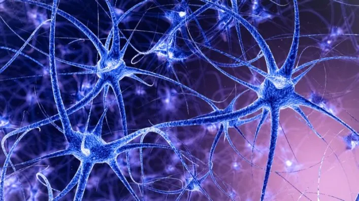
Telomere loss is a proposed reason we age

Telomere attrition—the wearing out of your chromosomes’ protective caps with age—is widely thought to be one of the major drivers of aging. With each division, telomeres shorten a little bit, and after 50-70 divisions, they become critically short. Once this threshold (the Hayflick limit) is hit, cells undergo replicative senescence, and their division comes to a grinding halt.

This limitation would quickly lead all cell lines to extinction if stem cells—the progenitors of each cell—didn’t express the enzyme telomerase, which allows them to replenish their telomeres and give rise to subsequent generations of new cells to replace dying ones.

Short telomeres have long been linked to age-related pathologies as well as to a number of other diseases not related to aging. Conversely, telomerase expression— and thus telomere elongation—where it’s not supposed to happen is what drives most human cancers. For these reasons, this field of research has been very active over the past few decades, and today, we’d like you to meet one of its big names.

A telomere pioneer

Dr. Blasco is a molecular biologist whose primary interests, since her university days, have been cancer and aging. After completing her Ph.D. at the Center of Molecular Biology in Madrid under the supervision of Professor Margarita Salas, she moved to Cold Spring Harbor, New York to work as a postdoc researcher in the lab of Dr. Carol Greider—the same Carol Greider who co-discovered telomerase with Elizabeth Blackburn in 1995. At the time, the link between cancer, aging, and telomerase was a mere hypothesis yet to be proven, and Blasco set to out clone the murine telomerase gene and generate telomerase-knockout mice [1] in order to study the effects that the lack of the enzyme would cause in the animals [2].

That same year, Dr. Blasco returned to Spain, her home country, where she started her own research group at the Centro Nacional de Biotecnología (National Biotechnology Centre) in Madrid. Eventually, she was recruited as the Head of the Telomeres and Telomerase Group at the Spanish CNIO—the National Center for Oncological Research. In addition to this position, which she still retains, she was also appointed Director of CNIO’s Molecular Oncology Program, then Vice-Director of Basic Research, and since 2011, she has been the CNIO Director. Additionally, she sits on the Scientific Advisory Board of the SENS Research Foundation together with many other luminaries in the field of aging research.

Dr. Blasco is also one of the co-authors of the well-known publication The Hallmarks of Aging [3], which was published in 2013 in the journal Cell, one of the most cited papers in biogerontology. This paper showed the essential lack of any meaningful biological distinction between “aging itself” and the pathologies of aging, and it reinvigorated the idea that a maintenance approach against aging may be the way to go.

She also co-authored the book Morir joven, a los 140: El papel de los telómeros en el envejecimiento y la historia de cómo trabajan los científicos para conseguir que vivamos más y mejor. (“Die young at 140: The role of telomeres in aging and the story of how scientists work to get us to live longer and better”)

Driving telomere research forward

The link between cancer, aging, and telomeres has always been the main target of Dr. Blasco’s laboratory. She and her team showed that telomere loss contributes to degenerative pathologies and that increased telomere fragility leads to higher cancer risk [4].

Other studies at her lab clarified the effect of telomerase activity and telomere length on the regenerative capacity of adult stem cells [5]; showed that the longest telomeres are a general feature of adult stem cell compartments and suggested that telomere loss may contribute to stem cell dysfunction with age [6]; found that cancer-resistant mice that overexpress telomerase experience delayed aging and have longer median lifespans [7]; and showed how DNA damage limits reprogramming of defective cells [8].

More recently, Dr. Blasco and her team developed a new model mouse for idiopathic pulmonary fibrosis that more accurately emulates the human disease and demonstrated that telomerase therapy reverses it [9].

What draws you to aging research over studying other fields?

For me, the aging research field is the most fascinating because aging is underlying the majority of diseases that are killing us today in developed countries: cancer, neurodegenerative diseases, lung, kidney, liver fibrosis, all this is the consequence of aging. So, understanding aging may be the way to one day be able to prevent or cure these diseases.

You and your team recently published a study showing that it is the rate of telomere shortening that predicts the lifespan of a species rather than the total length of telomeres. Does this discovery confirm the role of telomere attrition as a primary cause of aging rather than a consequence?

I think this study that means that telomeres are important in determining a species’ longevity. It’s not something that happens only in humans, where it’s already clear that in humans, telomere length matters, because there are humans that have mutations in telomerase, and they are going to have diseases associated with telomere shortening, which means that telomere shortening rates are very limiting for humans.

We didn’t know whether this was general to other species or only something particular to humans. In this study, we see that telomeres seem to matter across evolution in different species, from birds to mammals. It’s not the telomere length that matters but the rate of telomere shortening. So, we see that the rate of telomere shortening actually fits into a power law [regression] curve, and this predicts the longevity of a given species.

The study compared the telomeres of mice, goats, dolphins, gulls, reindeer, vultures, flamingos, elephants and humans, and it shows that species in which telomeres shorten faster live shorter lives. Could it mean that telomere shortening rate could be a suitable aging biomarker to test interventions against aging with?

I think so; telomere shortening rate is important in humans in order to determine if anyone is at risk of prematurely developing diseases associated with short telomeres. It’s not as important to measure telomeres once, because this probably is not going to be very informative, but the rate at which telomeres shorten may be more informative of the risk of developing any disease related to short telomeres.

The study also suggests that it is the rate at which telomeres shorten rather than the length that could be important. However, other studies show that telomeres form loops when they’re long enough, and they silence various age-associated genes and contribute to genomic stability through the telomere position effect (TPE). How does this reconcile with the idea that rate and not length is important in aging?

I think that this is a good point, but we already knew for instance that mice have very long telomeres and, still, they live very short lives, like two or three years. So this was already telling us that the length of the telomeres is not what is important to determine longevity; actually, later on, we found that the rate of telomere shortening of mice is like 1000 times faster than in humans. That’s where we had the idea of looking at other species and seeing whether there was any conservation of this idea that the rate of telomere shortening determines longevity.

So even though TPE may have an important role, it seems that throughout evolution, the rate of telomere shortening has been adjusted to determine lifespan, at least in the species that we included in the study. Of course, we should look at more species and see whether they still fit into this power law curve that we found.

Could negative effects from telomere shortening be related also to the release of shelterin proteins, since older humans do not have critically short telomeres across all tissues and cells?

It is a very interesting question. I think it will be worth looking at shelterins, because we have previously found and published that shelterin levels also go down with aging, and so it would be interesting to see what happens with shelterin proteins in different species. That would be a very interesting study.

Telomerase has many effects that are independent of telomeres. Can you see that they matter in aging?

Well, it is interesting because we have, in the past, demonstrated that we can extend the lifespan of mice by using telomerase, but it must be wild-type telomerase; if we use catalytically dead telomerase, then we don’t see this lifespan extension. So I would say that in order to see effects of telomerase in lifespan, you need it to be catalytically active telomerase, and this is the canonical pathway of telomerase, which is elongating the telomeres. At least in our hands, this is the mechanism by which telomerase can increase longevity: by extending short telomeres.

Some people suggest that mouse telomeres could work differently from humans because they are longer. So, what works in mice might not work the same in humans. Would you say that the telomere mechanisms and the dynamics are really that different between mice and people?

I think humans among mice are not that different. What is very different is the rate at which mice experience shortening telomeres, or in other words, mice are much worse than humans, at maintaining their telomeres. So, I think this makes a difference. So mice shorten their telomeres really fast, we still don’t understand why compared to humans, but now we also know that different species shorten their telomeres at different rates, and I think it’s very interesting to study that. We don’t know why. For example, the elephant and the flamingo have the same rate of telomere shortening and they have similar longevity; why is that? Then a mouse has a much faster rate of telomere shortening and a shorter longevity. I think this is a very interesting question to solve in the future.

Progeric mice are often criticized as not being representative of true aging and that if you perturb an aging hallmark as is done in these mice, then repairing that damage will, of course, increase their lifespan. What are your thoughts on using progeric mice strains as a way to inform the direction we take in aging research; are they still a useful model in that sense?

In general, I agree that a very fast model of aging may not represent physiological aging. In the case of telomeres, we know that also wild-type mice also suffer telomere shortening with aging, and I think this is a good model. Telomere shortening is something physiological that we and mice suffer, and it’s underlying aging and age-related diseases, so it’s a good model. A telomerase-deficient mouse, for instance, reflects a physiological process, which is telomere shortening associated with aging. I agree that if, for instance, you remove one shelterin, this causes very rapid aging, but I don’t think this is as physiological as telomerase deficiency, where we see something that we know is important for human aging.

Have you observed any change to the typical causes of death in the mice that have been treated with telomerase therapy?

At the end, the mice die of the same diseases. It’s that we are delaying the onset of these diseases which means we are delaying aging; aging is the cause of these diseases, and so if we delay aging, we delay the onset of these diseases. But, in the end, the mice die of the same diseases.

The goal is to translate telomerase therapies to mice that are not genetically modified to age faster, so the therapy can be tested against normal aging. Can you tell us about your experiences with telomerase therapy on normal-aging mice, and does this give us grounds for optimism in translating this therapy to humans?

Yes, actually the first experiment that we did was in normally aging mice. In this experiment that we did with telomerase gene therapy, we used wild-type mice, normal mice, and then treated them with telomerase, and we saw that we could delay aging and age-related diseases in normal mice. This was the first paper that we published in 2012 in EMBO Molecular Medicine.

Now, we are using mouse models for human diseases. We had to reproduce in mice pulmonary fibrosis associated with short telomeres, aplastic anaemia, because these are diseases that, in humans, originate due to short telomeres. So, we wanted to have mouse models in order to be able to try therapies, and the therapy that we have tried to use, of course, is telomerase activation, because the thing we are working on in my lab, mostly as a way to delay aging, is to activate telomerase.

In December 2016, Belmonte and his team successfully used OSKM partial cellular reprogramming in living mice to increase lifespan [10]. You and your team also published another example of OSKM induction in living animals published in January 2017 [11]. OSKM appears to rescue telomeres and restore their length. Some researchers also suggest that resetting telomeres can also reset the epigenetic alterations in the cell, as these two hallmarks are linked via TPE-OLD and other mechanisms. Do you think we need to target both telomere attrition and epigenetic changes in the cell, or can you get an effect by resetting one?

I think it is clear that when you reprogram and you go from a differentiated cell that has short telomeres to a pluripotent cell, an undifferentiated cell made by induced pluripotency, you are really changing everything. You are elongating the telomeres, activating telomerase, and changing the epigenetics. So, we are doing everything. This reprogramming is really able to reset telomeres to a pluripotent state, which is much longer, et cetera. So, it’s a way to start over.

Of course, using reprogramming right now, even though it showed some benefits in mice, has some concerns as well, because programming is something that cancer can use. Dedifferentiation of cells can produce teratomas.

The 2018 paper “Therapeutic effects of telomerase in mice with pulmonary fibrosis induced by damage to the lungs and short telomeres” [12] showed that telomerase therapy reverses the condition. How closely does this new fibrosis mouse model resemble the actual disease compared to older models, and does efficacy in this model mean that it can translate to human trials?

I think this model is a good model for the human pulmonary fibrosis associated with short telomeres; there may be other types of fibrosis that are not associated with short telomeres, but if a human has pulmonary fibrosis associated with short telomeres, then I think this is a good model, because these mice have short telomeres that per se cannot lead to fibrosis, but if you combine it with a low dose of a damaging agent to the lung, then they develop fibrosis. So, it’s a good model to combine short telomeres with the damaging agent to the lung, and this may be quite similar to what happens in humans, where short telomeres may synergize with smoking, pollution, or radiation.

What are the main challenges that telomerase therapy faces on the road to human use, and do you think we might see it reach humans in the next decade or so if all goes well?

I think it’s very likely that we will see telomerase activation for the treatment of telomere syndromes. These are diseases that are associated with mutations in telomerase, and the patients that have these diseases have no therapeutic options or very few. So I think there is likely that we will see telomerase activation therapies reaching clinical trials. This is my hope.

We have shown already in mouse models that telomerase activation can have therapeutic effects, slowing down the progression of diseases or even curing the disease. I think a logical step would be to go through clinical trials to see whether there is a clinical benefit in the treatment of these patients.

We would like to ask you a small favor. We are a non-profit foundation, and unlike some other organizations, we have no shareholders and no products to sell you. All our news and educational content is free for everyone to read, but it does mean that we rely on the help of people like you. Every contribution, no matter if it’s big or small, supports independent journalism and sustains our future.

Literature

[1] Blasco, M. A., Funk, W., Villeponteau, B., & Greider, C. W. (1995). Functional characterization and developmental regulation of mouse telomerase RNA. SCIENCE-NEW YORK THEN WASHINGTON-, 1267-1267.

[2] Blasco, M. A., Lee, H. W., Hande, M. P., Samper, E., Lansdorp, P. M., DePinho, R. A., & Greider, C. W. (1997). Telomere shortening and tumor formation by mouse cells lacking telomerase RNA. Cell, 91(1), 25-34.

[3] López-Otín, C., Blasco, M. A., Partridge, L., Serrano, M., & Kroemer, G. (2013). The hallmarks of aging. Cell, 153(6), 1194-1217.

[4] Martínez, P., Thanasoula, M., Muñoz, P., Liao, C., Tejera, A., McNees, C., … & Blasco, M. A. (2009). Increased telomere fragility and fusions resulting from TRF1 deficiency lead to degenerative pathologies and increased cancer in mice. Genes & development, 23(17), 2060-2075.

[5] Benetti, R., García-Cao, M., & Blasco, M. A. (2007). Telomere length regulates the epigenetic status of mammalian telomeres and subtelomeres. Nature genetics, 39(2), 243-250.

[6] Flores, I., Canela, A., Vera, E., Tejera, A., Cotsarelis, G., & Blasco, M. A. (2008). The longest telomeres: a general signature of adult stem cell compartments. Genes & development, 22(5), 654-667.

[7] Tomás-Loba, A., Flores, I., Fernández-Marcos, P. J., Cayuela, M. L., Maraver, A., Tejera, A., … & Viña, J. (2008). Telomerase reverse transcriptase delays aging in cancer-resistant mice. Cell, 135(4), 609-622.

[8] Marión, R. M., Strati, K., Li, H., Murga, M., Blanco, R., Ortega, S., … & Blasco, M. A. (2009). A p53-mediated DNA damage response limits reprogramming to ensure iPS cell genomic integrity. Nature, 460(7259), 1149-1153.

[9] Povedano, J. M., Martinez, P., Serrano, R., Tejera, Á., Gómez-López, G., Bobadilla, M., … & Blasco, M. A. (2018). Therapeutic effects of telomerase in mice with pulmonary fibrosis induced by damage to the lungs and short telomeres. Elife, 7, e31299.

[10] Ocampo, A., Reddy, P., Martinez-Redondo, P., Platero-Luengo, A., Hatanaka, F., Hishida, T., … & Araoka, T. (2016). In vivo amelioration of age-associated hallmarks by partial reprogramming. Cell, 167(7), 1719-1733.

[11] Marión, R. M., de Silanes, I. L., Mosteiro, L., Gamache, B., Abad, M., Guerra, C., … & Blasco, M. A. (2017). Common telomere changes during in vivo reprogramming and early stages of tumorigenesis. Stem cell reports, 8(2), 460-475.

[12] Povedano, J. M., Martinez, P., Serrano, R., Tejera, Á., Gómez-López, G., Bobadilla, M., … & Blasco, M. A. (2018). Therapeutic effects of telomerase in mice with pulmonary fibrosis induced by damage to the lungs and short telomeres. eLife, 7, e31299.

Neurons

T Cells Infiltrate the Aging Brain

As published in a recent study, researchers have discovered that neural stem cells are impeded by the invasion of T cells, immune cells that are not normally present in the neural stem cell niche [1].

The neural stem cell niches

Our brains contain neural stem cells (NSCs); like their name suggests, these cells are responsible for the formation of new neurons within the brain. This process, which continues throughout life, is known as neurogenesis. These stem cells live in particular niches, which contain a panoply of different cell types, including stem cells in different phases of development and multiple types of immune cells. However, the researchers discovered a startling fact: the brains of older mice contain many specific immune cells known as T cells, while the brains of younger mice contain very few – and, as the study explains, this is true for humans as well.

T cells and interferon-γ

NSCs have receptors for a signaling molecule called interferon-γ, which discourages them from performing their normal function, and the researchers suggest that this is likely to be the main reason why they fail to proliferate in aged brains. The invading T cells, which express interferon-γ, proliferate through clonal expansion; the researchers mention that this particular method of cellular reproduction is normally done in response to the presence of an antigen, a harmful molecule that causes immune cells to react. If there is such an antigen in the brains of aged mammals, what it is and why it is there are questions that have yet to be answered.

Abstract

The mammalian brain contains neurogenic niches that comprise neural stem cells and other cell types. Neurogenic niches become less functional with age, but how they change during ageing remains unclear. Here we perform single-cell RNA sequencing of young and old neurogenic niches in mice. The analysis of 14,685 single-cell transcriptomes reveals a decrease in activated neural stem cells, changes in endothelial cells and microglia, and an infiltration of T cells in old neurogenic niches. T cells in old brains are clonally expanded and are generally distinct from those in old blood, which suggests that they may experience specific antigens. T cells in old brains also express interferon-γ, and the subset of neural stem cells that has a high interferon response shows decreased proliferation in vivo. We find that T cells can inhibit the proliferation of neural stem cells in co-cultures and in vivo, in part by secreting interferon-γ. Our study reveals an interaction between T cells and neural stem cells in old brains, opening potential avenues through which to counteract age-related decline in brain function.

Conclusion

This particular study has discovered a possibly vital part of why neural stem cells in aging brains fail to proliferate, even though the reason for the T cell invasion remains unknown and the full relationship between the immune system and NSCs has not yet been studied. However, by simply announcing the fact of their presence, this study has opened the door to new lines of research in this area, which may one day lead to therapies that block the effects of interferon-γ, discourage T cells from invading NSC niches, or remove the fundamental reason why T cells invade such niches at all.

We would like to ask you a small favor. We are a non-profit foundation, and unlike some other organizations, we have no shareholders and no products to sell you. All our news and educational content is free for everyone to read, but it does mean that we rely on the help of people like you. Every contribution, no matter if it’s big or small, supports independent journalism and sustains our future.
Literature

[1] Dulken, B. W., Buckley, M. T., Negredo, P. N., Saligrama, N., Cayrol, R., Leeman, D. S., … & Wyss-Coray, T. (2019). Single-cell analysis reveals T cell infiltration in old neurogenic niches. Nature, 1.

Sergey Young interview

Sergey Young and the Longevity Vision Fund

We recently had the opportunity to interview Sergey Young, a board member of XPRIZE and the creator of the $100m Longevity Vision Fund.

When did you first become interested in healthy life extension, and why?

My interest began with a routine visit to a doctor. Five years ago, at the age of 42, my blood tests – which I neglected for 7 years, thinking I was in perfect health – showed that my cholesterol was extremely high, putting me at risk of one of the most common killers: heart disease. 

The only treatment offered by my doctor at the time was to take statins (cholesterol-reducing medication) for the rest of my life. However, this seemed unnatural and potentially dangerous for the body, and I definitely did not want to “live” on a pill forever. I refused to accept this as the only option (the doctor even made me sign a waiver for refusing treatment) and kept pushing for alternatives. Eventually, the doctor suggested I try a Mediterranean-style diet (based around healthy fats, cutting out sugar, etc.), which worked in bringing my cholesterol down to a normal range without any medication at all. 

It’s a pity that doctors, even well-meaning ones, do not start with dietary changes first. Since then, I developed an interest in diet and a lifestyle-based approach to health and longevity, and hope I can share this knowledge with as many people as possible. 

However, it was the meeting with Peter Diamandis last year in Vatican City at a conference on regenerative medicine, which was also attended by the Pope, that really kickstarted my mission in longevity. Peter is such an inspiring individual, and his XPRIZE Foundation served as a great example of how you can make a difference on a large scale. It was the perfect platform to make my interest in longevity serve to the benefit of society as a whole. 

In your opinion, what are the most important reasons why the pursuit of healthy longevity should be a priority for human society?

Our lives are like “Groundhog Day”: we spend most of our lives working until we decline into old age and illness – possibly without having had the time or health to enjoy life to the fullest. 

However, pursuing healthy longevity can potentially extend our lives by 25% or more. Having an extra 25 years of lifespan gives us the opportunity to pursue our dreams, spend more time with our children and grandchildren, and do the things that really matter – but that we have not had time for.

This cause is so close to your heart that you’re actually writing a book about it—no easy task. Is it your first book?

Yes, this is my first book. Hopefully not my last. 

I am putting a lot of time and effort into making longevity as practical and easy to read about as possible. I see it as a way of helping as many people as possible learn about life extension and making their lives more longevity-friendly. If this leads to follow-on books, covering longevity diets, or longevity exercises, for example, I would be happy to fulfill the readers’ requests.

What made you decide that it was time for you to write one?

When I first got into longevity, there were only two types the longevity books available: either 300-page books with a single hypothesis drawn out, or “encyclopedias” that were nearly impossible for the general population to read without a specialized degree. 

That’s why I decided to write “Growing Young: Simple Guide to Age Reversal” – something well-researched and comprehensive, yet simple and engaging for the general reader.   

I understand that we’re some way from finding your book on the shelves; can you give us a sneak peek by telling us a little about what topics you’re going to cover?

The book is heavy-packed with a lot of varied content. Here is a taster of some of the topics included:

  • Technological longevity breakthroughs – what is already available that can extend our lifespans by up to 10 years, and technologies that could emerge in the near future to extend our lifespans even further
  • Ethical trade offs of living to 200
  • Economics of longevity  

Has a release date, or time frame, for the book been decided yet?

Our planned release date is spring 2020 

You’re the founder of the Longevity Vision Fund, whose goal is to accelerate longevity breakthroughs and make them affordable for as many as possible as soon as possible. Many people fear that life extension would be only for the rich, so what’s your plan to make your vision come true, especially regarding affordability?

The focus of Longevity Vision Fund is on affordable and accessible technology. As with every area of technology, it undergoes a democratization process, becoming progressively more affordable to a wider range of people. The mission of Longevity Vision Fund is to enable and speed up the democratization of longevity-related technology by investing in companies and services that have the potential to become scalable, accessible and affordable for the general population. 

LVF is a rather new initiative; what has it achieved thus far?

Longevity Vision Fund was launched in February 2019, but we have already come a long way. For example, we raised most of the capital in just 3 months. We have assembled an impressive Advisory Board of five leading longevity scientists: Aubrey De Gray, Vadim Gladyshev, Joao de Magalhaes, Richard Faragher, and Morten Scheibye-Knudsen. 

We also entered into a collaboration partnership with BOLD Capital (Peter Diamandis’ fund), and LVF has already invested in four companies to date. 

What areas of life extension research, or life extension in general, will be LVF’s primary focus?

We like to invest in fields such as AI, diagnostics, wearables & devices, stem cell treatments, and organ regeneration.

Does LVF have a roadmap, or a tentative time frame, describing when specific goals should ideally be achieved?

As mentioned above, we have already achieved very significant milestones for a fund that has just been launched last year. Our main goal for the future is achieving our mission in stimulating progress in longevity breakthroughs to make them affordable to as many people as possible.

This is a progressive goal, and it would be impossible and irresponsible to make specific claims exactly as to when and by how much lifespans will be expanded. 

Let’s talk about XPRIZE a little bit. You’ve been on the XPRIZE Innovation Board for over a year now. What is your job as a member of this board?

In addition to being on the XPRIZE Innovation Board, I am also the Development Sponsor of Longevity XPRIZE. My job is to define the strategy of Longevity XPRIZE, help choose the areas of most impact, and attract and unite the brightest minds that could lead solutions to the world’s biggest problems – aging being one of them, since it affects us all. 

What led you to join XPRIZE?

As you probably already know, I am extremely passionate about longevity, and I want to use it to make a difference in the world. I want to help people live longer, healthier, and happier lives. Since meeting Peter Diamandis, XPRIZE has become the perfect high-impact platform for working on the world’s biggest issues

Speaking of XPRIZE, you took part in its recent Future of Longevity brainstorming session, in which our president, Keith Comito, had the pleasure to meet you. Can you share your impressions of the event?

As the Development Sponsor, I could be biased, so I would be more interested in hearing Keith Comito’s thoughts! 

On a serious note, I thought it turned out to be a great, collaborative event with a constellation of over 50 of the world’s Longevity Leaders. I am grateful to everyone who came and made it such a success.

The goal of the brainstorming session was to design a Longevity XPRIZE that may further catalyze the development of a thriving longevity industry; our readers already know something about it from Keith’s article, but, as an insider, is there anything more you could share with us, such as when the prize might launch or if more proposals are being evaluated?

We expect to launch next year, and the exact date will probably be announced during XPRIZE Visioneering in October. I am really looking forward to it, since it is a great big event where ideas for future XPRIZE initiatives are discussed. There are also lots of amazing people in attendance – last year, Pharrell Williams and Eric Schmidt were there, for example. 

Besides the upcoming Longevity XPRIZE, are you involved in other XPRIZE initiatives?

Yes, I was also involved in the Global Learning XPRIZE, where I coached five finalists in the competition. The result of their work was to give children in African countries a tool to learn English (or their native language, Swahili) by themselves, without an adult, in just 12 months, using an app.

Speaking again of affordability, even if life extension medicine eventually becomes affordable, we can’t expect that to happen overnight. How long, in your opinion, will it take before it’ll be cheap enough to be widespread?

Life extension is a very complex issue – if there were a simple solution, evolution would probably already have taken care of it.

I expect that various technological breakthroughs, scientific research, and the work of many people around the world (including our small team at Longevity Vision Fund) will collectively contribute to the continuous evolution of longevity and the prolongation of human lifespans. However, as I mentioned before, it would be impossible to predict when exactly this will happen and exactly how widespread we can make life extension medicine.

Let me ask a few personal questions again. Many people, even life extension enthusiasts, are not convinced that major breakthroughs will happen during their lifetimes. How about you; do you expect to “make the cut”?

I visualize myself living to 200, and whether I get to live to 200 or not, is arbitrary. The mindset of living to 200 myself stimulates me to live, work, and contribute to longevity to the best of my ability, since I am more passionate about improving the lives of others than focusing just on my own. As Peter Diamandis says, “the best way to become a billionaire is to help a billion people.”

Besides that, I definitely expect to see significant breakthroughs during my own lifetime and even bigger breakthroughs in the coming generations.

In your XPRIZE biography, I read that you’re a “happy co-founder of a diversified portfolio of 4 kids.” I take it as a funny way to say you’ve got four children; do you talk to them about life extension? What do they think of it?

I do talk about longevity and life extension to my two oldest kids: the 20-year-old and the 8-year-old. However, they see longevity as their father’s passion rather than something they are interested in as a topic. That’s understandable – hardly anyone under 30 thinks about aging. 

As for my two youngest kids aged 5 and 1, I tell them “longevity bedtime stories”: tales involving “good” healthy foods, and avoiding “evil” sugar, sweet drinks, fast food, etc.

You make no mystery of your wish to live to two hundred. Is it just two hundred or at least two hundred?

To most people, both sound equally unbelievable, so with my binary thinking, I just focus on the number “200”.

Many people out there wonder what we’d do if we lived much longer than the current status quo. What would you do with that kind of extra time?

Adding an extra 25 years to our lifespans gives us fundamentally new options: spending more time with children and grandchildren, redefining our lives, getting new careers, and working on solving the world’s problems.

I would, of course, spend more time with my family and continue my work in solving the problems and diseases of aging. But this is a question that everyone can ask themselves. What would you do if your life were extended by a quarter?

As a final question, are there other causes than life extension that you find very important or are personally involved in?

Apart from focusing on doing good on a global scale, as with my work on longevity, I like to make a difference on a smaller scale as well. While it’s important to work on solving global problems, it’s just as important to connect and support communities locally. For example, every New Year’s, my wife, my kids and I give away festive food sets to families in need. We started with 100 families, and last year, that number reached 300.  

I would also like to end this question by thanking LEAF and, in particular, its president, Keith Comito. Thank you for your dedication in promoting life extension and for building such a fantastic and comprehensive longevity platform like LEAF!

We would like to ask you a small favor. We are a non-profit foundation, and unlike some other organizations, we have no shareholders and no products to sell you. All our news and educational content is free for everyone to read, but it does mean that we rely on the help of people like you. Every contribution, no matter if it’s big or small, supports independent journalism and sustains our future.

A Step Closer to Regenerating the Aging Thymus

Researchers from the Monash Biomedicine Discovery Institute have made progress in the quest to rejuvenate the aging immune system by identifying the factors responsible for the age-related decline of the thymus. 

The thymus shrinks as we age

The thymus is one of the most important organs in the body, and it is where thymocytes produced in the bone marrow travel to become new T cells before being trained in the lymph nodes to become the defenders of the adaptive immune system. However, as we get older, the thymus increasingly turns to fat and starts to shrink, causing its ability to produce new T cells to fall dramatically. This process is known as thymic involution and actually begins shortly after puberty, so this is one aspect of aging that begins fairly early in life, although it is many decades later before its decline causes serious health issues.

The fall in production of new T cells from the thymus results in a decline of the adaptive immune system and is part of the collective decline of the immune system called immunosenescence. The end result of immunosenescence is that your body is no longer able to mount an effective defense against diseases and is inappropriately activated, leading to dysfunction and persistent inflammation. This inflammation contributes to inflammaging, a chronic smoldering background of low-grade inflammation, which other age-related sources contribute to as well.

The decline of the thymus has been linked to cancer risk, which rises dramatically as we age as part of the immunosenescence model of cancer. Immunosenescence is also strongly correlated with multiple age-related diseases, which is probably no surprise, given that the aged immune system is no longer able to respond effectively or even appropriately to invading pathogens.

While it has long been known that the thymus shrinks with age, the exact mechanisms behind this involution were not totally clear.

The downward spiral

The new study has shed new light on what drives the loss of thymus function in old age and the resulting failure of immune cell production [1]. Published in the journal Cell, the new study lays the foundation for developing therapies that may help the thymus to recover its ability to produce T cells and combat infections and diseases. 

The researchers show that BMP4 and activin are growth and differentiation factors that are key to the self-renewal and differentiation of thymic epithelial stem cells and that a change in their levels due to aging causes the loss of these epithelial cells. This loss then results in the decline of T cell production in the thymus, ultimately leaving us open to infection and disease. This study is a world first and finally identifies the core reason why we experience the loss of thymic epithelial stem cells and the molecules and mechanisms that drive this process. 

The researchers’ next step will be to find ways to reverse this decline and effectively turn the thymus back on again so that T cell production resumes. The researchers believe that age-related changes in the thymus can be reversed, and they are now investigating to see if a therapy to regenerate thymic epithelial stem cells can be created. 

A key feature of immune functional impairment with age is the progressive involution of thymic tissue responsible for naive T cell production. In this study, we identify two major phases of thymic epithelial cell (TEC) loss during aging: a block in mature TEC differentiation from the pool of immature precursors, occurring at the onset of puberty, followed by impaired bipotent TEC progenitor differentiation and depletion of Sca-1lo cTEC and mTEC lineage-specific precursors. We reveal that an increase in follistatin production by aging TECs contributes to their own demise. TEC loss occurs primarily through the antagonism of activin A signaling, which we show is required for TEC maturation and acts in dissonance to BMP4, which promotes the maintenance of TEC progenitors. These results support a model in which an imbalance of activin A and BMP4 signaling underpins the degeneration of postnatal TEC maintenance during aging, and its reversal enables the transient replenishment of mature TECs.

Conclusion

Effective targeting of these pathways could lead to restoration of not only thymic T cell production but also T cell diversity. This has the potential to dramatically improve health in older people, who generally have poor or nearly non-functional T cell production as a result of thymic involution. If a therapy can be developed that regenerates the thymus, restoring its function to more youthful levels, it has the potential to improve the health of older people and reduce their risk of contracting a myriad of age-related and non-age-related diseases. 

A number of companies and research teams are now focused on rejuvenation of the thymus, and the evidence to date, including animal studies and even tentative human trials, leads us to believe that regenerating the thymus is a quite possible near-future prospect.

We would like to ask you a small favor. We are a non-profit foundation, and unlike some other organizations, we have no shareholders and no products to sell you. All our news and educational content is free for everyone to read, but it does mean that we rely on the help of people like you. Every contribution, no matter if it’s big or small, supports independent journalism and sustains our future.

Literature

[1] Lepletier, A., Hun, M. L., Hammett, M. V., Wong, K., Naeem, H., Hedger, M., … & Chidgey, A. P. (2019). Interplay between Follistatin, Activin A, and BMP4 Signaling Regulates Postnatal Thymic Epithelial Progenitor Cell Differentiation during Aging. Cell Reports, 27(13), 3887-3901.

The Rejuvenation Market in Singapore

Singapore has one of the fastest-aging populations in the world. Senior citizens 65 years old or older are expected to make up almost half of Singapore’s population by 2050. Unfortunately, this swelling population is spending more time living with sickness, even though they live longer. While average lifespans have been extended, healthspans have not. [1] Singaporeans have an impressive average life expectancy of 84.8 years, but an average Singaporean born in 2017 is predicted to spend the last ten and a half years in sickness, compared to how a Singaporean born in 1999 is likely to spend only nine twilight years in deteriorating health.

This is becoming a massive concern for the Singaporean government because of the financial strain that this is imposing on Singapore’s budget. Having the world’s second-lowest birth rate coupled with a rapidly aging population means that the ratio of working adults to senior citizens is quickly shrinking. In 2007, there were 6.9 working adults for every senior citizen. By 2030, there will be 2.3 working adults per senior citizen.

In under a decade, Singapore’s healthcare budget more than doubled from S$4 billion in 2010 to S$10 billion in 2017. [2] Among the developed nations of the world, Singapore has a reputation for being one of the most fiscally conservative; there’s a socio-political stigma against the term “welfare state” in Singapore. Since its unprecedented independence in 1965, Singapore has had a general zeitgeist of “every man for himself,” as we are a nation with no natural resources. Our highly-educated workforce, along with our strategic geographical location, is the primary resource undergirding our knowledge-based economy.

Traditional values meet future medicine

However, the overwhelming majority of Singaporeans tend to balk at the suggestion of life extension, even the most liberal youngsters of my generation. The idea of life extension is still associated with speculative science fiction or fantastic, magical thinking. 84.6% of Singaporeans are religious, and religious conservatism still holds a degree of sway over Singapore’s mainstream socio-political zeitgeist. The thought of curing aging is seen as going against the natural order of things and makes most of the public squeamish.

Dr. Jan Gruber has personally sat on board discussions with the executives of Singapore’s Ministry of Health. As he anticipated, the response from the rest of the board was dismissive. At an annual budget meeting, the majority of the suggestions offered pertained to managing symptoms of aging through such means as hiring more foreign nurses or employing AI nurses. However, Dr. Gruber was the only one present to suggest actually tackling the problem at its core by treating biological aging itself. [3]

In spite of this, one of the Singaporean government’s current short-term goals is to encourage our senior citizens to remain in the workforce for as long as possible. Singapore has recently extended its official retirement age from 62 to 67. Our government has also begun to offer more incentives for employers who hire workers over the age of 54. For workers who are 55 or older earning up to S$4,000 a month, the government will provide wage offsets. [1]

Singapore’s stance towards curing biological aging can appear to be somewhat schizophrenic. Healthcare officials appear to openly reject it on the grounds of tradition and conservatism while still flirting with the idea that curing the hallmarks of aging is crucial to slashing its healthcare costs and maintaining its productivity. Singapore currently sits on this knife’s edge, but as its window of opportunity shrinks, it is becoming increasingly open to embracing biological cures for the hallmarks of aging.

Primed for the rejuvenation market

While there is a pervasive international stereotype that Singapore’s conservative political environment makes it inhospitable to cutting-edge medical therapies, Elena Milova begs to differ. The crux of Singapore’s willingness to embrace rejuvenative therapies is how these therapies are marketed. As the director of the Lifespan Extension Advocacy Foundation (LEAF), Elena is well-versed in the importance of framing cutting-edge radical medical therapies in the least provocative way possible. She is of the opinion that Singapore’s political conservatism is not analogous with medical conservatism. An article from a mainstream Singaporean newspaper, TODAY, states, “there is a need to increase life expectancy while simultaneously cutting down the amount of time people spend in poor health.” [1] This is entirely consistent with LEAF’s goal of making life extension therapies medically viable and publicly available.

In his most recent interview with Adam Ford on Science, Future, and Technology, the esteemed Aubrey de Grey gave a shout out to Brian Kennedy: “I want to give a real shout out here to Singapore, who have made the extremely smart move of hiring my good friend Brian Kennedy to run this very promising, prestigious centre for healthy life extension. I believe that’s going to be a massive global player in this area right now. Singapore is going to lead Asia into actually becoming a true partner in the crusade against aging.” [4] This is certainly a note for optimism, indeed.

In 2017, Professor Kennedy was featured in an article in The Straits Times, Singapore’s top newspaper. The article explicitly stated that Kennedy’s research was centered on “delaying aging.” Kennedy aims to establish a clinical approach to human aging in his lab at NUS, focusing on “compressed morbidity.” [5] Compression of morbidity refers to the reduction of the percentage of time that people spend in ill health due to aging by increasing their healthspans to make up a greater portion of their increasing lifespans. Kennedy is now working on bringing Singaporean clinicians and researchers together to begin human clinical trials. Kennedy himself has called the Singaporean government “progressive-minded” and “capable of thinking 10 years ahead.” “This island could be a testing ground for intervention to try to keep people healthy.”

Hopefully, this trend will perpetuate itself in the decades to come. As a self-sufficient knowledge-driven economy, Singapore must tackle its aging problem or risk losing its cherished competitive edge. Singapore has been ranked as the “most competitive economy in the world” in 2019 by IMD, outstripping both the US and Hong Kong. In IMD’s own words of praise, Singapore’s rise to this position can be accredited to its “advanced technological infrastructure, the availability of skilled labor, favorable immigration laws, and efficient ways to set up new businesses.” [6] All of these, save for our “favorable immigration laws”, are heavily dependent upon maintaining a highly educated working population with sharp cognitive capabilities.

The opportunity for Singapore to become a thriving regional market for rejuvenative therapies now seems to be growing ever-riper, and Kennedy is hoping that more of the coming generation of gerontologists will join him there soon.
We would like to ask you a small favor. We are a non-profit foundation, and unlike some other organizations, we have no shareholders and no products to sell you. All our news and educational content is free for everyone to read, but it does mean that we rely on the help of people like you. Every contribution, no matter if it’s big or small, supports independent journalism and sustains our future.

Literature

[1] Siau, M. E. (2017). Elderly to make up almost half of S’pore population by 2050: United Nations. TODAY Online.

[2] Ho, T. (2018). Singapore’s Ageing Population: The Financial Implications Of Our Country Growing Old. Dollars and Sense Online.

[3] Gruber, J. (2019). Personal interview with Sarah Lim.

[4] De Grey, A. (2019). Aubrey de Grey – Robust Mouse Rejuvenation only 3 years away? An update on Anti-Aging Research. YouTube video interview by Adam Ford.

[5] Goy, P. (2017). ‘Healthspan’ hasn’t grown as much as lifespan, says expert. The Straits Times Online.

[6] IMD Online. (2019). Singapore topples United States as world’s most competitive economy. IMD News.

Cytomegalovirus Infection Linked to Metabolic Syndrome

Today, we want to highlight a new publication that looks at cytomegalovirus (CMV) and its potential role in the development of metabolic syndrome.

What is cytomegalovirus? 

CMV is part of the β-subfamily of herpes viruses, a family of viruses that are believed to have been co-evolving with their hosts for around 180 million years [1]. CMV is spread primarily by exposure to infected secretions and subsequent mucosal contact, which then leads to the acute infection of various cell types. After this acute infection, in which the virus spreads, it enters a dormant state in the body.

In the United States, around one in three children are believed to be infected with CMV by the age of five. More than fifty percent of adults by the age of 40 are thought to be infected with CMV. CMV is a persistent virus; once it enters a person’s body, it stays there for life.

CMV infection is asymptomatic, meaning that it is latent and produces no symptoms; lying dormant in the cell, it waits for activation under certain set conditions [2].

Reactivation from this dormant state is believed to happen periodically, so it requires constant, lifelong immune surveillance to keep the body free from disease. Patients who have compromised immune systems are at greater risk for CMV reactivation, and this can lead to an increase of morbidity and mortality [3].

The need for lifelong immune surveillance to keep CMV in check puts a drain on the immune system as a whole, as ever-greater numbers of immune cells have to specialize in countering the virus. This draws off valuable immune cells, which could otherwise be fighting different pathogens, and contributes to the weakening of the immune system. This, combined with an ever-declining supply of fresh immune cells produced by sources such as the thymus and hematopoietic stem cells, ultimately results in the weakening of the immune system, greatly reducing its ability to react to threats.

In order to reactive and awaken from this dormant state, CMV, like all herpesviruses, progresses through an ordered cascade that starts with the expression of intermediate-early genes, which serve as a trigger for further (early and late) viral gene expression [4]. Studies have shown that CMV periodically expresses these intermediate-early genes at random during this dormant (latent) period [5-6]. This means that there is always a background level of CMV reactivation.

Once these initial genes are expressed, the cycle moves to express genes for host manipulation, DNA replication, and viral packaging. The expression of the intermediate-early genes is closely linked to pro-inflammatory transcription factors, such as nuclear factor kappa beta (NF-κB), TNFα, and interleukin-1β. Not surprisingly, these inflammatory signals can reactivate CMV as a result of this link [7-8].

In this way, CMV actively responds to its environment, and its behavior is dictated by the local inflammatory situation. Essentially, this means that injuries and any other sources of inflammation could awaken CMV from its dormant state just by the presence of inflammatory signals, and it uses this inflammation as a way to increasingly reproduce and spread throughout the body.

CMV and metabolic syndrome

Deregulated nutrient sensing is one of the hallmarks of aging, and metabolic syndrome is a part of this process as well as being a possible precursor to type 2 diabetes. Factors such as being sedentary or overweight can provoke metabolic syndrome to develop, particularly when excessive amounts of visceral fat are present. Being overweight when older is particularly problematic and is linked to senescent cell accumulation, another cause of aging, and systemic inflammation [9].

A new study explores the relationship between CMV infection, fat tissue, and the development of metabolic syndrome [10].

Cytomegalovirus (CMV) is a ubiquitous herpesvirus infecting most of the world’s population. CMV has been rigorously investigated for its impact on lifelong immunity and potential complications arising from lifelong infection. A rigorous adaptive immune response mounts during progression of CMV infection from acute to latent states. CD8 T cells, in large part, drive this response and have very clearly been demonstrated to take up residence in the salivary gland and lungs of infected mice during latency. However, the role of tissue resident CD8 T cells as an ongoing defense mechanism against CMV has not been studied in other anatomical locations. Therefore, we sought to identify additional locations of anti-CMV T cell residency and the physiological consequences of such a response. Through RT-qPCR we found that mouse CMV (mCMV) infected the visceral adipose tissue and that this resulted in an expansion of leukocytes in situ. We further found, through flow cytometry, that adipose tissue became enriched in cytotoxic CD8 T cells that are specific for mCMV antigens from day 7 post infection through the lifespan of an infected animal (> 450 days post infection) and that carry markers of tissue residence. Furthermore, we found that inflammatory cytokines are elevated alongside the expansion of CD8 T cells. Finally, we show a correlation between the inflammatory state of adipose tissue in response to mCMV infection and the development of hyperglycemia in mice. Overall, this study identifies adipose tissue as a location of viral infection leading to a sustained and lifelong adaptive immune response mediated by CD8 T cells that correlates with hyperglycemia. These data potentially provide a mechanistic link between metabolic syndrome and chronic infection.

Conclusion

This is yet more support for regular exercise and a balanced diet in order to avoid weight gain and the formation of an excessive amount of visceral fat.

We would like to ask you a small favor. We are a non-profit foundation, and unlike some other organizations, we have no shareholders and no products to sell you. All our news and educational content is free for everyone to read, but it does mean that we rely on the help of people like you. Every contribution, no matter if it’s big or small, supports independent journalism and sustains our future.

Literature

[1] McGeoch, D. J., Cook, S., Dolan, A., Jamieson, F. E., & Telford, E. A. (1995). Molecular phylogeny and evolutionary timescale for the family of mammalian herpesviruses. Journal of molecular biology, 247(3), 443-458.

[2] Fields, B. N., Knipe, D. M., Howley, P. M., & Griffin, D. E. (2007). Fields virology. 5th.

[3] Crough, T., & Khanna, R. (2009). Immunobiology of human cytomegalovirus: from bench to bedside. Clinical microbiology reviews, 22(1), 76-98.

[4] Hermiston, T. W., Malone, C. L., Witte, P. R., & Stinski, M. F. (1987). Identification and characterization of the human cytomegalovirus immediate-early region 2 gene that stimulates gene expression from an inducible promoter. Journal of Virology, 61(10), 3214-3221.

[[5] Grzimek, N. K., Dreis, D., Schmalz, S., & Reddehase, M. J. (2001). Random, Asynchronous, and Asymmetric Transcriptional Activity of Enhancer-Flanking Major Immediate-Early Genes ie1/3 andie2 during Murine Cytomegalovirus Latency in the Lungs. Journal of Virology, 75(6), 2692-2705.

[6] Henry, S. C., & Hamilton, J. D. (1993). Detection of murine cytomegalovirus immediate early 1 transcripts in the spleens of latently infected mice. Journal of Infectious Diseases, 167(4), 950-954.

[7] Hummel, M., Zhang, Z., Yan, S., DePlaen, I., Golia, P., Varghese, T., … & Abecassis, M. I. (2001). Allogeneic transplantation induces expression of cytomegalovirus immediate-early genes in vivo: a model for reactivation from latency. Journal of Virology, 75(10), 4814-4822.

[8] Cook, C. H., Trgovcich, J., Zimmerman, P. D., Zhang, Y., & Sedmak, D. D. (2006). Lipopolysaccharide, tumor necrosis factor alpha, or interleukin-1β triggers reactivation of latent cytomegalovirus in immunocompetent mice. Journal of virology, 80(18), 9151-9158.

[9] List, E. O., Jensen, E., Kowalski, J., Buchman, M., Berryman, D. E., & Kopchick, J. J. (2016). Diet-induced weight loss is sufficient to reduce senescent cell number in white adipose tissue of weight-cycled mice. Nutrition and healthy aging, 4(1), 95-99.

[10] Contreras, N. A., Sitnik, K. M., Jeftic, I., Coplen, C. P., Čičin-Šain, L., & Nikolich-Žugich, J. (2019). Life-long control of cytomegalovirus (CMV) by T resident memory cells in the adipose tissue results in inflammation and hyperglycemia. PLoS pathogens, 15(6), e1007890.