An in-depth preprint published in bioRxiv has thoroughly described how separate tissues lose proteostasis in different ways.
Segmental progeria
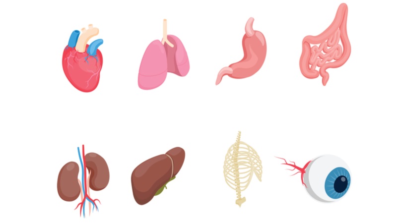
As a model of accelerated aging, the researchers used an already short-lived vertebrate, the killifish, with a mutation that impairs its ability to create telomerase. This results in telomere attrition and genomic instability [1], two other hallmarks of aging, just as it does in humans. The affected tissues are the ones with the most proliferative capacity, such as the gut, blood, skin, and testes.
This research compares these sorts of tissues in old wild-type, young wild-type, and old telomerase-deficient killifish, focusing on specific protein aggregates.
Aggregated proteins were tissue-specific
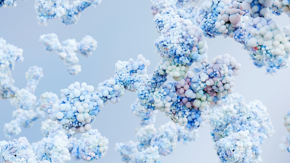
For the first part of their experiment, the researchers compared protein accumulation between old and young killifish, examining the brain, gut, heart, liver, muscle, skin, and testis. They found that there were many more differences than similarities between tissues in protein accumulation (as determined by tissue lysation), aggregation (determined by molecular weight), and propensity for aggregation, the latter two of which were increasingly tissue-specific. Some of these aggregated proteins are chaperones that are responsible for proteostasis, and this is not the first time that we’ve seen broken proteostasis machinery making the problem worse. Many of the increased proteins are also linked to specific diseases, including myopathies, mitochondrial dysfunction, mental retardation, and cirrhosis.
One key surprising finding is that the proteins that accumulated in the telomerase mutants and their naturally aging counterparts were nearly all completely different. Similarly to the first experiment, very few of the proteins that were differentially regulated in progeria and normal aging were the same between tissues. The researchers enumerated the very few proteins that formed aggregates in both progeria and normal aging, and none of these proteins were the same between tissues.
Interestingly, in many critical tissues and protein aggregates that were heavily upregulated in telomerase-deficient mutants, there was no significant difference between naturally aged and young killifish. As expected, the lamin protein LMNA, which is a factor in human progeria, was increased in the skin of the telomerase mutants, both in amount and in aggregation.
Conclusion
While extremely in-depth, this research also showed multiple interesting facts that are fundamental to how aging works on the molecular level.
The first is that it reinforced well-known information about the interplay between the hallmarks of aging. The loss of proteostasis does not occur in a vacuum, unrelated to other hallmarks. Rather, the aggregation of proteins is strongly dependent on the dysregulation of the machinery that keeps these proteins from aggregating.
The second is that models of accelerated aging often do not resemble actual aging, even in such models as killifish. If this fact holds true for more humanlike animals, such as mice, it brings into question the value of using such progeric mutants for research.

The third is the importance of telomerase and telomere attrition. While no single hallmark of aging is ultimately responsible for all of its aspects, it is clear that telomerase deficiency leads to a significant number of downstream problems, including problems related to entirely different hallmarks of aging.

Finally, the differences between protein accumulation in tissues point to the true complexity of aging. If we want to stop the aggregation of hundreds of different proteins, the only practical way to do this is to repair the fundamental mechanisms at their source.
Literature
[1] O’sullivan, R. J., & Karlseder, J. (2010). Telomeres: protecting chromosomes against genome instability. Nature reviews Molecular cell biology, 11(3), 171-181.
[2] Kirwan, M., & Dokal, I. (2009). Dyskeratosis congenita, stem cells and telomeres. Biochimica Et Biophysica Acta (BBA)-Molecular Basis of Disease, 1792(4), 371-379.