A new article published by the International Journal of Molecular Sciences sheds light on the differences between the extracellular matrix (ECM) of healthy people and people in end-stage heart failure.
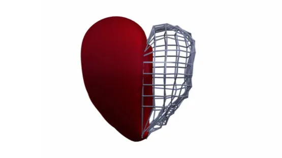
The key to the heart could be its ECM
Regeneration of heart tissue following a heart attack has been a challenging goal for scientists. At best, most interventions currently only limit damage after the blockage occurs but are not capable of reversing it. Of particular interest, the ECM of the heart is its physical scaffolding and provides a structure on which the cells of the heart can live. Donor heart tissues can be decellularized and used either as a biomaterial in heart regeneration strategies or to study heart physiology and pathology. However, much is unknown about the various components of heart ECM, the effect it has on cells, and how it changes with age and pathology. Recent research published by a joint collaborative effort in Italy has shed light on each of these issues. [1]
The researchers decellularized ECM from two different groups of human donors. The first group came from younger (average age of 36) patients who died from causes unrelated to the heart and were considered healthy heart donors. The second group consisted of heart donors who had pathologies. They were much older (average age of 59) and had end-stage heart failure, and their hearts were collected during heart transplant surgery. After decellularization, the researchers compared cardiac stromal primitive cells cultured on the two groups of ECM as well as cells cultured on 2D cell culture plastic.
The cells cultured on both ECM groups showed greater heart-specific behaviors when compared to cells cultured on 2D. Healthy ECM also demonstrated further benefits over pathological ECM. Cells cultured on the pathological ECM expressed more fibrotic factors, such as PDGF-AA, TGF-β2, and IGF-2. They also demonstrated an increase in compounds associated with a damage response, including GAT4A and hydrogen peroxide. Furthermore, cells cultured on healthy ECM exhibited more cardioprotective cytokines, such as HGF, and expression of angiogenic factors.
Additional studies exposed endothelial cells to conditioned culture media. This conditioned media contains paracrine factors excreted by the cells from the original ECM comparison experiments. The researchers found that endothelial cells cultured using conditioned media from the healthy ECM resulted in faster and greater capillary formation than cells that received conditioned media from the pathological ECM group.
Abstract
In conclusion, CPCs exposed in vitro to dECM-PH from HF myocardium displayed partially reduced cardiac commitment drive, defective paracrine support to angiogenesis, and increased release of pro-fibrotic cytokines, despite displaying some features of a rescue-like mechanism, similar to those described in vivo in pathological conditions. These observations shed novel insights on the crosstalk between ECM remodeling and cardiac stromal primitive cells, suggesting also caution and limitations in the use of non-healthy decellularized myocardium for cardiac tissue engineering approaches.
Conclusion
Overall, 3D cell culture has, many times over, been shown to be superior to 2D culture, but many novel conclusions can be drawn by the research’s comparisons between cells cultured on healthy and pathological ECM. This research suggests that ECM from unhealthy donors may have some limitations in cardiac regeneration strategies. Additionally, it provides evidence for potential mechanisms by which the ECM mediates cardiac cell behavior and some possible pathways for pathological cardiac remodeling. However, the authors did not discuss the difference in age between their two groups of tissue donors, instead primarily attributing their findings to the results of end-stage heart failure. Since the two groups of donors also differed significantly by age, further research is needed to disentangle which differences are caused by heart failure and which are the result of aging tissue.
Literature
[1] Belviso, I., Angelini, F., Di Meglio, F., Picchio, V., Sacco, A.M., … & Chimenti, I. (2020). The Microenvironment of Decellularized Extracellular Matrix from Heart Failure Myocardium Alters the Balance between Angiogenic and Fibrotic Signals from Stromal Primitive Cells. International Journal of Molecular Sciences, 21(21), 7903. https://doi.org/10.3390/ijms21217903