Neurons from Stem Cells Alleviate Parkinson’s Disease in Rats
- This experiment represents a substantial advancement over previous efforts.

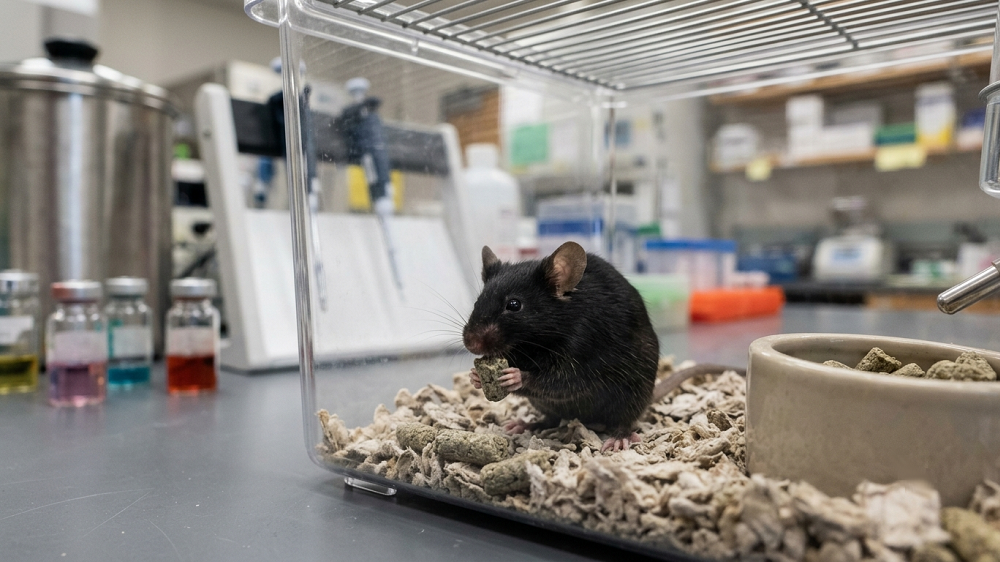
In a new study, a group of researchers report on their success in priming stem cells to differentiate into dopaminergic neurons in rat brains. Loss of these neurons is a major cause of Parkinson’s disease [1].
Less neurons, more Parkinson’s
Neuronal degeneration is the culprit behind several age-related disorders, including Parkinson’s disease (PD), which affects one million people in the US alone. PD stems from the gradual depletion of dopaminergic neurons, a subset of neurons that reside in a small part of the brain called the substantia nigra. Despite constituting only a fraction of all neurons, dopaminergic neurons perform the important task of dopamine production. Low levels of this vital neurotransmitter cause the motor symptoms of PD, such as tremor and stiffness.
Several decades ago, the prevalent opinion was that the central nervous systems of adults do not generate new neurons, and this misconception still lingers among the general public. Today, we know that neurogenesis does occur in the human brain but only in some parts of it. Simple organisms, on the other hand, often possess enviable neuroregenerative abilities.
Helping MSCs become neurons
Mesenchymal stem cells (MSCs) are multipotent stem cells found in bone marrow and several other tissues, and they can differentiate into neurons [2]. Attempts have been made to deploy MSCs against neurodegenerative diseases, with mostly unsatisfactory results. In this new study, scientists describe a method of priming MSCs in vitro in order to boost their differentiation into viable neurons in rat brains.
The compound used for priming is 22(R)-hydroxycholesterol (22-HC). Cholesterols are usually mentioned in the context of cardiovascular diseases, but these chemicals and their derivatives play an important role in multiple processes, such as hormone production. As much as 25% of the body’s total cholesterol is located in the brain, where it participates in the maturation and survival of dopaminergic neurons [3].
Previous studies have shown that oxysterols such as 22-HC can facilitate the differentiation in vitro of human embryonic stem cells (hESCs) into dopaminergic neurons. The main advantage of this new study is that it uses human MSCs instead of hESCs. MSCs have been recently gaining in popularity, being much more available and also safer (due to their pluripotency, ESCs are prone to forming ghastly tumors called teratomas). The researchers used MSCs of slightly different kinds sourced from three human tissues: bone marrow, adipose tissue, and dental pulp.
Detecting a cell type is not always an easy task, as scientists have to rely on morphological and chemical analysis. Soon after being treated with 22-HC, the MSCs began to strongly express several neuron-specific chemical markers. Moreover, their shapes became very neuron-like, with distinctive somas (bodies), axons, and neurites. 22-HC also increased the expression of transcription factors responsible for maturation and survival of dopaminergic neurons. The scientists rigorously checked for other neuronal markers, such as mitochondrial activity: neurons are extremely energy-hungry, and producing such amounts of energy requires a concerted effort of swarms of mitochondria [4]. MSCs from dental pulp were the clear winner in neurogenerative potential.
Motor function restored
The primed cells were then transplanted into the brains of rats suffering from a surgically induced PD-like condition. Tests showed that the cells had engrafted well. The rats’ performance was assessed prior to the PD-inducing surgery, after the surgery, and then after the transplantation. Three subgroups of rats received primed MSCs of the three types (one for each subgroup). Another three subgroups received naïve (untreated) MSCs. The control group received a sham treatment, an inactive transplant that did not contain MSCs. Following the PD-inducing surgery, the rats’ condition predictably deteriorated, showing distinct motor symptoms. Primed MSCs, especially the ones from dental pulp, demonstrated spectacular results, restoring rats’ motor function almost to pre-surgery levels. Naïve MSCs, on the other hand, made little difference, in line with previous experiments.
The researchers contend that their results are unprecedented in several aspects, such as the percentage of stem cells that differentiated into neurons (as much as 80%). The type of stem cells used and the protocol itself are highly cost-effective, which can help in advancing this new technique.
Conclusion
Stem cells are the stars of longevity research because, theoretically, they can provide us with an unlimited supply of young cells for our tissues. This study makes an important step by successfully creating functioning dopaminergic neurons and transplanting them into rat brains. Record-breaking results, combined with the pressing need to counter Parkinson’s disease, mean that we might see clinical trials soon.
Literature
[1] Singh, M., Jain, M., Bose, S., Halder, A., Nag, T. C., Dinda, A. K., & Mohanty, S. (2021). 22 (R)-hydroxycholesterol for dopaminergic neuronal specification of MSCs and amelioration of Parkinsonian symptoms in rats. Cell Death Discovery, 7(1), 1-17.
[2] Urrutia, D. N., Caviedes, P., Mardones, R., Minguell, J. J., Vega-Letter, A. M., & Jofre, C. M. (2019). Comparative study of the neural differentiation capacity of mesenchymal stromal cells from different tissue sources: An approach for their use in neural regeneration therapies. PloS one, 14(3), e0213032.
[3] Sacchetti, P., Sousa, K. M., Hall, A. C., Liste, I., Steffensen, K. R., Theofilopoulos, S., … & Arenas, E. (2009). Liver X receptors and oxysterols promote ventral midbrain neurogenesis in vivo and in human embryonic stem cells. Cell stem cell, 5(4), 409-419.
[4] Misgeld, T., & Schwarz, T. L. (2017). Mitostasis in neurons: maintaining mitochondria in an extended cellular architecture. Neuron, 96(3), 651-666.