A recent paper published in Immunity has described the accumulation of exhausted microglia in the brains of people who are vulnerable to Alzheimer’s, potentially spurring and worsening the disease.

Proteostasis and inflammation
Alzheimer’s is characterized by amyloids, but these misfolded protein accumulations are not its only characteristic. Neuroinflammation, which is linked to helper cells called microglia, occurs as well [1]. Microglia play multifarious roles in Alzheimer’s, which can be helpful or harmful [2].
The well-known Apolipoprotein E (APOE) gene is also key to Alzheimer’s. APOE3 is the most common, people with APOE2 are far less likely to get Alzheimer’s, and people with APOE4 are far more likely to get it [3]. APOE also strongly affects immunity, including responses to cancer [4]. In this experiment, the researchers sought to determine if this immune response was part of why people with APOE4 are so much more likely to get Alzheimer’s disease.
A mouse model of APOE alleles
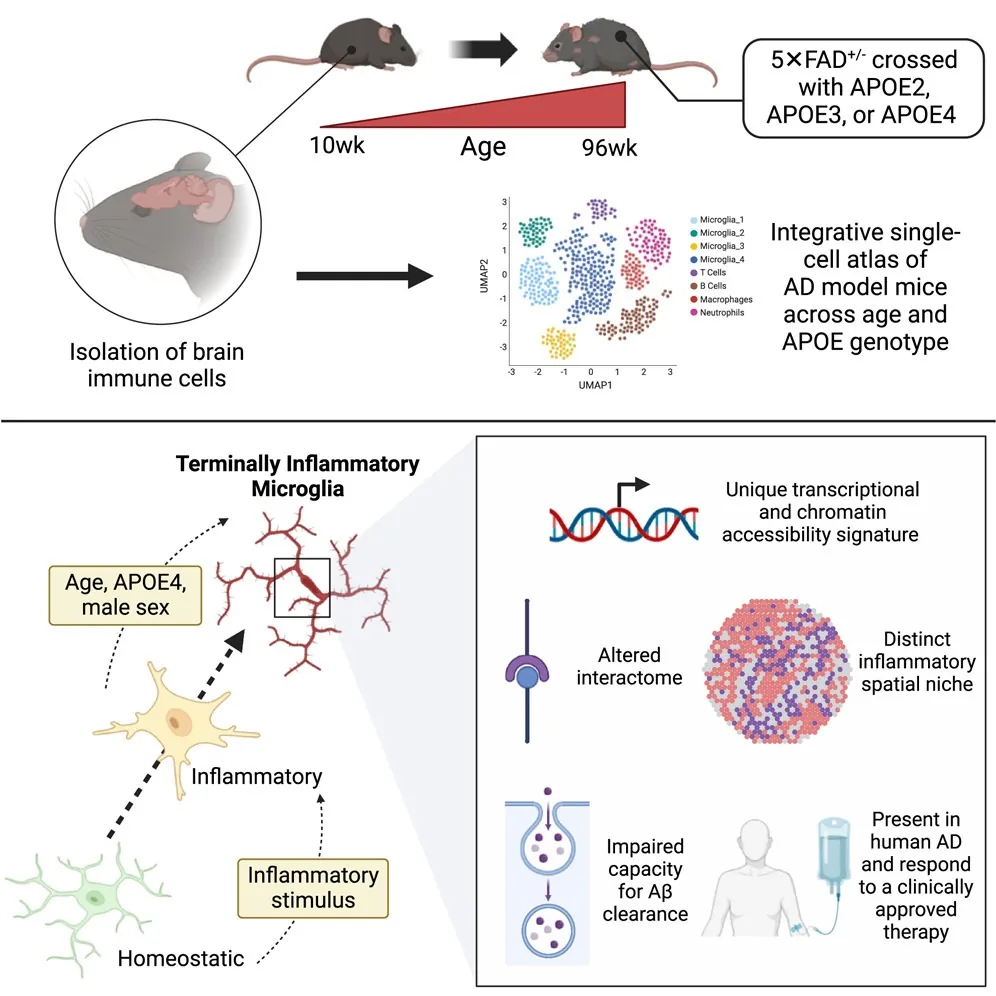
Mice don’t naturally get Alzheimer’s, so the researchers crossbred a common Alzheimer’s model mouse with various strains of mice that produce the three common human APOE alleles. These experiments were performed on cells derived from the mice when they were 96 weeks old, which is roughly equivalent to 80-year-old humans.
By using RNA sequencing to differentiate the populations of microglia, the researchers found a very specific subpopulation of terminally inflammatory microglia (TIMs), which were characterized both by their consistent expression of inflammation-related genes and their presence only in aged mice.
However, not all TIMs are created equal. The mice with APOE2 were heavy in “effector-hi” TIMs, and the mice with APOE4 were heavy in “effector-lo” TIMs. The “effector-lo” TIMs were far heavier in stress markers. All TIMs were inflammatory in nature, but the specific inflammatory factors they secreted were unusual for microglia, being rich in some and far less rich in others. “Effector-lo” and “effector-hi” TIMs also differed from one another in this respect.
Also pertains to people
With these murine results in hand, the researchers turned to RNA sequencing results derived from human brains and compared them. They found that TIM-related genes were enriched both in people with Alzheimer’s and in people who had the APOE4 allele but did not have the disease. These robust results were consistent across ten different sample sets, regardless of how the data was derived. Further analysis suggested that TIMs followed the same trends between people and Alzheimer’s-prone mice.
Looking closely at brains donated by people who had suffered from Alzheimer’s disease, the researchers found that APOE4 was correlated with increases both in TIMs and their proximity to gray matter. Additionally, more TIMs were found in regions with more amyloid-beta plaques.
They also found that TIMs, in general, are worse than normal microglia in dealing with amyloid-beta proteins. However, the researchers found that all effector-lo TIMs, which are more frequently found with APOE4, are worse at clearing this protein than normal microglia. They also found that the effector-lo TIMs with the APOE4 allele are uniquely poor at this task.
While this research is unlikely to lead directly to any new therapies, it does suggest targets and options for researchers interested in targeting various aspects of Alzheimer’s disease. If microglia in the TIM state can be targeted for removal or replaced with healthier microglia, it might represent a new front in fighting against this brain-destroying disease.
Literature
[1] Hansen, D. V., Hanson, J. E., & Sheng, M. (2018). Microglia in Alzheimer’s disease. Journal of Cell Biology, 217(2), 459-472.
[2] St-Pierre, M. K., VanderZwaag, J., Loewen, S., & Tremblay, M. È. (2022). All roads lead to heterogeneity: The complex involvement of astrocytes and microglia in the pathogenesis of Alzheimer’s disease. Frontiers in Cellular Neuroscience, 16, 932572.
[3] Liu, C. C., Kanekiyo, T., Xu, H., & Bu, G. (2013). Apolipoprotein E and Alzheimer disease: risk, mechanisms and therapy. Nature Reviews Neurology, 9(2), 106-118.
[4] Ostendorf, B. N., Bilanovic, J., Adaku, N., Tafreshian, K. N., Tavora, B., Vaughan, R. D., & Tavazoie, S. F. (2020). Common germline variants of the human APOE gene modulate melanoma progression and survival. Nature medicine, 26(7), 1048-1053.


