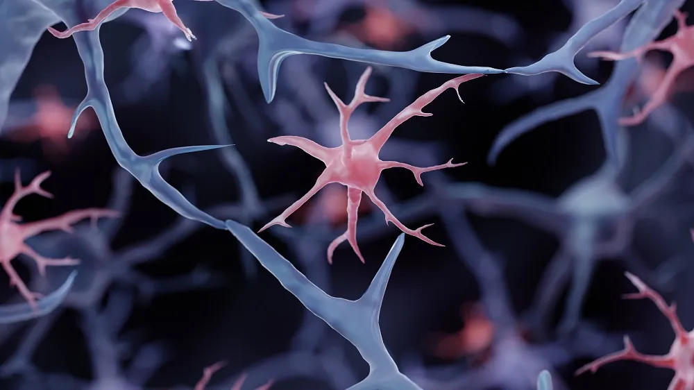
In Cell Stem Cell, researchers have described how genetically engineered microglia can be used to deliver therapeutic proteins to the brain.
The blood-brain barrier
Delivering therapeutic compounds to the brain has a concern that is absent for other organs: the blood-brain barrier (BBB), which retains strict controls over the kinds of compounds that can reach the brain and thus protects it from contamination. Deterioration of the BBB can lead to significant neurological problems [1]. However, the BBB can also prevent therapeutic compounds from entering it, which has represented a significant problem in drug delivery [2].
Getting around the BBB may involve invasive methods, such as directly injecting compounds or neural stem cells (NSCs) into brain tissue, or virus-based gene therapy. However, all of these methods have their own limitations and concerns; NSCs may form tumors [3], and virus-based approaches may cause inflammation [4]. If the goal is simply to deliver a protein into the brain, these sorts of methods may do more harm than necessary.
These researchers, therefore, have chosen mature microglia, helper cells in the brain, as their therapeutic vector. These cells do not form tumors [5], and previous work has found that they can properly engraft into models [6].
Responsive to plaques
The researchers developed a mouse model that does not create its own microglia and accumulates amyloid plaque. They also engineered induced pluripotent stem cells (iPSCs) to create microglia (iMG) that generate neprilysin, an enzyme that degrades amyloid beta, upon sensing these plaques through the receptor CD9.
Initial analysis suggested that this approach worked. These iMG only expressed CD9 alongside plaques rather than throughout the brain. Further work found that this extended to neprilysin as well; the researchers also noted that a secreted-neprilysin (sNEP) approach, as opposed to simply generating neprilysin on the membrane (NEP), led to more distribution of this therapeutic compound.
This secretion approach also improved the microglia’s ability to clear these plaques through phagocytosis in vitro. Compared to ordinary human microglia, NEP microglia consumed amyloids at 1.5 times the speed, and sNEP microglia consumed amyloids twice as fast. These benefits were found in the mouse model as well; these microglia were often able to penetrate and degrade amyloid beta, decreasing amyloid burden in general and reducing the sizes of amyloid plaques in the brain.
There were benefits for synapses, which disintegrate rapidly in these model mice [7], as measured by the crucial synaptic protein synaptophysin (SYP). NEP microglia had no statistically significant effect on SYP; however, sNEP microglia restored levels of this protein approximately to those of a control group. These levels were found to be significantly correlated with the expression of neprilysin.
Like human Alzheimer’s patients, these model mice develop astrogliosis [8], an inflammatory increase in the prevalence of astrocytes. In the hippocampus, mice given sNEP microglia enjoyed a significant reduction in GFAP, a protein related to astrogliosis; however, this reduction was not to the level of the control group.
Fortunately, other proteins that are known to be targets of neprilysin were not affected in unrelated regions of the brain, showing that the localization was effective in this model. The researchers also found that “widespread engraftment of sNEP-microglia is not necessary to achieve brain-wide reductions in amyloid species”; targeting the hippocampus and cortex with precise injections of iMG were enough.
The reduction in amyloid was also accompanied by a tremendous reduction in inflammation. Key inflammatory proteins, including interleukins, had levels indistinguishable from those of a control group, despite these proteins normally being highly elevated in this Alzheimer’s model.
This is extremely early-stage research, and the authors describe their study as a proof of principle. Nearly every element of this study was carefully controlled at the genetic level; wild-type mice were not involved. Whether or not iPSC microglia can be made suitable for human use is still an open question, but if successful, this approach may open the door for the delivery of otherwise unfeasible drugs.
Literature
[1] Profaci, C. P., Munji, R. N., Pulido, R. S., & Daneman, R. (2020). The blood–brain barrier in health and disease: Important unanswered questions. Journal of Experimental Medicine, 217(4), e20190062.
[2] Terstappen, G. C., Meyer, A. H., Bell, R. D., & Zhang, W. (2021). Strategies for delivering therapeutics across the blood–brain barrier. Nature Reviews Drug Discovery, 20(5), 362-383.
[3] Amariglio, N., Hirshberg, A., Scheithauer, B. W., Cohen, Y., Loewenthal, R., Trakhtenbrot, L., … & Rechavi, G. (2009). Donor-derived brain tumor following neural stem cell transplantation in an ataxia telangiectasia patient. PLoS medicine, 6(2), e1000029.
[4] Prasad, S., Dimmock, D. P., Greenberg, B., Walia, J. S., Sadhu, C., Tavakkoli, F., & Lipshutz, G. S. (2022). Immune responses and immunosuppressive strategies for adeno-associated virus-based gene therapy for treatment of central nervous system disorders: current knowledge and approaches. Human gene therapy, 33(23-24), 1228-1245.
[5] Zong, H., Parada, L. F., & Baker, S. J. (2015). Cell of origin for malignant gliomas and its implication in therapeutic development. Cold Spring Harbor perspectives in biology, 7(5), a020610.
[6] Chadarevian, J. P., Hasselmann, J., Lahian, A., Capocchi, J. K., Escobar, A., Lim, T. E., … & Blurton-Jones, M. (2024). Therapeutic potential of human microglia transplantation in a chimeric model of CSF1R-related leukoencephalopathy. Neuron, 112(16), 2686-2707.
[7] Oakley, H., Cole, S. L., Logan, S., Maus, E., Shao, P., Craft, J., … & Vassar, R. (2006). Intraneuronal β-amyloid aggregates, neurodegeneration, and neuron loss in transgenic mice with five familial Alzheimer’s disease mutations: potential factors in amyloid plaque formation. Journal of Neuroscience, 26(40), 10129-10140.
[8] Oakley, H., Cole, S. L., Logan, S., Maus, E., Shao, P., Craft, J., … & Vassar, R. (2006). Intraneuronal β-amyloid aggregates, neurodegeneration, and neuron loss in transgenic mice with five familial Alzheimer’s disease mutations: potential factors in amyloid plaque formation. Journal of Neuroscience, 26(40), 10129-10140.


