×

The Blog

Building a Future Free of Age-Related Disease

Fighting Aging and Hurricane Harvey: AgeMeter Campaign Extension and Matching Funds Announced

Aging remains the number one killer, taking the lives of 100,000 people per day due to a number of severe age-related diseases like cancer, heart disease, and Alzheimer’s. Still, this is not the only danger we have to protect ourselves from: the recent developments in the US with the devastating Hurricane Harvey clearly show how important is to unite our efforts to gain more control over our lives.

This storm has influenced our activities at Lifespan.io in an unexpected and unprecedented way: Elliott Small, the project leader of AgeMeter, was called in by the Federal Emergency Management Agency (FEMA) to help with the disaster relief effort, helping local people to deal with the consequences of the storm. During this time he had to postpone all campaign activities in order to lend a hand.

It is partly for this reason that we at Lifespan.io have decided to extend the AgeMeter campaign by two weeks, to allow the project team to make up for lost time and hopefully achieve 100% funding! If you considered donating to AgeMeter to get this useful device to measure functional age developed, now you can do it until September 15.

We are also happy to announce the support of two initiatives for matching donations for this campaign.  The first is an anonymous donor who will match the next $1,000 donated to the campaign, making your contribution count double.  

The community at LongeCity has also decided to support the campaign with a matching fundraiser to get an AgeMeter for use in their affiliate labs!

We are very proud of Elliott for helping those in desperate need and confident that our community will carry his important project to the success it deserves.


MitoSENS Update August 2017

The following is an update from MitoSENS scientist Dr. Matthew “Oki” O’Connor:

Hi, everyone! Long time, no update – my fault!

As you all saw, our manuscript that was supported by Lifespan.io donors was published last fall. Dr. Boominathan and I have been presenting our results at conferences around the world, and I can tell you that they have been very well received.

The second part of the project has been completed (testing a library of MTSes on ATP6), and that does not seem to have solved the problem of substantially increased ATP6 expression and import.

We have, however, learned more about the problem and have made progress on solving it. We’ve discovered that there was a problem at both the mRNA expression level and at the protein level.

We think we’ve solved the mRNA problem, but we’re still working on the protein problem. The mRNA solution is a bit of a long story, so I’m going to save the details for a future update, but it’s pretty cool.

Additionally, it’s been an exciting summer. The highlight of our summer is always the SRF Summer Scholars program, in which talented undergraduate biology students join our various projects for 12 weeks.

This summer has been twice as special for the Mito project because we got not one but two amazing interns to pioneer new work. This is due to a timely grant from Michael Antonov (one of the visionary co-founders of Oculus) that allowed us to expand operations this summer to push a new project that I’ll talk about in my next update. But first…

Optimizing the Allotopic Expression of ATP6 to Mitochondria in Mutant Cells

Jasmine Zhao joined us this summer from UCLA, where she will be a senior this fall. In Jasmine’s words:

This summer, my project will be conducted under the mentorship of Dr. Amutha Boominathan and Dr. Matthew O’Connor at the SRF Research Center in Mountain View. The goal of this project is to design and test different constructs that can potentially improve the allotopic expression of ATP6 to mitochondria in mutant cell lines.

Mitochondria are double-membrane bound organelles that provide energy in the form of ATP to power the biochemical reactions of a cell. Unlike other organelles, however, mitochondria have their own DNA separate from the nucleus, and 13 out of those 37 genes encode for oxidative phosphorylation complex proteins.

Due to possible leakage of the high-energy electrons of the respiratory chain, which results in the formation of reactive oxygen species, the oxidative stress mitochondrial-DNA (mtDNA) is subjected to can lead to mutations, aging, and cell death. For instance, the ATP6 gene encodes for subunit a of the Fo structural domain of ATP synthase, also known as Complex V. The Fo structural domain is embedded in the inner membrane of the mitochondria and contains the membrane proton channel that allows for the synthesis of ATP.

Mutations of ATP6 have been implicated in different human diseases that affect neural development, vision, and motor movement, such as Leigh syndrome and Neuropathy, Ataxia, and Retinitis Pigmentosa (NARP).


Thanks

Oki

Want to know more?

You can learn more about this project at the SENS website here!

False Positives in Senescent Cell Detection

Macrophages with markers of cellular senescence may not be senescent 

Researchers commonly determine the presence of senescent cells through the use of the p16ink4a (p16) and β-galactosidase biomarkers. Senescent cells are known to exhibit both of these biomarkers, but it seems that they are not the only cells that do this, and the concern is that therapies that target these biomarkers may also be removing cells that are not senescent.

A study was published recently, and it shows that some cells that are not necessarily senescent share these two biomarkers [3]. This new study is by the same research team that investigated cellular senescence in the immune system last year with a focus on macrophages [4].

So, could this mean that older people have a partially senescent macrophage population, or does it mean that these cells are not harmful and just exhibit some of the same markers that senescent cells do?

This new study suggests that the latter may be the case, although it has not yet been determined if the macrophages exhibiting these commonly shared biomarkers are harmful.

So is this an issue for senolytics?

Fortunately, we are aware of no research group developing senolytics that target β-galactosidase. The use of β-galactosidase is limited to assessing the presence and numbers of senescent cells but is not actually the target of therapies.

However, p16 is a potential issue, as at least one company, Oisin Biotechnology (we interviewed its CEO here) does use this biomarker to target senescent cells. Its approach uses target genes and their level of expression in order to target cells for destruction. Oisin will no doubt be doing its due diligence in determining the effect of removing p16-positive macrophages as part of its ongoing development process.

That said, one of the first proof-of-concept studies used p16 as the target for senescent cell removal, and positive health results were seen [5]. So, it is worth bearing in mind that, in this case, p16 cells, regardless of the source, were removed in a broad stroke in this study, yet positive benefits were observed.

Therefore, it may be that case that removing them is not a significant problem, and earlier studies strongly suggest that removing senescent cells is beneficial, even taking into account the risk of removing non-senescent cells.

Conclusion

The paper’s authors conclude that the importance of senescent cells in the aging process and removing senescent cells is not in question, and their findings do not suggest that developing senolytics is a bad idea.

The points raised here are that we need to better refine how we identify senescent cells and that we need to improve our understanding of how to determine if a cell is senescent. Therefore, we need to develop better biomarkers to identify senescent cells.

Considering the research community’s intense focus on senescent cells and the considerable funding behind the development of senolytics, those gaps in our knowledge will likely not remain in place for long.

Given the pace of progress in this particular area, it is reasonable to believe that such issues may be resolved in the near future once more studies are concluded.

Literature

[1]  López-Otín, C., Blasco, M. A., Partridge, L., Serrano, M., & Kroemer, G. (2013). The hallmarks of aging. Cell, 153(6), 1194-1217.

[2]  van Deursen, J. M. (2014). The role of senescent cells in ageing. Nature, 509(7501), 439-446.

[3] Hall, B. M., Balan, V., Gleiberman, A. S., Strom, E., Krasnov, P., Virtuoso, L. P., … & Leonova, K. I. (2017). p16 (Ink4a) and senescence-associated β-galactosidase can be induced in macrophages as part of a reversible response to physiological stimuli. Aging.

[4] Hall, B. M., Balan, V., Gleiberman, A. S., Strom, E., Krasnov, P., Virtuoso, L. P., … & Leonova, K. (2016). Aging of mice is associated with p16 (Ink4a)-and β-galactosidase-positive macrophage accumulation that can be induced in young mice by senescent cells. Aging (Albany NY), 8(7), 1294.

[5] Baker, D. J., Wijshake, T., Tchkonia, T., LeBrasseur, N. K., Childs, B. G., Van De Sluis, B., … & Van Deursen, J. M. (2011). Clearance of p16Ink4a-positive senescent cells delays ageing-associated disorders. Nature, 479(7372), 232.


How Misfolded Proteins Cause Age-related Diseases

Coming from a fusion of the words ‘protein’ (a molecule that a cell uses as a machine or scaffolding) and ‘stasis’ (meaning to keep the same), the term ‘proteostasis’ can essentially be simplified into “Each function reliant on proteins is running as it should. There are enough proteins to serve a function, and the concentrations of proteins are being maintained at healthy levels.”

Proteostasis is the process that cells perform in order to have all their proteins functioning properly; this, in turn, allows cells to work properly. Since cells are the building blocks of our bodies, when they work properly, we are healthy.

When proteostasis is not maintained, cells become dysfunctional and can die; this failure can lead to aging, cancer, neurodegenerative diseases, inflammation, developmental defects, and other problems. The loss of proteostasis is thought to be a primary reason we age, and we discuss how this happens in more detail here.

Why do cells need proteins?

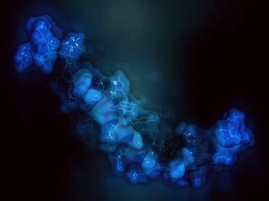
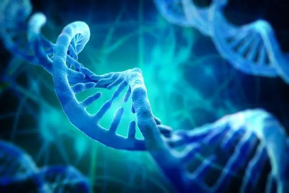
Proteins are perhaps the most important molecules in cells. DNA’s main purpose is to act as a ‘protein library’, storing and passing on information about how to build proteins.

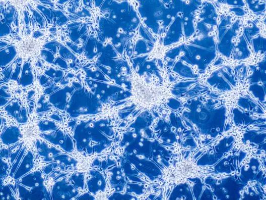
If a cell does something, you can be fairly certain that a protein is behind it at some level. In a neuron, for instance, proteins are important for the creation, movement, release, and recognition of neurotransmitters, the chemicals that allow brain cells to talk to each other. Proteins are also used to help cells determine which direction is up or down, left or right, or front and back.

For instance, to discern its front from its back, a cell uses 3 proteins, BCD, HB-M, and NOS. The side to become the ‘front’ becomes high in BCD and HB-M, while production of NOS is blocked in that region. Meanwhile, the side to become the back becomes high in NOS, with BCD and HB-M inhibited in that region. When the cell divides, this balance shifts ever so slightly, depending on the direction that the two ‘daughter cells’ are facing.

Through detecting how the proteins are distributed, the cell knows where it is in the body and so produces the right structures in the right places. It is because of this system that our noses only form on the front of our heads, for instance; a breakdown in this system could lead to a nose forming on the back of your head or not forming on your face.

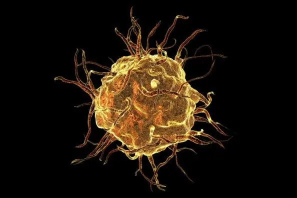
Proteins are also vital for allowing the sheer amount of chemical reactions necessary in a cell to be carried out. It may not feel like it, but 37 degrees C (body temperature) is extremely cold for chemistry; hardly anything can happen at this temperature. To get around this, our bodies turn protein strings into tiny machines called ‘enzymes’ that physically ram molecules together, forcing chemical reactions to happen at temperatures at which they never would naturally.

Not only does this allow our cells to cause the reactions necessary for their own survival and life in general, it also allows our cells to control these reactions. Through disabling a protein’s creation, for instance, a cell can turn off a reaction and use that disabled reaction as a signal to activate (or deactivate) something else in the cell.

Through increasing the amount of an enzyme being produced, a cell can speed up a reaction, and through decreasing the amount of enzyme being produced, it can slow it down. This perfect control is one method that our bodies use to maintain  ‘homeostasis’; in other words, it helps us to keep our bodies within the narrow conditions necessary for life. For example, homeostasis is how we stop ourselves from overheating or freezing, from becoming too acidic or too alkaline, and from having too high blood pressure or too low. In summary, homeostasis is the most important process in the body for its own survival.

So where does it all go wrong?

Unfortunately, proteostasis can be lost during many disease states, including, but not limited to, aging[1]. Various toxins can, for instance, inhibit the proteins in a person’s cells[2]. This inhibition can cause a loss of proteostasis.

Incurable infectious agents (which cause disease) known as prions can also form spontaneously out of any newly forming protein in any healthy individual at any time, acting just like zombies – converting any healthy version of the protein that they were meant to be into another prion upon touch, causing death through loss of proteostasis.

During the aging process, proteostasis is lost for other reasons; for instance, while a protein is being assembled, it can sometimes fold incorrectly, forming a useless glob of junk, which can accumulate unless the cell gets rid of it through autophagy, breaks it down with a general ‘recycling bin’ (the proteasome), or refolds it into its proper shape through the use of ‘chaperone proteins’ in what is known as “chaperone mediated folding”.

Exposure to heat (otherwise known as “heat shock”) can also cause a protein to misfold. When the human body detects this heat, it responds by using the SIRT1 family of proteins to ‘turn on’ ‘heat shock proteins’, such as hsp70. These proteins are chaperone proteins; they can refold the proteins to their previous shape and prevent them from clumping together into harmful masses called ‘aggregates’.

Additionally, cross-linking between proteins can occur[3], which causes these proteins to stick together, increasing their stiffness and causing them to lose some of their function.

Why does this matter?

Numerous age-related diseases have their roots in the loss of proteostasis, including macular degeneration[4] (a form of blindness), the increased risk of heart attack and stroke that comes with age, Alzheimer’s disease[5], and Parkinson’s disease[6]. Finding ways to restore proteostasis could potentially allow us to prevent or reverse many of these age-related diseases.

How can we restore proteostasis?

There are a number of promising routes towards the restoration of proteostasis; we will discuss a few below.

The first approach, AmyloSENS, was originally funded by the SENS Research Foundation at UT Houston at the Sudhir Paul lab and has now moved to the hands of biotechnology company Covalent Biosciences. This company is currently working on immunotherapeutic candidates that target amyloid proteins for treating central nervous system and systemic amyloidosis.

The GlycoSENS program, which is related to amyloids, is being developed by the SENS Research Foundation and researchers at Yale. This program is focused on tackling the protein crosslinks that accumulate in tissue as we age. This cross-linking creates a weblike structure of fused proteins outside of cells, leading to loss of tissue mobility, elasticity, and repair ability.

The aim of GlycoSENS is to take enzymes from bacteria that decompose bodies in graveyards and use these enzymes as a therapy. Enzymes for the breakdown of proteins have to be very specific, and since we do not see the misfolded proteins responsible for the loss of proteostasis flooding the soil of graveyards, these misfolded proteins must be biodegradable.

This means that at least some of the bacteria present in graveyards must have enzymes capable of breaking down the misfolded proteins, and if we can identify these enzymes, we can provide our cells with them so they can break down the problem proteins themselves.

The second possible approach is the use of the recently invented ‘adPROM’ system, which was affectionately named the ‘Protein Missile System’ [7] by its inventors.  This system essentially hijacks the body’s own ‘protein destruction’ system, allowing it to recognize and target a specific protein of choice within the cell. The system achieves this by creating a molecule which binds the protein targeted for destruction to a ‘CRL’ protein, which then binds a ‘ubiquitin ligase’ protein also within the ‘CRL’ system.

This ubiquitin ligase (which can be loosely translated into “an enzyme that adds the protein ‘ubiquitin’ to its target”) effectively marks the targeted protein for destruction by adding a string of ubiquitin molecules to it. If the body’s protein-recycling system (the proteasome) detects these molecules, the proteins attached to these molecules are broken down into individual amino acids. While this approach is promising, it is still in its early days.

The technology involved is not even a year old at this point; no attempts that we know of are being made to use the Protein Missile System to reverse loss of proteostasis at this time. However, this is not to say that the Protein Missile System will not be used in this manner in the future, and we are keeping an eye on developments.

Finally, the removal of misfolded proteins may be possible using the General Amyloid Interaction Motif (GAIM) approach[8-13].

GAIM is able to target multiple common amyloids, including Aβ, tau, α-synuclein, and prions, by recognizing a common amyloid protein conformation shared by them all. Therefore, it could be used to prevent the formation of misfolded proteins involved in the development of a number of neurodegenerative diseases and help to restore proteostasis. GAIM also shows promise in targeting key stages of amyloid assembly, preventing their formation in the first place and blocking the cell-to-cell spread of misfolded proteins.

The GAIM approach currently relies on a protein found on the outer coat of a bacteriophage, which is a virus that only infects bacteria. This protein can prevent misfolded proteins from clumping together into harmful plaques and has been shown to increase the amount of tyrosine hydroxylase in cells. This is a vital precursor to dopamine, and a deficiency can cause Parkinson’s disease.

While GAIM is early in development, it has enjoyed some positive results thus far, and the first and second generation candidates are in Phase 1 and preclinical testing at this time. We will be watching developments closely.

Conclusion

Given the sheer number of misfolded proteins and crosslinks to break down, any approach to this issue must be methodical, focusing on the most frequent cross-links and misfolded proteins and continuing on to less common problems.

With monoclonal antibodies for glucosepane finally being produced at Yale, we appear to be taking our first steps down this long path, thanks to the efforts of the SENS Research Foundation.

If advances continue in this area, we may see a significant decrease in average blood pressure as well as a reduction in many of the classical diseases of old age, such as Alzheimer’s disease and Parkinson’s disease. Beyond the direct benefits of restoring proteostasis, the reduction in blood pressure should decrease the rate of loss of non-renewing quiescent cells as well as reduce the load on the remaining stem cell pool in aged individuals. Therefore, restoring proteostasis should indirectly have a beneficial impact towards the treatment of two other hallmarks.

Literature

[1] López-Otín, C., Blasco, M., Partridge, L., Serrano, M., & Kroemer, G. (2013). The Hallmarks of Aging. Cell, 153(6), 1194-1217. https://dx.doi.org/10.1016/j.cell.2013.05.039

[2] enzyme inhibitors. (2017). Chemguide.co.uk. Retrieved 8 August 2017, from https://www.chemguide.co.uk/organicprops/aminoacids/enzymes3.html

[3] Sell, D., Biemel, K., Reihl, O., Lederer, M., Strauch, C., & Monnier, V. (2005). Glucosepane Is a Major Protein Cross-link of the Senescent Human Extracellular Matrix. Journal Of Biological Chemistry, 280(13), 12310-12315. https://dx.doi.org/10.1074/jbc.m500733200

[4] Athanasiou, D., Aguilà, M., Bevilacqua, D., Novoselov, S., Parfitt, D., & Cheetham, M. (2013). The cell stress machinery and retinal degeneration. FEBS Letters, 587(13), 2008-2017. https://dx.doi.org/10.1016/j.febslet.2013.05.020

[5] Murphy, M., & LeVine, H. (2010). Alzheimer’s Disease and the Amyloid-β Peptide. Journal Of Alzheimer’s Disease, 19(1), 311-323. https://dx.doi.org/10.3233/jad-2010-1221

[6] Wakabayashi, K., Tanji, K., Mori, F., & Takahashi, H. (2007). The Lewy body in Parkinson’s disease: Molecules implicated in the formation and degradation of α-synuclein aggregates. Neuropathology, 27(5), 494-506. https://dx.doi.org/10.1111/j.1440-1789.2007.00803.x

[7] Fulcher, L., Macartney, T., Bozatzi, P., Hornberger, A., Rojas-Fernandez, A., & Sapkota, G. (2016). An affinity-directed protein missile system for targeted proteolysis. Open Biology, 6(10), 160255. https://dx.doi.org/10.1098/rsob.160255

[8] Tsubery, H., Krishnan, R., Proschitsky, M., Asp, E., Lulu, M., Gilead, S., … & Cullen, V. (2014). IMMUNOGLOBULIN-GENERAL AMYLOID INTERACTION MOTIF (IG-GAIM) MOLECULES TARGET BETA AMYLOID AND NEUROFIBRILLARY TANGLES IN VITRO AND IN VIVO. Alzheimer’s & Dementia: The Journal of the Alzheimer’s Association, 10(4), P494.

[9] Levenson, J. M., Asp, E., Carroll, J., Cullen, V., Dodiya, H., Proschitsky, M., … & Tsubery, H. (2014). Reduction of β-amyloid and phospho-tau in transgenic mice by a novel fusion protein bivalent for a general amyloid interaction motif (gaim). Alzheimer’s & Dementia: The Journal of the Alzheimer’s Association, 10(4), P866.

[10] Levenson, J. M., Asp, E., Carroll, J., Cullen, V., Dodiya, H., Proschitsky, M., … & Tsubery, H. (2014). Reduction of β-amyloid and phospho-tau in transgenic mice by a novel fusion protein bivalent for a general amyloid interaction motif (gaim). Alzheimer’s & Dementia: The Journal of the Alzheimer’s Association, 10(4), P866.

[11] Masliah, E., Rockenstein, E., Fisher, R., Levenson, J., & Gannon, K. (2013). A general amyloid interaction motif (GAIM) approach to targeting multiple types of misfolded protein deposits in models of neurodegenerative disease. Alzheimer’s & Dementia: The Journal of the Alzheimer’s Association, 9(4), P809.

[12] Levenson, J., Asp, E., Becker, M., Krishnan, R., Masliah, E., Mufson, E., … & Tsubery, H. (2013). Novel fusion protein bivalent for a general amyloid interaction motif reduces beta-amyloid aggregates in transgenic mice. Alzheimer’s & Dementia: The Journal of the Alzheimer’s Association, 9(4), P810.

[13] Krishnan, R. (2013). A novel protein motif that targets misfolded protein assemblies. Prion, 7, 97.

Restoring Youthful Plasticity in the Brains of Old Mice

As we age, our brains lose their flexibility; this, in turn, affects our ability to learn, to remember things and to adapt to new situations. The classic example is of older people who are stuck in a rut and unable to change how they think.

Some people raise this possibility when any discussion of healthy, longer lives is mentioned. The concern is that if we had a world of people who lived for decades longer than they do now, they would become so set in their ways that society would stagnate.

However, many proponents of rejuvenation biotechnology refute this and suggest that mental plasticity could be rejuvenated just the same as cells and tissues could be. A new study offers us a hint of what might be possible, although the focus here is specifically on the visual cortex[1].

Rejuvenating brain plasticity

Scientists at University of Utah Health have reported that they can rejuvenate the visual cortex and restore plasticity in mice. This affects the ability of mice to adapt to new situations and to learn based on experience. They achieved this by manipulating a single gene known as Arc.

This study builds upon this team’s earlier research, which showed that Arc is essential for visual plasticity and that young mice without the Arc gene cannot adapt to new experiences as normal mice do.

Given their earlier results, they wondered if Arc was essential for controlling visual plasticity during normal brain development. After all, if there is no visual plasticity in mice lacking Arc, then could the gene be essential for that process? The expression of Arc rises and falls with visual plasticity, peaking in teenaged mice and falling quickly at middle age, supporting the link between the two.

They tested this link by giving mice an improved supply of Arc as they aged; by middle age, these mice were able to respond to visual deprivation as well as younger mice. Thus, having access to Arc for a longer period allowed the mice to maintain plasticity for longer.

The next step was even more interesting. They delivered Arc to middle-aged mice using gene therapy once the usual age-related decline had begun. Following therapy, the mice were able to respond to visual deprivation in just the same way that younger mice do. Therefore, increasing the expression of Arc was able to restore plasticity in aged mice, almost as though a door that opens in youth and closes at middle age was reopened again.

Conclusion

This research shows that a single gene manipulation in adult mouse brains can boost brain plasticity. This has implications for combating age-related cognitive decline and even helping the brain recover from injuries caused by strokes or trauma.

Of course, that all hinges on plasticity being regulated in the same way in humans as it is in mice, so further research is needed to ascertain this. However, it offers a tantalizing hint that brain aging is not a one-way process and that, in time, it may be possible to rejuvenate an aged brain just as you might do with any other organ or tissue. Certainly, this is the goal of rejuvenation biotechnology, and it is a worthy goal in our view.

Literature

[1] Jenks, K. R., Kim, T., Pastuzyn, E. D., Okuno, H., Taibi, A. V., Bito, H., … & Shepherd, J. D. (2017). Arc Restores Juvenile Plasticity In Adult Mouse Visual Cortex. bioRxiv, 130880.

Senolytics as a Potential Treatment for Lung Fibrosis

Today, we have a new study in which researchers focus on pulmonary fibrosis and the role of cellular senescence.

What is pulmonary fibrosis?

Pulmonary fibrosis causes scarring of lung tissue, which results in the progressive loss of lung function over time. When the disease’s origin is unknown, we talk about idiopathic pulmonary fibrosis, or IPF. Currently, the treatment options for this disease are very limited, so the researchers are attempting to understand how the disease begins in order to target the cause.

During the study, the research team has found another piece of the puzzle and is one step closer to solving how IPF begins. The team showed in the experimental model, and in the lungs of IPF patients, that a number of alveolar cells in the epithelial tissue had the biomarkers of cellular senescence. The researchers already suspected that this was the case, as the risk for IPF rises with age.

How cellular senescence drives the development of pulmonary fibrosis

As you grow older, an increasing number of your cells enter into a state known as senescence. Senescent cells no longer divide or support the tissue they are part of; instead, they spew out a cocktail of harmful chemical signals that cause inflammation and drive the aging process.

This pro-inflammatory collection of proteins, cytokines, and signals, is known as the senescence associated secretory phenotype (SASP). The SASP blocks various important cellular processes, prevents stem cells from repairing damaged tissue efficiently, and is implicated in the development of age-related diseases[2-3].

If that was not bad enough, SASP from senescent cells can also encourage other nearby healthy cells to become senescent, initiating a downward spiral of increasingly poor tissue repair. This means that a small number of these cells can have a dramatic effect.

In the case of IPF, senescent cells prevent the lung cells from dividing when they need replacing, and the inflammatory SASP also promotes the further spread of fibrosis.

Pulmonary fibrosis halted in culture

The researchers realized that, as this mechanism also plays a role in cancer, they could potentially use drugs that are known to remove senescent cells, known as senolytics, to treat fibrosis.

The team members have so far tested their approach in a 3D cell culture where they observed the effect of senolytic drugs on fibrotic cells. They observed a reduction in inflammatory factors secreted by the cells and a fall in the mass of connective tissue proteins which rise dramatically during disease progression.

This study demonstrates that senescent cells play a key role in the progression of IPF and that treatment with senolytic drugs can help to halt disease progression by targeting it at the root cause – cellular senescence.

Conclusion

Senescent cells appear to be key players in the development and progression of IPF; therefore, the removal of senescent cells is a promising approach to halting this disease.

Removing senescent cells represents the low-hanging fruit, and given that the number of senolytics is steadily increasing and these drugs are becoming more refined, we can expect to see more progress in this area in the years ahead.

With Unity moving into human clinical trials of senolytics later this year and the huge amount of interest and research about this topic, it is not unreasonable to believe that we will likely see senolytic human trials to reverse fibrosis in the next few years.

Literature

[1]Lehmann, M., Korfei, M., Mutze, K., Klee, S., Skronska-Wasek, W., Alsafadi, H. N., … & Wagner, D. E. (2017). Senolytic drugs target alveolar epithelial cell function and attenuate experimental lung fibrosis ex vivo. European Respiratory Journal, 50(2), 1602367.

[2]Coppé, J.-P., Desprez, P.-Y., Krtolica, A., & Campisi, J. (2010). The Senescence-Associated Secretory Phenotype: The Dark Side of Tumor Suppression. Annual Review of Pathology, 5, 99–118.

[3]Freund, A., Orjalo, A. V., Desprez, P.-Y., & Campisi, J. (2010). Inflammatory Networks during Cellular Senescence: Causes and Consequences. Trends in Molecular Medicine, 16(5), 238–246.

George Church – Turn Back Time to End Age-related Diseases

Many of you will likely already know who Professor George Church is, and that he is an important and senior member of the research community engaged in treating the aging processes to prevent or reverse age-related diseases, not to mention all kinds of other applications for genetic engineering. For those who are not familiar with him, a short bio follows.

George Church is a professor at Harvard & MIT, the co-author of over 425 papers, 95 patent publications and the book Regenesis. He developed the methods used for the first genome sequence back in 1994 and he was instrumental in reducing the costs since then using next-generation sequencing and nanopores plus barcoding, DNA assembly from chips, genome editing, writing and re-coding.

He co-initiated the Genome projects in 1984 and 2005 to create and interpret the world’s only open-access personal precision medicine datasets. He was also involved in launching the BRAIN Initiative in 2011.

We had the opportunity to catch up with him and he was kind enough to answer some questions we had about his work and his vision of what breakthroughs we might expect in the field of aging research in the near future.

Hello Professor Church, you were recently featured in the Toronto Sun suggesting that you predict “we are about to end the aging process. In the next five years no less!” Whilst progress has indeed been rapid in the field of rejuvenation biotechnology, could you clarify, is this five years to achieving this in human cells, to clinical trials, or what exactly?

Within five years it seems plausible to have some gene therapies in FDA approved clinical trials in dogs – aimed at general aging reversal, but quite likely, labeled for specific diseases (and in humans soon thereafter).

How do you propose to bring the various aging processes under medical control?

Combinations of gene therapies aimed at most of the known major aging pathways, though there are major challenges in efficient delivery.

Do you agree that epigenetic alterations as described in Hallmarks of Aging are a primary driver of the aging process, and if so, do you think we can safely use cell reprogramming factors OSKM (OCT4, SOX2, KLF4 and MYC) and possibly additional factors to effectively reverse cell age in humans, like Belmonte and his team recently did in mice?

Yes. Epigenetics are important drivers, but it is only part of the Hallmarks of Aging — and OSKM would, in turn, be only part of that. Other examples are factors behind heterochronic parabiosis.  Efficacy may depend on the various tissue types.

Note: Cells can be reprogrammed to induced pluripotent stem cells (iPS) by ectopic expression of OCT4, SOX2, KLF4 and MYC (OSKM). This restores them to an earlier developmental state, making them more flexible and easier to transform into other kinds of cells. The work last year by Belmonte and his team saw them transiently induce these four factors in cells briefly enough to reset their age but not so much that it changed the type of cells they were. It allowed specific tissues and organs to preserve their structure and function. This resulted in functionally younger cells and increased lifespan in mice[1].

With OSKM the mice have to be engineered to react to doxycycline, an antibiotic, in order to express these factors. Is there an elegant solution that does not involve small molecules and all the side effects that come with them?

Since both the small molecules and their coupling to the age-related genes can be modified, we can choose particularly innocuous small molecules. For example, we have been developing alternatives to doxycycline, based on sucralose and dozens of other Generally Recognized As Safe (GRAS) molecules.

Note: This means the scientists can design custom molecules to induce OSKM without the side effects. This opens the door to reprogramming the cells in mammals and resetting cell age without the need to genetically engineer the mammal first. Ultimately this could lead to the restoration of more youthful cell and tissue function in humans once the technique passes through clinical trials in the future.

DNA damage is proposed to be a primary reason we age. Can it be repaired by targeting TFAM (Transcription factor A, mitochondrial precursor) to increase NAD (a coenzyme in all living cells that facilitates the production of energy) levels that are known to facilitate DNA repair?

We have targeted TFAM and consequently raised NAD successfully. The NAD-facilitated repair is not the only route – we can prevent DNA damage (via the management of radical oxygen species), prevent the impact of such damage (e.g. duplicating tumor suppressor genes), favor specific types of repair (gene conversion vs Non-homologous end joining – a pathway that repairs double-strand breaks in DNA), or induce apoptosis in cells which appear to acquire potentially oncogenic mutations.

Note: Transcription factor A, mitochondrial precursor (TFAM) is a mitochondrial precursor that regulates mitochondrial function and facilitates the creation of cellular energy via Nicotinamide adenine dinucleotide (NAD), a coenzyme found in all living cells that plays a role in DNA repair.

Cancer is caused by an unstable genome resulting from DNA damage and could be considered the poster child of aging diseases. Can we use CRISPR to defeat it?

Genome editing (TALENs, CRISPR, etc.) and transgenic methods (CART) are being ‘successfully’ applied, but proof of generality and long remission is not here yet.  Effective alternatives are preventative – vaccines against some of the 11 infectious, cancer-causing agents (e.g. HPV), inherited genome sequencing, genetic counseling, prophylactic surgery and avoiding environmental risk factors.

Some strategies which work to preventatively reduce cancer in mice might benefit from engineering germline or more efficient delivery of gene therapies (since single untreated cells matter more for cancer than other diseases).

A recent paper suggested CRISPR-cas 9 causes many unwanted mutations; do you believe we can solve such issues by using CRISPR-cpf1 or other variants that are better suited to mammalian cells?

Three groups, including ours, have pointed out serious issues with their conclusions here, here and here. Many groups have been studying unwanted mutations since the first paper on using algorithms to avoid off-target [2]. Unwanted mutations can be lower than the spontaneous mutation rate and probably less than 0.01% of these would be deleterious.

As we age, the thymus shrinks and loses the ability to produce T cells, leaving us vulnerable to infection and disease. What would your solution to this be?

We have been developing improved methods for making transplantable cells and organs (e.g. at Juno and Egenesis). These will be initially aimed at organ failures and cancers, but as part of that and in parallel include engineering the immune system to handle Immunological tolerance, inflammation, senescence and pathogens.

Note: Immunological tolerance is the failure to mount an immune response to an antigen and can be in two forms:

  • Natural or “self” tolerance. This is the failure to attack the body’s own proteins and other antigens. If the immune system should respond to “self”, an autoimmune disease may result.

  • Induced tolerance. This is tolerance to external antigens. Examples of this include: Manipulating the immune system to reduce excessive immune responses from allergies, reducing the immune response to transplanted organs and preventing useful bacteria in the gut being attacked.

What do you think is currently the best biomarker of aging in humans?

It is important to use a full range of biomarkers – from molecular (DNA 5mC, SA-beta-gal, telomeres) to system functions (immune, muscle strength, damage recovery time  and cognitive tests).

Note: Basically a broad panel of biomarkers is best as each of them has potential shortcomings, so using more quality markers helps to build a more consistent and reliable picture of what is going on. We talk about this topic in detail in an earlier article here.

Do you think we can learn useful knowledge that can be applied to humans from the whole-genome sequencing of long-lived species, such as the 400-year-old greenland shark?

The most promising sequencing insights will probably come from genomes closest to average humans, such as naked mole rat, bowhead whales and human supercentenarians.

Even more crucial is low-cost, high-accuracy testing of hypotheses flowing from those sequences, plus already hundreds of hypotheses from model organisms and cell biology (see the GenAge database).

In your opinion, can diseases like the senile form of Alzheimer’s be managed through gene therapy? If not, what other techniques look most promising for neurodegenerative diseases?

Yes. Genetic counseling as a preventative strategy is likely more cost-effective, ethical and humane than current alternatives.  In addition, several gene therapies are being developed and tested (e.g. NGF, NEU1, NGFR, miR-29b, BACE1-siRNAs, anti-amyloid antibodies, APP-sα). And additional ones might be discovered and tested using in vitro neural AD models, like those from the Yankner lab.

We would like to thank Professor Church for taking the time to speak with us today and wish him every success in the many endeavors he is involved in. His work is an inspiration to us here at LEAF, and when he says the aging processes are something we can eventually bring under medical control we cannot help but be excited about what the future holds.

Literature

[1] Ocampo, A., Reddy, P., Martinez-Redondo, P., Platero-Luengo, A., Hatanaka, F., Hishida, T., … & Araoka, T. (2016). In vivo amelioration of age-associated hallmarks by partial reprogramming. Cell, 167(7), 1719-1733.

[2] Mali, P., Yang, L., Esvelt, K. M., Aach, J., Guell, M., DiCarlo, J. E., … & Church, G. M. (2013). RNA-guided human genome engineering via Cas9. Science, 339(6121), 823-826.

New Study Suggests Previous Concerns about CRISPR Safety are Questionable

Gene editing aims to make precise changes to the target DNA whilst avoiding altering other parts of the DNA. The objective of this is to remove undesirable genetic traits and introduce desirable changes in both plants and animals. For example, it could be used to make crops more drought-resistant, prevent or cure inherited genetic disorders, or even treat age-related diseases.

As some of you may recall, back in May a study was published which claimed that the groundbreaking gene-editing technique CRISPR caused thousands of off-target and potentially dangerous mutations[1]. The authors of the paper called for regulators to investigate the safety of the technique, a move that could potentially set back research years if not decades.

This publication has been widely blasted by the research community due to serious questions about the study design being raised. One of the problems with this original paper was that it involved only three mice, an extremely poor number to make the kind of conclusions the paper did. There have been calls for the paper to be withdrawn and critical responses to the study.

Now a new paper published this month proposes a more reasonable explanation for the questionable results of the controversial paper[2]. If this new paper is correct, it means the original damning paper was in fact wrong, and if so it should be retracted as it is misleading and gives the impression this technique is unsafe.

Unexpected mutations

Back in May this year, concerns were raised about the safety of CRISPR when Stephen Tsang of Columbia University Medical Center and his team compared the genomes of two mice that had been edited using CRISPR with a regular mouse. The researchers discovered that the two edited mice shared thousands of DNA mutations.

The researchers assumed these mutations were due to CRISPR, and as a result, they published a press release suggesting that CRISPR was far more dangerous than the myriad of previous studies suggested.

However, it has long been known that CRISPR does have the potential to cause off-target alterations, and indeed efforts to improve its accuracy are ongoing. The off target changes usually happen when the CRISPR tools bind to DNA sequences that closely match the intended target sequence.

Past studies never raised red flags

In the past, studies on the safety of CRISPR have investigated if any sequences similar to the target sequence had been changed. The majority of these studies found that there were few unwanted changes, suggesting that CRISPR is safe to use.

In his study, Tsang and his team claimed they found thousands of off-target mutations that earlier studies had missed as they only looked at sites that were similar to the target sequence and not the wider genome. This does indeed at first to appear to be cause for great concern about the safety of gene editing.

However, the latest study suggests that the explanation is far simpler: the two CRISPR-edited mice were just more closely related, and so they shared more gene mutations than the control mouse Tsang’s team compared them to.

Tsang and colleagues made the assumption that the three mice they used in their study were genetically identical, as their parents were closely related. However, the way the mouse colony was maintained means this is likely not so and the assumption could well be wrong.

This makes sense, as the shared mutations in both CRISPR-edited mice were nowhere near DNA sequences that were similar to the target sequence that was edited. Thus it is unclear why CRISPR would cause mutations in the exact same sites in two different mice.

Conclusion

It seems that the original study authors may have made a hasty conclusion about the safety of CRISPR and that media hype has done nothing positive either. Given the two mice were very closely related and more likely to share common mutations, plus the way the mouse colony was managed, these new study results casts serious doubt on the validity of the research by Tsang and his team.

CRISPR is of course not free of risks, but it does seem that the risks may not be anywhere near as high as suggested in the Tsang study, which may need amending or retracting in light of this new information.

Literature

[1] Schaefer, K. A., Wu, W. H., Colgan, D. F., Tsang, S. H., Bassuk, A. G., & Mahajan, V. B. (2017). Unexpected mutations after CRISPR-Cas9 editing in vivo. Nature methods, 14(6), 547-548.

[2] Lareau, C., Clement, K., Hsu, J. Y., Pattanayak, V., Joung, J. K., Aryee, M. J., & Pinello, L. (2017). “Unexpected mutations after CRISPR-Cas9 editing in vivo” are most likely pre-existing sequence variants and not nuclease-induced mutations. bioRxiv, 159707.

The real cost of life extension advocacy

There is a persistent view that life extension advocacy is something that does not require any investment and can be done in your spare time. Fundraising for overhead expenses is like the proverbial elephant in the room: it is hard not to notice it is there, but people try to avoid talking about it.

The truth is that it all depends on how ambitious the goal of that advocacy is. Without a doubt, talking to friends about the promise of rejuvenation technologies or reposting research news on your Facebook feed is useful and can be done for free.

However, what if the goal is more ambitious – to change local legislation to become more longevity-friendly, to convince decision makers in a government grant system to allocate more money to rejuvenation research, or to reach out to wealthy individuals who can fund more studies? These activities require money. In this article, we will help you become more familiar with the notion of advocacy and the expenditures behind it.

First, let’s make it clear what we mean by life extension advocacy

Advocacy is an activity that aims to influence decision making within political, economic, and social systems and institutions to promote a specific cause and thus ensure positive changes in relation to this cause on multiple levels.

Advocacy implies dealing with the general public as well as academia, business, and government agency representatives who have the formal authority to make policy decisions. The scope of advocacy initiatives can range between regional, national, and global. The suggested changes are usually made on the basis of existing evidence, so data that proves the importance of the problem and the potential of the proposed solution is necessary for successful advocacy.

Life extension advocacy combines two well-known categories: health advocacy (including patient advocacy) and research advocacy.

Health advocacy was born from the early patient rights movement, and today its main concerns involve directly serving the individual or family in order to ensure the fulfillment of their right to health as well as activities that promote health and better access to healthcare for the general public or among specific groups and communities. Public education regarding a specific health issue, such as aging, its social implications, and potential solutions, is an important part of health advocacy.

Research advocacy means efforts to raise public, academia, business and policymaker awareness about the importance of a specific field of research in order to ensure sufficient resources (including funding, workforce and equipment) and a positive regulatory landscape for fundamental and applied research and the development of new technologies. In our case, it is fundamental and translational gerontology that are the focus.

Goals of life extension advocacy

It is probably reasonable to say that every member of our community hopes for rejuvenation therapies to be developed, implemented, and delivered at an affordable price as soon as possible, preferably in their and their relatives’ lifetime. Even though there is steady progress, it would be good to see it speed up. How? Mostly by removing things that are holding us back.

The list of bottlenecks include the following aspects:

  • Insufficient research funding due to rejuvenation projects being innovative and not well understood by the decision-makers of different funding bodies

  • Flaws within the grant system: unnecessarily detailed grant applications and reports, making the scientists spend time on them, rigid rules on how money should be used during the project, and delays in funding delivery

  • Young scientists turning to mainstream topics like single diseases instead of rejuvenation to avoid reputational risks and problems with funding

  • A need for many more basic studies and unwillingness of business to fund basic science that does not produce a final product that can be sold in order to provide a return on the investment

  • Local legislation that can be unfriendly towards animal studies or some specific topics of research like stem cells or gene therapy and local legislation that slows down the process of foreign scientific equipment acquisition and delivery

  • Authors needing to pay high costs to be published in a peer-reviewed journal

  • Paywalls (paid access to full-text articles) blocking scientific communication and dissemination of knowledge from scientists to the public

  • Lack of public awareness on the promise of rejuvenation technologies and the positive aspects for our society of their wide implementation

  • Usage of counterproductive messages by comminuty members, leading to rejection of the idea by the members of the general public

One could call a life extension advocate successful if he or she were gradually removing or mitigating some of these bottlenecks so that the overall situation in the field were measurably improving.

So, what exactly are life extension advocates doing to achieve that? There are three main aspects: study of the field to acquire appropriate expertise, presenting the cause to the public, and internal activities to ensure the widest coverage possible.

An analysis of the field

For any activity, be it education, fundraising, or lobbying  for a new law, a reliable body of evidence should be at hand in order for an advocate to be able to explain and justify suggested changes and initiatives.

Study and analysis can include many focal points, but here are the most important ones for the life extension field. Yes, being a good advocate means to study them all, at least to some extent.

  • Biological aging as a phenomenon (the hallmarks of aging, the relationship between aging and age-related diseases, the stage of development of different interventions to address different aging processes, data on healthspan and lifespan extension in animal studies, and equivalent data in human studies)

  • Demography (population structure and dynamics, corresponding morbidity and mortality dynamics both locally and globally, dependence ratio dynamics)

  • Economy (economic development in relation to population aging, social welfare, and healthcare expenditure dynamics)

  • Core decision makers, specificially in the fields of healthcare and aging (people and organizations) as well as the process of decisionmaking

  • Core aging researchers and research organizations (their topic, their progress) along with bottlenecks in rejuvenation research

  • Global and local policies related to healthcare and scientific activities

  • The problems of scientific development (decisionmaking in science development, research priorities, the grant system, other sources of funding, the dynamics of funding, scientific publications, and related problems)

  • The main global and state programs for older persons.

  • Public perception of rejuvenation research and methods to influence it.

A few examples of the time investment needed to prepare different advocacy materials

To write one popular but accurate article about aging research progress in a specific field, an activist has to spend 2-3 hours to familiarize himself with the latest publications on the topic. Writing 2-3 pages with scientific references can also take several hours. So, one article usually takes a half of the working day.

If the writer is also involved in social media development (which requires posting new original materials every day) this can no longer be considered a hobby: it becomes at the least a part-time job, and that should be paid. Have a look at the level of salaries of scientific writers for financial reference.

To write a large educational article like the ones relating to public concerns about life extension, the activist has to spend about a week to gather appropriate materials from different fields and then has to spend a few days writing the article.

An article on a topic such as overpopulation requires collecting official reports and scientific publications regarding topics such as population dynamics, food production and consumption, water production and consumption, different sources of energy and the forecast for their development, agricultural development, ecology, transportation, comparison of different technologies, and many others. It should be no surprise that one article of this type may need a couple of weeks of part-time activity to prepare.

To prepare a proposal for changes to a law, the activist has to read all related laws (5-10 items that are each 20-30 pages long), find and get familiar with the corresponding body of evidence (for instance, demography dynamics and forecasting), and compose a detailed document covering the propositions of the existing law and suggesting new revisions.

Aside from that, to present these changes at a public event where the law will be discussed, the activist has to prepare a presentation and a speech to inform and engage the audience. Analysis of these laws can take several weeks or even months of full-time work (such a position is called a legislative analyst), and preparation of the document takes at least several days. This does not include the process of lobbying itself; this additional time expenditure will be discussed below.

As you can see, when an activist is deeply involved with advocacy activities, it requires a significant time investment to get familiar with different aspects of our cause that do not really fit into the frame of volunteering. To ensure sufficient progress in the activities involving analytics and writing, an organization has to consider spending money on salaries for its team.

Presenting the cause and networking

The usual places to promote rejuvenation research and corresponding policies are scientific conferences, public events, and meetings of working groups discussing necessary changes in a law. While the last type of events are most often free (thanks to the principles of democracy), this cannot be said of conferences.

Here are the fees of large conferences that relate to aging and biotechnologies as well as several innovation and VC events to give you an understanding of the expenditures that an advocacy organization has to make to send a representative there (on-site registration for a regular visitor):

The 21st IAGG World Congress of Gerontology and Geriatrics (June 2017): $899

Global Biotechnology Congress (July 2017, Session Speaker Corporate): $1690

Global Innovation Forum (November 2017): €2000

20th Annual Venture Capital + Innovation Conference (February 2018): $350

2018 Aging in America Conference (March 2018): $945

In addition to the fee (which is normally paid way in advance to reduce the cost), going to a conference implies travel expenditures and booking a hotel, which can cost several hundred to a few thousand dollars per person, depending on the region where the conference takes place and its duration. Promotion of a cause on a regular basis means that an organization has to be represented at 10-20 events per year and often even more. Even if half of them do not have a registration fee, it means spending around $10k on the registration and up to $20k on travel and accommodation per person per year.

The truth is, sudden phone calls from charities to the directors of a company (who can make decisions about donations) are most often declined, and mails from charities can be ignored multiple times unless they come from someone whom the director already knows. So, to have an opportunity to talk to a potential philanthropist face-to-face, we have to attend conferences and other public events where we can meet in person.

So, before asking representatives of a life extension advocacy group “Why don’t you guys engage wealthy people to support rejuvenation research?”, it is probably useful to remember these expenditures related to advocacy activities and compare them to the organization’s current budget.

Costs aside, going to a conference for advocacy reasons is a significant workload. First of all, the advocate has to be properly prepared to freely discuss the topic with other participants. Most of the networking, such as talking with decisionmakers, is done during coffee breaks and lunch, which does not leave a chance to rest in between sessions. Gala dinners and culture events for the speakers represent an additional working period during the evenings. Not only are such activities a full-time job, they also require a certain level of mental and physical stamina.

After the conference is over, the activist has to engage in follow-up activities, exchanging emails, preparing new proposals for collaboration, and setting and going to meetings with new partners – or engaging other team members to do so.

In the case of lobbying for changes in the law (which can take several years), the activist has to attend from 5 to 20 meetings of the working group per year to ensure that the proposed changes are still being considered and keep being included in the new version of the law. Each meeting can take a half of a working day and implies some follow-up analytical and networking activities. You can check these links to have an estimate of salaries of professional lobbyists or government relations managers.

Internal activities

As we previously discussed, one of the main bottlenecks in rejuvenation research is funding. Because government and business do not provide sufficient support to these studies, the only alternative source of funding is the general public. However, due to rejuvenation research being a relatively novel topic, not many people know about it and consider it important enough to express their support. This means that activities aimed at dissemination of knowledge are crucial for faster progress.

Educational tool development includes writing popular articles, recording video interviews, writing books, designing infographics, making educational and promotional movies, organizing and broadcasting public discussions and webinars, and hosting conferences. To ensure that this coverage is sufficient to promote the cause, all the materials and all the events should be promoted in social media channels – the development and maintenance of which are an independent part-time to full-time activity.

While a non-profit can engage volunteer support and avoid some (but not all) expenditures on video production and social media management, certain aspects inevitably imply a significant monetary investment.

For instance, organization of a decent scientific conference requires $30-50k (the size of an NIA grant for a scientific conference). This money is spent on booking a conference hall, appropriate sound equipment, arranging meals for the participants, travel and accommodation for the top speakers, translation services, video recording, broadcasting, and technical support.

Editing of the video coming from a one-day conference usually requires a few weeks of part-time work. This is why in case the organization is hosting events on a regular basis, or is producing other video content, it can mean that a video editing specialist should be a part of the core team.

If the conference venue and the list of speakers are not impressive, it is often hard to persuade government representatives to pay a visit, and without that, the interest of newsmakers to report about the event and the cause can be weaker – which, in turn, reduces the impact. This is why while most advocacy organizations look at the costs very carefully, they do not always agree on the cheapest scenario.

The event management, depending on how large the conference is, can require part-time commitment of 2-5 team members for about a year and full-time commitment during the month when the conference takes place. Volunteering in event management is usually not an option: when dealing with VIP participants and partner organizations, it is better to avoid sudden breaks of communication.

This means that, apart from the expenditures of the conference hall and participants’ travel and accommodation, the advocacy group also has to find resources for several part-time salaries for the event managers in order to ensure that the conference will be prepared in time and to a good standard. All of us welcome events where a registration fee is absent, but have you ever thought where else the money for the conference comes from?

It is provided by fundraising for administrative needs (you can check the salaries of fundraisers here), running projects bringing some revenue, or capitalizing on the assets that an organization has (for instance, advertisement on the website); most often, the funds come from all of these activities combined.

Conclusion

Looking at all these costs above, it is really hard to understand why some members of the public expect an advocacy group to effectively promote the cause while also condemning any fundraising efforts to meet these administrative expenditures. Isn’t it more logical to ensure enough resources for a company to operate to its maximum potential in order to have a bigger impact?

American humanitarian activist Dan Pallotta, whose successful campaigns for AIDS and breast cancer research (which later became a case study of Harvard Business School) were explained by significant investments into fundraising activities, was crucified “for greed” and had to go out of business for having spent 40% for overhead while only 5-10% are considered “normal” for a charity. Watch his TEDx Talk to learn this sad story.

Then again, nobody questions the commitment of volunteers. But the studies show that “healthy” volunteering takes no more than 2 hours per week [1], and if it is more, a volunteer can burn out quickly and leave the organization.

This amount of time, even if spent regularly, is not enough to acquire and maintain the necessary level of expertise for networking and lobbying projects. It is also not enough to ensure regular educational activities either.

Life extension advocacy groups are constantly seeking grant opportunities to cover their administrative needs. All of the same problems that impede scientists trying to receive grants for rejuvenation research also impede advocacy projects in our field. Due to the novelty of the idea of aging prevention, not many grant givers are keen to provide resources for its promotion.

So, before you ignore the “Donate” button that you see on the site of a life extension advocacy group, and before you frown at the administrative costs in their annual report, consider this: you and other members of our community are, so far, the only part of population who dislikes aging strongly enough to invest in the solution. The best time to step in is always the same: now.

There is too much work for volunteers to do, and sustained high-quality advocacy and activism comes at a cost; if you agree and want to help us make a real difference, then perhaps consider supporting our work.

Literature

[1] Grant, A. (2014). Give and take: Why helping others drives our success. Penguin books.

Reprogramming Cells to Seek and Destroy Brain Cancer

A new type of cell that can seek and destroy brain cancer and then dispose of themselves has just been successfully tested in mice. The cells are able to home in on brain tumors and reduce them to between 2 to 5% of their original size[1].

This new approach could potentially give doctors a new weapon against aggressive cancers like brain cancer (glioblastoma), which normally kills in 12-15 months.

Interestingly, it only took the researchers four days to create and deploy these cells in the mice, which is an amazing accomplishment.

As we have mentioned in previous articles, the approach of reprogramming cells in situ to combat diseases is gaining traction in the research community. Earlier this year, a study saw researchers change astrocytes (a common type of brain cell) into dopamine-producing neurons to replace the losses caused by Parkinson’s.

Homing in on cancer

Glioblastomas are particularly difficult cancers to treat as they are very challenging to remove from the brain. They spread through the brain creating a complex network of cancer cells, somewhat like a spider web, and this is what makes it almost impossible to extract safely.

As glioblastomas grow, they give off signals that tell the body there is an injury, and so the immune system sends in roaming stem cells that detect these signals to make repairs. Stem cells are cells able to create various types of specialized cells on demand, and can be thought of like the Swiss army knives of the cell world.

The research team here believe they can take advantage of stem cells and their ability to detect injury signals that allow them to home in on cancer cells. They could adapt these cells to carry anticancer drugs and deliver them with precision to the target cancer cells. This could allow doctors to make surgical strikes against aggressive brain cancers.

Building on previous research

This is not even the first time researchers have hijacked this homing ability of stem cells either. Researchers have previously used the method using neural stem cells to seek and destroy brain cancer in mice and deliver a cancer drug payload[2].

However, this approach has not been widely tested in people due to it being difficult to obtain these neural cells. Currently, to collect such cells you would have to either harvest them from another person or reprogram other adult cells in a two-step process using reprogramming factors to change the cell type.

Unfortunately, harvesting cells requires risky invasive surgery, and reprogramming adult cells and making them into stem cells runs the risk of the cells becoming cancerous. Finally, using cells from other people often triggers an immune rejection response from the immune system.

Taken together, these issues make the approach impractical and risky, so researchers needed a better way to achieve the same results.

Cellular reprogramming

In answer to the issues mentioned, the research team in this new study decided to see if they could skip a step in the cell reprogramming process. Normally, during the process you have to turn an adult cell into a regular stem cell and then into the desired cell, but the researchers tried something different.

They treated skin cells with a mix of factors that promote neural stem cell properties and it appeared to work. The skin cells turned into the desired cells with only one step, confirming that the process worked as desired.

The researchers then wanted to know if these new cells would home in on tumors. The good news was they did. The researchers observed the cells moving towards the tumors and that they were able to dig into tumors grown in the lab. The cells moved a distance of 500 microns in 22 hours.

Next, the team engineered the cells to deliver payloads of common cancer treatments to tumors in mice. Mouse tumors that were injected directly with the reprogrammed cells shrank between 20 to 50 fold in no more than 28 days.

Moving to clinical trials

The big question now is how effective this approach will be in a human brain. In the brain, the cells would have to travel much longer distances to reach tumors and may have to move many millimeters or even centimeters, many times farther than the 500 micron distance observed in lab dishes.

The research team is currently working on this question and is now testing how far their homing cells can move using larger animal models. They are also obtaining skin cells from glioblastoma patients to ensure the method works in the people they need to treat.

The research team is hoping to move to human clinical trials as soon as possible and are doing everything they can to speed up the process and ensure their therapy is as safe and efficient as possible.

Conclusion

Cancer is one of the diseases of aging and caused by the hallmark genomic instability. Therefore, progress in bringing cancer under effective control is of great interest to LEAF. This approach so far is very promising.

If we wish to enjoy healthy and longer lives through rejuvenation biotechnology then it is paramount that solutions to cancer are found, and research progress like this makes us extremely hopeful.

The traditional approaches to cancer are moving to more novel and innovative methods using the power of cellular reprogramming and boosting the body’s own defences to fight it.

President Nixon started the war on cancer in 1971; it could be that, in the not-so-distant future, the war will finally be won.

If you enjoyed this article and would like to support us to create more articles, events, livestream panels, talks and scientific advocacy please consider becoming a Lifespan Hero.

Literature

[1] Bagó, J. R., Okolie, O., Dumitru, R., Ewend, M. G., Parker, J. S., Vander Werff, R., … & Hingtgen, S. D. (2017). Tumor-homing cytotoxic human induced neural stem cells for cancer therapy. Science translational medicine, 9(375), eaah6510.

[2] Aboody, K. S., Najbauer, J., Metz, M. Z., D’Apuzzo, M., Gutova, M., Annala, A. J., … & Garcia, E. (2013). Neural stem cell–mediated enzyme/prodrug therapy for glioma: Preclinical studies. Science translational medicine, 5(184), 184ra59-184ra59.

[3] López-Otín, C., Blasco, M. A., Partridge, L., Serrano, M., & Kroemer, G. (2013). The hallmarks of aging. Cell, 153(6), 1194-1217.

Adjusting Macrophages to Heal Peripheral Nerve Injury

Researchers studying peripheral nerve damage have adjusted the behaviour of macrophages to facilitate healing. [1]

What is peripheral nerve injury?

The peripheral nerves are located outside of the brain and spinal cord. They carry signals to the muscles to initiate movement as well as transporting sensory information back from the extremities and other tissues to the spinal cord and brain.

When peripheral nerves are injured, they distort or interrupt the messages between the brain and the rest of the body and are a major source of disability. Peripheral nerve damage often results in painful neuropathies which can impair sensation, movement, gland or organ function and other aspects of health, depending on the type of nerve that is damaged.

Macrophages to the rescue

Macrophages are derived from monocytes, a type of immune cell which can change into one of a number macrophage types, depending on the local signalling environment when they arrive at an injury site. The macrophage types of interest here are the M1 and M2 cells.

In general, M1 macrophages produce pro-inflammatory signals and high levels of oxidative metabolites, and M2 macrophages make the environment supportive for tissue repair by producing anti-inflammatory signals that facilitate tissue regeneration.

As discussed in our previous article, adjusting the ratios of M1 and M2 type macrophages is the focus of the therapeutic approach here. By changing the populations of macrophages to favour the M2 type, the researchers here are seeking to repair nerve damage.

The researchers here demonstrate that injuries even in tissue types not normally repaired reliably, such as nerve tissue, can be encouraged to heal by altering the population of macrophages in the tissue.

Conclusion

There is now a good amount of supporting evidence showing that macrophages play a key role in tissue repair and regeneration [2]. Researchers have demonstrated in a number of studies that adjusting the ratio of macrophages to favour the M2 “healing” cell type can improve healing and outcomes and encourage regeneration that would not usually occur reliably in nerve tissues [3].

The study discussed today is another example of the direction researchers are heading, i.e. encouraging the body’s own repair systems to heal injuries when it would not normally do so.

Literature

[1] Mokarram, N., Dymanus, K., Srinivasan, A., Lyon, J. G., Tipton, J., Chu, J., … & Bellamkonda, R. V. (2017). Immunoengineering nerve repair. Proceedings of the National Academy of Sciences, 201705757.

[2] Simkin, J., Gawriluk, T. R., Gensel, J. C., & Seifert, A. W. (2017). Macrophages are necessary for epimorphic regeneration in African spiny mice. elife, 6, e24623.

[3] Mokarram, N., Merchant, A., Mukhatyar, V., Patel, G., & Bellamkonda, R. V. (2012). Effect of modulating macrophage phenotype on peripheral nerve repair. Biomaterials, 33(34), 8793-8801.

Alexandra Elbakyan is the creator of Sci-Hub.

Alexandra Elbakyan – Science Should Be Open to All, Not Behind Paywalls

A few days ago, at the international exhibition Geek Picnic 2017 in Moscow, LEAF director Elena Milova had the opportunity to talk with the founder of Sci-Hub, Alexandra Elbakyan. The Sci-Hub website is devoted to providing scientists, students, and researchers with free access to scientific publications that would otherwise be locked behind paywalls.

In this exclusive interview, Alexandra talks about the history of the project and shares her vision on how to stimulate the transition to an open access system globally.

Before you watch the interview, we would like to remind you why progress in this direction is so important for science, particularly rejuvenation biotechnology.

70% of scientific publications are behind paywalls

A typical scientific project requires a review of everything that was investigated previously. Depending on the subject, this can mean collecting up to several hundreds of full-text scientific publications and having them at hand for analysis for several years.

However, up to 70% of these publications are paid ones, which means having to obtain the journals for a fee ($30-50 per publication) or under the terms of a subscription (several hundred dollars per year for an individual user, or 5-30 thousand dollars per year or even more for an institution). An individual researcher may need to pay around $15,000 out of pocket just to collect all the information necessary for a mandatory review of the literature.

This amount is comparable to a small research grant in size; if the money is used this way, then what remains to run the study itself?

Research institutions have to buy subscriptions to get access to these publications for their scientific groups, but they often struggle to do so, because there are many journals covering each area of science, so they need to subscribe to many journals at once, which drains their modest budgets even more.

This has provoked several attempts to boycott the big publishing houses in order to force them to change their price policies.

Open access research – better for progress

There is an alternative type of publication, called ‘open access’. Under this model, scientific publications are available for free to whoever needs them, but, in exchange, the authors of the project have to pay a fee of 2 to 5 thousand dollars to the journal per publication – money usually taken from the funds of their research grant.

This system again greatly benefits the scientific journals financially, as most of the work preparing it for publication and peer review is done by the authors for free and not by the journal.

So, what does this mean in terms of the distribution of scientific information within academia and the general public? And, most importantly, what are the consequences for scientific progress?

The system is hindering scientific progress

Well, most scientists who face paywalls in their work agree that this system hinders progress and disrupts both scientific communication and the distribution of up-to-date scientific knowledge among the general public.

They are almost certainly right: imagine that you are a young medical practitioner and read a news article about an exciting study to reverse some age-related damage with a well-known intervention.

You want to learn more, so you look for the original publication on Pubmed, but the only part you can see is a small summary (the abstract), which often provides nothing but the description of the study goals, and the rest is locked behind a hefty fee.

How would this help you to adjust what health and longevity advice that you offer to patients?

The right to benefit from scientific advancement

The Universal Declaration of Human Rights contains a section dedicated to setting an appropriate standard for the results of scientific advancement:

Article 27 1. Everyone has the right freely to participate in the cultural life of the community, to enjoy the arts and to share in scientific advancement and its benefits. 2. Everyone has the right to the protection of the moral and material interests resulting from any scientific, literary or artistic production of which he is the author.

This set of standards together with the growing tension within the scientific community requires changes to local and global legislation. These changes should enable scientists all over the world to freely exchange the results of their work, and they should remove inequalities related to access so that the representatives of federal agencies, civil society organizations (including civil scientists), and the general public can benefit from evidence-based data as much as the members of research institutions.

Thanks to the activities of the movement for open access, the issue was recently raised as high as the European Commission. All projects receiving Horizon 2020 funding are required to make sure that any peer-reviewed journal article that they publish is openly accessible free of charge.

Bypassing the system

However, the changes in legislation to remove the barriers to dissemination of scientific information need time to be implemented, and the researchers need access right now. It is no surprise that they are seeking ways to bypass these obstacles.

One of the most successful initiatives in this direction is Sci-Hub – a site allowing people to get free access to scientific publications regardless of their open or paid status. Sci-Hub was founded back in 2011 by Alexandra Elbakyan, a neuroscience researcher from Kazakhstan.

As she mentions in her interview with LEAF, she first faced the problem of closed publications when she was still a university student, and, since then, she has been trying to find a way to help her fellow researchers solve this problem.

Sci-Hub now claims to contain more than 60 million publications and proceed hundreds of thousands of requests per day.

While publishing houses in several countries are seeking to sue Alexandra and stop the activities of Sci-Hub under the pretext of copyright violation, the scientific community does nothing but welcome the initiative and spread the word about this open access platform around the world.

Despite the common expectation that the service would be mostly used by researchers in the least developed countries (as they can least afford access to publications), a recent study shows that a great share of the Sci-Hub audience comes from the most developed countries and is located at big universities.

Another recent study of Sci-Hub was performed by Bastian Greshake, researcher at the Institute of Cell Biology and Neuroscience, Goethe University Frankfurt (Germany), and has shed light on what areas of science require more accessibility and which journals are the most requested – the leaders being Elsevier, Springer Nature and Wiley-Blackwell.

Alexandra Elbakyan speaks to LEAF

We spoke to Alexandra about the history of Sci-Hub and her vision of why science should be freely available to all in this exclusive interview, and we hope you will enjoy it as much as we enjoyed spending time with Alexandra and learning about her work. The original interview is also available here for Russian speakers.

What the future holds for scientific knowledge access

While the legal status of open access initiatives like Sci-Hub remain controversial (unlike the robust moral grounds of these initiatives), we should not forget just how much we all depend on the pace of scientific and technological progress.

The availability of scientific information to researchers as well as to advocacy groups can influence the pace at which we can proceed to clinical trials and then to the implementation of rejuvenation technologies into clinical practice.

In closing, it is worth keeping in mind that a longevity-friendly legislation landscape should promote open access by default and that we should all be pushing to change the current status quo for the betterment of scientific knowledge.

If you enjoyed this article and would like to support us to create more articles, events, livestream panels, talks and scientific advocacy, please consider becoming a Lifespan Hero.

Dr. Jose Luis Cordeiro – Cryonics and Rejuvenation Biotechnology

During the recent International Longevity and Cryopreservation Summit in Madrid, LEAF Board member Elena Milova had the opportunity to speak with Dr. Jose Luis Cordeiro, new fellow of the World Academy of Art and Science (WAAS) and long-term proponent of innovation technologies in many fields.

Jose shared his vision on how public perception of rejuvenation technologies is changing over time and what are the main outcomes of the groundbreaking show he and his team managed to organize.

Dr. Cordeiro got his B.Sc. and M.Sc. degrees in Mechanical Engineering at the Massachusetts Institute of Technology (MIT) in Cambridge, USA, with a minor in Economics and Languages. He is President Emeritus of the Future World Society (Venezuela), and since its foundation about two decades ago, Jose managed to become an influential futurist.

He is a founding faculty at the Singularity University, created by NASA in Silicon Valley. The goal of the research centre is to tackle global problems such as health, nutrition, poverty, and education, using the medium of technology. He is also on the board of directors for the Lifeboat Foundation.

Jose is part of Fundacion VidaPlus, promoting rejuvenation technologies as well as cryonics, as he believes that people who are too old to make use of the emerging biotechnologies should be granted a plan B in form of cryopreservation.

Apart from travelling all over the world to promote innovative ideas in his inspiring talks, Jose has written more than 10 books and co-written over 20 more in five languages, including sections of the State of the Future by the Millennium Project.

His extensive associations and achievements are far too numerous to list in this short article, and we invite you to read more about Jose here and also watch his awesome TEDx talk here.

Boosting Autophagy to Treat Atherosclerosis

Today, we decided that it was a good time to take a look at a new study that demonstrates that increasing autophagy is a good approach to slowing aging and could be the foundation for a variety of therapies to treat age-related diseases.

What is Autophagy?

Autophagy is an intracellular degradation system that delivers unwanted cell components to a cellular garbage disposal system known as the lysosome. The lysosome uses powerful enzymes that break down the unwanted material for recycling.

However, as we age, the lysosomes become clogged up with materials that are so fused together that not even the potent enzymes can destroy them. This causes the lysosomes to become dysfunctional, and eventually the cell dies.

This is a particular problem for long-lived cells with a very low rate of replacement, such as the heart, the back of the eyes, nerve cells, and other cells that rarely divide if at all. Ultimately, as more and more cells become dysfunctional over time due to lysosome dysfunction, tissue function becomes impaired and age-related disease sets in.

Macrophages and heart disease

Macrophages are responsible for cleaning up many kinds of cellular waste, including misfolded proteins, excess fat droplets, and dysfunctional organelles, and they are the housekeepers of the body. They protect our blood vessels from damage by the toxic byproducts of cholesterol, and they work by surrounding these toxic byproducts and breaking them down in their lysosomes into useful materials.

However, macrophages can become dysfunctional from the accumulation of lysosomal waste that they cannot break down. Over time, macrophages consume ever more amounts of toxic materials, and eventually their lysosomes become filled with insoluble waste that cannot be destroyed.

This causes the macrophages to eventually stop functioning and either become trapped and immobile in the artery wall or simply die. It is the buildup of trapped macrophages in the arterial wall that is the basis of arterial plaques, which lead to heart disease. Eventually, once the plaques grow too large, the injury swells and bursts, sending out clots that trigger strokes and heart attacks.

One of the potential ways to address this problem is by increasing autophagy in macrophages, which makes them better at dealing with the toxic waste and helps them to resist stress. It is the hope of some researchers to find ways to improve autophagy, thereby making macrophages more robust and slowing the accumulation of lysosomal waste, thus reducing the risk of heart disease.

Improving autophagy could help combat heart disease

A new study published in Nature Communications demonstrates that finding ways to make macrophages more efficient and more resistant to stress can help to slow the progression of atherosclerosis [1]. The approach also has the potential to treat other diseases, such as fatty liver disease and type 2 diabetes.

The research team found that a natural sugar known as trehalose boosts autophagy in macrophages, encouraging them to improve their housekeeping efforts. These enhanced macrophages are then better able to deal with the toxic materials and break down the atherosclerotic plaques that have built up inside arteries and cause heart disease.

In the study, the researchers showed that mice prone to atherosclerosis had reduced plaque in their arteries after being injected with trehalose. The sizes of the plaques measured at the aortic root were variable, but on average, the plaque size measured 0.35 square millimeters in control mice versus 0.25 square millimeters in the mice given trehalose. This was approximately a 30 percent reduction of plaque size and is therefore statistically significant.

The effect was not observed when mice were given trehalose orally or when they were injected with other types of sugar, even ones that are structurally similar. The sugar is broken down by the digestive system when eaten, so its ability to trigger autophagy is destroyed as well.

So what is trehalose?

Trehalose is a naturally occurring sugar that consists of two glucose molecules bound together. It is approved by the Food and Drug Administration for human consumption and is commonly used as an ingredient in various pharmaceuticals.

Past work by many research groups has shown trehalose triggers autophagy [2-3]. However, exactly how it boosts autophagy has remained unknown until now. The study authors showed that trehalose activates a molecule called transcription factor EB or TFEB. TFEB is a master regulator of lysosomal biogenesis, the creation of lysosomes in the cell, as well as autophagy in mice and humans.

Once activated by trehalose, TFEB then goes into the nuclei of macrophages and binds to the DNA. When the molecule binds to the DNA, this causes various genes to be expressed, instructing the cell to create additional housekeeping components – in this case, more lysosomes to gobble up toxic waste.

So, interestingly, this process isn’t just enhancing the existing cellular machinery already in place: it actually triggers the cell to make new housekeeping machinery, boosting cellular autophagy.

The researchers are continuing to study trehalose and its potential as a therapy for heart diseases, particularly since it is safe for human consumption. The researchers are hoping to overcome the need for injections, potentially by blocking the digestive enzyme that breaks trehalose down when eaten. This would allow trehalose to retain its structure, and thus its ability to trigger autophagy, and would offer a convenient way to deliver the sugar to the macrophages.

Conclusion

It is important to note that this work is in preclinical testing and has some way to go before it could move into human phase 1 clinical trials. Certainly, if such a therapy can be translated to humans, and there is reason to be optimistic that it might, it may offer a potentially valuable approach to treating heart disease and other diseases caused by plaque accumulation, such as Alzheimer’s and Parkinson’s.

Another, more direct, approach might be to remove the toxic waste in the first place before it has a chance to accumulate to dangerous levels, and this is the approach that the SENS Research Foundation is taking with its LysoSENS program. Which of the two approaches will arrive first is anyone’s guess, but either would be a good step for treating heart disease and helping people to continue living healthy, independent, and long lives.

If you enjoyed this article and would like to support us to create more articles, events, livestream panels, talks and scientific advocacy, please consider becoming a Lifespan Hero.

Literature

[1] Sergin I, Evans TD, Zhang X, Bhattacharya S, Stokes CJ, Song E, Ali S, Dehestani B, Holloway KB, Micevych PS, Javaheri A, Crowley JR, Ballabio A, Schilling JD, Epelman S, Weihl CC, Diwan A, Fan D, Zayed MA, Razani B. Exploiting macrophage autophagy-lysosomal biogenesis as a therapy for atherosclerosis. Nature Communications. June 7, 2017.

[2] Sarkar, S., Davies, J. E., Huang, Z., Tunnacliffe, A., & Rubinsztein, D. C. (2007). Trehalose, a novel mTOR-independent autophagy enhancer, accelerates the clearance of mutant huntingtin and α-synuclein. Journal of Biological Chemistry, 282(8), 5641-5652.

[3] Aguib, Y., Heiseke, A., Gilch, S., Riemer, C., Baier, M., Ertmer, A., & Schätzl, H. M. (2009). Autophagy induction by trehalose counter-acts cellular prion-infection. Autophagy, 5(3), 361-369.

Friday June 9th 13:00 EST/18:00 UK “How to Promote Longevity?”

LEAF/Lifespan.io is teaming up with the Major Mouse Testing Program (MMTP) for a special longevity panel with Dr. Alexandra Stolzing, Dr. Aubrey de Grey, Dr. Oliver Medvedik and a number of guests. The MMTP will be streaming the panel live to their Facebook page and we invite you to join us. The panel will also be recorded and will be made available later to view on the LEAF/Lifespan.io Youtube channel and on the MMTP Facebook page. This was originally scheduled for the 6th June, but had to be rescheduled due to technical problems. We apologize for the inconvenience.