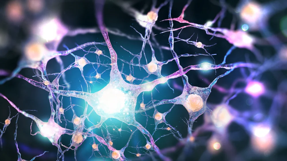
Researchers have published a study in Aging Cell on how inhibiting the death of axons in the brain protects the brains of old mice from inflammation.
Necroptosis in the brain
It is well-known that aged organisms have problems with cognitive function, and this is strongly linked to losses in the number and availability of synapses [1] and the degradation of axons in the aged brain [2], including the human brain [3].
Necroptosis refers to a form of programmed cellular death that is due to tumor necrosis factor (TNF) [4]. Under normal circumstances, it is a response to cellular injury, and previous work has found that it is a key factor in axonal degeneration [5]. However, in the aged brain, it contributes to inflammation [6].
The mechanisms and downstream effects of axonal necroptosis had not yet been thoroughly explored. This paper builds upon previous work, using mouse models with and without key elements of necroptosis.
Necroptosis and axonal degeneration
This study examined three different age groups of mice: adult (3-6 months), old (12-15 months) and aged (over 20 months). Examining the brains of these mice, the researchers found that with aging, mice begin to lose axonal integrity in the hippocampus: the area of neuronal fibers significantly drops, and markers of axonal damage increase.
These negative changes occur alongside the increase of two factors associated with necroptosis: the phosphorylated (p) forms of MLKL and RIPK3, which barely existed in the youngest group studied but became more prevalent with aging, particularly in its movement from the nucleus to the cytoplasm. This increase was found throughout the hippocampus and, in the case of pMLKL, in other brain regions as well.
Unsurprisingly, necroptosis was also found to contribute to the senescent-associated secretory phenotype (SASP), which characterizes the spread of senescent cells.
Preventing degeneration through genetics and pharmacology
With these results in hand, the researchers then determined if axonal degeneration can be suppressed by deactivating MLKL in a mouse model, and the results were striking. Aged mice that did not produce MLKL had similar numbers of degenerated axons as adult wild-type mice, biomarkers of inflammation were significantly reduced, and the increase in microglia that normally occurs with aging alongside these degenerated axons [6] did not occur in these modified mice.
Their brains responded to electric stimulus as if they were much younger. Their performance on the famous Morris water maze task matched these results as well, and their SASP biomarkers were reduced. The researchers concluded that a loss of MLKL results in the maintenance of learning and memory abilities with aging.
The researchers then sought to determine if these results could be replicated through pharmacology. They administered GSK’872, an inhibitor of RIPK3, to 23-month-old mice for four weeks, and administered the same tests.
The results were similarly striking. Like with the MLKL-free mice, inflammatory and SASP biomarkers were significantly decreased compared to their untreated counterparts, axonal degeneration was significantly decrased, Morris water maze performance was substantially enhanced, and electrical stimulus response was similar to that of younger mice.
Conclusion
This study has presented significant evidence showing that preventing what appears to be a maintenance process gone haywire seems like a viable strategy in combating neurodegeneration. However, while these results are strong and promising, this is still one mouse study. Only clinical trials can determine if RIPK3 or MLKL inhibitors can be useful in preventing necroptosis and axonal degeneration in human beings.
Literature
[1] Rosenzweig, E. S., & Barnes, C. A. (2003). Impact of aging on hippocampal function: plasticity, network dynamics, and cognition. Progress in neurobiology, 69(3), 143-179.
[2] Stahon, K. E., Bastian, C., Griffith, S., Kidd, G. J., Brunet, S., & Baltan, S. (2016). Age-related changes in axonal and mitochondrial ultrastructure and function in white matter. Journal of Neuroscience, 36(39), 9990-10001.
[3] Radhakrishnan, H., Stark, S. M., & Stark, C. E. L. (2020). Microstructural alterations in hippocampal subfields mediate age-related memory decline in humans, Front. Aging Neurosci. 0 2020.
[4] Seo, J., Nam, Y. W., Kim, S., Oh, D. B., & Song, J. (2021). Necroptosis molecular mechanisms: Recent findings regarding novel necroptosis regulators. Experimental & Molecular Medicine, 53(6), 1007-1017.
[5] Arrázola, M. S., Saquel, C., Catalán, R. J., Barrientos, S. A., Hernandez, D. E., Martínez, N. W., & Catenaccio, A. (2019). Axonal degeneration is mediated by necroptosis activation. Journal of Neuroscience, 39(20), 3832-3844.
[6] Thadathil, N., Nicklas, E. H., Mohammed, S., Lewis, T. L., Richardson, A., & Deepa, S. S. (2021). Necroptosis increases with age in the brain and contributes to age-related neuroinflammation. Geroscience, 43, 2345-2361.


