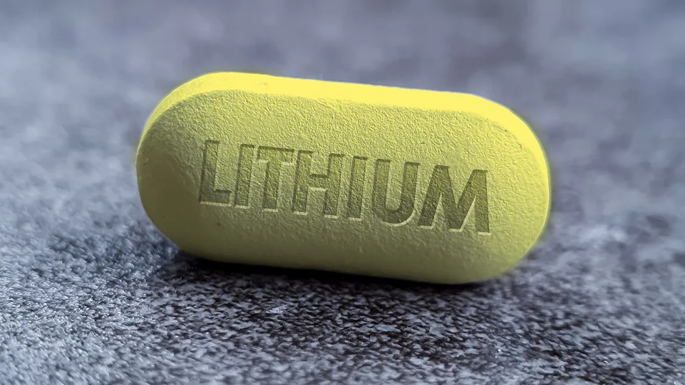
In a recent study, researchers identified the critical role that lithium plays in brain health and the development of mild cognitive impairment and Alzheimer’s disease. Supplementing with a lithium salt called lithium orotate can reverse many of its cognitive decline-related changes on the molecular and cellular levels [1].
The road less traveled
Understanding the underlying causes and factors related to Alzheimer’s disease is essential to developing effective therapies, which do not yet exist. The researchers of this paper focused on less explored factors such as metals, which play essential roles in the brain’s functioning and have not been deeply studied in the context of Alzheimer’s disease.
They started by assessing 27 metals in the brain and blood of aged people with different levels of cognitive abilities, including no cognitive impairment, mild cognitive impairment, and Alzheimer’s disease. They specifically focused on the prefrontal cortex, which is usually affected in Alzheimer’s disease, and used the cerebellum, the brain region that is not usually affected, for comparison.
Lithium and cognition
They identified that the levels of one metal, lithium, were significantly reduced in the prefrontal cortex of people with mild cognitive impairment and Alzheimer’s disease but not in the cerebellum. Lithium was also present in the amyloid beta (Aβ) plaques, and Alzheimer’s patients had higher concentrations of lithium in Aβ plaques compared to those with mild cognitive impairment.
In the next step, the researchers divided prefrontal cortex samples into two fractions: a plaque-enriched fraction and a fraction not containing amyloid plaques. Compared to people without cognitive impairment, there was less lithium in the prefrontal cortex non-plaque fraction of Alzheimer’s patients. They also noted a correlation between some cognitive abilities and lithium levels in the non-plaque cortical fraction.
Further data from older mouse models showed lower lithium levels in the non-plaque cortical fractions and lithium concentrations in the Aβ deposits, suggesting the isolation of lithium in the Aβ deposits that result in lower bioavailability.
Restricting lithium
Since previous experiments suggested decreased lithium bioavailability, the researchers imitated that state through restricting the lithium in the mouse diet by 92%, which led to a 89% drop in mean serum lithium and a 47–52% decrease in mean cortical lithium in the non-plaque fraction, suggesting the dietary approach was working in lowering lithium levels.
In mouse models prone to forming Aβ deposits, mice with reduced dietary lithium had increases in Aβ deposition and phospho-tau isoforms, Alzheimer’s disease-associated proteins, in the hippocampus compared to the age-matched, normally fed controls, which started to appear in relatively young mice and continued to accumulate with age.
A similar trend was observed in the wild-type mice; however, here the researchers observed an increase specifically in cortical and hippocampal Aβ42, the primary pathogenic Aβ form. Together, these results suggest that lithium deficiency accelerates Aβ deposits and phospho-tau accumulation.
A lithium-deficient diet also affected cognition in the mouse models prone to forming Aβ deposits and in the aging wild-type mice. It impaired learning, long-term memory, and novel-object recognition memory but didn’t impact spatial learning, locomotor activity, and exploratory behavior.
Lithium deficiency and Alzheimer’s similarities
Analysis of gene expression in the hippocampus, a part of the brain that is one of the first to be affected by mild cognitive impairment and Alzheimer’s disease, indicated many cell-type-specific changes in lithium-deficient mice that were prone to forming Aβ deposits.
Many of those changes overlapped with changes observed incortical biopsies obtained from people showing early-stage Aβ deposition, and even higher levels of overlap were observed when the researchers analyzed cortical biopsies from patients who were diagnosed with Alzheimer’s disease before or within one year of biopsy and who showed both Aβ and phospho-tau pathology.
Similarly, an analysis of microglia, the immune cells of the central nervous system, significantly overlapped the gene expression patterns of microglia in Alzheimer’s disease, including similarities to a reactive pro-inflammatory microglial state specific to Alzheimer’s disease and impaired Aβ clearance. This occured when either Alzheimer’s-prone mice or wild-type mice were deprived of lithium.
Lithium restriction also led to decreases in gene expression and proteins that relate to synaptic signaling and structure along with myelin, which forms a protective sheath around nerve fibers, in the aging mouse brain. This led to a loss of myelin itself, creating thinner myelin sheaths and a reduced number of certain types of cells in the nervous system.
The mediator of changes
The researchers analyzed differentially expressed genes to identify signaling pathways that lead to the outcomes of lithium deficiency. They identified a molecular target of lithium, serine-threonine kinase GSK3β, as a protein that regulated some of the affected signalling pathways and has a direct connection to Alzheimer’s disease, as tau is phosphorylated by GSK3β in Alzheimer’s disease. Activated GSK3β levels were increased in the hippocampal cells of lithium-deficient mice [2, 3].
When the researchers inhibited GSK3β in lithium-deficient animals or cell cultures, many of the lithium deficiency-related features were reversed, “including Aβ deposition, phospho-tau accumulation, myelination and microglial pro-inflammatory activation, as well as restoring the ability of microglia to clear Aβ.”
Reversing the decline
The researchers reasoned that since lithium is being sequestered by amyloid plaques, using lithium salts with reduced amyloid binding might have therapeutic potential. They identified lithium orotate as having the highest therapeutic potential and compared it with the clinical standard, lithium carbonate.
Compared to mice receiving lithium carbonate, the Alzheimer’s-prone mice that received low doses of lithium orotate had lower concentrations of lithium in Aβ plaques, more lithium in the non-plaque fraction, an almost complete absence of Aβ plaque deposition and phospho-tau accumulation, a reversed expression of lithium deficiency-related genes, a nearly complete reversal of memory loss, and improved learning and spatial memory.
The effect of low-dose lithium was further investigated with a focus on normal brain aging in wild-type mice. The researchers noted the positive impact of lithium orotate on brain age-related conditions, specifically, a reduction in pro-inflammatory cytokines, a restoration of the ability of microglia to degrade Aβ, synapse maintenance, and a reversal of learning and memory decline without any toxic effects.
A new and promising idea
“The idea that lithium deficiency could be a cause of Alzheimer’s disease is new and suggests a different therapeutic approach,” said senior author Bruce Yankner, professor of genetics and neurology in the Blavatnik Institute at HMS. “What impresses me the most about lithium is the widespread effect it has on the various manifestations of Alzheimer’s. I really have not seen anything quite like it all my years of working on this disease,” Yankner adds.
“One of the most galvanizing findings for us was that there were profound effects at this exquisitely low dose,” Yankner adds. This is especially important since higher doses of lithium could lead to kidney and thyroid toxicity in aged individuals. Still, such toxicity was not detected in mouse models treated with low lithium doses [4].
He also adds that lithium treatment is much different than current Alzheimer’s approaches: “My hope is that lithium will do something more fundamental than anti-amyloid or anti-tau therapies, not just lessening but reversing cognitive decline and improving patients’ lives,” he said.
However, Yankner also cautions and reminds people that human trials are imperative to ensure this approach is a viable treatment: “You have to be careful about extrapolating from mouse models, and you never know until you try it in a controlled human clinical trial,” Yankner said. “But, so far, the results are very encouraging.”
Literature
[1] Aron, L., Ngian, Z. K., Qiu, C., Choi, J., Liang, M., Drake, D. M., Hamplova, S. E., Lacey, E. K., Roche, P., Yuan, M., Hazaveh, S. S., Lee, E. A., Bennett, D. A., & Yankner, B. A. (2025). Lithium deficiency and the onset of Alzheimer’s disease. Nature, 10.1038/s41586-025-09335-x. Advance online publication.
[2] Folke, J., Pakkenberg, B., & Brudek, T. (2019). Impaired Wnt Signaling in the Prefrontal Cortex of Alzheimer’s Disease. Molecular neurobiology, 56(2), 873–891.
[3] Leroy, K., Yilmaz, Z., & Brion, J. P. (2007). Increased level of active GSK-3beta in Alzheimer’s disease and accumulation in argyrophilic grains and in neurones at different stages of neurofibrillary degeneration. Neuropathology and applied neurobiology, 33(1), 43–55.
[4] Kakhki, S., & Ahmadi-Soleimani, S. M. (2022). Experimental data on lithium salts: From neuroprotection to multi-organ complications. Life sciences, 306, 120811.


