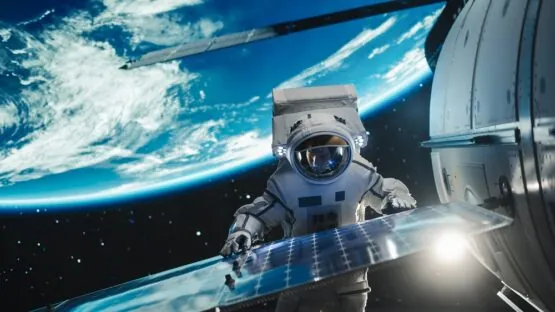
Scientists have found that human heart tissue is harmed by even a short stint in orbit. This might have implications for future space travel [1].
Fly me to the Moon
Space exploration is cool, but the human body was obviously not made for it. All its systems have evolved to function under normal gravity and the protection of Earth’s atmosphere against cosmic radiation. Since the early days of space flight, scientists have known that it alters human biology in troubling ways. Now, thanks to novel technologies, we are closer to understanding how and why.
Cardiac function is known to be altered by microgravity. In a twin study, an astronaut who spent almost a year in space exhibited changes associated with deconditioning, probably because the heart muscle doesn’t have to work as hard when there’s no gravity [2].
Another study documented cardiac arrhythmias during long space flights in otherwise notoriously healthy astronauts [3]. Yet another found decreased cardiac output and cardiac muscle atrophy [4]. However, since not many humans go to space, the sample sizes are small, so scientists have to look for other research modalities.
Adding a dimension
Heart muscle cells (cardiomyocytes) have already traveled to space, but a two-dimensional cell culture is not the best model of heart function. In this new study, published in Proceedings of the National Academy of Sciences, scientists used more sophisticated 3D organoids that are better at imitating a real heart.
These tiny patches of cardiac muscle were built in an intricate process, using scaffolds made of decellularized myocardial extracellular matrix and an electroconductive synthetic material to better recreate the contractile function. Each organoid was placed between two posts to allow it to contract freely, and tiny magnets were used to measure contractions.
Less twitchy, less healthy
After twelve days of space flight on board the International Space Station (ISS), the organoids began showing a decline in cardiomyocyte twitch forces, which produce contraction, compared to both baseline values and a control group of organoids that remained on Earth. The twitch force values continued to decline for the whole month of the flight and remained low until the end of the nine-day follow-up period.
Sarcomeres are the smallest functional contractile units inside muscle cells, consisting of several proteins. Their length is associated with the maximal twitch force. The organoids that went to space had sarcomeres that were significantly shorter and less organized. Interestingly, their length did not rebound after the samples returned from orbit, which might explain the persistent decline in twitch forces.
Since cardiomyocytes are so energy-hungry, their function depends heavily on mitochondria, which form large networks. The researchers found that in the organoids that went to space, mitochondria were more fragmented and swollen, which could indicate increased production of reactive oxygen species (ROS), harmful byproducts of energy generation. The cells also abnormally accumulated lipid droplets, another sign of mitochondrial dysfunction.
RNA sequencing showed significant differences in gene expression between spaceflight samples and controls. Pathways associated with the formation of cardiac muscle were downregulated, while some pathways associated with heart failure and inflammation were upregulated. The latter included the cGAS pathway, which senses mitochondrial DNA emitted by dysfunctional mitochondria and summons an immune response.
What about Mars?
Future long flights, such as to Mars, also worry researchers. A study found that Apollo program astronauts who traveled to the Moon were five times more likely to die from cardiovascular disease than their colleagues who only spent time in low orbit [5], despite the relatively short durations of Moon missions. The reason is probably the deadly cosmic radiation. Scientists must find ways to shield space travelers from it, and working with organoids might help get quality data.
What about future colonies on Mars? Their inhabitants can be shielded from radiation, which is much stronger on Mars due to the thinner atmosphere (for instance, by building underground), but gravity on Mars is only about one-third of what we are accustomed to. It remains to be seen how this will impact the human body.
In this study with EHTs, spaceflight was found to cause impaired contractility, detrimental subcellular structural modifications, mitochondrial structural changes, and increased oxidative stress. RNA-seq analysis of spaceflight samples indicated transcriptional changes associated with metabolic dysfunction, increased inflammatory cytokine production, and heart failure pathway up-regulation. Additional RNA-seq analysis and in silico modeling indicated that oxidative stress and mitochondrial dysfunction may have led to downstream tissue damage and cardiovascular dysfunction.
Literature
[1] Mair, D. B., Tsui, J. H., Higashi, T., Koenig, P., Dong, Z., Chen, J. F., … & Kim, D. H. (2024). Spaceflight-induced contractile and mitochondrial dysfunction in an automated heart-on-a-chip platform. Proceedings of the National Academy of Sciences, 121(40), e2404644121.
[2] Garrett-Bakelman, F. E., Darshi, M., Green, S. J., Gur, R. C., Lin, L., Macias, B. R., … & Turek, F. W. (2019). The NASA Twins Study: A multidimensional analysis of a year-long human spaceflight. Science, 364(6436), eaau8650.
[3] Anzai, T., Frey, M. A., & Nogami, A. (2014). Cardiac arrhythmias during long-duration spaceflights. Journal of Arrhythmia, 30(3), 139-149.
[4] Vernice, N. A., Meydan, C., Afshinnekoo, E., & Mason, C. E. (2020). Long-term spaceflight and the cardiovascular system. Precision Clinical Medicine, 3(4), 284-291.Chicago
[5] Delp, M. D., Charvat, J. M., Limoli, C. L., Globus, R. K., & Ghosh, P. (2016). Apollo lunar astronauts show higher cardiovascular disease mortality: possible deep space radiation effects on the vascular endothelium. Scientific reports, 6(1), 29901.


