×

The Blog

Building a Future Free of Age-Related Disease

Rhesus macaques

Caloric Restriction Slows Ovarian Aging in Monkeys

In Aging, researchers have published their discovery that three years of caloric restriction in rhesus macaques that are beginning to enter menopause slows their ovarian aging.

Closer to people than mice

The ovary is one of the first human organs to be seriously affected by aging, with the follicles becoming depleted long before most causes of age-related mortality become common [1]. However, the associated loss of estrogen has downstream consequences on many other organs [2].

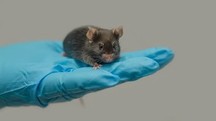
There are crucial caveats to studying ovarian health in mice, as their estrous cycle is different from that of women; mice do not menstruate. Rhesus macaques, on the other hand, are much closer to people; they live much longer, share 93% of our genome, and have a menstrual cycle that, while having a seasonal component [3], is broadly similar to that of women. Previous work has found that caloric restriction improves the healthspan and lifespan of these monkeys [4], but that work did not investigate its effects on ovarian health.

Benefits just before menopause

These researchers tested the effects of three years of 30% caloric restriction on female rhesus macaques. The young group began the study at 7-10 years old (a few years after puberty), and the old group began the study at ages 16-23. All of the young animals began this study with regular menstrual cycles, while only one old monkey in the control group and none in the caloric restriction group had regular cycles. This was not a lifespan or healthspan study; the monkeys’ organs were harvested after completion.

Ovarian CR study macaques

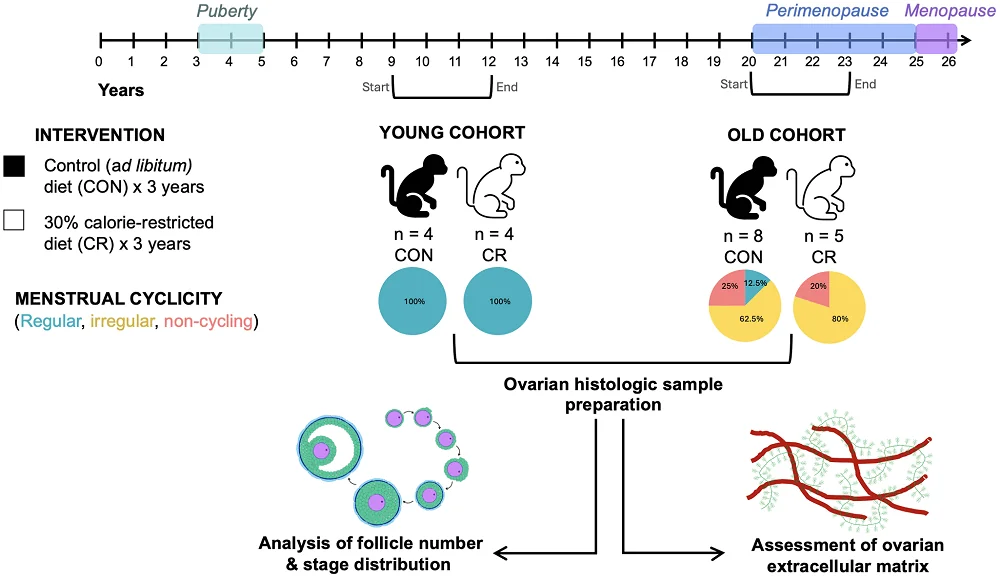
The comparisons between young and old animals were entirely as expected: older animals had significantly fewer follicles of every type than their younger counterparts, and menopause visibly shrank the ovaries. Unsurprisingly, there was an extremely strong correlation between follicle count and menstruation status.

In irregularly cycling (perimenopausal) animals, the number of quiescent (primordial) and multilayer follicles was greater in monkeys on caloric restriction than in the control group, with positive trends in many other follicle groups, leading to an increase in overall follicle density and suggesting a general increase in ovarian activity. Additionally, the proportion of follicle classes in the caloric restriction group was more like that of young animals. However, no benefit to follicle density was found in menopausal monkeys.

The ovaries normally become more fibrotic with age, which was found to occur in the control group but not in the caloric restriction group, as measured by tissue staining. Similarly, there is a loss of hyaluronic acid with age, which was only significant in the control group. These results suggest that caloric restriction delays age-related ovarian fibrosis.

A small study that confirms existing knowledge

This was a relatively small study, and the researchers note that their three years of caloric restriction are, in the research world, considered short term, as other studies on lifespan and healthspan have involved lifelong dietary restrictions. They acknowledge that it is difficult to impossible for human beings to stay on such a restrictive diet, and they suggest that such techniques as intermittent fasting may be more practical and that caloric restriction mimetic drugs may be of use, although further study is required in both of these areas.

Furthermore, most of this study’s results were based on computer analysis of histological slides; there was no investigation into the biochemistry behind the effects of caloric restriction on ovarian function. The researchers hypothesize that the effects are similar between people, monkeys, and mice, such as the sirtuin pathway [5].

The researchers hold that the timing of caloric restriction may be crucial. The significant effects were seen in monkeys whose ovaries were beginning to experience age-related decline. Caloric restriction to this degree did not restore the function of fully menopausal monkeys, and it did not seem to have any significant effects on the ovaries of younger monkeys; it is likely that these results are also true for humans. Therefore, for comparable interventions to be appropriate for the maintenance of ovarian health, they would need to be administered at a very specific time in a woman’s life.

We would like to ask you a small favor. We are a non-profit foundation, and unlike some other organizations, we have no shareholders and no products to sell you. All our news and educational content is free for everyone to read, but it does mean that we rely on the help of people like you. Every contribution, no matter if it’s big or small, supports independent journalism and sustains our future.

Literature

[1] Hansen, K. R., Knowlton, N. S., Thyer, A. C., Charleston, J. S., Soules, M. R., & Klein, N. A. (2008). A new model of reproductive aging: the decline in ovarian non-growing follicle number from birth to menopause. Human reproduction, 23(3), 699-708.

[2] Cui, J., Shen, Y., & Li, R. (2013). Estrogen synthesis and signaling pathways during aging: from periphery to brain. Trends in molecular medicine, 19(3), 197-209.

[3] Beltran-Frutos, E., Casarini, L., Santi, D., & Brigante, G. (2022). Seasonal reproduction and gonadal function: A focus on humans starting from animal studies. Biology of Reproduction, 106(1), 47-57.

[4] Mattison, J. A., Colman, R. J., Beasley, T. M., Allison, D. B., Kemnitz, J. W., Roth, G. S., … & Anderson, R. M. (2017). Caloric restriction improves health and survival of rhesus monkeys. Nature communications, 8(1), 14063.

[5] Long, G. Y., Yang, J. Y., Xu, J. J., Ni, Y. H., Zhou, X. L., Ma, J. Y., … & Luo, L. L. (2019). SIRT1 knock-in mice preserve ovarian reserve resembling caloric restriction. Gene, 686, 194-202.

Sunlight

Vitamin D Rescues Telomere Attrition in Leukocytes

A sub-study, which was part of the large-scale VITAL trial, determined that vitamin D supplementation slows telomere attrition in leukocytes almost to a halt. This could have real-life clinical implications [1].

The chromosome guardians

Attrition of telomeres, repetitive sequences that cap chromosomes, is one of the original hallmarks of aging. Our cells’ replication machinery does not copy the last few base pairs at chromosomes’ ends. These telomeres act as “surplus” DNA that does not contain valuable information and can be sacrificed. This also means that with each division, telomeres become shorter, which can eventually trigger cellular senescence. Previous research has found that telomere attrition in leukocytes, such as T cells, can predict chronic disease and mortality [2].

The attrition of attrition

A new study co-led by researchers at Mass General Brigham and the Medical College of Georgia has its roots in the VITAL trial, a large, nationwide, randomized, double-blind, placebo-controlled study that tested daily supplementation with vitamin D₃ (2 000 IU) and/or marine omega-3 fatty acids (1 g fish oil) for primary prevention of cancer and cardiovascular disease. VITAL lasted for five years and enrolled almost 26,000 US females and males aged at least 50 years.

VITAL already yielded some impressive results, showing statistically significant reductions in cancer mortality, the incidence of autoimmune decease, and circulating C-reactive protein levels (a marker of inflammation) in people who took vitamin D.

This new sub-study included slightly more than one thousand VITAL participants who had had their leukocyte telomere length measured at least twice during the study. At baseline, the mean leukocyte telomere length (LTL) was 8,700 base pairs, and the mean LTL loss in the placebo group was 160 base pairs over four years.

Vitamin D abrogated LTL loss almost completely, bringing it down to just about 20 base pairs over four years on average. Omega-3 supplementation did not significantly affect any outcome. While impressive, these results were just barely statistically significant because of the overall slow LTL loss and the limitations of current methods of measuring telomere length. No clinical outcomes were measured.

The association remained robust even after accounting for key demographic, behavioral, and cardiometabolic treatment variables. Those included age, sex, race, body-mass index (BMI), smoking status, hypertension medication use, diabetes, and high cholesterol.

“VITAL is the first large-scale and long-term randomized trial to show that vitamin D supplements protect telomeres and preserve telomere length,” said co-author JoAnn Manson, MD, principal investigator of VITAL and chief of the Division of Preventive Medicine at Brigham and Women’s Hospital. “This is of particular interest because VITAL had also shown the benefits of vitamin D in reducing inflammation and lowering risks of selected chronic diseases of aging, such as advanced cancer and autoimmune disease.”

Vitamin D Leukocytes

It’s the shortest telomere that counts

These results require some unpacking. While the average annual LTL loss of 40 base pairs seems minuscule compared to a baseline LTL of 8,700, it might still be clinically significant because of the way cellular senescence works.

It’s the single most-eroded telomere in a cell that triggers the DNA damage response and pushes the cell into senescence. Once that “sentinel” telomere falls below a critical length, the cell stops dividing, regardless of how long all the others are [3]. A small shift in the mean LTL of about 40 base pairs per year might be a sign of a steady increase in the fraction of critically short telomeres across the cell population, increasing the probability that any given cell will cross that threshold.

Faster rates of LTL attrition in adults predict higher risks of adverse events even after adjusting for lifestyle and inflammation markers. Those epidemiological associations indicate that even small differences in attrition rate carry real-world health implications.

“Our findings suggest that targeted vitamin D supplementation may be a promising strategy to counter a biological aging process, although further research is warranted,” said Haidong Zhu, Ph.D., first author of the report and a molecular geneticist at the Medical College of Georgia, Augusta University.

We would like to ask you a small favor. We are a non-profit foundation, and unlike some other organizations, we have no shareholders and no products to sell you. All our news and educational content is free for everyone to read, but it does mean that we rely on the help of people like you. Every contribution, no matter if it’s big or small, supports independent journalism and sustains our future.

Literature

[1] Zhu, H., et al. (2025). Vitamin D₃ and marine omega-3 fatty acids supplementation and leukocyte telomere length: 4-year findings from the VITAL randomized controlled trial. The American Journal of Clinical Nutrition, 0(0), 000–000.

[2] Epel, E. S., Merkin, S. S., Cawthon, R., Blackburn, E. H., Adler, N. E., Pletcher, M. J., & Seeman, T. E. (2008). The rate of leukocyte telomere shortening predicts mortality from cardiovascular disease in elderly men. Aging (Albany NY), 1(1), 81.

[3] Berardi, P., Martinez-Fernandez, V., Rat, A., Rosas Bringas, F. R., Jolivet, P., Langston, R., … & Teixeira, M. T. (2025). The shortest telomere in telomerase-negative cells triggers replicative senescence at a critical threshold length and also fuels genomic instability. bioRxiv, 2025-01.

Outdoor mouse

Common Laboratory Mice Age Faster in a Natural Environment

In Aging Cell, researchers have found that exposing ordinary Black 6 mice to a more natural environment accelerates rather than slows the aging of their livers.

When natural doesn’t mean better

It is a well-known fact that laboratory animals live in controlled conditions beyond that of wild animals or even most pets. Temperature and food are regulated, and social interactions between animals are limited [1]. The lack of predation and competition means that captive animals, such as in zoos, often live far longer [2].

It has been suggested that these conditions have an effect on aging as well. In Wales, a population of house mice, which are similar to the standardized Black 6, were captured and epigenetically analyzed through their feces; these mice were found to age more rapidly than laboratory mice in a handful of ways [3]. Of course, these mice are still not quite the same at the genetic level, so the results of such a comparison will always be murky.

For a cleaner look at environmental effects, the same mice must be exposed to different environments, which is exactly what these researchers did. At two weeks of age, a population of mice was taken into a field enclosure that protected them from predators but not the elements or other environmental effects. The livers of these mice were compared to those of a standard, laboratory-raised control group.

Faster aging in nearly every way

Animals reared in the laboratory, as expected, mostly age in similar ways to animals in a less controlled environment; nearly all of the changes occur in the same way, with slightly over 11% of age-related epigenetic changes occurring in opposite directions; nearly all of these were epigenetic sites becoming hypermethylated in field animals but hypomethylated in lab animals. Fewer than 1% of the total sites were hypomethylated in the field while being hypermethylated in the lab.

However, for nearly all of the sites that were methylated in the same way, the animals in the field were found to age faster; in some ways, much faster. For hypermethylations, 96% of the sites aged nearly twice as rapidly; every single analyzed site aged more rapidly in the field than the lab. For hypomethylations, 66% of the sites aged an average of 28% more quickly, with 94% of these sites aging more rapidly in the field than the lab.

The researchers took a close look at the specific sites involved. Hypermethylated sites were largely related to epigenetic changes with replication [4] along with stem cell proliferation, and these data suggested an increased risk of cancer [5, 6]. Hypomethylated sites were related to transcription factors that govern the function of liver cells.

More stress on the liver

In mice that were introduced into the field during adulthood rather than infancy, the results were largely similar but with crucial differences. Specifically, these mice were suspected of having more rapid DNA damage, as determined by epigenetic methylation happening more rapidly in sites bound by two DNA repair genes [7]. There was also hypermethylation of sites related to the binding ISL1, an insulin regulator, in a way that suggests that the mice were burning more fat [8].

Re-wilding animals exposes them to many different sources of stress. This work focused specifically on the liver, which the researchers recognize as being central in processing environmental toxins. However, it is also possible that inter-social stresses in mice helped to bring about the overall outcome, and the mice also faced challenges related to natural weather.

For the public, the most crucial revelation about this study may be the confirmation that rather than living a supposedly high-stress life, normal laboratory mice live in an environment with some of the least stress possible and exposure to stress ages them more rapidly, at least in the liver. The researchers expressed their intention to do future work on other tissues, some of which are less environmentally sensitive. Future work that focuses on toxin processing or repairing environmental-related damage may find a more promising start in outdoor-raised mice rather than mice raised in strict laboratory conditions.

We would like to ask you a small favor. We are a non-profit foundation, and unlike some other organizations, we have no shareholders and no products to sell you. All our news and educational content is free for everyone to read, but it does mean that we rely on the help of people like you. Every contribution, no matter if it’s big or small, supports independent journalism and sustains our future.

Literature

[1] Zipple, M. N., Vogt, C. C., & Sheehan, M. J. (2023). Re-wilding model organisms: opportunities to test causal mechanisms in social determinants of health and aging. Neuroscience & Biobehavioral Reviews, 152, 105238.

[2] Tidière, M., Gaillard, J. M., Berger, V., Müller, D. W., Bingaman Lackey, L., Gimenez, O., … & Lemaître, J. F. (2016). Comparative analyses of longevity and senescence reveal variable survival benefits of living in zoos across mammals. Scientific reports, 6(1), 36361.

[3] Hanski, E., Joseph, S., Raulo, A., Wanelik, K. M., O’Toole, Á., Knowles, S. C., & Little, T. J. (2024). Epigenetic age estimation of wild mice using faecal samples. Molecular ecology, 33(8), e17330.

[4] Zhou, W., & Reizel, Y. (2024). On correlative and causal links of replicative epimutations. Trends in Genetics.

[5] Teschendorff, A. E., Menon, U., Gentry-Maharaj, A., Ramus, S. J., Weisenberger, D. J., Shen, H., … & Widschwendter, M. (2010). Age-dependent DNA methylation of genes that are suppressed in stem cells is a hallmark of cancer. Genome research, 20(4), 440-446.

[6] Schlesinger, Y., Straussman, R., Keshet, I., Farkash, S., Hecht, M., Zimmerman, J., … & Cedar, H. (2007). Polycomb-mediated methylation on Lys27 of histone H3 pre-marks genes for de novo methylation in cancer. Nature genetics, 39(2), 232-236.

[7] Schumacher, B., Pothof, J., Vijg, J., & Hoeijmakers, J. H. (2021). The central role of DNA damage in the ageing process. Nature, 592(7856), 695-703.

[8] Zhao, F., Ke, J., Pan, W., Pan, H., & Shen, M. (2022). Synergistic effects of ISL1 and KDM6B on non-alcoholic fatty liver disease through the regulation of SNAI1. Molecular Medicine, 28(1), 12.

LSF Grant Aging Brain

Grant Award Announcement: Rejuvenating the Aging Brain Study

The Longevity Science Foundation (LSF), a nonprofit organization dedicated to funding research aimed at extending the healthy human lifespan, is proud to announce a grant award to the University of Copenhagen’s Center for Healthy Aging within the Department of Cellular and Molecular Medicine, for the study “Rejuvenating the Aging Brain.” The research is led by Dr. Morten Scheibye-Knudsen, a globally recognized expert in aging and neurodegeneration. The Foundation’s grant will fund a key project component over three years beginning in 2025.

The research aims to reverse brain aging by developing compounds that selectively eliminate senescent astrocytes, which are damaged brain cells that accumulate with age while preserving healthy neurons. These senescent cells are believed to contribute to cognitive decline and neurodegenerative diseases. Combining AI-driven screening with high-throughput compound testing, the research team will identify promising molecules, refine their specificity and pharmacokinetics, and validate their therapeutic potential through rigorous in vitro and in vivo testing.

The LSF’s support is essential in enabling this groundbreaking work, which could lead to the development of entirely new classes of treatments for age-related brain conditions. The project also reinforces the Foundation’s commitment to funding translational science that bridges the gap between laboratory discovery and real-world medical application.

“We are thrilled to support Dr. Morten Scheibye-Knudsen and his team at the University of Copenhagen,” said Joshua C. Herring, President and CEO of the Longevity Science Foundation. “This project reflects our belief that targeted, innovative research can lead to meaningful interventions in aging and neurodegeneration. We are committed to enabling discoveries that extend life and enhance its quality.”

This partnership is a step in achieving the Foundation’s broader mission of democratizing access to cutting-edge longevity research and ensuring that the most promising science receives the resources it needs to thrive.

If you are interested in supporting the groundbreaking research conducted by the Scheibye-Knudsen Lab, donating to the LSF, or supporting our other research initiatives, please reach out to our COO, Lev Dvornik, at ld@longevity.foundation, and our CEO, Joshua Herring, at jh@longevity.foundation. All donations are tax-deductible up to IRS limits and directly fund research, dollar for dollar.

About the Longevity Science Foundation

The Longevity Science Foundation (LSF) is a nonprofit organization advancing human longevity by funding research and development of medical technologies to extend the healthy human lifespan. The long-term mission of the Foundation is to prevent all chronic and age-related diseases and to help make longevity-focused care accessible to everyone, no matter their background, by bringing cutting-edge science on aging out of the laboratory and into the mainstream. To learn more, visit www.longevity.foundation. Our work is made possible by our generous donors. To donate to the Longevity Science Foundation, visit https://longevity.foundation/support-us#donate.

Department of Cellular and Molecular Medicine, University of Copenhagen

The focus of the department is the functional cell, its genetic components and molecular cellular mechanisms in a medical context. With a firm foundation in the basic function of the normal and differentiating cell an understanding of the molecular, cellular and genetic mechanisms behind disease and aging is sought. Learn more at healthyaging.ku.dk.

We would like to ask you a small favor. We are a non-profit foundation, and unlike some other organizations, we have no shareholders and no products to sell you. All our news and educational content is free for everyone to read, but it does mean that we rely on the help of people like you. Every contribution, no matter if it’s big or small, supports independent journalism and sustains our future.
Mouse in hand

Dietary Methionine Restriction Improves Healthspan in Mice

In a recent study, researchers investigated how restricting dietary methionine and inhibiting the tyrosine degradation pathway affects healthspan in aged mice. While affecting tyrosine didn’t show any benefits, methionine restriction improved many, but not all, measures of healthspan, including frailty, pathological disease burden, and neuromuscular function [1].

Aging of metabolism

Changes in metabolism accompany aging and are associated with many age-related diseases. Reprogramming metabolism to its youthful state could help to alleviate the signs and symptoms of aging or even reverse some aspects of it.

The authors of this study focus on two metabolic pathways that are dysregulated in aging: methionine metabolism and the tyrosine degradation pathway.

Methionine is an important metabolite, as it is the initiating amino acid during protein synthesis. Improving methionine metabolism has shown positive effects on healthspan and lifespan in model organisms, such as fruit flies, rodents, and human cell lines [2-4]. Still, the role of methionine restriction started late in life hasn’t been sufficiently explored.

Similarly, previous research in fruit flies indicated increased levels of enzymes in the tyrosine degradation pathway during aging, but the levels of tyrosine and tyrosine-derived neurotransmitters decrease. Reducing the levels of those enzymes leads to positive effects on healthspan and lifespan in fruit flies and worms [5, 6]. However, there is a lack of similar research in aged mice.

Metabolic interventions

In a population of aged (18-month-old) mice, the researchers either restricted methionine by decreasing the concentration of methionine in the diet from 0.86% (as a proportion of protein) to 0.17% or inhibited the tyrosine degradation pathway using nitisinone, a compound that elevates circulating tyrosine levels. This 6-month intervention was intended to test healthspan.

Unsurprisingly, a control group of young mice gained weight throughout the experiment, while aged mice with restricted methionine lost weight to levels similar to young mice at the start of the experiment, with a stronger effect observed in males. The weight loss of aged mice was caused by a loss of fat mass, but their lean mass was increased. No impact of nitisinone was observed.

After two weeks of treatment, the researchers tested blood plasma to determine whether these treatments were indeed impacting the levels of methionine and tyrosine. Compared to young mice, methionine levels were increased in aged mice, but only in males, which might suggest why males experienced a stronger effect of methionine restriction on weight loss. Dietary methionine restriction led to decreased plasma methionine levels in males and females, compared to aged animals of the same sex. Additionally, dietary methionine restriction improved hormonal markers of metabolic health in male mice.

Tyrosine levels were not significantly different between young and old mice. Inhibiting the tyrosine degradation pathway increased plasma tyrosine levels but didn’t affect hormonal markers.

Improved physical health

While testing molecular markers is important to understand effects, a successful treatment must improve the quality of life in an aged organism. To assess that, the resarchers conducted several tests on these mice at baseline and after 6 months of the treatment.

For a broad look at healthspan, the researchers assessed pathological disease burden scores of organs along with a frailty index that encompasses 26 different assessments and can address “the effect of treatment on different aspects of healthspan and predict life expectancy and the efficacy of lifespan–extending interventions up to a year in advance.”

The researchers reported more frailty and higher pathological disease burden scores in older animals. Frailty was significantly decreased in aged animals whose methionine was restricted, while a methionine-restricted diet reversed disease burden in female mice to that of young (10-month-old) mice.

Compared to old, normally-fed mice, methionine restriction improved neuromuscular function in aged mice, as measured by coordination, balance, grip strength, and time spent on exploratory activity.

Aged animals on a methionine-restricted diet also had improved lung functions compared to their aged, normally fed counterparts. However, clarity of vision, short-term spatial working memory, cardiovascular function, age-related hearing loss, or enlarged prostate were not improved in that group.

There were also no changes in any of those measures in the nitisinone-treated group, even though the researchers confirmed sufficient inhibition of the tyrosine degradation pathway. This is in contrast to previously observed lifespan extension in fruit flies upon inhibition of this pathway. In that study, the effect was even more substantial when tyrosine degradation pathway inhibition was neuron-specific [5]. For future studies, the researchers propose inhibiting the tyrosine degradation pathway in mouse neurons or using different drug concentrations.

Metabolism and cognition

Previous research had linked methionine to age-related changes in cognitive health. Therefore, these researchers aimed to determine whether dietary methionine restriction can reduce amyloid plaque deposition. They used aged genetically engineered mice that exhibit many features of human Alzheimer’s disease.

After measuring multiple biomarkers, the researchers noted improvements in renal and neuromuscular functions in aged mice that underwent methionine restriction for 6 months but didn’t observe a significant effect on plasma amyloid levels. Conversely, it increased the levels of insoluble (intracellular) amyloid brain deposits.

The study’s authors suggest that to obtain beneficial effects, methionine levels might need to be in a specific concentration range, and levels that are too high or too low would not have fully beneficial effects and might even cause more problems.

Still biologically aged

Since the researchers determined that there were improvements in healthspan, they used epigenetic clocks to determine if the animals were biologically younger. However, mice on a restricted methionine diet did not have significant epigenetic changes compared to aged controls.

These surprising results prompted the researchers to also analyze human blood samples from a clinical trial. During that double-blind 8-week study, participants received either low or high sulfur amino acids (methionine and cysteine). The results were similar to those obtained in mice, with no significant effects on biological age.

The researchers suggested a few reasons for this absence of epigenetic changes. First, epigenetic clocks may have higher sensitivity to lifespan extension than healthspan improvements. As this experiment began methionine restriction late in life, it might not lead to a lifespan increase, but lifespan was not measured in this study.

Second, since many epigenetic clocks are built from blood samples, they might not catch beneficial changes in single organs, such as muscle, as observed in this study.

Third, methionine is the essential building block of a metabolite that delivers methyl groups for methyltransferases, which can methylate DNA. It is possible that lower levels of dietary methionine can affect this process, thus disturbing the measurements of epigenetic clocks that rely on DNA methylation patterns.

Overall, this study found that dietary methionine restriction, even when started later in life, can benefit healthspan in mice. Clinical trials are necessary to test whether these benefits will translate to humans.

We would like to ask you a small favor. We are a non-profit foundation, and unlike some other organizations, we have no shareholders and no products to sell you. All our news and educational content is free for everyone to read, but it does mean that we rely on the help of people like you. Every contribution, no matter if it’s big or small, supports independent journalism and sustains our future.

Literature

[1] Hernández-Arciga, U., Stamenkovic, C., Yadav, S., Nicoletti, C., Albalawy, W. N., Al Hammood, F., Gonzalez, T. F., Naikwadi, M. U., Graham, A., Smarz, C., Little, G. J., Williams, S. G., McMahon, B., Sipula, I. J., Vandevender, A. M., Chuan, B., Cooke, D., Pinto, A. F. M., Flores, L. C., Hartman, H. L., … Parkhitko, A. A. (2025). Dietary methionine restriction started late in life promotes healthy aging in a sex-specific manner. Science advances, 11(16), eads1532.

[2] Parkhitko, A. A., Wang, L., Filine, E., Jouandin, P., Leshchiner, D., Binari, R., Asara, J. M., Rabinowitz, J. D., & Perrimon, N. (2021). A genetic model of methionine restriction extends Drosophila health- and lifespan. Proceedings of the National Academy of Sciences of the United States of America, 118(40), e2110387118.

[3] Kozieł, R., Ruckenstuhl, C., Albertini, E., Neuhaus, M., Netzberger, C., Bust, M., Madeo, F., Wiesner, R. J., & Jansen-Dürr, P. (2014). Methionine restriction slows down senescence in human diploid fibroblasts. Aging cell, 13(6), 1038–1048.

[4] Orentreich, N., Matias, J. R., DeFelice, A., & Zimmerman, J. A. (1993). Low methionine ingestion by rats extends life span. The Journal of nutrition, 123(2), 269–274.

[5] Parkhitko, A. A., Ramesh, D., Wang, L., Leshchiner, D., Filine, E., Binari, R., Olsen, A. L., Asara, J. M., Cracan, V., Rabinowitz, J. D., Brockmann, A., & Perrimon, N. (2020). Downregulation of the tyrosine degradation pathway extends Drosophila lifespan. eLife, 9, e58053.

[6] Ferguson, A. A., Roy, S., Kormanik, K. N., Kim, Y., Dumas, K. J., Ritov, V. B., Matern, D., Hu, P. J., & Fisher, A. L. (2013). TATN-1 mutations reveal a novel role for tyrosine as a metabolic signal that influences developmental decisions and longevity in Caenorhabditis elegans. PLoS genetics, 9(12), e1004020.

Amyloids between neurons

Nanostructures Trap Amyloid Beta, Rescuing Neurons

Scientists have created engineered nanostructures that bind monomers and oligomers of harmful amyloid beta (Aβ) protein, preventing them from entering neurons and drastically increasing the cells’ survival in vitro [1].

Don’t let them into cells!

Misfolded proteins are thought to be behind diseases like Alzheimer’s and amyotrophic lateral sclerosis (ALS). The most recognizable hallmark of Alzheimer’s is the aggregation of amyloid plaques between brain cells. However, removing those plaques has only very limited impact on the disease.

Recently, evidence has been growing that soluble Aβ early-stage fibrils and oligomers, which can enter cells, are more damaging than plaques and more tightly linked to cognitive decline [2]. Moreover, plaques might act as a sink, pulling harmful oligomers out of circulation [3]. Scientists have tried targeting these harmful proteins using antibodies, but clearly, new, better chemical tools are needed.

A new study from Northwestern University, published in the Journal of the American Chemical Society (ACS), explores one such tool: engineered peptide amphiphiles (TPAs). These are molecules that can self-assemble into long nanofibers through a process called supramolecular polymerization. Some TPAs, such as semaglutide, are already used in therapies.

Neuronal death averted

The researchers ingeniously combined several building blocks to create custom fibers designed to bind to Aβ. Those included short chains of amino acids (peptides) and a natural sugar called trehalose.

“The advantage of peptide-based drugs is that they degrade into nutrients,” said Dr. Samuel I. Stupp, the study’s senior author. “The molecules in this novel therapeutic concept break down into harmless lipids, amino acids, and sugars. That means there are fewer adverse side effects.”

The researchers thought that trehalose might stabilize misfolded proteins, since it is known as a protein chaperone that can protect proteins from misfolding, denaturation, and aggregation [4].

“Trehalose is naturally occurring in plants, fungi, and insects,” said Zijun Gao, a Ph.D. candidate in Stupp’s laboratory and the paper’s first author. “It protects them from changing temperatures, especially dehydration and freezing. Others have discovered that trehalose can protect many biological macromolecules, including proteins. So, we wanted to see if we could use it to stabilize misfolded proteins.”

To the scientists’ surprise, trehalose actually destabilized the nanofibers, making them “metastable” and more prone to binding to surrounding molecules, specifically Aβ42 peptides, a particularly harmful subspecies. Instead of just blocking the process, the nanofibers physically trapped the Aβ42 peptides by incorporating them into their structure.

“Unstable assemblies of molecules are very reactive,” Stupp explained. “They want to interact with and bond to other molecules. If the nanofibers were stable, they would happily ignore everything around them.”

TPA Amyloid Beta

The researchers co-cultured Aβ42 with human neurons derived from induced pluripotent stem cells (iPSCs). Using fluorescence microscopy, they observed that in the presence of TPA, Aβ42 did not accumulate in neuronal lysosomes, directly correlating with dramatically improved survival of neurons and demonstrating that the entrapment blocked Aβ42 uptake into them. While co-culturing with Aβ42 but not TPA caused more than 60% of cells to die, the presence of TPA reduced cell death to that of healthy controls.

“Our study highlights the exciting potential of molecularly engineered nanomaterials to address the root causes of neurodegenerative diseases,” said Stupp. “By trapping the misfolded proteins, our treatment inhibits the formation of those fibers at an early stage. Early-stage, short amyloid fibers, which penetrate neurons, are believed to be the most toxic structures. With further work, we think this could significantly delay progression of the disease.”

Lots of further questions

While these results are encouraging, significant questions remain. First, is it possible to deliver TPA structures, which might be too big to cross the blood-brain barrier, into the central nervous system? The researchers suggest that one of Alzheimer’s symptoms, increased blood-brain barrier permeability, might help. Alternatively, delivery via the intranasal route, which forgoes the BBB entirely, might be used.

Whether clearance of TPA-Aβ42 conjugates from the brain would be required, and if so, how this can be achieved, is another potential issue. Finally, the study did not assess effects on the extracellular environment beyond blocking Aβ42 internalization by neurons. In particular, there was no measurement of inflammatory signaling, extracellular toxicity markers, membrane disruption, or interactions with non-neuronal glial cells.

The researchers note that their invention might revolutionize Alzheimer’s treatment, especially at an early stage before large amounts of Aβ42 have accumulated inside neurons. This highlights the need for early-stage Alzheimer’s screening, a hot research topic in which some recent advances have been made [5].

We would like to ask you a small favor. We are a non-profit foundation, and unlike some other organizations, we have no shareholders and no products to sell you. All our news and educational content is free for everyone to read, but it does mean that we rely on the help of people like you. Every contribution, no matter if it’s big or small, supports independent journalism and sustains our future.

Literature

[1] Gao, Z., Qiu, R., Dave, D. R., Chandravanshi, P., Soares, G. P., Smith, C. S., … & Stupp, S. I. (2025). Supramolecular Copolymerization of Glycopeptide Amphiphiles and Amyloid Peptides Improves Neuron Survival. Journal of the American Chemical Society.

[2] Hermann, D., Both, M., Ebert, U., Gross, G., Schoemaker, H., Draguhn, A., … & Nimmrich, V. (2009). Synaptic transmission is impaired prior to plaque formation in amyloid precursor protein–overexpressing mice without altering behaviorally-correlated sharp wave–ripple complexes. Neuroscience, 162(4), 1081-1090.

[3] Fagan, A. M., Mintun, M. A., Mach, R. H., Lee, S. Y., Dence, C. S., Shah, A. R., … & Holtzman, D. M. (2006). Inverse relation between in vivo amyloid imaging load and cerebrospinal fluid Aβ42 in humans. Annals of neurology, 59(3), 512-519.

[4] Vinciguerra, D., Gelb, M. B., & Maynard, H. D. (2022). Synthesis and application of trehalose materials. Jacs Au, 2(7), 1561-1587.

[5] Palmqvist, S., Warmenhoven, N., Anastasi, F., Pilotto, A., Janelidze, S., Tideman, P., … & Hansson, O. (2025). Plasma phospho-tau217 for Alzheimer’s disease diagnosis in primary and secondary care using a fully automated platform. Nature Medicine, 1-8.

Alzheimer's doctor

Results of a Phase 1 Trial of Senolytics for Alzheimer’s

The results of a Phase 1 trial of the well-known senolytic combination of dasatinib and quercetin (D+Q) in patients with Alzheimer’s disease have been published in Neurotherapeutics.

Building on substantial previous work

The researchers introduce this study by discussing the relationship of senescent cells to Alzheimer’s and its related pathologies; for example, senescent cells in the brain are associated with tau aggregation [1], and senescent astrocytes have been linked to Alzheimer’s [2]. Dasatinib and quercetin are very well-known to reduce cellular senescence, and they have been reported to reduce both tau [1] and amyloid beta plaques [3] in mouse models. However, mice don’t naturally get Alzheimer’s, and without human testing, it’s not clear how much good senolytics could do against this disease in people.

This group has previously conducted a feasibility trial using D+Q in people with Alzheimer’s, finding that the dasatinib had successfully infiltrated the brain and that it was well-tolerated [4]. Here, they build on that trial with more work, aiming to develop a standardized system of biomarker analysis in order to determine if senescent cells are actually being effectively cleared in this population, thus paving the way for Phase 2 studies.

Largely negative results

This trial utilized only five people, who were between the ages of 70 and 82, were in the early stage of clinical Alzheimer’s disease, and received 100 milligrams of dasatinib and 1 gram of quercetin on an intermittent schedule for three months.

Fractalkine, an inflammatory chemokine, appeared to be increased in plasma by the D+Q treatment, although this result was not considered statistically significant after multiple comparisons correction, nor were any others. Urinary analysis of metabolites also showed no statistically significant changes. This was likely due to the low number of participants involved in this study. The researchers estimated that, if only one SASP factor (such as the inflammatory biomarker IL-6) were considered, only 25 participants would need to be included in a future study in order to gain statistically significant results.

Unfortunately, this lack of statistical significance also applied to markers of AD pathology. The researchers examined a great many biomarkers relating to tau and amyloids, finding statistically significant changes to none of them. One person with more substantial neurodegeneration than the other four had more dasatinib uptaken into the brain, which may be due to reduced function of the blood-brain barrier.

The fat balance (lipidome) of the blood was somewhat affected by D+Q. Phosphatidylcholine, which makes up lipoprotein membranes and is usually tightly controlled, decreased by a sixth after treatment. Lysophosphatidylcholine, which is associated with inflammation and cellular death [5], decreased by 24%; this result was close to statistical significance. However, none of the lipid classes were significantly affected.

Overall cellular stress, as measured by transcriptomic analysis, was also somewhat affected. Of 19 inflammation-related genes in peripheral blood mononuclear cells (PBMCs), 7 of them were downregulated, including the SASP-related IL8 and IL1β.

This small study was meant to test safety and was never meant to show efficacy. It only lasted for 12 weeks, which is unlikely to be long enough for disease modification; however, the lack of any discernible signal in amyloid or tau biomarkers may suggest that this particular combination of senolytics, if not senolytics as a whole, may be the wrong approach in dealing with Alzheimer’s despite a documented link in mouse studies.

With some promising study results, a larger and longer study may be useful in verifying efficacy; here, with this lack of effect on crucial neurological amyloids, the opposite is likely to be true, and a Phase 2 study would probably yield the same largely negative results. Ultimately, the evidence suggests that Alzheimer’s is not a disease that relies on senescence to propagate, and entirely different methods are likely to be necessary for dealing with this proteostasis disorder.

We would like to ask you a small favor. We are a non-profit foundation, and unlike some other organizations, we have no shareholders and no products to sell you. All our news and educational content is free for everyone to read, but it does mean that we rely on the help of people like you. Every contribution, no matter if it’s big or small, supports independent journalism and sustains our future.

Literature

[1] Musi, N., Valentine, J. M., Sickora, K. R., Baeuerle, E., Thompson, C. S., Shen, Q., & Orr, M. E. (2018). Tau protein aggregation is associated with cellular senescence in the brain. Aging cell, 17(6), e12840.

[2] Bhat, R., Crowe, E. P., Bitto, A., Moh, M., Katsetos, C. D., Garcia, F. U., … & Torres, C. (2012). Astrocyte senescence as a component of Alzheimer’s disease.

[3] Zhang, P., Kishimoto, Y., Grammatikakis, I., Gottimukkala, K., Cutler, R. G., Zhang, S., … & Mattson, M. P. (2019). Senolytic therapy alleviates Aβ-associated oligodendrocyte progenitor cell senescence and cognitive deficits in an Alzheimer’s disease model. Nature neuroscience, 22(5), 719-728.

[4] Gonzales, M. M., Garbarino, V. R., Kautz, T. F., Palavicini, J. P., Lopez-Cruzan, M., Dehkordi, S. K., … & Orr, M. E. (2023). Senolytic therapy in mild Alzheimer’s disease: a phase 1 feasibility trial. Nature medicine, 29(10), 2481-2488.

[5] Chang, M. C., Lee, J. J., Chen, Y. J., Lin, S. I., Lin, L. D., Liou, E. J. W., … & Jeng, J. H. (2017). Lysophosphatidylcholine induces cytotoxicity/apoptosis and IL-8 production of human endothelial cells: Related mechanisms. Oncotarget, 8(63), 106177.

LIN Report

Longevity Investor Network 2024 End of Year Update

Developing technologies to defeat age-related diseases by keeping people biologically younger is the goal of the rejuvenation biotechnology field. LRI created the Longevity Investor Network (LIN) to connect promising longevity tech companies with investors to get this technology to the clinic.

Looking back at a successful 2024 for the LIN

The Longevity Investor Network (LIN) has experienced significant growth in the last year. We wanted to showcase some of our successes with you in this annual LIN update.

Membership growth & network expansion

In 2024, the Longevity Investor Network experienced significant growth, nearly doubling our investor membership from 205 to 411 members. This expansion has strengthened our ability to connect investors with cutting-edge longevity biotechnology startups, furthering our mission to accelerate innovation in the field.

Investment activity & capital deployment

Our network actively invested in six promising longevity biotech companies, deploying over $650,000 USD in 2024. These investments reflect our commitment to backing innovative solutions that address aging-related diseases and extend human healthspan. The companies we supported in 2024 include:

XM TherapeuticsXM therapeautics logo. : ECM drives tissue regeneration and wound healing in all human organs. In acute or chronic organ injuries, especially in the aging population, abnormal ECM healing leads to fibrosis, inflammation & ischemia. XM Therapeutics’ platform technology provides a micro-molded non-adhesive substrate to culture cells in 3D, which allows human ECM particles produced in vitro to repair the abnormal ECM. It has shown that ECM induces recovery of ischemic myocardium by upregulating the protein networks involved in cellular contractility and metabolism.

Bioio logo.BioIO: BIOIO is a drug mechanism of action platform company challenging the traditional way of drug discovery by focusing on chemical classes with a disputed MoA currently in use for diseases of aging. Its lead candidate, BIOIO-1001, targets a unique NAD+-dependent pathway, modulating sirtuin 3. This mechanism induces fatty acid oxidation without relying on peroxisome proliferator-activated receptors (PPARs). This makes BIOIO-1001 different from existing metabolic drugs such as fibrates, glitazars, and thiazolidinediones. The compound’s mode of action restores cellular energy balance and improves metabolic function, resulting in significant insulin sensitization. Its drug has great potential to treat type 2 diabeties by addressing insulin resistance at its root.

Oviva logo.Oviva Therapeutics: Oviva is focused on enhancing healthspan for women through the creation of innovative therapies that improve ovarian function and longevity. The decline in ovarian function due to aging can result in various health issues for women. As women age, the ovaries produce fewer hormones, particularly estrogen and progesterone. This hormonal shift can lead to a variety of health issues, both physical and emotional.

One of the most commonly recognized consequences is the onset of menopausal symptoms, which can include hot flashes, night sweats, mood swings, and irregular periods. These symptoms can vary in intensity and duration, impacting daily life and overall quality of life. The decline in estrogen levels also has significant implications for bone health. By promoting ovarian health, Oviva seeks to mitigate the adverse effects of menopause, ultimately prolonging women’s healthspan.

BE therapeautics logo.BE Therapeutics: BE Therapeutics is focused on creating neocortical tissue to combat brain aging. Its goal is to use technology that can produce functional brain tissue to replace areas of the brain which have been damaged by aging. It has established a technological platform that addresses various challenges, allowing for the engineering of “ready-to-use” cortical replacement tissue aimed at reversing neurodegeneration, starting with stroke as an initial proof-of-concept. The company has secured $3.6 million in seed round funding.

LEAH Labs logo.

LEAH Labs: LEAH Labs is building a full stack engine for CAR-T cell therapy discovery and commercialization. It is the first in the world to visualize CAR-T cells in a living patient (dog or human) with cancer. Its CAR-T cells have already induced remission in pet dogs with lymphoma. CAR-T cells will bring a revolution to companion animal oncology, as there are 6,000,000 cases of cancer in “human’s best friend” every year. Market approval for lymphoma CAR-T is attainable in under 3 years for under $10M. The company’s bigger vision is to help pets, learn, and apply the technology to people. Its ‘Bits to Therapies’ pipeline identifies novel CAR-T assets faster than biology and uses dogs as a spontaneous disease model to increase the CAR-T cell therapy hit rate.

Vivian Therapeutics logo.

Vivan Therapeutics: Vivan Therapeutics is a biotechnology company focused on developing innovative therapies for age-related diseases and conditions. It aims to leverage advancements in science and technology to enhance longevity and improve quality of life. One particular focus for the company is cancer, and it uses an approach called the Personal Discovery Process (PDP).

PDP gives people research-based treatment on a large scale, acting like a big clinical trial for one patient. It creates a genetic model of each patient’s unique tumor using 500,000 fruit fly “avatars.” Using robotics, this approach can test up to 2,000 FDA/NICE approved drugs, including non-cancer drugs, in order to find drug combinations that work well for the patient’s avatar group. The Icahn School of Medicine at Mount Sinai developed the PDP method over many years of clinical research and found that real patient tumors can resist treatments that only use one drug. This resistance is caused by changes in several genes, some of which were not previously connected to cancer.

Investor engagement & educational initiatives

To foster deeper engagement and informed investing in longevity biotech, we have hosted 25 investor pitch sessions featuring 70+ longevity companies presenting their innovative solutions to our network. These sessions have provided valuable deal flow opportunities and insights into the latest advancements in the field.

Additionally, we conducted five Investor Education Seminars in collaboration with leading organizations and experts, covering critical industry topics.

graphic showing the events the LIN has been involved in.

A growing portfolio of rejuvenation biotech companies

Since 2020, the Longevity Investor Network has invested in 19 companies with over $5,525,000 in promising longevity startups worldwide. Our portfolio companies include:

Logos of the companies the LIN has supported with investment funds.

We are building a pipeline to the clinic

It certainly has been an exciting year for the LIN, and we look forward to continued growth in 2025. LIN project lead Javier Noris had this to say about what the network has achieved so far:

Image of Javier, the project lead of the LIN.

We are living through a remarkable turning point in biomedical history. The longevity biotechnology industry, once considered speculative, is now entering a phase of real-world traction. Investment is accelerating, driven by a new class of sophisticated funds and visionary investors who recognize both the economic potential and humanitarian promise of targeting the root causes of aging. 

We’re seeing a wave of therapies in clinical trials, from senolytics to cellular reprogramming, inch closer to public availability. The pipeline is no longer theoretical—it’s operational. What was once science fiction is quickly becoming science fact, and the commercialization landscape is opening doors not just for patient impact, but for sustainable, scalable business models. It’s an incredibly exciting time to be part of this movement.

As the leader of the Longevity Investor Network, I’m thrilled to be doing my part to support and accelerate this progress. If you’re passionate about investing in early-stage startups shaping the future of healthspan and lifespan, I warmly invite you to reach out and join our growing investment community.

Thanks to the work of the LIN, we are moving closer to the day when rejuvenation therapies could be available to the healthcare system.

The Longevity Investor Network is an excellent place to connect with upcoming rejuvenation biotech companies and explore this exciting field. If you are an investor and potentially interested in joining the LIN, please complete the contact form below, and we will be in touch.

Join Longevity Network

Peter Lidsky Op-Ed

Is Aging Part of the Immune System?

What is aging? Sadly, we don’t know. If we ask the experts in the field, they will give us different answers [1]. Consensus has not been reached on even the trivial definition of aging, let alone its primary mechanisms. But, can we cure aging without understanding it? I think not. Knowledge is essential to target the fundamental causes of a disease; otherwise, we risk focusing on its symptoms.

Why is a paradigm important?

Let’s do a thought experiment: try curing COVID-19 without knowing it is caused by a virus. We may attempt to reduce the patient’s temperature, headache, nausea, and cough, which will help a little. Encouraged by this minor progress, we will keep improving our remedies and likely stay busy forever, without any major results. Moreover, we can easily construct twelve “hallmarks of COVID” without having any idea about infection and try to use this “paradigm” to develop further treatments. Would AI-driven analysis of multi-omics datasets suggest the ultimate cure for COVID-19, vaccination, if it could not identify the virus under the conditions of our thought experiment? Also no.

How can we come up with the idea of vaccination? Only with a fundamental scientific model of infectious diseases and immunity. One can replace COVID-19 with any autoimmune or genetic disorder and see that, in most cases, a basic understanding of the underlying process is essential for identifying mechanisms and developing a cure. Why should aging be different?

This is why I believe our journey toward curing aging has not even truly begun and will not begin until we find out what aging is, or, in other words, build a scientific paradigm. How can such a paradigm be built?

Building the paradigm

Most researchers attempt to explain aging via its cellular and molecular mechanisms. However, biological systems are incredibly complex, and there are so many degrees of freedom that it is possible to build an internally non-contradictory aging model around virtually any biological process or random gene in the genome. Testing such models is not trivial: for example, disproving Harman’s oxidative theory of aging took several decades. Do we have time to test hundreds of other mechanistic models?

A better approach would be to prioritize understanding aging as an ecological phenomenon. Aging has evolved for a reason, and since “nothing in biology makes sense except in the light of evolution,” this approach is more stringent, allowing us to discard incorrect theories. Only a handful of models attempt to explain the evolution of aging. We in our team believe all of them are flawed; their assumptions are unrealistic, and nobody has found evolutionarily stable pleiotropic genes or the fundamental physiological trade-offs on which these theories rely. Also, the basic predictions of these models do not hold. Moreover, they cannot explain puzzling ecological observations such as the correlation between longevity and flight, extreme longevity in naked mole rats, or huge differences in lifespan between castes of eusocial insects [2, 3].

In our lab, we are developing an alternative evolutionary model that may become a new scientific paradigm for aging and pave the way to curing it.

The two types of models

The crucial point is the choice between damage-centered and program-centered models. Is aging an accumulation of entropic damage or a genetic program that kills us? These two alternatives are mutually exclusive, and choosing the correct one is key for designing strategies for anti-aging interventions.

If aging is the accumulation of damage, we need to fix this damage. If aging is a genetic program, we need to localize it in our genome and switch it off. In the first case, we fix something; in the second, we break something. These are two fundamentally different projects, and if we make the wrong choice, we risk wasting all the time and resources invested.

Currently, most scientists consider aging a damage accumulation process. However, no experiments proving that aging results from damage exist. Programmatic models can rationalize all the experiments and observations made so far. Why, then, are scientists so confident that aging is not a program? Because they have strong reasons to believe it cannot be one.

There are two seasoned arguments against programmed aging:

  1. It is not clear what evolutionary purpose programmed aging could serve.
  2. If aging is a genetic program, there should be genes that execute it. Mutations in these genes should make animals non-aging, but we do not observe such mutants.

Pathogen control hypothesis

To make a long story short, we have constructed a model that overcomes these arguments and supports the view of programmed aging as an evolutionary adaptation [4].

First, what is aging good for? Imagine an epidemic of infectious disease in a population of animals. This disease does not kill, but it cannot be removed by the immune system, resulting in chronic infection. It still damages the organism, impairing its ability to reproduce. Examples of such chronic sterilizing diseases in humans include gonorrhea, syphilis, genital herpes, and other sexually transmitted disorders. An animal infected with such a disease is useless for evolution; it cannot reproduce. Moreover, it can infect relatives who live nearby, potentially harming the propagation of its own genes.

In such cases, removing infected individuals may become evolutionarily advantageous. The probability of being infected with a chronic disease increases with age. Therefore, after a certain age, most individuals are expected to be infected and thus detrimental to the dissemination of their own genes. Evolution may have developed mechanisms to remove such individuals, using age as a proxy for infection risk. In this view, aging may have evolved as a primitive but universal immune mechanism, protecting kin from chronic infections that accumulate with age.

Second, why are non-aging mutants so rare? Such mutants must exist in principle: negligibly aging naked mole rats share common ancestors with other rodents. Thus, at some point during evolution, an aging mother must have produced a nearly immortal pup. Yet, for some reason, aging is extremely stable, and such cases are very rare.

Why might this be? Let’s look at species where aging is less stable: eusocial insects. Queens in ants, bees, and termites live far longer than workers, suggesting that the aging program is plastic in these species. Surprisingly, some parasites allow infected workers to age at the queens’ pace, almost fifty times slower [5]. Why do parasites do this? Because they benefit from their hosts living longer and continuing to disseminate the parasite’s progeny.

If aging evolved as a disease control mechanism, pathogens may have evolved ways to inhibit it. In response, hosts may have developed “security harnesses” that prevent manipulation of aging by pathogens. These might involve coupling aging to vital functions that would be damaged if aging were switched off. As a result, inhibiting aging, whether by pathogens, mutations, or pharmaceuticals, might harm or even kill the organism. These evolutionary stabilizers can explain the rarity of non-aging mutants.

Thus, the pathogen control hypothesis overcomes the classic arguments against programmed, adaptive aging, removing key reasons to believe that damage is the primary cause of aging.

Unlike other models, the pathogen control hypothesis relies on realistic and straightforward assumptions: the existence of chronic sterilizing epidemics and population viscosity. It also explains puzzling phenotypes not accounted for by damage-based models, such as the correlation between longevity and flight, slow aging in naked mole rats, semelparity, and caste-specific lifespans in eusocial insects [2].

By conventional epistemological criteria, pathogen control should be considered a leading candidate among models of aging.

Practical takeaways

If this model is correct, what consequences does it have for the field?

The pathogen control hypothesis proposes that aging is a functional part of immunity. Therefore, immune aspects should become a central focus of longevity research. In line with recent discoveries [6], aging may involve a gain-of-function mechanism, where aged immune cells damage tissues.

If the pathogen control hypothesis is correct, rejuvenating the immune system should be the first and most immediate priority in our quest to defeat aging and death.

We would like to ask you a small favor. We are a non-profit foundation, and unlike some other organizations, we have no shareholders and no products to sell you. All our news and educational content is free for everyone to read, but it does mean that we rely on the help of people like you. Every contribution, no matter if it’s big or small, supports independent journalism and sustains our future.

Literature

[1] Gladyshev VN, Anderson B, Barlit H, Barré B, Beck S, Behrouz B, Belsky DW, Chaix A, Chamoli M, Chen BH. Disagreement on foundational principles of biological aging. PNAS nexus. 2024;3(12):pgae499.

[2] Lidsky PV, Yuan J, Rulison JM, Andino-Pavlovsky R. Is aging an inevitable characteristic of organic life or an evolutionary adaptation? Biochemistry (Moscow). 2022;87(12):1413-45.

[3] Lidsky PV, Andino R. Could aging evolve as a pathogen control strategy? Trends in Ecology & Evolution. 2022;37(12):1046-57.

[4] Lidsky PV, Andino R. Epidemics as an adaptive driving force determining lifespan setpoints. Proceedings of the National Academy of Sciences. 2020;117(30):17937-48.

[5] Beros S, Lenhart A, Scharf I, Negroni MA, Menzel F, Foitzik S. Extreme lifespan extension in tapeworm-infected ant workers. Royal Society Open Science. 2021;8(5):202118.

[6] Yousefzadeh MJ, Flores RR, Zhu Y, Schmiechen ZC, Brooks RW, Trussoni CE, Cui Y, Angelini L, Lee K-A, McGowan SJ, Burrack AL, Wang D, Dong Q, Lu A, Sano T, O’Kelly RD, McGuckian CA, Kato JI, Bank MP, Wade EA, Pillai SPS, Klug J, Ladiges WC, Burd CE, Lewis SE, LaRusso NF, Vo NV, Wang Y, Kelley EE, Huard J, Stromnes IM, Robbins PD, Niedernhofer LJ. An aged immune system drives senescence and ageing of solid organs. Nature. 2021;594(7861):100-5. doi: 10.1038/s41586-021-03547-7.

Investor

Longevity Investment More Than Doubled to $8.5bn in 2024

London, UK – May 13, 2025 – Industry analysts at Longevity.Technology today published the 2024 Annual Longevity Investment Report – a full-year report on the state of investment in the longevity sector – with total financing reaching USD $8.49 billion across 331 deals.

The report analyses the companies and investors behind technologies designed to extend the number of years we live in good health, breaking down the industry into 25 domains ranging from senotherapeutics and reproductive longevity to partial cellular reprogramming and immunity. It tracks investment data in public and private markets over the past 10 years, segmenting financing activity by location, domain and stage, addressing recent trends, and identifying the top segments and key players in longevity.

This year’s figures marked a strong rebound from a sharp downturn 2023, with the $3.74 billion raised in Q1 setting the tone for a steady upward trajectory through to Q4, which closed at $1.75 billion. Later stage VC continued to dominate, making up around a third of all funding.

The US retained its position at the epicenter of longevity innovation, home to 57% of longevity companies and accounting for 84% of total deal volume. Investors heavily favored platform technologies, with longevity discovery platforms alone attracting more than $2 billion, reinforcing the field’s strategic pivot toward foundational tools that support therapeutic discovery, and pipeline efficiency.

“2024 was a year of renewed momentum for the longevity sector, reflecting growing investor confidence and a more selective, mature market approach,” said Phil Newman, CEO of Longevity.Technology. “Together, these trends reflect an industry moving beyond early-stage hype and into its execution phase.”

Investment highlights from the world of longevity biotech in 2024 included IPOs for BioAge Labs and Jupiter Neurosciences, as well as big funding rounds for senotherapeutics startup Rubedo Life Sciences and dog longevity biotech Loyal. But longevity isn’t just about drug development, and investors also showed a keen interest in companies focused on consumer longevity applications, including a $200 million Series D round for smart ring leader ŌURA and a star-studded $53 million Series A for personalized prevention platform Function Health.

“While investment activity is growing, it still falls short of the capital required to address the problem of mitigating the diseases of aging,” added Newman. “Annual US healthcare expenditure hit $4.9tn in 2023, 85% of which went to chronic disease; every developed society is experiencing the same financial burden – the opportunity to develop and approve aging therapies remains untapped.”

Key report highlights:

  • 2024 total financing: $8.49bn (up from $3.82bn in 2023).
  • 2024 Total deal count: 325 deals (down from 331 deals in 2023).
  • Notable quarters in 2024: $3.74bn in Q1.
  • Top financing by deal type: Later stage VC (31% of total).
  • Longevity companies by location: USA (57% of total).
  • Deals by location: USA (84% of total).
  • Top financing domain: Longevity discovery platforms ($2.65bn).

The full report is free and available at: https://longevity.technology/investment/report/annual-longevity-investment-report-2024/

About Longevity.Technology

Longevity.Technology is a UK-headquartered research, media and investment organisation exclusively focused on the longevity industry. Longevity.Technology bridges the gap between scientific advancements and investors, fostering collaboration among researchers, entrepreneurs, policymakers, and clinical practitioners to drive innovation and investment in healthspan extension and longevity. www.longevity.technology

We would like to ask you a small favor. We are a non-profit foundation, and unlike some other organizations, we have no shareholders and no products to sell you. All our news and educational content is free for everyone to read, but it does mean that we rely on the help of people like you. Every contribution, no matter if it’s big or small, supports independent journalism and sustains our future.
Doctor and patient

Well-Known Researchers Discuss Personalized Aging Treatments

The Hallmarks of Aging team has returned to Cell, publishing a detailed review discussing how future methods of dealing with aging might be highly personalized.

Two more hallmarks?

In the original and very heavily cited 2013 paper [1], Dr. López-Otín and colleagues outlined nine hallmarks of aging. In their 2023 update [2], three more were added, reflecting the enhanced state of knowledge on the subject. Here, they add two more, bringing the total to 14: changes to the extracellular matrix (ECM), which are well-documented to occur with aging [3], along with psychosocial isolation, which is both a cause and consequence of age-related mental and physical enfeeblement [4].

New Hallmarks

However, this paper notes the fundamental limitations of the hallmarks, one of the largest being that they are so heavily intertwined. They give the example of restoring autophagy, which assists genomic stability, restores mitochondrial function, improves intestinal barrier function, and reduces inflammation. The downstream effects of dealing with senescent cells are similar. Therefore, they refer to the hallmarks as “entry points” that researchers can use to attack aging from multiple angles.

The reviewers also lament the difficulty of using individual hallmarks as biomarkers. Beccause of senescent cells’ heterogeneity, detailed -omics techniques are the only useful way of measuring them, and the same is true for inflammation and intestinal dysbiosis. There is no established way to measure whole-organism autophagy at all. This lack of standardization makes it difficult to establish continuity between experiments.

Gerosuppressors versus gerogenes

Because of the difficulty of using hallmarks as diagnostic tools, the researchers offer an entirely different method of evaluating aging: one focused on genes [5]. This method allows for more precise evaluation and is not dependent on molecular pathways.

The authors point out that these genes are not evolved to promote or fight aging; rather, the functions they perform have downstream consequences that lead to longevity or its inverse. Some of these genes are well-known, such as the APOE variants associated with Alzheimer’s or its prevention [6] and a mutation of the lamin A gene leading to progeria [7], but many genetic mutations can accelerate aging in some way. To be defined as a gerogene or a gerosuppressor, a gene must both be associated with human aging and have effects when intervened upon in animal studies. Ideally, confirmation will be established once these treatments have been tested in human clinical trials.

The reviewers also express the hope that the massive amount of data in biomarkers can be functionally evaluated by an AI system [8]. These encompass epigenetic clocks along with a great many -omics, including evaluations of cell ratios, microbiomes, plasma proteins, and metabolism. It is unknown whether or not an algorithm trained on these masses of data will be better than more foundational biology in identifying and evaluating molecular targets; however, they may be more immediately useful in diagnosing the effects of existing drugs and the various drugs currently in clinical trials.

Additionally, aging need not always affect the whole body at the same time. The reviewers describe the phenomenon of localized aging, in which individual organs age faster than the rest of the body, increasing mortality risk in otherwise generally healthy people [9]. They also discuss ageotypes, molecular signatures that describe how someone is aging [10], although these ageotypes are not currently well-defined. Future, AI-driven analysis may better describe this phenomenon.

Bringing precision geromedicine to the clinic

The reviewers offer a three-pronged strategy for evaluating and treating aging on the individual level. The first is to use a systems biology approach, using a full suite of -omics technologies and clinical evaluations in order to suggest particular treatments. Second, premature aging (“precocious derangements”) should be discovered and treated in advance of clinical pathology. Third, biomarker analysis should be used to detect specific problems, such as cancer and blood vessel blockages, well in advance.

Obviously, much of this evaluation and prevention is part of ordinary clincal practice today; however, such diagnostics and treatment are often performed by specialists in individual areas rather than being based on systemic evaluations. The reviewers also strongly differentiate their systemic approach from ‘longevity clinics’, which frequently offer untested therapies. Instead, the reviewers argue, a ‘longevity clinic’ should be part of a hospital and rely solely on treatments and diagnostics that are supported by evidence and will not be harmful to their patients.

The reviewers also offer a method by which this approach might be approved by the FDA. They propose that, alongside a control group receiving the current standard of medical care, healthy people should be given access to care that uses individualized multi-omics and interventions, some of which are non-medical in nature (such as diet and exercise). The evaluation criteria would be quantifiable differences in aging biomarkers. Naturally, such a trial would be extremely expensive, which the reviewers acknowledge, suggesting that it should be conducted with resources in multiple countries.

Personalized geromedicine

Ultimately, this is a blueprint and a suggestion for updating the existing medical framework with technological advancements that may extend patients’ healthy lifespan. If -omics approaches can be brought to the clinic and widely adopted by doctors, it may be easier and more feasible for functional anti-aging interventions to reach the public.

We would like to ask you a small favor. We are a non-profit foundation, and unlike some other organizations, we have no shareholders and no products to sell you. All our news and educational content is free for everyone to read, but it does mean that we rely on the help of people like you. Every contribution, no matter if it’s big or small, supports independent journalism and sustains our future.

Literature

[1] López-Otín, C., Blasco, M. A., Partridge, L., Serrano, M., & Kroemer, G. (2013). The hallmarks of aging. Cell, 153(6), 1194-1217.

[2] López-Otín, C., Blasco, M. A., Partridge, L., Serrano, M., & Kroemer, G. (2023). Hallmarks of aging: An expanding universe. Cell, 186(2), 243-278.

[3] Mavrogonatou, E., Papadopoulou, A., Pratsinis, H., & Kletsas, D. (2023). Senescence-associated alterations in the extracellular matrix: deciphering their role in the regulation of cellular function. American Journal of Physiology-Cell Physiology, 325(3), C633-C647.

[4] López-Otín, C., & Kroemer, G. (2024). The missing hallmark of health: psychosocial adaptation. Cell stress, 8, 21.

[5] López-Otín, C., Maier, A. B., & Kroemer, G. (2024). Gerogenes and gerosuppression: the pillars of precision geromedicine. Cell Research, 34(7), 463-466.

[6] Neel, J. V. (1962). Diabetes mellitus: a “thrifty” genotype rendered detrimental by “progress”?. American journal of human genetics, 14(4), 353.

[7] Gordon, L. B., Rothman, F. G., López-Otín, C., & Misteli, T. (2014). Progeria: a paradigm for translational medicine. Cell, 156(3), 400-407.

[8] Biomarkers of Aging Consortium, Herzog, C. M., Goeminne, L. J., Poganik, J. R., Barzilai, N., Belsky, D. W., … & Gladyshev, V. N. (2024). Challenges and recommendations for the translation of biomarkers of aging. Nature Aging, 4(10), 1372-1383.

[9] Oh, H. S. H., Rutledge, J., Nachun, D., Pálovics, R., Abiose, O., Moran-Losada, P., … & Wyss-Coray, T. (2023). Organ aging signatures in the plasma proteome track health and disease. Nature, 624(7990), 164-172.

[10] Ahadi, S., Zhou, W., Schüssler-Fiorenza Rose, S. M., Sailani, M. R., Contrepois, K., Avina, M., … & Snyder, M. (2020). Personal aging markers and ageotypes revealed by deep longitudinal profiling. Nature medicine, 26(1), 83-90.

2060 PR

2060 Longevity Forum: Future Health Meets Smartest Capital

Aix-en-Provence, France – August 30–31, 2025 – The South of France will host the first edition of the “2060 Longevity Forum” (https://forum.2060.life), a groundbreaking event designed to position longevity as the greatest investment opportunity of our time. Taking place in one of the most beautiful, culturally rich, and longevity-friendly regions in the world, this high-level conference is positioned as the “World Economic Forum” of Longevity.

Held on August 30th and 31st, 2025, the event will gather a curated community of world-class scientists, visionary entrepreneurs, and influential investors – from HNWIs and family offices to business angels and institutional players – to discuss and shape the future of human health and lifespan.

Participants will dive into the latest breakthroughs in regenerative medicine, stem cell and gene therapies, cellular reprogramming, and more. The event will also feature startup pitch sessions, where early-stage biotech ventures focused on life extension will raise capital from global investors.

The conference has a list of prestigious speakers, who will come and share their vision and work on longevity:

  • Sebastian Brunemeier, CEO @ ImmuneAGE & GP @ Healthspan Capital
  • Brian Kennedy, Director, Centre for Healthy Longevity, National University of Singapore
  • Liz Parrish, CEO of Bioviva
  • and so many more, see the full list on https://forum.2060.life

2060 Longevity Forum Attendees

Beyond the cutting-edge content, the Forum offers a rare opportunity to combine intellectual exploration with Mediterranean leisure — think wine tastings, spa retreats, and rooftop dinners with leaders shaping the future of health.“If you believe the next trillion-dollar opportunity lies in longevity, this is where your journey begins.”Join us in Aix-en-Provence — where science, capital, and quality of life converge.

Learn more and request your invitation: https://forum.2060.life

Press inquiries: gabriel@2060.life

We would like to ask you a small favor. We are a non-profit foundation, and unlike some other organizations, we have no shareholders and no products to sell you. All our news and educational content is free for everyone to read, but it does mean that we rely on the help of people like you. Every contribution, no matter if it’s big or small, supports independent journalism and sustains our future.
Artichokes

How Apigenin May Reduce Senescence and Cancer

Screening of a natural compounds library has revealed the senomorphic properties of apigenin. This natural flavonoid also demonstrated rejuvenating effects on many aging-associated molecular features as well as physical and cognitive performance, and it has a beneficial impact on cancer treatment in mice and cells [1].

Drugs for old cells

Senotherapeutics are therapeutic agents that have demonstrated health and lifespan benefits in animals and humans. They can be divided into senolytics, which kill senescent cells, and senomorphics, which suppress the pro-inflammatory senescence-associated secretory phenotype (SASP) of senescent cells. Some agents can have both activities, and their action might depend on context or dose.

The paper’s authors discuss the ineffectiveness of the current drug development pipeline and believe that drug repurposing can be an efficient strategy to overcome some drug development pitfalls. To search for repurposing candidates, they screened a library of 66 natural medicinal agents derived from plants and microbes. They tested the effect of those natural metabolites on primary human prostate stromal cells that were induced to become senescent.

This screen failed to identify new senolytics but did identify some potential senomorphic agents: rutin, resveratrol and apigenin. This study focuses on apigenin, a natural flavonoid found in fruits and vegetables that exhibits antioxidant, anti-inflammatory, antiviral, and anticancer properties.

Reducing inflammation

The initial screen was focused on one SASP factor, so the researchers conducted a more in-depth analysis to investigate the impact of apigenin on senescence. First, in a cellular study, they learned that apigenin treatment didn’t impact senescence biomarkers. On the other hand, it reduced pro-inflammatory cytokines and chemokines along with many of the SASP factors upregulated by senescence. These effects are independent of senescence type or organ type.

The researchers aimed to understand the molecular mechanism behind the apigenin action. For this, they investigated how apigenin disturbs molecular pathways in senescent cells. They explain that SASP activation starts from the translocation of ATM to the cytoplasm, which leads to TAK1 activation. This results in activation of p38MAPK, followed by the involvement of the PI3K/Akt/mTOR pathway.

They found that apigenin doesn’t affect the ATM and TAK1 steps in this pathway, but further components were affected, suggesting that apigenin’s targets are downstream of ATM and TAK1 but upstream of p38MAPK and PI3K/Akt/mTOR.

Then, using bioinformatics tools and cellular analysis of protein profiles, they searched for proteins that can possibly interact with both ATM and p38MAPK. Among 19 identified molecules, they decided to focus on HSPA8.

HSPA8 is a member of the heat shock protein 70 family and is known to be involved in autophagy, which, when compromised, disrupts protein homeostasis, leading to cellular senescence. Based on its functions, the authors hypothesized that HSPA8 is essential in orchestrating multiple factors involved in maintaining senescence and SASP development.

Experiments suggested that apigenin interferes with crosstalk between ATM, p38MAP, and HSPA8, leading to attenuated SASP expression; however, as the researchers point out, the direct apigenin targets are still unknown.

Therefore, they performed biochemical experiments on lysed senescent cells to discover the potential molecules that directly interact with apigenin in the cells. Narrowing down the search, they identified PRDX6, a protein that could directly bind to apigenin. PRDX6 is an enzyme that is essential for the maintenance of lipid peroxidation repair, inflammatory signaling, and antioxidant damage response.

They found that apigenin’s ability to reduce SASP response was correlated with a decrease in PRDX6’s phospholipase A2 (PLA2) activity, which allows cells to produce arachidonic acid, a molecule involved in inflammation. Further experimentation confirmed this observation and also pointed to an interaction between HSPA8 and PRDX6.

These data suggest that treating senescent cells with apigenin reduces pro-inflammatory responses because apigenin directly binds to PRDX6 and blocks its PLA2 activity and HSPA8 activation. Blocked HSPA8 has a negative impact on all the downstream events.

Apigenin 1

Helping chemotherapeutics

Previous research linked SASP factors to promoting tumor progression [2]. Therefore, the authors tested whether apigenin treatment can inhibit this progression instead.

They gathered the medium in which senescent cells live (and into which they secrete SASP factors) and added it to prostate cancer cells. These cells’ proliferation, migration, and invasion capacity increased, which was attenuated by adding apigenin. Apigenin also reduced the resistance of cancer cells to chemotherapeutics, which was probably accomplished through apigenin’s pro-apoptotic effect on cancer cells. This suggests that it has potential in improving the efficacy of anticancer treatments.

The researchers further tested apigenin’s antitumor potential by experimenting on mice. They induced tumors in mice with non-obese diabetes and severe combined immunodeficiency. Experiments that combined chemotherapeutic treatment with apigenin showed that using apigenin led to further reduction in tumor size compared to chemotherapeutic treatment alone (75% vs 58% reduction).

In cancer cells, when the chemotherapeutic agent was used alone, it increased DNA damage response signaling and cell death by apoptosis. Apigenin increased the levels of those processes even further, suggesting increased cytotoxicity.

Rejuvenating effects

Apart from cancer, the researchers also studied apigenin’s effects on aging mice. To test that, they aged the animals prematurely by irradiation and divided them into two groups, one given apigenin and the other not.

Irradiated mice have shown many aging-associated features that were improved upon apigenin treatment, such as reduction of expanded lung air sacs, which contribute to pulmonary dysfunctions; improvements in the structure of the spleen, a major immune organ; and a reversal of the decrease in spleen-derived immune cells.

Apigenin didn’t prevent the occurrence of senescent cells but decreased the levels of several SASP factors, whose levels were increased in the irradiated mice. Irradiation led to decreased muscle strength and impaired exercise performance. Treating the prematurely aged mice with apigenin significantly, but not fully, reversed this damage.

Reversal of cognitive impairment was also observed following apigenin treatment. Specifically, the researchers observed short-term memory recovery and anxiety alleviation in prematurely aged mice.

Apigenin 2

While performed in cell cultures and mice, this study demonstrates the potential of using apigenin as a senotherapeutic agent to alleviate age-related conditions and as an agent in combination with chemotherapeutics. These results are particularly promising since the researchers didn’t observe any severe cytotoxicity with apigenin treatment; however, further optimization and human testing are needed.

We would like to ask you a small favor. We are a non-profit foundation, and unlike some other organizations, we have no shareholders and no products to sell you. All our news and educational content is free for everyone to read, but it does mean that we rely on the help of people like you. Every contribution, no matter if it’s big or small, supports independent journalism and sustains our future.

Literature

[1] Zhang, H., Xu, Q., Jiang, Z., Sun, R., Wang, Q., Liu, S., Luan, X., Campisi, J., Kirkland, J. L., Zhang, W., & Sun, Y. (2025). Targeting Senescence with Apigenin Improves Chemotherapeutic Efficacy and Ameliorates Age-Related Conditions in Mice. Advanced science (Weinheim, Baden-Wurttemberg, Germany), e2412950. Advance online publication.

[2] Dong, Z., Luo, Y., Yuan, Z., Tian, Y., Jin, T., & Xu, F. (2024). Cellular senescence and SASP in tumor progression and therapeutic opportunities. Molecular cancer, 23(1), 181.

T cells

Gamma Delta T Cells Show Promise Against Cellular Senescence

Scientists from the Lifespan Research Institute have discovered that a subset of T cells effectively targets senescent cells and improves outcomes in a mouse model of idiopathic pulmonary fibrosis [1].

How do we harness the immune system against senescence?

Cellular senescence, which occurs when cells subjected to stress stop dividing and start emitting pro-inflammatory signals, is a double-edged sword. Senescence plays a beneficial role in various processes, such as embryonic development, wound healing, and early cancer surveillance. However, with age, the increasing burden of senescent cells starts contributing to various pathological phenotypes [2], which earned cellular senescence a place on the Hallmarks of Aging list.

Even in an aged organism, senescent cells are few and far between, and they are also highly heterogeneous, which is why going after them, as promising as it seems, is also not easy. Senescent cells are targeted by our immune system, but it suffers from its own age-related decline, becoming less efficient in dealing with threats [3]. On top of that, senescent cells can be good at evading immunosurveillance.

In a new preprint study, scientists from the Lifespan Research Institute, Cedars-Sinai Medical Center, University of Washington, and the Buck Institute for Research on Aging describe targeting senescent cells using a distinct gamma delta (γδ) subset of T cells.

The Swiss Army knife of T cells

In general, T cells are part of the adaptive immune system, but γδ T cells have certain innate-like properties. While conventional alpha-beta T cells respond to infected cells that display foreign peptides on major histocompatibility complex (MHC) molecules, γδ T cells recognize cells that are under various types of stress and respond quickly. The targets include senescent and cancer cells, which is why γδ T cells are also studied for potential anti-cancer applications.

“γδ T cells are widely recognized for their ability to interact and remove stressed cells,” said Gabriel Meca-Laguna from the Lifespan Research Institute, the study’s first author. “They have features from the adaptive and innate immune system and have received a lot of attention in the field of cancer because of their safety and efficacy.”

The researchers started by isolating peripheral blood mononuclear cells (PBMCs) from multiple human donors. They then selectively expanded a subtype of γδ T cells called Vγ9Vδ2 T cells, which are the most abundant in human blood.

The cells were then co-cultured with senescent and non-senescent cells. The scientists found that γδ T cells effectively kill senescent human fibroblasts and endothelial cells (senolysis) while mostly sparing non-senescent ones.

Comparing γδ T cell-enriched cultures with total PBMCs, the researchers found that the γδ T cells were the best at eliminating senescent cells. Cells from multiple donors were tested, and even with donor variability, most demonstrated consistent selectivity for senescent cells. Removing residual αβ T cells from the cultures further reduced off-target killing of non-senescent cells while preserving senolysis.

The killing was mediated via both γδ T cell receptors (TCR) and other receptors, NKG2D, which bind to stress-induced molecules on senescent cells. Blocking either receptor reduced cytotoxicity; blocking both nearly abolished it.

“Gamma delta cells are the Swiss Army knife of the immune system,” said Dr. Amit Sharma from the Lifespan Research Institute, the study’s corresponding author. “They appear to employ multiple redundant mechanisms in recognizing senescent cells, which may make them more resistant to the immune evasion mechanisms used by senescent cells.”

The researchers propose that metabolic rewiring in senescent cells, particularly changes in the mevalonate pathway, primes them for recognition by γδ T cells. In senescent cells, this pathway is disrupted in a way that leads to the accumulation of IPP, a small molecule that acts as a stress signal. This buildup appears to trigger the surface expression of BTN3A1, a key ligand that activates γδ T cells, making senescent cells visible targets for immune clearance.

Increased survival in an IPF model

Idiopathic pulmonary fibrosis (IPF) is a deadly and currently incurable disease, one of the few in which cellular senescence is known to play a key role. To test their γδ T cells “on the battlefield,” the researchers used a bleomycin-induced pulmonary fibrosis mouse model, a well-established model that mimics human IPF. There was a significant increase in γδ T cells in bronchoalveolar lavage (BAL) fluid, both in number and percentage. This confirmed that γδ T cells are recruited or expand in response to senescence-associated lung injury.

The researchers isolated and expanded mouse γδ T cells and transferred them into mice with bleomycin-induced fibrosis on days 8 and 11 post-injury. Compared to control mice, mice receiving γδ T cells showed less inflammation, lower fibrosis scores, and better survival.

Gamma delta T cells

“In this paper, we show that both human and mouse γδ T cells are very selective in killing senescent cells in cell culture assays,” said Sharma. “γδ T cells can be an essential therapeutic tool because they are considered allogeneically safe, so one can envision cord blood-derived γδ T cells or partially haplo-matched donor cells, similar to ongoing Phase 2 and 3 clinical trials where these cells are being used to target cancer.”

Sharma noted that it was somewhat surprising to see reversal of fibrosis, since not many treatments have been able to do that. “We plan to investigate the mechanisms more thoroughly, in addition to the cells’ senolytic functions,” he said.

“Ultimately, our hope is to help patients who suffer from age-related diseases driven by senescent cells, and this discovery is a big step forward,” Meca-Laguna added. “We are very excited to share these finding with the field and contribute to extending healthy aging.”

We would like to ask you a small favor. We are a non-profit foundation, and unlike some other organizations, we have no shareholders and no products to sell you. All our news and educational content is free for everyone to read, but it does mean that we rely on the help of people like you. Every contribution, no matter if it’s big or small, supports independent journalism and sustains our future.

Literature

[1] Meca-Laguna G, Admasu TD, Shankar A, et al. γδ T cells target and ablate senescent cells in aging and alleviate pulmonary fibrosis. bioRxiv. 2025 May 9.

[2] Childs, B. G., Durik, M., Baker, D. J., & Van Deursen, J. M. (2015). Cellular senescence in aging and age-related disease: from mechanisms to therapy. Nature medicine, 21(12), 1424-1435.

[3] Ventura, M. T., Casciaro, M., Gangemi, S., & Buquicchio, R. (2017). Immunosenescence in aging: between immune cells depletion and cytokines up-regulation. Clinical and Molecular Allergy, 15, 1-8.

Wrinkled skin

A New Approach to Treating Aging Skin

Researchers publishing in Aging Cell have found a biochemical pathway that leads skin cells to become senescent along with a potential target for future therapies.

Fibroblasts have a purpose

Dermal fibroblasts are common subjects in aging research, particularly in the context of senescence. These cells have a critical purpose: they are responsible for maintaining the integrity and collagen of the skin [1]. When these cells become senescent, the skin becomes visibly older, becoming thinner and saggy [2].

Targeting the fundamental factors involved in senescence has yielded some results in early-stage research. For example, p53, which is associated with tumor suppression, is a key player in senescence [4], and inhibiting this protein has been found to reduce senescence [5]. However, these fundamental factors serve critical functions, and simply attempting to deplete them is likely to do more harm than good and has never been demonstrated to have broad benefits in later-stage research.

Another of these key factors is USP7, which is responsible for protein maintenance. Upregulating the USP7/p300 pathway increases p53 and activates another well-known senescence factor, p21 [6]. Turning off USP7 kills off senescent cells (a senolytic effect) [7] but causes Drosophila flies to die more quickly [8].

These researchers may have found a fundamental factor that is more amenable to targeting. Sequestosome1, also known as p62, is crucial throughout the human body; depletion is linked to neurological problems and perhaps the earliest stages of Alzheimer’s [9], and it is even vital in fighting pathogens [10]. These researchers, therefore, investigated whether restoring this important protein to youthful levels may have benefits against senescence in the skin.

A senescence-preventing protein

The Kyoto Encyclopedia of Genes and Genomes (KEGG) confirmed that p62 in the skin is indeed depleted with age in human beings. These findings were recapitulated in mice; while the senescence-related biomarkers p53, p21, and p16 were all increased in older mouse skin, p62 levels decrease instead. Driving cells senescent through radiation or cancerous oncogene activation also decreases p62.

To verify p62’s importance, the researchers created a mouse model that does not express it in keratinocytes, the cells that bind the dermis and the outer epidermal layer. Unsurprisingly, the skin of these mice aged quickly compared to unmodified counterparts, rapidly becoming wrinkled and thin; the skin of young p62-less mice was roughly as thin as that of old wild-type mice. The inflammatory biomarkers associated with senescence, including interleukins and TNF-α, were also dramatically increased, giving young mice inflammation similar to old wild-type mice.

These findings were recapitulated in cells. Fibroblasts and keratinocytes that did not produce p62 became senescent under UV radiation much more quickly than their unmodified counterparts, raising both USP7 and p53. Cells that overexpressed p62, on the other hand, were much more resilient, becoming senescent at roughly half the rate of unmodified cells and expressing far less p53, p21, and p16.

The researchers suggested that these effects are related to the crucial cellular maintenance process known as autophagy. USP7 levels are controlled by protein degradation in the lysosome, an autophagic process. Reducing autophagy, therefore, causes USP7 to rise. Further work found that p62 has a direct, binding effect on USP7, restraining its senescence- promoting effects. Mutations in the genes responsible for either compound, unsurprisingly, prevented this interaction and thus promoted senescence.

These findings make p62 a very tempting target for future research. Unlike with other fundamental proteins, it is the lack of p62 that is a cause of senescence. The goal is to replace a dwindling necessity, not remove one in an effort to kill senescent cells. If p62 levels can be maintained, it may be possible to reduce the rate at which skin cells become senescent, thus keeping skin healthier for longer. However, this is still very early-stage work, no p62 promoter has been suggested, and it is unknown what increasing p62 systemically or in skin cells may do to mice or to people.

We would like to ask you a small favor. We are a non-profit foundation, and unlike some other organizations, we have no shareholders and no products to sell you. All our news and educational content is free for everyone to read, but it does mean that we rely on the help of people like you. Every contribution, no matter if it’s big or small, supports independent journalism and sustains our future.

Literature

[1] Bahudhanapati, H., Tan, J., Apel, R. M., Seeliger, B., Schupp, J., Li, X., … & Kass, D. J. (2024). Increased expression of CXCL6 in secretory cells drives fibroblast collagen synthesis and is associated with increased mortality in idiopathic pulmonary fibrosis. European Respiratory Journal, 63(1).

[2] Zhang, J., Yu, H., Man, M. Q., & Hu, L. (2024). Aging in the dermis: Fibroblast senescence and its significance. Aging Cell, 23(2), e14054.

[3] Xu, L. W., Sun, Y. D., Fu, Q. Y., Wu, D., Lin, J., Wang, C., … & Li, Q. F. (2024). Unveiling senescence-associated secretory phenotype in epidermal aging: insights from reversibly immortalized keratinocytes. Aging (Albany NY), 16(18), 12651.

[4] Wu, D., & Prives, C. (2018). Relevance of the p53–MDM2 axis to aging. Cell Death & Differentiation, 25(1), 169-179.

[5] Kim, J., Nakasaki, M., Todorova, D., Lake, B., Yuan, C. Y., Jamora, C., & Xu, Y. (2014). p53 Induces skin aging by depleting Blimp1+ sebaceous gland cells. Cell death & disease, 5(3), e1141-e1141.

[6] Zeng, M., Zhang, X., Xing, W., Wang, Q., Liang, G., & He, Z. (2022). Cigarette smoke extract mediates cell premature senescence in chronic obstructive pulmonary disease patients by up-regulating USP7 to activate p300-p53/p21 pathway. Toxicology Letters, 359, 31-45.

[7] He, Y., Li, W., Lv, D., Zhang, X., Zhang, X., Ortiz, Y. T., … & Zhou, D. (2020). Inhibition of USP7 activity selectively eliminates senescent cells in part via restoration of p53 activity. Aging cell, 19(3), e13117.

[8] Cui, L., Song, W., Zeng, Y., Wu, Q., Fan, Z., Huang, T., … & Fan, X. (2020). Deubiquitinase USP7 regulates Drosophila aging through ubiquitination and autophagy. Aging (Albany NY), 12(22), 23082.

[9] Ramesh Babu, J., Lamar Seibenhener, M., Peng, J., Strom, A. L., Kemppainen, R., Cox, N., … & Wooten, M. W. (2008). Genetic inactivation of p62 leads to accumulation of hyperphosphorylated tau and neurodegeneration. Journal of neurochemistry, 106(1), 107-120.

[10] Lee, Y. J., Kim, J. K., Jung, C. H., Kim, Y. J., Jung, E. J., Lee, S. H., … & Kwon, Y. T. (2022). Chemical modulation of SQSTM1/p62-mediated xenophagy that targets a broad range of pathogenic bacteria. Autophagy, 18(12), 2926-2945.