×

The Blog

Building a Future Free of Age-Related Disease

Older people walking

7,000 Steps a Day Are Enough for Most Benefits

A massive new meta-analysis confirms that 10,000 daily steps are not required for most of the health benefits of walking. Around 7,000 seems to be the sweet spot [1].

A step in the right direction

“10,000 steps a day” is some of the most frequently heard health advice. It is hard to say why this particular number has become the Holy Grail of walking [2], but some recent studies have suggested that there is nothing special about it and that you can get most of the health benefits (and there are many) by clocking less steps [3].

A new review, coming from the University of Sydney and published in the journal Lancet Public Health, summarizes the current state of knowledge by synthesizing data from 57 studies from 2014 to 2025 that were conducted in more than ten countries, including Australia, the US, the UK, and Japan. This makes it the most comprehensive analysis to date on the relationship between daily steps and a wide range of health outcomes.

The sweet spot

For many outcomes, including all-cause mortality, cardiovascular mortality, cardiovascular disease incidence, dementia, and falls, the researchers discovered an inverse non-linear association, with inflection points at around 5000-7000 steps per day. This means that after this threshold, the “return on investment” was diminished. Interestingly, after the plateau at around 7,000 steps a day, all-cause, cardiovascular, and cancer mortality risks took another dip when approaching 12,000 steps.

However, several outcomes showed a linear reduction in risk as step counts increased, without a noticeable plateau. Those included cancer incidence, type 2 diabetes incidence, and the risk of depressive symptoms. The latter relationship was quite strong, suggesting that long walks are indeed good for mental health.

Compared to a baseline of 2,000 steps per day, walking 7,000 steps per day was associated with a 47% lower risk of all-cause and cardiovascular mortality, 25% lower risk of cardiovascular disease incidence, 37% lower risk of cancer mortality, 38% lower risk of dementia, 14% lower risk of type 2 diabetes, 22% lower risk of depressive symptoms, and 28% lower risk of falls. The authors emphasize that even moving from 2,000 to 4,000 steps a day was linked to substantial health benefits, including a 36% lower risk of all-cause mortality.

Steps per day

“Aiming for 7,000 steps is a realistic goal based on our findings, which assessed health outcomes in a range of areas that hadn’t been looked at before,” said Professor Melody Ding from the School of Public Health, a lead author of the study. “However, for those who cannot yet achieve 7,000 steps a day, even small increases in step counts, such as increasing from 2,000 to 4,000 steps a day, are associated with significant health gain. We know daily step count is linked to living longer, but we now also have evidence that walking at least 7,000 steps a day can significantly improve eight major health outcomes – including reducing the risk of cardiovascular disease, dementia and depressive symptoms.”

“For people who are already active, 10,000 steps a day is great,” said Dr. Katherine Owen, co-author and chief analyst of the study from the School of Public Health. “But beyond 7000 steps, the extra benefits for most of the health outcomes we looked at were modest.”

An achievable goal

Populational studies are notoriously noisy and cannot definitively prove causality. Combining data from several studies might be even trickier. While the researchers tried to account for this, they also admit several limitations of their study. One potential problem constantly lurking in such studies is reverse causality: people with health conditions are less capable of or amenable to physical activity, which might partially explain their prevalence in the low-activity part of the spectrum. However, researchers made an honest attempt to control for such confounding factors.

This study adds important knowledge, since 7,000 steps a day is a much more achievable everyday goal than 10,000. This does not mean that people should stop after 7,000 steps, but knowing that it is possible to get most of the health perks of walking with less effort might offer encouragement, especially to populations who might benefit from physical activity the most. “Our research helps to shift the focus from perfection to progress,” says Professor Ding. “Even small increases in daily movement can lead to meaningful health improvements.”

We would like to ask you a small favor. We are a non-profit foundation, and unlike some other organizations, we have no shareholders and no products to sell you. All our news and educational content is free for everyone to read, but it does mean that we rely on the help of people like you. Every contribution, no matter if it’s big or small, supports independent journalism and sustains our future.

Literature

[1] Ding, D., Nguyen, B., Nau, T., Luo, M., del Pozo Cruz, B., Dempsey, P. C., … Owen, K. (n.d.). Daily steps and health outcomes in adults: a systematic review and dose-response meta-analysis. The Lancet Public Health.

[2] Stamatakis, E., Ahmadi, M., Murphy, M. H., Chico, T. J., Milton, K., Cruz, B. D. P., … & Gill, J. (2023). Journey of a thousand miles: from ‘Manpo-Kei’to the first steps-based physical activity recommendations. British Journal of Sports Medicine, 57(19), 1227-1228.

[3] Paluch, A. E., Bajpai, S., Bassett, D. R., Carnethon, M. R., Ekelund, U., Evenson, K. R., … & Fulton, J. E. (2022). Daily steps and all-cause mortality: a meta-analysis of 15 international cohorts. The Lancet Public Health, 7(3), e219-e228.

Blood genetics

A Gene That Keeps Cells Under Control

Researchers publishing in Cell Stem Cell have investigated the function of the gene DNMT3A and found that it has wide-ranging effects beyond methylation.

More effects than expected

Clonal hematopoiesis (CH), which occurs when stem cells create large numbers of cells with the same mutation, is linked to blood cancers [1]. DNMT3A is the most commonly mutated gene in CH [2], it has been directly linked to cancer itself [3], and we have previously reported on DNMT3A mutants being able to outcompete unmutated cells in the body.

These researchers note that as DNMT3A has been described as a methyltransferase enzyme due to its function in embryonic stem cells [4], most work has been done on its relationship to methylation. However, other evidence suggests that this function may not be relevant in the context of CH, cancer, and other age-related diseases; the methylation directly affected by DNMT3A may have little to do with the actual downstream consequences of its mutations [5]. The researchers of this study, therefore, wanted to know what this gene does, and does not, actually do in adult organisms.

Methylation-deficient variants still have strong effects

The mouse gene Dnmt3L works with Dnmt3a to methylate DNA. Therefore, in their first experiment, the researchers used lentiviral transduction to overexpress Dnmt3L in blood stem cells (HSPCs) derived from mice along with another group that overexpresses Dnmt3a as well as a control group. As expected, the Dnmt3L group had very different methylation from either group, and overexpressing Dnmt3L in Dnmt3a-deficient cells yielded no statistically significant difference, showing that its effects are dependent on Dnmt3a.

Changing the expression of Dnmt3a, on the other hand, yielded completely different results. Death by apoptosis significantly increased in cells that overexpressed this gene. Cells that did not express Dnmt3a reproduced out of control, creating far more colony-forming units. By creating Dnmt3a variants that had impaired methylation activity, the researchers ascertained that its restraining effects are not due to methylation. Cells that had one of these variants had far less methylation than cells with normal Dnmt3a, yet their reproduction was equally controlled.

Mouse experiments found that, while some level of DNA methylation is required for normal embryonic development, the low-methylation variants did not have significant amounts of CH compared to wild-type cells when given as transplants to irradiated mice. Further experimentation involving transplants found that cells that don’t express Dnmt3a at all were significantly tilted towards self-renewal, while the methylation-affected variants were more restrained.

Concordant with their other experiments, the researchers analyzed the effects of these variants and found that function and methylation had no significant correlations. This even extended to gene expression, which would theoretically be directly related; instead, gene expression and methylation had no significant correlation in this experiment.

Effects on telomeres and reproduction

Previous work has also found that Dnmt3a loss is linked to longer telomeres in mice [7] and that DNMT3A mutations are linked to lengthened telomeres in cancers in people [8]. Therefore, the researchers examined the telomeres of their transplanted cells and found that this is indeed the case; both telomere length and the telomere-lengthening gene Tert were increased in cells that do not express Dnmt3a. This length was also increased in the short term in the methylation-impaired variants but was reduced over time, suggesting both methylation-related and unrelated mechanisms.

A lack of Dnmt3a was also found to keep cells proliferating when telomere-related mechanisms suggested that they should not. The DNA damage response is activated when telomeres become too short, causing senescence; however, a lack of Dnmt3a was found to cause these cells to continue to proliferate, and this had nothing to do with methylation. Furthermore, both telomerase and alternative mechanisms of lengthing telomeres were caused by a lack of Dnmt3a.

This study illuminates key facts about cellular senescence and proliferation. Uncontrolled growth is perhaps even more immediately dangerous in the context of aging than unwelcome senescence. The proliferation of unhealthy cells is a driving force behind cancer and other age-related diseases, and this study may be followed up by future work that seeks to restore DNMT3A in order to stop runaway clonal expansion of mutated cells.

We would like to ask you a small favor. We are a non-profit foundation, and unlike some other organizations, we have no shareholders and no products to sell you. All our news and educational content is free for everyone to read, but it does mean that we rely on the help of people like you. Every contribution, no matter if it’s big or small, supports independent journalism and sustains our future.

Literature

[1] Jaiswal, S., Fontanillas, P., Flannick, J., Manning, A., Grauman, P. V., Mar, B. G., … & Ebert, B. L. (2014). Age-related clonal hematopoiesis associated with adverse outcomes. New England Journal of Medicine, 371(26), 2488-2498.

[2] Challen, G. A., & Goodell, M. A. (2020). Clonal hematopoiesis: mechanisms driving dominance of stem cell clones. Blood, The Journal of the American Society of Hematology, 136(14), 1590-1598.

[3] Ley, T. J., Ding, L., Walter, M. J., McLellan, M. D., Lamprecht, T., Larson, D. E., … & Wilson, R. K. (2010). DNMT3A mutations in acute myeloid leukemia. New England Journal of Medicine, 363(25), 2424-2433.

[4] Okano, M., Xie, S., & Li, E. (1998). Cloning and characterization of a family of novel mammalian DNA (cytosine-5) methyltransferases. Nature genetics, 19(3), 219-220.

[5] Spencer, D. H., Russler-Germain, D. A., Ketkar, S., Helton, N. M., Lamprecht, T. L., Fulton, R. S., … & Ley, T. J. (2017). CpG island hypermethylation mediated by DNMT3A is a consequence of AML progression. Cell, 168(5), 801-816.

[6] Jia, D., Jurkowska, R. Z., Zhang, X., Jeltsch, A., & Cheng, X. (2007). Structure of Dnmt3a bound to Dnmt3L suggests a model for de novo DNA methylation. Nature, 449(7159), 248-251.

[7] Gonzalo, S., Jaco, I., Fraga, M. F., Chen, T., Li, E., Esteller, M., & Blasco, M. A. (2006). DNA methyltransferases control telomere length and telomere recombination in mammalian cells. Nature cell biology, 8(4), 416-424.

[8] Myllymäki, M., Redd, R., Reilly, C. R., Saber, W., Spellman, S. R., Gibson, C. J., … & Lindsley, R. C. (2020). Short telomere length predicts nonrelapse mortality after stem cell transplantation for myelodysplastic syndrome. Blood, The Journal of the American Society of Hematology, 136(26), 3070-3081.

Human organs

Organ-Specific Aging Analysis Reveals Disease Connections

A recent study explored the differences in the speed of organ aging. The researchers have built models that can predict the odds of diseases and mortality risk based on organ-specific proteins found in plasma [1].

Not all organs age equally

Since each organ in the human body is different, the way they age also differs, with some ceasing to function properly early in life, such as the female reproductive system. Monitoring organ health and developing interventions to improve individual organs may be a viable approach to extend healthspan and lifespan.

In this study, the researchers estimated the biological age of 11 organs: adipose tissue, artery, brain, heart, immune tissue, intestine, kidney, liver, lung, muscle, and pancreas. They used data from the UK Biobank, analyzing almost 3,000 proteins found in the plasma of over 44,000 people between the ages of 40 and 70 years.

First, they defined which plasma proteins were likely to be derived from a specific organ. Based on this data, they trained models to predict participants’ biological age for each organ separately. Their results suggested organ-specific differences in biological age that were only weakly correlated with each other, implying that organs age at different rates.

Organ age estimates allowed the researchers to predict future diseases in specific organs, including highly significant associations between heart aging and atrial fibrillation (irregular and rapid heart rhythms) and heart failure, pancreatic aging and kidney aging with chronic kidney disease, brain aging with Alzheimer’s disease, and lung aging with chronic obstructive pulmonary disease (COPD).

“We’ve developed a blood-based indicator of the age of your organs,” said Tony Wyss-Coray, PhD, professor of neurology and neurological sciences and director of the Knight Initiative for Brain Resilience. “With this indicator, we can assess the age of an organ today and predict the odds of your getting a disease associated with that organ 10 years later.”

An extremely young brain is protective against dementia

Since the organs didn’t age at the same speed, the researchers turned to look at ‘extreme organ agers’ who had one or more organs that aged much faster along with another group of people who had extremely youthful organs.

Extreme agers of 2 to 4 organs had a significantly elevated risk for every disease examined by the researchers. On the other side, people who had between 2 and 4 extremely youthful organs were protected from many diseases.

The researchers evaluated the speed of brain aging and Alzheimer’s association more deeply. They learned that extreme brain aging led to a 3.1-fold increase in the risk of Alzheimer’s disease. In contrast, an extremely young brain reduced the risk by 74% independent of age, sex, APOE4 (a strong genetic risk factor for Alzheimer’s), and APOE2 (a genetic variant linked to lower Alzheimer’s risk). As they conclude in the paper, “individuals with aged brains exhibited a 13.5-fold increased risk of developing Alzheimer’s disease compared to those with youthful brains.”

To put this risk in perspective, the researchers compared it to the APOE genotype. The risk of developing Alzheimer’s for a person with an aged brain was similar to carrying one copy of APOE4. In contrast, a youthful brain was as protective as carrying two copies of APOE2. Brain age was very weakly correlated with APOE genotype.

The brain as a central lifespan regulator

The researchers also found a link between organ biological age and mortality risk, with brain aging having the most potent predictive properties, “suggesting that the brain may be a central regulator of lifespan in humans“.

The researchers note an association between each organ age and the risk of death. An aged single organ was associated with a 1.5- to 3-fold increased risk of death, and having more aged organs led to an even higher risk of death. Having 2-4, 5-7, or 8+ extremely aged organs was associated with a 2.3-fold, 4.5-fold, and 8.3-fold increased risk of death, respectively.

Surprisingly, youthful organs were not protective regarding mortality risk, and the researchers were not clear about the reasons behind this. They suggest that the sample size might have been too small to see statistically significant differences.

A significant reduction in mortality risk was observed only in participants with youthful brains and immune systems. This brain- and immune-system-related longevity was probably due to the preservation of the brain extracellular matrix, which was caused by reduced degradation by peripheral inflammatory factors and reduced chronic inflammation.

“The brain is the gatekeeper of longevity,” Wyss-Coray said. “If you’ve got an old brain, you have an increased likelihood of mortality. If you’ve got a young brain, you’re probably going to live longer.”

This seems unsurprising, given that the brain regulates many age-related bodily functions, such as circadian rhythms, blood pressure, energy homeostasis, and stress response. The immune system is also linked to aging processes, with a strong role of chronic inflammation in aging.

Lifestyle matters for organ aging

The researchers tested 18 lifestyle factors, such as diet, alcohol, smoking, exercise, and insomnia; socioeconomic factors, including education and the Townsend Deprivation Index, a metric of material deprivation; and 137 drugs and supplements, adjusted for each other, age, and sex.

The researchers reported an association between age acceleration across several organs and smoking, alcohol, processed meat intake, the Townsend Deprivation Index, and insomnia. Youthful organs were found to be associated with vigorous exercise, oily fish consumption, poultry consumption, and higher education. Premarin, ibuprofen, glucosamine, cod liver oil, multivitamins, and vitamin C were significantly associated with youth in at least two organs.

Premarin’s association was especially interesting, since Premarin is an estrogen medication used by women experiencing postmenopausal symptoms. Observing this, the researchers asked whether the timing of menopause might impact organ aging, thus affecting lifespan and disease risk. Among the small sample of women they were able to analyze, they noted an association between earlier menopause and accelerated aging across most organs, while estrogen treatment was associated with youthful immune, liver, and artery profiles.

Preventing organ-specific diseases

The researchers would like to expand on this topic by, for example, defining the sequence of organ aging for individuals and on the population level. It would also be interesting to delve deeper into sex-specific differences since the data showed that men had older kidneys, immune systems, and intestines than women, while women’s adipose tissue, arteries, and hearts were older than men’s.

The authors hope that these tools can help to monitor organ health and to test organ-specific medical interventions that could expand longevity. “This approach could lead to human experiments testing new longevity interventions for their effects on the biological ages of individual organs in individual people,” Wyss-Coray said.

We would like to ask you a small favor. We are a non-profit foundation, and unlike some other organizations, we have no shareholders and no products to sell you. All our news and educational content is free for everyone to read, but it does mean that we rely on the help of people like you. Every contribution, no matter if it’s big or small, supports independent journalism and sustains our future.

Literature

[1] Oh, H. S., Le Guen, Y., Rappoport, N., Urey, D. Y., Farinas, A., Rutledge, J., Channappa, D., Wagner, A. D., Mormino, E., Brunet, A., Greicius, M. D., & Wyss-Coray, T. (2025). Plasma proteomics links brain and immune system aging with healthspan and longevity. Nature medicine, 10.1038/s41591-025-03798-1. Advance online publication.

Finding the right drug

FDA-Approved Drug Combo Rescues Alzheimer’s in Mice

Scientists have creatively used large databases of existing FDA-approved drugs and electronic medical records to locate candidates that are potentially effective against Alzheimer’s [1].

New approaches needed

Many previously discovered drugs may be effective beyond their original indications, but it is challenging to match them to new ones. Thankfully, ever-growing computing power, new data, and novel analytical tools make repurposing drugs easier.

In a new study published in the journal Cell, scientists from the University of California San Francisco set out to find existing drugs that would be effective against Alzheimer’s disease. Billions of dollars have been poured into Alzheimer’s drug development, but successes are very rare. Even a handful of the particularly expensive drugs that have been approved only slightly delay the disease’s progression, which makes finding new therapeutic options a pressing need.

One problem is Alzheimer’s complex etiology. Scientists do not know the exact causes, but it seems to be an amalgam of many factors, such as the accumulation of amyloid-β and tau proteins along with neuroinflammation [2]. To complicate things further, various types of brain cells behave differently in this disease.

Different cells, different drugs

The researchers used data from three large, publicly available single-nucleus RNA sequencing (snRNA-seq) datasets from human post-mortem brain tissue to determine which proteins are expressed differently in Alzheimer’s-affected brains vs healthy brains. Gene expression changes in six major brain cell types were included in the analysis: excitatory neurons, inhibitory neurons, microglia, astrocytes, oligodendrocytes, and oligodendrocyte precursor cells (OPCs).

The team found that each cell type had a unique Alzheimer’s-related gene expression signature, with some genes showing opposite changes in different cell types. For example, in people with Alzheimer’s, the well-known risk-related gene APOE was upregulated in microglia but downregulated in astrocytes and OPCs.

The next step was to find existing drugs that could reverse these changes. The researchers used the Connectivity Map (CMap), a database of gene expression changes in human cell lines caused by different drugs, to screen for potential therapeutic candidates. By looking for molecules that produced gene expression changes in opposition to the Alzheimer’s signatures for each cell type, they identified several potential hits.

25 repurposed drugs significantly reversed cell-type-specific Alzheimer’s-associated gene expression profiles in multiple cell types. The list included a wide variety of compounds, such as antibiotics, anti-inflammatories, and antipsychotics. The immunosuppressant rapamycin (sirolimus), known for its anti-aging properties, also made the cut.

The team then validated their findings using a large Electronic Medical Records (EMR) database from the University of California health system by assessing the risk of Alzheimer’s in patients who had been prescribed these drugs for other conditions, such as cancer, to a matched control group with similar demographic and health characteristics.

The researchers decided to move on with two compounds, letrozole and irinotecan, as a combination therapy. Letrozole is a hormone therapy used for breast cancer, while irinotecan is a chemotherapy drug.

Letrozole was chosen to target neuronal Alzheimer’s signatures, and irinotecan is for glial cell signatures. The EMR analysis revealed that both were associated with a significantly lower risk of Alzheimer’s. Letrozole showed a relative risk of 0.466, while irinotecan had a risk of 0.195, meaning that patients who took irinotecan had an 80.5% lower risk of being diagnosed with Alzheimer’s compared to their matched control group.

“Alzheimer’s is likely the result of numerous alterations in many genes and proteins that, together, disrupt brain health,” said Yadong Huang, MD, Ph.D., senior investigator and director of the Center for Translational Advancement at Gladstone, professor of neurology and pathology at UCSF, and co-senior author of the paper. “This makes it very challenging for drug development – which traditionally produces one drug for a single gene or protein that drives disease.”

Sex-specific effects in mice

For in vivo validation, the researchers used a mouse model (5xFAD/PS19) that develops both amyloid plaques and tau tangles, the two key pathologies of AD, to closely mimic the human condition. The mice were treated for three months with either letrozole, irinotecan, or their combination.

The team assessed the mice’s spatial learning and memory using the Morris water maze test. Only the mice on the combination therapy demonstrated a significant improvement in both short-term and long-term memory compared to controls. The single-drug treatments produced much less pronounced effects.

The effect was also sex-dependent, with female mice showing much less improvement in the tests. Interestingly, letrozole works by targeting the estrogen pathway, which might explain the disparity. It is also known that in humans, Alzheimer’s prevalence is much higher in women, although this might be due to their longer average life expectancy [3]. On the other hand, EMR analysis showed no sex differences in letrozole’s effect on Alzheimer’s prevalence. The authors caution, however, that this finding is inconclusive because the number of male patients taking letrozole was very small.

After the behavioral tests, the researchers examined the mice’s brains for Alzheimer’s-related pathologies. The combination therapy group showed significant reductions in hippocampal atrophy, amyloid-beta plaque load, phosphorylated tau (p-tau) pathology, neuroinflammation, and neuronal loss. The treatment also reversed the expression of many of the disease’s signature genes that were initially identified in humans.

“Alzheimer’s disease comes with complex changes to the brain, which has made it tough to study and treat, but our computational tools opened up the possibility of tackling the complexity directly,” said Marina Sirota, Ph.D., the interim director of the UCSF Bakar Computational Health Sciences Institute, professor of pediatrics, and co-senior author of the paper. “We’re excited that our computational approach led us to a potential combination therapy for Alzheimer’s based on existing FDA-approved medications.”

We would like to ask you a small favor. We are a non-profit foundation, and unlike some other organizations, we have no shareholders and no products to sell you. All our news and educational content is free for everyone to read, but it does mean that we rely on the help of people like you. Every contribution, no matter if it’s big or small, supports independent journalism and sustains our future.

Literature

[1] Li, Y., Pereda Serras, C., Blumenfeld, J., Xie, M., Hao, Y., Deng, E., … Sirota, M. (n.d.). Cell-type-directed network-correcting combination therapy for Alzheimer’s disease. Cell.

[2] Breijyeh, Z., & Karaman, R. (2020). Comprehensive review on Alzheimer’s disease: causes and treatment. Molecules, 25(24), 5789.

[3] Mielke, M. M. (2018). Sex and gender differences in Alzheimer’s disease dementia. The Psychiatric times, 35(11), 14.

Deer antlers

Vesicles From Antler Cells Restore Bone in Monkeys

Researchers publishing in Nature Aging have discovered that extracellular vesicles (EVs) derived from antler blastema progenitor cells (ABPCs) restore bone mass to rhesus macaques.

Finding the most effective EVs

In the world of rejuvenation research, EVs are nothing new. We have covered them extensively, as studies have repeatedly found benefits for the heart and effectiveness against cellular senescence. Because they are derived from stem cells rather than being cells themselves, there are no concerns with immunorejection [1].

Deer antlers are the only organ that regenerates fully in adulthood, and so antler cells have been an attractive source of pro-regeneration EVs. One study found that ABPCs remain robust even after 50 cellular cycles and that their EVs are a potential treatment for arthritis [2]. These researchers have previously found a population of ABPCs with particularly strong potential [3]; this study uses those cells’ EVs against multiple age-related targets.

More effective than bone marrow cells

In their first experiment, the researchers compared their ABPCs to bone marrow stem cells (BMSCs) derived from aged and fetal rats. The ABPCs proliferated far faster than either BMSC group, growing at a rate nearly six times that of the adult cells and over three times as fast as the fetal cells. They had significantly lower senescence markers as well. These differences were due to cell type rather than species; BMSCs derived from young male deer had roughly the same proliferation rates as fetal rat BMSCs.

ABPCs also produced more EVs than BMSCs, producing almost ten times as many as adult BMSCs and twice as many as fetal BMSCs. An RNA analysis of ABPC-derived EVs found many beneficial factors, including directly pro-regeneration factors along with maintenance of the cell cycle, telomere length and proteostasis. Inflammatory factors were downregulated in these EVs, and they were better than fetal BMSCs at promoting cellular function.

ABPC-derived EVs were more effective than fetal BMSC-derived EVs in attenuating aging in adult BMSCs. Compared to the fetal group, the ABPC group had far less of the DNA damage marker γ-H2AX, greatly reduced senescence markers, and significantly more proliferation. Additionally, aging causes BMSCs to shift from bone to fat, a cause of age-related bone loss; ABPC-derived EVs were more effective than BMSC-derived EVs in reversing this tendency.

ABPC effectiveness

The researchers identified a single, crucial mRNA in EVs that was responsible for a substantial portion of this effect: Prkar2a, which is involved in the cell cycle and in cellular development. Adult ABPCs that had impaired response to Prkar2a received far fewer benefits.

Effective in mice

The researchers then administered the various EVs to aged mice for four weeks. Here, again, ABPCs outperformed the other groups, substantially increasing bone strength and mineral density. While osteoclasts, which degrade bone, were similar among all the tested groups, the ABPC group had much more osteoblast activity, which represents more bone formation.

Once more, isolating Prkar2a had substantial benefits: removing it from ABPC-derived EVs significantly diminished their effectiveness, while adding it to adult BMSC-derived EVs greatly enhanced theirs.

There were also substantial systemic benefits from ABPC-derived EV administration. Mice given these EVs had better balance and less fatigue along with a reduction in SASP-related inflammatory markers such as interleukins. Gene expression related to oxidative stress was reduced, and epigenetic aging markers showed evidence of rejuvenation in this area as well.

There were multiple benefits across multiple organs. The ABPC EV-treated mice’s kidneys had less evidence of cellular death by apoptosis, and their livers and kidneys had less fibrosis. There was also a neuroprotective effect, as these mice’s brains had less evidence of DNA damage. Systemic benefits were found in both male and female mice.

The effects on the brain were examined more closely, and the researchers found evidence of better brain function; the treated mice were more interested in new things, as measured by the Y maze test and the novel object recognition test. They also spent more time in the open arms in an elevated maze, suggesting reduced anxiety.

Monkeys received benefits as well

Because of their similarity to human beings, non-human primates, such as the rhesus macaques used in this study, are commonly used to test the effectiveness of promising approaches. Compared to a control group that only received saline, treating older female monkeys with ABPC-derived EVs greatly increased their willingness to move without affecting their ability to sleep. Improvements in motor dexterity were also noted.

The benefits to cellular function found in mice were also observed in these monkeys. Prkar2a was upregulated, inflammatory cytokines and senescent cells were reduced, and epigenetic age was reduced in bone marrow. While the monkeys’ intelligence was not analyzed, brain imaging found substantial improvements to their grey matter, including in the cerebral cortex.

While the researchers do not yet recommend their ABPC-derived EVs for human use, suggesting that there may be a possibility of tumors over the long term, they note that their findings provide a foundation for potential treatments. It may be possible to isolate the key factors that make ABPC-derived EVs so effective, such as Prkar2a, and administer them directly.

We would like to ask you a small favor. We are a non-profit foundation, and unlike some other organizations, we have no shareholders and no products to sell you. All our news and educational content is free for everyone to read, but it does mean that we rely on the help of people like you. Every contribution, no matter if it’s big or small, supports independent journalism and sustains our future.

Literature

[1] Zhang, K., & Cheng, K. (2023). Stem cell-derived exosome versus stem cell therapy. Nature Reviews Bioengineering, 1(9), 608-609.

[2] Lei, J., Jiang, X., Li, W., Ren, J., Wang, D., Ji, Z., … & Wang, S. (2022). Exosomes from antler stem cells alleviate mesenchymal stem cell senescence and osteoarthritis. Protein & cell, 13(3), 220-226.

[3] Qin, T., Zhang, G., Zheng, Y., Li, S., Yuan, Y., Li, Q., … & Qiu, Q. (2023). A population of stem cells with strong regenerative potential discovered in deer antlers. Science, 379(6634), 840-847.

AI Reveals a Hidden Effect in a Failed Alzheimer’s Trial

Scientists have created an AI model that stratifies Alzheimer’s patients into subgroups that progress slowly or rapidly. When applied to a real-world failed trial, it revealed a robust effect in the former subgroup [1].

Stratify and conquer

Drugs don’t work for everyone equally. Unfortunately, clinical trials are not always able to account for that, which creates several potential problems: what if a drug shown to be ineffective in a trial actually works for a subset of patients? How do we identify this subset and make sure that a useful treatment does not get discarded?

Scientists have suspected for quite a while that this is the case for some experimental treatments for Alzheimer’s disease (AD). The success rate in AD trials is abysmal. Might this be due to the heterogeneity of the patient population?

In a new study from the University of Cambridge, published in Nature Communications, scientists trained their Predictive Prognostic Model (PPM) on data from the ADNI (Alzheimer’s Disease Neuroimaging Initiative) study in order to differentiate their subgroups. The sample size (256 patients) and the number of parameters (just three: β-amyloid, APOE4, and medial temporal lobe grey matter density), were rather small, but according to the authors, it was enough to achieve 91.1% classification accuracy.

The hidden effect

The team then applied their tool to AMARANT, a real-world clinical trial that had failed to show efficacy of lanabecestat, a BACE1 inhibitor designed to reduce the production of β-amyloid plaques in the brain. The AI model analyzed each patient’s baseline data and assigned a prognostic score with which to judge the progression of Alzheimer’s.

“Our AI model gives us a score to show how quickly each patient will progress towards Alzheimer’s disease,” said Professor Zoe Kourtzi in the University of Cambridge’s Department of Psychology, senior author of the study. “This allowed us to precisely split the patients in the clinical trial into two groups: slow- and rapid-progressing, so we could look at the effects of the drug on each group.”

When the researchers analyzed the drug’s efficacy for the slow-progressing subgroup, they found a 46% slowdown in the disease’s progression in patients that received the higher 50mg dose. This is significantly more than what the best treatments that passed their trials achieved for the entire patient population. When the researchers lumped the subgroups back together, the cognitive benefit disappeared, confirming the original outcome and proving that the effect was masked by the heterogeneity of the initial trial population.

Alzheimer's subgroups

Alzheimer’s escape velocity

The team then asked another question: did the treatment prevent patients from transitioning from slow progress to more rapid progress? The answer was positive, as high-dose lanabecestat kept patients in the slow-progressing, hence probably more treatable, subgroup for longer. For the placebo group, 60% of “slow progressors” transitioned to “rapid,” while for the 50mg lanabecestat group, only 33.3% did.

This outcome might be highly relevant considering that new treatments for dementia are nearing approval and will likely be more effective in slowly progressing patients. Combined with better early-stage diagnostics, this might create a sort of “Alzheimer’s escape velocity,” when one treatment slows the disease’s progression enough for upcoming treatments to take over.

“AI can guide us to the patients who will benefit from dementia medicines, by treating them at the stage when the drugs will make a difference, so we can finally start fighting back against these cruel diseases,” said Kourtzi. “Making clinical trials faster, cheaper and better, guided by AI, has strong potential to accelerate the discovery of new precise treatments for individual patients, reducing side effects and costs for health care services.”

Cheaper trials

Stratifying patients with this new AI tool from the start might help make Alzheimer’s clinical trials cheaper and likelier to succeed. The researchers calculated that to detect the drug’s effect in an AI-selected slowly progressing group, a future trial would only need 82 patients per group (treatment vs. placebo). In contrast, to find an effect in a mixed group, a trial would require 762 patients per group. This amounts to a 90% reduction in the required sample size, which could save hundreds of millions of dollars and years of time in drug development. This might seem like abandoning the rapid-progressing patient population, but as this study shows, many of them are former “slow progressors.”

“Promising new drugs fail when given to people too late, when they have no chance of benefiting from them,” Kourtzi said. “With our AI model, we can finally identify patients precisely and match the right patients to the right drugs. This makes trials more precise, so they can progress faster and cost less, turbocharging the search for a desperately needed precision medicine approach for dementia treatment.”

Literature

[1] Vaghari, D., Mohankumar, G., Tan, K., Lowe, A., Shering, C., Tino, P., & Kourtzi, Z. (2025). AI-guided patient stratification improves outcomes and efficiency in the AMARANTH Alzheimer’s Disease clinical trial. Nature Communications, 16(1), 1-12.

Lungs

Engineered Stem Cells Reduce Lung Fibrosis in Mice

In Molecular Therapy, researchers have described their creation of cells that express the regenerative factor GDF11 and found that they ameliorate fibrosis in a mouse model.

Context-dependent benefits

Like sirtuins and klotho, GDF11 is a biochemical factor that has been heavily investigated in the context of age-related diseases [1]. However, research on it has been contradictory, with some research finding that it is detrimental for muscle regeneration [2] and other research finding that it is beneficial [3]. Some research has found that GDF11 mitigates fibrosis [4], while other research has found that it causes it [5].

These researchers offer several explanations as to why. There are measurement problems involving its similarity to GDF8, but its effects are also extremely context-dependent, varying by dose, disease, fibrosis amount, and tissue type [6].

All of these caveats, along with GDF11’s high cost of manufacture and short half-life in the body, make using it as a drug extremely difficult. The proper dose at the right time may provide significant benefits, but improper dosing can be very dangerous [7].

Therefore, these researchers have developed an embryonic stem cell (ESC) line that endogenously produces GDF11 and can be triggered to secrete it. They differentiated lung progenitors from these engineered cells, which have been made safer with a kill switch that removes harmful cells that proliferate too rapidly [8].

Beneficial for lung cells

In their first experiment, the researchers confirmed that older mice express the Gdf11 gene much less than their younger counterparts. Interestingly, 12-month-old and 24-month-old mice do not differ much in this respect, and so 12-month-old mice were chosen as the “old” group in future experiments; the young mice they were compared to were only 8 to 10 weeks old.

Some of these mice were subjected to a lung injury that involes bleomycin, which induces fibrosis. This is a well-known model that mimics idiopathic pulmonary fibrosis (IPF) in human beings. Unsurprisingly, the older mice could not recover from fibrosis 28 days after bleomycin exposure in the same way that the young mice did.

This injury decreased Gdf11 exposure in the old mice compared to healthy age-matched controls. The researchers also discovered a negative association between Gdf11 and the fibrosis gene S100a4, a gene that was accompanied by an increase in the senescence-related gene p16.

The researchers then returned to cells, examining alveolar type II cells (AEC-IIs) that reside in the distal lung. Exposing old AEC-11s to recombinant GDF11 restored their expression of surfactant protein C, which is vital for these cells’ function, and restored mitochondrial function as well, reducing the effects of oxidative stress.

The telomeres of these AEC-11s were lengthened by GDF11 as well, and there were no effects in this respect on young cells. DNA damage appeare to be reduced, and senescence genes were reduced as well. This was accomplished without killing the senescent cells, making GDF11 a senomorphic compound rather than a senolytic one for these cells.

Then, the researchers further described the cells they had created for this study. They made sure that these particular cells were committed to being lung progenitors and that they would only produce GDF11 when prompted to do so by the administration of doxycycline, a drug that does not occur in nature. These cells were designated as SC-GDF11 cells.

The researchers then compared what happens to bleomycin-injured lung cells in the presence of either recombinant GDF11 or SC-GDF11 cells and doxycycline, along with other groups cultured alongside other cells that did not express GDF11. Compared to those groups, the GDF11-exposed cells fared much better, with significantly reduced signs of cellular senescence; the SC-GDF11 cells were even more effective than the recombinant GDF11.

Significantly reduced fibrosis in mice

Finally, the researchers tested their cells in older mice. Two weeks after bleomycin administration, they administered SC-GDF11 cells to one population, alongside a group that received cells that do not express GDF-11 along with a bleomycin-only group and a control group with uninjured lungs. The lungs of the SC-GDF11-treated mice looked much more like those of the control group compared to the non-GDF11 groups, with normal alveola and lung density along with far less fibrosis. The treatment group’s lungs were also able to inhale normal amounts of air, demonstrating a preservation of function.

These findings were confirmed with a gene expression analysis. Not only was Gdf11 restored, a variety of key senescence markers, including those responsible for the senescence-associated secretory phenotype (SASP), were significantly reduced, with some markers at the levels of the control group and others only slightly above it.

In total, these are strong results that suggest that this is a potential treatment. Of course, this is still only a murine and cellular study, the created cells were made for mice rather than human beings, and bleomycin-induced injury is still only a model of IPF. There is also a question of immune rejection; although the researchers have a potential remedy to this problem [9], it was not implemented in this particular study. Therefore, further work on designing cells for human use must be done before clinical trials can begin.

We would like to ask you a small favor. We are a non-profit foundation, and unlike some other organizations, we have no shareholders and no products to sell you. All our news and educational content is free for everyone to read, but it does mean that we rely on the help of people like you. Every contribution, no matter if it’s big or small, supports independent journalism and sustains our future.

Literature

[1] Zhang, Y., Wei, Y., Liu, D., Liu, F., Li, X., Pan, L., … & Chen, D. (2017). Role of growth differentiation factor 11 in development, physiology and disease. Oncotarget, 8(46), 81604.

[2] Egerman, M. A., Cadena, S. M., Gilbert, J. A., Meyer, A., Nelson, H. N., Swalley, S. E., … & Glass, D. J. (2015). GDF11 increases with age and inhibits skeletal muscle regeneration. Cell metabolism, 22(1), 164-174.

[3] Sinha, M., Jang, Y. C., Oh, J., Khong, D., Wu, E. Y., Manohar, R., … & Wagers, A. J. (2014). Restoring systemic GDF11 levels reverses age-related dysfunction in mouse skeletal muscle. Science, 344(6184), 649-652.

[4] Dai, Z., Song, G., Balakrishnan, A., Yang, T., Yuan, Q., Möbus, S., … & Sharma, A. D. (2020). Growth differentiation factor 11 attenuates liver fibrosis via expansion of liver progenitor cells. Gut, 69(6), 1104-1115.

[5] Pons, M., Koniaris, L. G., Moe, S. M., Gutierrez, J. C., Esquela-Kerscher, A., & Zimmers, T. A. (2018). GDF11 induces kidney fibrosis, renal cell epithelial-to-mesenchymal transition, and kidney dysfunction and failure. Surgery, 164(2), 262-273.

[6] Zhang, F., Yang, X., & Bao, Z. (2022). Bioinformatics network analyses of growth differentiation factor 11. Open Life Sciences, 17(1), 426-437.

[7] Sutherland, B. A., Hadley, G., Alexopoulou, Z., Lodge, T. A., Neuhaus, A. A., Couch, Y., … & Buchan, A. M. (2020). Growth differentiation factor-11 causes neurotoxicity during ischemia in vitro. Frontiers in Neurology, 11, 1023.

[8] Liang, Q., Monetti, C., Shutova, M. V., Neely, E. J., Hacibekiroglu, S., Yang, H., … & Nagy, A. (2018). Linking a cell-division gene and a suicide gene to define and improve cell therapy safety. Nature, 563(7733), 701-704.

[9] Pavan, C., Davidson, K. C., Payne, N., Frausin, S., Hunt, C. P., Moriarty, N., … & Parish, C. L. (2025). A cloaked human stem-cell-derived neural graft capable of functional integration and immune evasion in rodent models. Cell Stem Cell, 32(5), 710-726.

Immortal Dragons logo

Immortal Dragons Launches $40M Longevity Fund

Immortal Dragons, a purpose-driven longevity fund headquartered in Singapore, today announced its unique approach to investing in radical life extension technologies. With $40 million in assets under management (AUM), Immortal Dragons is poised to redefine how capital fuels scientific breakthroughs in longevity and healthspan.

At its core, Immortal Dragons is driven by a profound philosophy: to view aging and death not as inevitability, but as technical challenges that can be overcome through scientific innovation. This conviction underpins every investment decision, prioritizing impacts over financial returns.

Boyang PR 1

Moonshot Ventures

Immortal Dragons targets diverse areas within the longevity sector. The fund has already invested in over 15 startups that are at the forefront of these paradigm shifts, exploring technologies across several strategic pillars:

  • Replacement & Regeneration: Including pioneering research in xenotransplantation, cryopreservation, replacement or regeneration of biological components.
  • Gene Therapy: Gene therapies aimed at addressing the root causes of aging and age-related diseases.
  • 3D Bioprinting: 3D bioprinting of tissues and organs for therapeutic and regenerative purposes.
  • Longevity Infrastructure: Foundational ecosystem for longevity science, accelerated clinical trials, and regulatory sandboxes.

Boyang Wang, the founder of Immortal Dragons, articulated the fund’s approach: “Whether it’s cutting-edge science or creating better environments for research, we need to see the opportunity for real impact.” [2] This commitment to diverse areas guides the fund towards supporting what Boyang calls “moonshot projects push the boundaries of science, while infrastructure work – like special economic zones – creates conditions for broader success.” [2]

Purpose-Driven Capital

Operating with the flexibility of a single-LP structure, Immortal Dragons directs its own capital towards projects it is most passionate about, enabling swift and decisive action. This model allows the fund to support underfunded but transformative research that traditional venture capital might overlook.

“We say we are a purpose-driven fund, and the key implication is that Immortal Dragons values impact over economic returns,” Boyang explained in his recent interview. “I’m investing in the field of longevity because I want to see progress and breakthroughs in the sector.” [1]

This commitment extends to personal conviction. Boyang Wang is notably among the first 300 global recipients of Minicircle’s follistatin gene therapy, underscoring the fund’s willingness to embrace and test frontier science. “The gene therapy is a personal attempt, but this does reflect our risk profile and support for the cause,” Boyang explained, “We have strong motivation to support the first wave of longevity companies to make profit, so as to propel the investment flywheel.”

Boyang PR 2

Global Longevity Advocacy

Beyond conventional investments, Immortal Dragons is dedicated to fostering a global longevity advocacy. The fund actively engages in educational outreach, and community-building initiatives, such as translating scientific talks, translating and publishing longevity-themed books, hosting leading chinese longevity podcast channel, sponsorships and grants to longevity initiatives like Vitalist Bay, ARDD 2025. This approach recognizes that progress requires not only financial investment but also public awareness and a robust infrastructure.

The fund’s advocacy approach has been praised by leading academics in the field.

Professor Peter Lidsky of the City University of Hong Kong’s Biomedical Science department commented, “I was excited to meet Immortal Dragons team members at the Vitalist Bay, Berkeley earlier this year. These are young, brave and energetic people committed to resolving the main challenge humanity faces: aging. Their foundation helped me a lot in translating one of my lectures to Chinese, and I hope our collaboration will prosper in the future.”

With deep roots in both east and west, the fund is committed to bridge markets, capital, research and institution through various evangelism efforts.

Aubrey de Grey, president of the LEV Foundation commented, “It has been a great pleasure to get to know the Immortal Dragons team. I have been frustrated for many years that there has been too little longevist activity in China, and I am delighted that that is now changing, with the work of a group that is bringing my and others’ work to the Chinese public. I am also very happy that Immortal Dragons are investing in promising longevity startups.”

Immortal Dragons stands as a testament to the power of purpose-driven investment. By differentiating from the status quo and offering a new perspective to human healthspan, the fund is on a mission to push boundaries and carry on the torch or human life extension.

About Immortal Dragons:

Immortal Dragons (https://www.id.life/) is a purpose-driven longevity fund headquartered in Singapore. The fund invests in cutting-edge, high impact technologies by supporting 15+ portfolio companies. Beyond conventional investments, the fund also puts effort into longevity advocacy, including: book translation and publishing, translation of longevity leader’s talks, hosting leading Chinese longevity podcast, sponsorships and grants to longevity initiatives and conferences.

Contact:

Boyang Wang

Founder of Immortal Dragons

Mailto: contact@id.life

We would like to ask you a small favor. We are a non-profit foundation, and unlike some other organizations, we have no shareholders and no products to sell you. All our news and educational content is free for everyone to read, but it does mean that we rely on the help of people like you. Every contribution, no matter if it’s big or small, supports independent journalism and sustains our future.

References:

[1] Lifespan.io. (2025, June 9) Boyang Wang on Targeting Underfunded Longevity Projects. https://www.lifespan.io/news/boyang-wang-on-targeting-underfunded-longevity-projects

[2] Longevity.Technology. (2025, June 30). Inside the longevity fund that aims to ‘make death optional’. https://longevity.technology/news/inside-the-longevity-fund-that-aims-to-make-death-optional/

Psilocybin mushrooms

A Hallucinogenic Mushroom Compound Extends Mouse Lifespan

Researchers investigated psilocybin, a psychedelic compound found in hallucinogenic mushrooms, for its anti-aging properties. They found that it extends cellular and organismal lifespan, even when administered later in life [1].

From ancient times to contemporary therapies

Hallucinogenic mushrooms have a long history of use, reaching back to ancient times when people often used them for religious or spiritual reasons. Today, their naturally occurring psychedelic compound, psilocybin, has been investigated for its therapeutic value, especially in psychiatric and neurodegenerative diseases [2, 3].

“There have been a number of clinical studies that have explored the therapeutic potential of psilocybin in psychiatric conditions such as depression and anxiety; however, few studies have evaluated its impacts outside the brain,” said Dr. Louise Hecker, associate professor of medicine at Baylor and senior author of the study.

Extending cellular lifespan

Those researchers set out to test the effect of psilocybin on biological aging. In their cell culture studies, they used psilocin, the active metabolite of psilocybin and a product of psilocybin digestion.

First, they tested replicative senescence using human fetal lung fibroblasts. In these experiments, cells were allowed to grow and divide in the presence of psilocin until they reached replicative senescence and became unable to divide further.

Cells treated with higher doses of psilocin had cellular lifespan extended by 57% compared to untreated controls. Treated cells also had delayed senescence; decreased levels of cell cycle arrest, DNA damage, oxidative stress, and senescence markers; increased markers of proliferation and DNA replication; and increased levels of Sirtuin 1, a protein that plays an essential role in aging, metabolism, and stress responses.

Based on their results and the results of previous studies, the authors suggest that psilocybin, through interaction with serotonin receptors, induces the expression of the Sirtuin 1 gene (SIRT1), which increases antioxidant enzymes, leading to a reduction of oxidative stress and neuroprotection. Additionally, SIRT1, through the regulation of senescence, extends longevity.

These results suggest that psilocin impacts multiple aging-associated signaling pathways and processes, leading to delayed senescence and increased cellular lifespan.

The psilocybin-telomere hypothesis

This study was also the first to show experimental evidence supporting the “psilocybin-telomere hypothesis,” [4] which states that psilocybin can positively impact telomere length.

When cultured cells reached the senescent state, the untreated cells had reduced telomere length compared to young cells. However, that did not happen in psilocin-treated age-matched cells, which preserved their telomere length.

While these results shed light on the age-related molecular pathways affected by psilocybin, future studies are necessary to dive deeper to understand the molecular mechanism behind the positive impact of psilocybin on aging-related phenotypes and investigate some pathways and molecular processes that were not examined in this study, including the possible geroprotective impact of psilocybin on epigenetic changes, especially since psychedelic treatments were previously linked to chromatin remodeling and DNA methylation [5, 6].

Improved survival

The positive results of the cellular experiments prompted the researchers to test the effects of psilocybin on mice. They used aged (19-month-old) female mice, equivalent to 60-65 human years, and treated them with psilocybin once per month for 10 months, first with a low and later with a high psilocybin dose. After that time, the psilocybin-treated group reached median survival, and the remaining mice were euthanized.

Psilocybin significantly improved the survival of aged mice compared to untreated mice. It also improved overall fur quality, hair growth, and reduced white hair; however, this was not quantified.

“This is a very exciting and clinically relevant finding that suggests that even when intervention is initiated late in life, it can have dramatic impacts,” said Dr. Kosuke Kato, lead author of the study and assistant professor of medicine at Baylor.

Further optimization is needed

While these results are promising, there is a need to optimize further and test the possibility of initiating the treatment earlier in life, which can possibly lead to greater effects.

“It is important to note that additional research is needed to validate these findings in human studies,” Kato said. “There is still a lot to understand, including optimal dosing protocols that will lead to maximal efficacy. We also need to better understand the potential risks of long-term psilocybin treatment before this type of treatment is ready for public use.”

Future studies also need to address the sex-specific effect of psilocybin. Some studies reported sex-specific effects of psilocybin in rodents, but the results are inconsistent and need clarification [7].

To minimize the effect of sex as a variable, in this study, the researchers studied only female mice; therefore, there is a need to test whether the described effect is also present in male mice.

What’s promising is that psilocybin seems to have minimal adverse side effects and has received FDA’s designation as a breakthrough therapy: a “process designed to expedite the development and review of drugs that are intended to treat a serious condition and preliminary clinical evidence indicates that the drug may demonstrate substantial improvement over available therapy on a clinically significant endpoint(s).”

Anti-aging potential

The researchers point out that their results, for the first time, show psilocybin’s impact on multiple hallmarks of aging (cellular senescence, telomere attrition, genomic stability, and altered intracellular communication) and suggest psilocybin’s potential as an anti-aging agent and a potential therapeutic for age-related diseases.

“Our findings open an exciting new chapter in psychedelic research beyond its neurological and psychological benefits,” Hecker says. “Psilocybin may represent a disruptive agent that promotes healthy aging. The next steps need to explore the therapeutic effects across multiple age-related diseases.”

We would like to ask you a small favor. We are a non-profit foundation, and unlike some other organizations, we have no shareholders and no products to sell you. All our news and educational content is free for everyone to read, but it does mean that we rely on the help of people like you. Every contribution, no matter if it’s big or small, supports independent journalism and sustains our future.

Literature

[1] Kato, K., Kleinhenz, J. M., Shin, Y. J., Coarfa, C., Zarrabi, A. J., & Hecker, L. (2025). Psilocybin treatment extends cellular lifespan and improves survival of aged mice. npj aging, 11(1), 55.

[2] Raison, C. L., Sanacora, G., Woolley, J., Heinzerling, K., Dunlop, B. W., Brown, R. T., Kakar, R., Hassman, M., Trivedi, R. P., Robison, R., Gukasyan, N., Nayak, S. M., Hu, X., O’Donnell, K. C., Kelmendi, B., Sloshower, J., Penn, A. D., Bradley, E., Kelly, D. F., Mletzko, T., … Griffiths, R. R. (2023). Single-Dose Psilocybin Treatment for Major Depressive Disorder: A Randomized Clinical Trial. JAMA, 330(9), 843–853.

[3] Goodwin, G. M., Aaronson, S. T., Alvarez, O., Arden, P. C., Baker, A., Bennett, J. C., Bird, C., Blom, R. E., Brennan, C., Brusch, D., Burke, L., Campbell-Coker, K., Carhart-Harris, R., Cattell, J., Daniel, A., DeBattista, C., Dunlop, B. W., Eisen, K., Feifel, D., Forbes, M., … Malievskaia, E. (2022). Single-Dose Psilocybin for a Treatment-Resistant Episode of Major Depression. The New England journal of medicine, 387(18), 1637–1648.

[4] Germann C. B. (2020). The Psilocybin-Telomere Hypothesis: An empirically falsifiable prediction concerning the beneficial neuropsychopharmacological effects of psilocybin on genetic aging. Medical hypotheses, 134, 109406.

[5] de la Fuente Revenga, M., Zhu, B., Guevara, C. A., Naler, L. B., Saunders, J. M., Zhou, Z., Toneatti, R., Sierra, S., Wolstenholme, J. T., Beardsley, P. M., Huntley, G. W., Lu, C., & González-Maeso, J. (2021). Prolonged epigenomic and synaptic plasticity alterations following single exposure to a psychedelic in mice. Cell reports, 37(3), 109836.

[6] Inserra, A., Campanale, A., Cheishvili, D., Dymov, S., Wong, A., Marcal, N., Syme, R. A., Taylor, L., De Gregorio, D., Kennedy, T. E., Szyf, M., & Gobbi, G. (2022). Modulation of DNA methylation and protein expression in the prefrontal cortex by repeated administration of D-lysergic acid diethylamide (LSD): Impact on neurotropic, neurotrophic, and neuroplasticity signaling. Progress in neuro-psychopharmacology & biological psychiatry, 119, 110594.

[7] Tylš, F., Páleníček, T., Kadeřábek, L., Lipski, M., Kubešová, A., & Horáček, J. (2016). Sex differences and serotonergic mechanisms in the behavioural effects of psilocin. Behavioural pharmacology, 27(4), 309–320.

2025 Longevity Summit Dublin

The 2025 Longevity Summit Dublin

The 2025 Longevity Summit Dublin was held in July, and we have the highlights from the event for you along with the latest research updates and news from the conference.

The Summit was founded by Aubrey de Grey and Martin O’Dea in 2022 and combines a plethora of noteworthy researchers and advocates in the rejuvenation biotechnology field.

Hosted in Dublin at the historic Trinity College, it is Ireland’s leading university, ranked No. 1 in Ireland and 87th best in the world. It was founded in 1592 and has a rich history and a reputation for excellence in education, research and innovation. Famous writers Oscar Wilde and Samuel Beckett, and scientists William Rowan Hamilton and Ernest Walton, attended Trinity College.

This makes it the ideal place to host a conference focused on the frontiers of biology, technology and human innovation. Since its inception in 2022, the Summit has cemented its place in the longevity event landscape.

Day one: Longevity goes public

The event was a four-day affair, with the first day free and open to the public and devoted to women’s health and aging. This area of research is neglected, in particular that of female reproductive aging and female aging in general. It was good to see that this was the theme of the first day.

Having the first day of the conference open to the public is a clever move. Like it or not, aging and rejuvenation research is a niche topic, and it’s not one that the public is likely going to pay to learn about. So, having a day free to attend means that curious people can whet their appetites, and it helps to spread the word.

While we are some years away from the first therapies reaching the healthcare system, there are quite a few rejuvenation therapies in clinical trials right now. Mass public awareness and support is unlikely until these therapies begin to arrive. At that moment, the acceptance that aging decline is something we can address is likely to become widespread.

Day two: Longevity talks begin

The second day saw the start of scientific talks that were targeted at researchers and people who are invested in the field. There was a mix of speakers this year, and some of the talks were particularly impactful.

Healthspan and lifespan are mutual goals

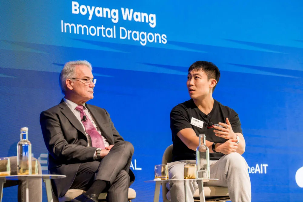
Martin O’Dea welcomed guests with his opening talk. Martin is a well-known figure in the longevity community, an experienced businessman, an author, and the CEO of Longevity Events Limited, which hosted the Longevity Summit Dublin.

Longevity Summit Dublin 1

He reflected on the recent trend of longevity and how the conference was trying to combine public interest in healthspan and well-being with the actual science of rejuvenation. He also cautioned that while he believed the two things could work together, it was important to ensure that science remained the focus for the event.

Martin suggested that lifespan and healthspan aren’t in opposition but that they need to be combined to work together and not in opposition. He said, “We can chew gum and walk; we can do healthspan and lifespan together.”

The idea that lifespan and healthspan are separate and competing goals makes little sense when you think about the actual biology at play here. It is not impossible to increase one but not the other, the modern healthcare system has achieved this by increasing lifespans but not healthspans, but the two are strongly linked.

The goal of our field is not to increase one over the other. The aim is to provide both longer and healthier lifespans for everyone. Ultimately, this is about quality and quantity, and this is what Martin was driving at in his opening talk.

Nanotics could really shake up rejuvenation biotechnology

The first presentation was by Lou Hawthorne from biotech company NaNotics, which pushes the envelope in trying new things. Its approach is to use NaNots to remove target molecules from blood without the use of drugs or external filtering devices.

Longevity Summit Dublin 2

NaNots are tiny artificial structures that lock in the target molecules away, making them ready to be excreted by the body. One advantage of NaNots is that they can remain in blood for days while absorbing these molecules.

This company’s current focus is on tumor necrosis factor (TNF), a regulator of the immune system and inflammatory response. NaNots only target the soluble TNF in blood and ignore membrane TNF, which is important as the membrane-bound form of TNF is very important for healthy cell function. NaNots are able to be selective in this manner, unlike traditional drugs that can target both forms.

Lou mentioned that the soluble form of TNF is linked to multiple sclerosis (MS) and that the company is hoping to move to the clinic to try to cure it. He reported that the mouse data they have for the approach is promising enough to move towards a clinical trial.

NaNots may also potentially be applied to deal with cancer metastases and tumors. Lou said that the company is also looking at PDL-1 with cancer treatment in mind. Additionally, the company intends to tackle sepsis and the resulting cytokine storm it causes, and in addition to TNF, it hopes to use NaNots to hoover up soluble interleukin-1β and interleukin-6 cytokines.

More generally, inhibiting unwanted pro-inflammatory cytokines could potentially be used to address inflammaging, the smoldering background of inflammation present in the majority of older people. NaNotics plans to use its product to target these inflammatory signals, which are secreted by senescent cells and created by other sources. Effectively, NaNots might be used as a form of senotherapeutic.

If the chronic inflammation observed in older people could be effectively reduced without harming healthy cell function, it might delay or even prevent a number of age-related diseases. NaNots are a new and highly selective approach to old problems.

Life Biosciences is bringing partial cellular reprogramming to the clinic

Michael Ringel from Life Biosciences had some interesting news about his company’s work on Yamanaka factors.

Longevity Summit Dublin 3

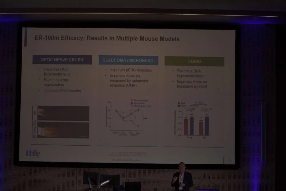
The expression of the Yamanaka factors Oct4, Sox2, Klf4, and c-Myc (OSKM) has been found to restore youthful gene expression patterns, reverse epigenetic age, and make old cells and tissues functionally younger.

Life Biosciences has been exploring these so-called reprogramming factors for a number of years, hoping to reverse epigenetic alterations, one of the reasons we age.

It is known that exposing old cells to these factors can make them functionally young again; however, doing so reverts their types to a developmental state. This is a problem because we don’t want the target cells to have their identities erased; heart cells forgetting they are heart cells would be a big problem!

Fortunately, researchers worked out that if cells are only exposed to the Yamanaka factors for just a brief period of time, it is enough to reverse their age without erasing their identities. This is known as partial cellular reprogramming.

The tricky thing is achieving this transient exposure in living animals. Life Biosciences has worked out a way of achieving this short-term exposure.

The company has been using just three of these factors in its research: Oct4, Sox2, and Klf4 (OSK). There was a concern that c-Myc could encourage the onset of cancer, and, as fortune would have it, not using it still allowed partial reprogramming to occur.

Michael explained that the company is now focusing its efforts on bringing partial cellular reprogramming to the clinic later this year. It is planning to move to Phase 1 for optic neuropathy, particularly glaucoma and non-arteritic anterior ischemic optic neuropathy (NAION).

The company is using doxycycline, something not found in nature, to turn on OSK gene copies delivered to target cells. This allows them to transiently turn the OSK genes on or off based on the presence of doxycycline. Effectively, this acts as a kill switch if things go wrong and controls exposure just enough to partially reprogram the old cells back to being young.

Life Biosciences has demonstrated that this works in mice with age-related optical neuropathy and reports that primate study results have been positive. It seems that partial cellular reprogramming is finally approaching the clinic after decades of research. If successful, it could offer a potential solution to repairing and rejuvenating aged cells and tissues.

Positive results for therapeutic plasma exchange in human trials

Dobri Kiprov from Global Apheresis was on hand to talk about the latest results from his research in therapeutic plasma exchange (TPE). He discussed how his results support that the rejuvenation effect seen in TPE is due to a dilution effect on the negative factors in aged blood, rather than there being some secret sauce in young blood.

Longevity Summit Dublin 4

He went on to explain that fresh albumin is anti-inflammatory and that replacing the old albumin improves patient outcomes. So far, his company has conducted two human trials for Alzheimer’s using TPE, with the results suggesting the procedure is safe.

The second, larger trial involved 40 people and included sham apheresis. Patients were either hooked up to an apheresis machine to have albumin exchanged or connected to a noise-making machine behind a curtain that did not actually exchange plasma. In this way, the participants had no idea if they were receiving actual TPE or just a mock treatment. This second trial also had two treatment frequencies in the test group.

Dobri reported that grip strength and balance improved in all treatment groups but not in the sham one. This trial used an impressive 35 aging clocks, and, broadly speaking, there was a significant reversal of aging markers. It seems that TPE rejuvenates the stem cell niche and makes the signaling environment more like that seen in younger people.

The take-home here is that the effects of TPE, as demonstrated in mouse studies by people such as Irina and Michael Conboy, appear to translate to humans. This means that TPE could potentially help us to stay healthier and biologically younger for longer.

However, for a rejuvenation technology to be truly successful, it needs to be both cost effective and scalable. As TPE likely needs to be done 3-4 times a year, based on what Dobri suggested, there must be a lot of procedures conducted. As many people would likely want TPE for its anti-aging effects, there is a question of scalability. Clearly, that is a consideration that will require creative solutions.

Perhaps even more intriguing is the possibility we could find out exactly what it is about aged plasma that is harmful and repair it in situ, instead of replacing it with new albumin. NaNots and other new technologies might be harnessed to that effect to achieve this, depending on what needs fixing in the albumin of course. One thing is certain, now we know that TPE works for people, the race is on to find scalable solutions.

A new early type of stem cell

Yuta Lee from Accelerated Biosciences announced that his company has the earliest form of stem cell free from ethical issues. Human trophoblast stem cell (HTSC) stem cells are gathered from ectopic pregnancies, which occur when a fertilized egg implants itself outside of the womb, usually in one of the fallopian tubes.

These stem cells are the earliest stem cells without ethical concerns, as the embryo is non-viable in these pregnancies. They are between embryonic stem cells and mesenchymal stem cells in terms of lineage and potency. Yuta suggested that these HTSC stem cells are also apparently clean of endogenous viruses.

One of the advantages of HTSCs is they are highly scalable due to the number of potential cell passages compared to other types of stem cells.

Yuta reported that HTSC cell secretions, like those of other types of stem cells, inhibit the inflammatory SASP secreted by senescent cells. That suggests that they may find application in the treatment of inflammatory diseases and conditions.

The company has already been successful in its good manufacturing practice (GMP) requirements. GMP describes the minimum standard that a medical manufacturer must meet in their production processes. Accelerated Biosciences wants to work with others to bring solutions for age related diseases.

Cyclarity Therapeutics progressing with human clinical trials

Earlier this year, we interviewed Matthew (Oki) O’Connor after Cyclarity launched human trials to Cure atherosclerosis, and he was at the Summit with an update.

Longevity Summit Dublin 5

The good news is that the initial part of Cyclarity’s Phase 1 clinical trial in Adelaide is done. The first five dosing levels are complete, and no adverse reactions have been observed with UDP-003.

The next step is about to begin; this will be the ascending dose group. The purpose of this is to gain an initial insight into the pharmacokinetics of a drug’s single dose and its safe dosage range. It is recommended to administer doses to participants one after another, allowing sufficient observation time between each.

Oki also mentioned that Cyclarity is planning for a 150-person trial for Phase 2 in Europe. While there is no date on that yet, they are pushing hard on Phase 1 and so it could even be this year.

Finally, he revealed that his company is developing an AI-based system to optimize cyclodextrin drug development. While the plaques in atherosclerosis are the target of UDP-003, Cyclarity is interested in removing other harmful molecules using this system.

Cyclarity is also working on finding solutions to nanoplastics, things like BPA and PFAS that the company believes its technology could potentially address. This could potentially address unhealthy levels of nanoplastics in people and remove them from the blood, cells, and tissues where they have accumulated.

We are looking forward to hearing more from Cyclarity and are proud that we helped this company to be founded. Heart disease is the number one killer worldwide; to have a solution to treat it effectively would be game-changing.

Our knowledge of female biology and aging is lacking

Jennifer Garrison from the Buck Institute waded in on female biology and aging. She believes that a loss of homeostasis systems in the brain regulates aging. Jennifer highlighted that male biology is better understood than female biology.

Longevity Summit Dublin 6

She said that females have a shorter healthspan than males. Some of this is likely due to how fast ovaries age, up to twice as fast as other organs. Ovaries appear to be part of a wider signaling system, which isn’t well understood but appears to promote female healthspan and lifespan.

Jennifer talked about how perimenopause causes a breakdown of this communication and promotes aging, and menopause does even more of this.

She said that HRT is a band-aid and can reduce all-cause mortality by up to 30% if given within ten years of menopause. Also, on average, if menopause is later in life, then lifespan is often longer as well. She believes that if we can extend ovarian function by delaying aging of the organ, we could increase female healthspan.

More funding for female aging is urgently needed. Women’s health is seeing research cutbacks by the NIH at a time when it badly needs to be improved. It is more important than ever that more focus is put into understanding how women age and the additional functions ovaries play in that aging.

There are now a number of companies exploring female aging. If their efforts to rejuvenate or extend the healthspan of ovaries succeeds, this will be a great demonstration that aging is not a one-way street.

Lifespan Research Institute: A new org, a new direction

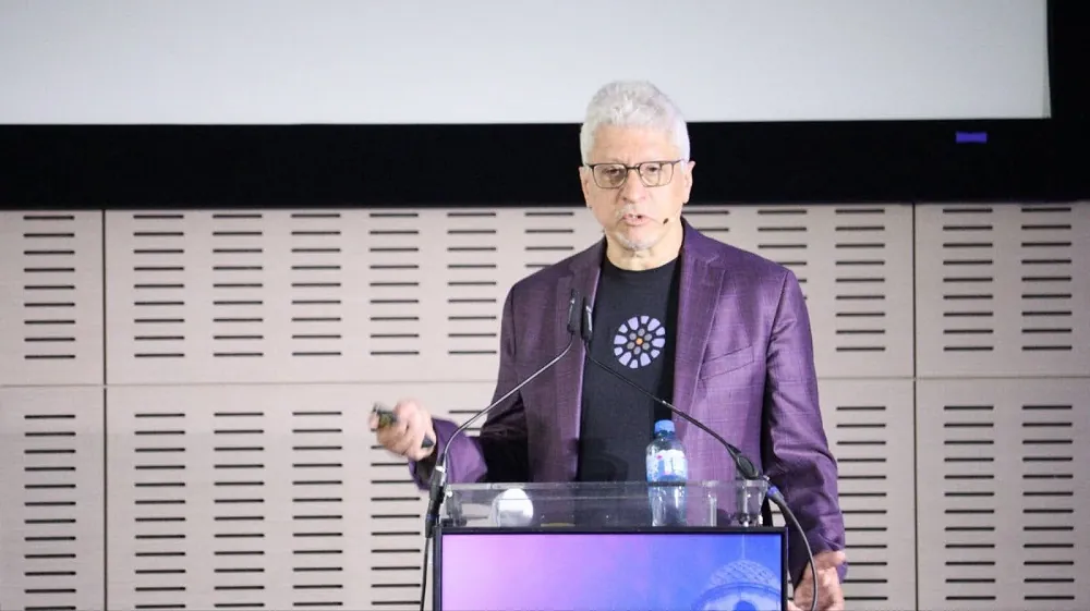
Lifespan Research Institute (LRI) President Keith Comito focused on the always-important need to work together to face the challenges our field presents. Solving aging is the greatest challenge humanity has ever faced, and, as a relatively young industry, it is critical that we collaborate where possible to make rapid progress.

Longevity Summit Dublin 7

Among the challenges we face is getting wide public support for rejuvenation biotechnology. Keith said, “Bringing the public along with us on the journey is important. We need to meet them where they are, rather than assume they will just get on board.”

Regarding public engagement, he ventured that developing AI-based tools could also help support effective advocacy and determine public sentiment towards rejuvenation biotechnologies. Keith is well known for his work with AI and other disruptive technologies and he feels they could be used to great effect for our field.

He gave four historical examples where interest in rejuvenation appeared to peak:

Longevity Summit Dublin 8

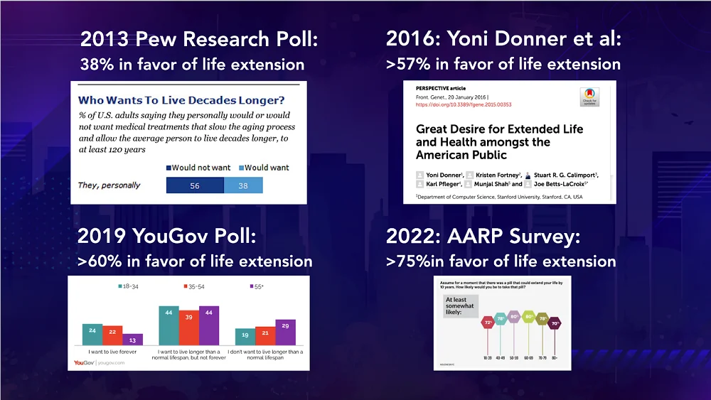
Imagine if we had AI-based tools to help interpret these peaks in public interest and to help identify what approaches work best. This would mean we could more effectively engage with people about the field. We will have more to say about new public advocacy tools in the near future.

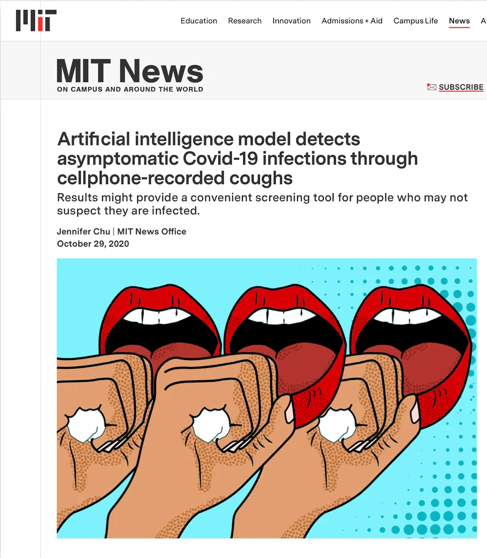
Keith also highlighted the importance of non-invasive biomarkers of aging and how machine learning might be a useful tool in our longevity arsenal. He gave an example of when machine learning was used as a detection method for COVID-19.

Longevity Summit Dublin 9

He suggests that machine learning and AI more generally may be adapted in the context of aging research. For instance, it isn’t hard to imagine how an AI based biomarker system may be useful in the context of functional aging.

Consider a system that could examine gait and body movement and identify trends associated with age-related changes. Combined with clinical biomarkers, these non-invasive biomarkers could potentially help round out more comprehensive biomarker panels.

Finally, Keith took the opportunity to talk about the LRI, our new organization created by the merger of LEAF and SENS in October last year. We have launched a new website that showcases our work and explains how we are adapting to the changing landscape of longevity and rejuvenation research.

Longevity Summit Dublin 10

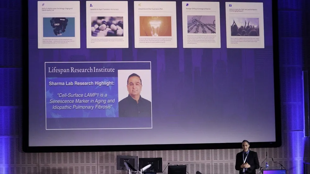
LRI has five broad guiding principles behind its research.

  • Ability To Boldly Impact the Biology of Aging and Extend Healthy Life
  • Capability for Rapid Translation into Humans
  • Uniqueness and Non-Duplicative Effort
  • Paradigm-Shifting Technological Advances
  • Ability to Inspire the Public and Lead the World to Prioritize Aging Research

At our Mountain View, California research center, we have two pioneering labs working on repair-based solutions to age-related diseases.

The Boominathan Lab: Dr. Amutha Boominathan’s research team focuses on exploring mitochondrial biology, creating gene therapies to treat mitochondrial issues, and improving therapies for conditions associated with mitochondrial DNA mutations and aging processes.

The Sharma Lab: Dr. Amit Sharma’s research team is exploring the impact of aging and cellular senescence on immunity with a focus on creating approaches to utilize immune responses for identifying and eliminating senescent cells.

The emphasis is very much on actionable research that helps propel the field forward as rapidly as possible. If you would like to support our non-profit mission for longer and healthier lives, see how you can help us.

See you next year for more longevity and rejuvenation

There were many more talks during the conference, too many to list and discuss here. We have picked out the ones that most resonated with us and our mission of accelerating technologies to overcome age-related disease and extend healthy human lifespan.

The Lifespan team was delighted to be a part of this year’s conference, and we would like to thank our hosts for inviting us. We are looking forward to the 2026 Longevity Summit in Dublin and wish Martin and the team the best in making that happen!

Blood vessel in bone

Non-Toxic Stem Cell Transplantation Prevents Cancer in Mice

Scientists have developed a protocol for hematopoietic stem cell transplantation that reconstructs a healthy blood system and prevents blood cancers in old mice while also reducing toxicity [1].

The blood factory

Hematopoietic stem cells (HSCs) work hard for our entire lives, producing vast numbers of various blood cells. As we age, this process, like many others in the body, gets dysregulated [2]. This contributes to the decline of the immune system and to the development of several harmful conditions, including blood cancers.

HSC transplantation is a feasible strategy, but it requires depleting the host’s own stem cells to make space in the bone marrow niche for the donor’s cells to engraft. Today, this is achieved mostly by chemo- and radiotherapies, which are notorious for their powerful side effects [3]. In a new study published in Nature Communications, researchers at the Lund Stem Cell Center at Lund University, Sweden, attempted to develop a safe and effective method for transplanting young, healthy HSCs into aged recipients.

“Conventional transplantation requires patients to undergo chemotherapy or irradiation to eliminate malignant cells, suppress immune rejection, and make room for new stem cells in the bone marrow,” explained David Bryder, Professor of Molecular Hematology at Lund University. “But these treatments are highly toxic, especially for older individuals, who are also the ones most likely to benefit.”

More cells, more drugs, less toxicity

First, the researchers tested an existing non-genotoxic conditioning agent, CD45-saporin (CD45-SAP), an immunotoxin that selectively eliminates HSCs without widespread toxicity, in both young (2-month-old) and aged (16-month-old) mice. Then, the team transplanted young HSCs and monitored the success of the transplant.

The team found that the conditioning was much less effective in aged mice. While CD45-SAP successfully depleted HSCs in young mice, a significant number of the host’s own HSCs remained in the aged mice. Consequently, when young HSCs were transplanted, they engrafted successfully and created new blood cells in the young mice but not in the aged group.

Having found that standard doses of HSCs fail to engraft well in aged mice, the researchers explored whether using a much larger number of donor HSCs could force a successful transplant. They expanded a small number of young HSCs into a large population ex vivo and then transplanted these cells into young mice, hypothesizing that the available niches for HSCs in the bone marrow might be more reliably populated if “flooded” with sufficient numbers of donor cells.

At this stage, the researchers used only young mice to prove that an increase in HSC numbers can improve transplantation outcomes. Transplanting large numbers of expanded HSCs indeed led to successful, long-term multilineage blood cell production even in unconditioned young hosts. Combined with CD45-SAP conditioning in young mice, the result was a robust and near-complete reconstitution of the blood system.

“In particular, the mice showed a strong resurgence in the production of naïve B and T lymphocytes, cells that play a key role in immune defense, which usually decline with age,” said Bryder. “These young cells didn’t just survive; they reshaped the entire immune landscape. We were especially encouraged to see that the new cells maintained long-term function and diversity, even within an aged environment.”

The researchers hypothesized that the primary problem with aged hosts was the failure to clear out enough of the old, resident HSCs. To fix this, they developed a more potent conditioning regimen by adding to the CD45-SAP immunotoxin a two-drug mobilization regimen (G-CSF/AMD3100), which forces the remaining host HSCs out of their protective bone marrow niches and into the bloodstream.

“Instead of using conventional chemotherapy, we deployed antibody-toxin conjugates, also known as immunotoxins, that selectively target and deplete the recipient’s own HSCs while sparing surrounding tissues,” explained Anna Konturek-Ciesla, postdoctoral researcher and first author of the study. “This was paired with a drug-based mobilization strategy that temporarily displaces stem cells from the bone marrow, creating space for incoming donor cells.”

Successful prevention in vivo

Finally, the researchers tested their strategy in a disease context, using a transgenic mouse model (NHD13) that is genetically predisposed to developing myelodysplastic syndrome (MDS) and acute leukemia, common blood disorders in elderly humans. They treated two-month-old mice with the combined conditioning regimen and transplanted healthy, wild-type bone marrow cells. The mice were then monitored for their entire lifespan.

In the untreated group, 75% of the mice developed blood malignancies, versus just 33% in the study group. Most strikingly, while 25% of untreated mice developed aggressive acute leukemia, none of the transplanted mice did.

The treatment was initiated at this young age because these transgenic mice develop symptoms early. The researchers also position their invention as “a prophylactic tool to delay or even prevent” the onset of age-associated hematological disorders. Potentially, non-toxic HSC transplantation can be used in an even wider context in order to prevent age-related immunosenescence, which is considered a major cause of aging and mortality.

“While these findings are currently limited to animal models, and there are many more steps to take before this can be applied in humans, they offer a proof-of-concept, that aged or malfunctioning stem cells can be safely replaced (without the toxicity of traditional conditioning) and that youthful blood production can be restored even in an older body,” summarized Konturek-Ciesla.

Literature

[1] Konturek-Ciesla, A., Zhang, Q., Kharazi, S., & Bryder, D. (2025). A non-genotoxic stem cell therapy boosts lymphopoiesis and averts age-related blood diseases in mice. Nature Communications, 16(1), 5129.

[2] Kim, M. J., Kim, M. H., Kim, S. A., & Chang, J. S. (2008). Age-related deterioration of hematopoietic stem cells. International journal of stem cells, 1(1), 55-63.

[3] Gyurkocza, B., & Sandmaier, B. M. (2014). Conditioning regimens for hematopoietic cell transplantation: one size does not fit all. Blood, The Journal of the American Society of Hematology, 124(3), 344-353.

Astrocytes on blood vessel

How Blood-Brain Barrier Leaks Make Parkinson’s Worse

Researchers have discovered how α-synuclein (α-syn), a key protein in Parkinson’s disease and Lewy body dementia, leads to inflammation and disruption of the axons in the brain.

Failure of the barrier

Unlike other organs, the brain is heavily protected from many compounds in the bloodstream in order to prevent damage, with a unique combination of cells and junction proteins involved in this layered defense: this is the blood-brain barrier (BBB) [1]. As expected, damage to the BBB is directly related to neurodegenerative diseases [2].

This relationship has been very heavily studied for Alzheimer’s disease [3], but only limited work has been done to tie together the BBB and α-synucleinopathies [4]. While most work on α-syn has focused on how it directly damages cells in the context of Parkinson’s [5], these researchers note that BBB damage is likely to have its own negative effects that need to be included for accurate drug development [6].

Aggregates cause damage to cells

For accurate results, the researchers investigated both the usual, monomeric form of α-syn along with an aggregate called preformed fibril α-syn (PFF). They then introduced each of these proteins to endothelial cells that line the BBB (HBMVECs) in vitro.

Both regular α-syn and PFF were taken up by the HBMVECs. While these cells did not react to the aggregate within the first hour, the PFF fibrils were carried to lysosomes for processing within a day. Before it could be properly processed, however, PFF was found to lead to disruption of vascular endothelial cahedrin (VE-cadherin), a core protein of the BBB. This disruption led to increased penetration of a form of dextran thaat does not normally penetrate the BBB. Unaggregated α-syn, which is associated with normal function, did not have such effects.

A gene expression analysis revealed a link to inflammation. While there were no interesting differences between the monomeric α-syn group and the control group, the PFF group had significant differences. At 24 hours, a gene cluster that is related to inflammatory factors, such as NF-κB and TNF-α, was strongly upregulated compared to controls. Cellular proliferation and growth were also impeded according to this analysis.

Because genes are strongly inter-related and the underlying biology is exceptionally complicated, the researchers used an AI algorithm to determine what upstream pathways were responsible for this change in gene expression; many of these genes were well-known in the literature for being related to inflammation. They also found that TNF-α was uniquely upregulated, becoming elevated by nearly 250-fold within one hour compared to controls.

Critically, inhibiting TNF-α in these cells, while it did not completely reverse the effects, led to lower permeability of dextran. This suggests potential benefits for the BBB.

Heavy BBB leakage in mice

The researchers used mice that were modified to aggregate α-syn (G2-3 mice), first confirming that these aggregates were indeed found in both the brain tissue and in the vasculature. They then tested for claudin 5, a core protein responsible for BBB integrity. Unsurprisingly, they found that claudin 5 in G2-3 mice was significantly decreased from that of wild-type mice, although it took 13 months of aging for this damage to appear to a statisticallly significant degree. An antibody for immunoglobulin G (IgG) demonstrated a tremendous amount of BBB leakage: at 13 months, approximately six times as much IgG had worked its way into the brains of the G2-3 mice than those of wild-type mice.

Pericytes, along with the endfeet of astrocytes, are also part of BBB maintenance. Compared to wild-type controls, the G2-3 mice had more astrocytic activity, showing that they were working harder to compensate for the porous BBB. However, aquaporin 4, a protein involved in disposing of potentially dangerous waste, was depleted, which suggests that the astrocytes were overwhelmed. Similarly, a marker of pericyte activity, PDGFRβ, was upregulated, suggesting that these cells were also working much harder to defend the leaky BBB.

Small vessel disease (SVD) is a BBB failure that leads to nerve damage. Microglial inflammation near the vasculature, which is found in SVD, was also found in the G2-3 mice. Degeneration of the extracellular matrix was discovered, and there was evidence of axonal damage near the vasculature as well.

A potential treatment may already exist

In another experiment, the researchers used a wild-type strain of mice and injected their brains with PFF. They then dosed some of the mice with etanercept, a TNF-α inhibitor that is used to treat arthritis and does not normally penetrate the BBB. Compared to the mice that received PFF but not etanercept, the treated mice had significantly less IgG infiltration, nearly to the levels of the control group. Very significant effects were also found when etanercept was given to G2-3 mice, including benefits against the effects of SVD.

Etanercept was also found to have downstream benefits in mice. α-syn aggregation was found to cause damage to both novel object recognition, which measures cognitive function, and the rotarod test, which measures balance ability. Both of these metrics were improved with etanercept.

While there is still no evidence that this could work as a treatment for Parkinson’s in human beings, it is clear that BBB disruption and the resulting inflammation are likely to strongly contribute to Parkinson’s pathology. A clinical trial could validate whether etanercept or another drug that disrupts TNF-α could blunt the effects of this debilitating disease.

We would like to ask you a small favor. We are a non-profit foundation, and unlike some other organizations, we have no shareholders and no products to sell you. All our news and educational content is free for everyone to read, but it does mean that we rely on the help of people like you. Every contribution, no matter if it’s big or small, supports independent journalism and sustains our future.

Literature

[1] Jeon, M. T., Kim, K. S., Kim, E. S., Lee, S., Kim, J., Hoe, H. S., & Kim, D. G. (2021). Emerging pathogenic role of peripheral blood factors following BBB disruption in neurodegenerative disease. Ageing research reviews, 68, 101333.

[2] Baloyannis, S. J., & Baloyannis, I. S. (2012). The vascular factor in Alzheimer’s disease: a study in Golgi technique and electron microscopy. Journal of the neurological sciences322(1-2), 117-121.

[3] Ryu, J. K., & McLarnon, J. G. (2009). A leaky blood–brain barrier, fibrinogen infiltration and microglial reactivity in inflamed Alzheimer’s disease brain. Journal of cellular and molecular medicine, 13(9a), 2911-2925.

[4] Pediaditakis, I., Kodella, K. R., Manatakis, D. V., Le, C. Y., Hinojosa, C. D., Tien-Street, W., … & Karalis, K. (2021). Modeling alpha-synuclein pathology in a human brain-chip to assess blood-brain barrier disruption. Nature communications, 12(1), 5907.

[5] Michel, P. P., Hirsch, E. C., & Hunot, S. (2016). Understanding dopaminergic cell death pathways in Parkinson disease. Neuron, 90(4), 675-691.

[6] Elabi, O., Gaceb, A., Carlsson, R., Padel, T., Soylu-Kucharz, R., Cortijo, I., … & Paul, G. (2021). Human α-synuclein overexpression in a mouse model of Parkinson’s disease leads to vascular pathology, blood brain barrier leakage and pericyte activation. Scientific reports, 11(1), 1120.

Mitochondrion in cell

Scientists Successfully Edit Mitochondrial DNA

A new study demonstrates that novel gene-editing tools can correct disease-causing mutations in mitochondrial DNA in primary human cells [1].

Smaller editing tools needed

Genome-editing tools such as CRISPR were one of the greatest scientific breakthroughs of this century. However, they are only good for editing nuclear DNA.

Mitochondria, the energy-producing organelles, have their own circular DNA (mtDNA) that resides inside each mitochondrion and codes for a number of essential proteins. Mutations in mtDNA cause several diseases and are also associated with aging [2]. Until very recently, there was no easy way to edit mtDNA since CRISPR-based tools are too large to enter mitochondria.

The situation began to change with the introduction of smaller editing tools, but more research is needed to test and refine them. In a new study published in PLOS Biology, scientists from the University Medical Center Utrecht in the Netherlands used the double-stranded DNA deaminase toxin A-derived cytosine base editor (DdCBE), paired with guiding proteins called TALE, “to develop in vitro disease models and assess therapeutic strategies for mitochondrial diseases in primary human cells.”

Creating a disease model

First, the team used DdCBE to introduce a loss-of-function mutation (m.15150G>A) in human primary adult liver stem cell-derived organoids. This particular mutation has not been associated yet with any known disease, but other mutations in the same gene (MT-CYB) have. The researchers report that their editing tool successfully introduced the mutation.

This is an important step in creating models of mitochondrial diseases so that they can be studied and cured. “Mitochondrial dysfunction and mtDNA alterations are implicated in several age-associated pathologies, however, our ability to understand the underlying mechanisms is limited by lack of appropriate models,” said Dr. Amutha Boominathan, a senior researcher at the Lifespan Research Institute, who was not involved in this study.

While a cell has only two copies of the nuclear DNA, one from each parent, there can be hundreds of thousands of mitochondria in each cell, each one with its own circular DNA. Therefore, an edit needs to be introduced to as many of those copies as possible. The presence of more than one type of mtDNA within a single cell is called heteroplasmy.

When the researchers introduced a pathogenic mutation into healthy liver organoids, they did not create cells that were 100% mutated. Instead, by isolating and growing single cells, they generated a collection of organoid lines with a wide range of heteroplasmy levels (from 0% to 80% mutated). This allowed them to study the effects of different levels on the severity of the disease, as naturally occurring DNA diseases also manifest themselves only past a certain heteroplasmy threshold.

Mutation fixed

The next step was to try fixing a known harmful mutation. In fibroblasts from a patient, the DdCBE system successfully corrected the pathogenic m.4291T>C mutation, which is linked to Gitelman-like syndrome, a group of inherited kidney disorders.

Heteroplasmy remained a challenge: when the researchers grew out colonies from single edited cells, they found a wide range of DNA correction levels. On the bright side, those levels remained stable over 50 days of follow-up and even slightly increased, showing that the corrected mitochondria were healthy and not at a selective disadvantage within the cell.

In cell lines with a high level of correction (76% and 81%), the mitochondrial membrane potential was successfully restored to the level of healthy control cells, suggesting functional rescue. In a line with low correction (35%), there was no improvement.

The results for overall energy production were more modest and inconsistent. While slight improvements were observed in some experiments, the effect was not as strong or reliable as the restoration of the membrane potential. The authors note that this warrants further study.

Initially, the team used a tried-and-true method of delivery: DNA carried by viral vectors. In later experiments, they showed that a better method was to deliver the editor as modified RNA (modRNA). The modifications included tweaking RNA nucleotides for greater stability and shielding the molecule from being detected by the immune system. Compared to DNA delivery, modRNA demonstrated much higher efficiency and less cytotoxicity.

The modified RNA molecules were delivered using lipid nanoparticles (LNPs). This is the same state-of-the-art technology used to deliver the mRNA in COVID-19, considered the most advanced non-viral system for in vivo delivery.

“Adapting precision DNA editing tools such as base editors to target the mitochondrial genome holds significant promise for both modeling and treating mitochondrial DNA (mtDNA) mutation-associated diseases,” said Dr. Boominathan. “However, this approach faces several challenges, including the high number of edits required per cell (due to the large mtDNA copy number), achieving homogeneous editing across cell populations, and minimizing off-target effects. In this study, the authors successfully generated a pathogenic mutation in liver organoids and corrected the m.4291T>C mutation in patient-derived fibroblasts. Nonetheless, limitations such as variability in editing efficiency – both in the extent and uniformity of edits – persist and warrant further optimization.”

We would like to ask you a small favor. We are a non-profit foundation, and unlike some other organizations, we have no shareholders and no products to sell you. All our news and educational content is free for everyone to read, but it does mean that we rely on the help of people like you. Every contribution, no matter if it’s big or small, supports independent journalism and sustains our future.

Literature

[1] Joore, I. P., Shehata, S., Muffels, I., Castro-Alpízar, J., Jiménez-Curiel, E., Nagyova, E., … & Koppens, M. A. (2025). Correction of pathogenic mitochondrial DNA in patient-derived disease models using mitochondrial base editors. PLoS biology, 23(6), e3003207.

[2] Sprason, C., Tucker, T., & Clancy, D. (2024). MtDNA deletions and aging. Frontiers in Aging, 5, 1359638.

Neural connections

Senolytics May Affect Inflammation-Related Cognitive Decline

Researchers have found that inflamed, senescent microglia prune too many synapses in the hippocampus and demonstrated that a senolytic compound can ameliorate this process in Aging Cell.

Some synapse pruning is normal

During brain development, the resident immune cells of the brain (microglia) prune unneeded synapses as a form of maintenance. This is normally a beneficial process both in young children [1] and mature adults [2], as the removal of unnecessary circuits facilitates the formation of more meaningful connections.

However, as with any of the body’s cleanup processes, disease states can send it into overdrive, causing damage. For example, during the inflammation brought on by blood sepsis, microglia tear apart functional synapses, leading to cognitive decline [3].

Like other cells, microglia can become senescent and unable to proliferate further. However, this state does not mean that they are turned off completely. While the two appear to be related, senescent microglia and disease-associated microglia are not quite the same [4].

Inflamed microglia express genes differently

This experiment began by exposing 8- to 10-week-old Black 6 mice to lipopolysaccharides (LPS) for one week in order to cause neuroinflammation. A gene expression analysis revealed that, of the 20 most upregulated genes, a full eight were related to debris clearing (phagocytosis), including genes related to Complement 1q, a compound related to synaptic pruning. Five more upregulated genes were related to senescence.

These findings were confirmed with an examination of lysosomal and activity markers. The microglia in the LPS-exposed mice were significantly more involved in phagocytosis and were also more senescent according to the p16 biomarker, which was significantly increased in the active microglia, and another examination showed that the senescent microglia in LPS-exposed mice had some morphological distinctions from the senescent microglia in the control group. Astrocyte activity was also increased by LPS. Interestingly, this phagocytosis appeared to be only limited to excitatory, rather than inhibitory, synapses, which were unaffected by this chemical.

Senolytics appear to be effective

As expected, the LPS treatment resulted in measurable levels of cognitive decline. The mice given LPS were less able to navigate a Y maze, less interested in novel objects, and less willing to navigate an open field. However, treatment with the senolytic compound ABT-737 reversed this decline, making most of their measurements indistinguishable from those of the control group.

This improvement was not due to benefits in neuroinflammation; multiple fundamental inflammatory biomarkers, including SASP biomarkers, were unaffected by ABT-737. Instead, it affected markers more directly related to senescence, such as p16 and p21. This reduction was accompanied by a decrease in the number of senescent microglia in the hippocampi of these mice.

Most importantly, ABT-737 treatment appeared to do what it set out to do. The phagocytosis of excitatory synapses was reduced in the treated mice, although, like with the behavioral analysis, not all markers were reduced to the levels of the control group. The number of dendritic spines, which decreases with LPS, was restored with ABT-737, and neuroplasticity, as measured by postsynaptic potential, also appeared to be partially restored.

These experiments used mice that were treated with an inflammatory compound, not aged mice. Further work will need to be done to determine if ABT-737 or any other senolytic is able to ameliorate the cognitive decline brought on by senescent microglia in the context of aging.

We would like to ask you a small favor. We are a non-profit foundation, and unlike some other organizations, we have no shareholders and no products to sell you. All our news and educational content is free for everyone to read, but it does mean that we rely on the help of people like you. Every contribution, no matter if it’s big or small, supports independent journalism and sustains our future.

Literature

[1] Bohlen, C. J., Friedman, B. A., Dejanovic, B., & Sheng, M. (2019). Microglia in brain development, homeostasis, and neurodegeneration. Annual Review of Genetics, 53(1), 263-288.

[2] Colonna, M., & Butovsky, O. (2017). Microglia function in the central nervous system during health and neurodegeneration. Annual review of immunology, 35(1), 441-468.

[3] Chung, H. Y., Wickel, J., Hahn, N., Mein, N., Schwarzbrunn, M., Koch, P., … & Geis, C. (2023). Microglia mediate neurocognitive deficits by eliminating C1q-tagged synapses in sepsis-associated encephalopathy. Science advances, 9(21), eabq7806.

[4] Rachmian, N., Medina, S., Cherqui, U., Akiva, H., Deitch, D., Edilbi, D., … & Schwartz, M. (2024). Identification of senescent, TREM2-expressing microglia in aging and Alzheimer’s disease model mouse brain. Nature neuroscience, 27(6), 1116-1124.

Gabriel Cian Interview

Gabriel Cian on Investment and the 2060 Longevity Forum

In this Lifespan interview, we speak with Gabriel Cian, founder of the 2060 Longevity Forum, about how his background in software shaped his views on healthspan innovation, the forum’s approach to scientific and investment credibility, and what he sees as the biggest bottlenecks and opportunities facing the field of longevity today.

Hi, Gabriel. Let’s start with your background. Can you introduce yourself and explain how your path led from software entrepreneurship into the longevity space?

When I sold my last startup in the tech space, I had just turned 40. According to Western health standards, I could expect to live in relatively good health until around 65 followed by 15 years of chronic illness.

That would mean I would die sometime around 80. That was it, just 25 more healthy years, with so much still to experience and achieve. I wanted more. That’s when I discovered longevity research.

I began to realize that the relentless pace of entrepreneurship had taken a serious toll on my health. I was prediabetic, under chronic stress, had a misaligned vertebra in my spine, and showed inflammatory markers comparable to those of a 72 year old.

It was a serious wakeup call for me!

Picturing myself dedicating the second half of my life to longevity, helping Humanity overcome aging felt fully aligned with myself.

This is how the 2060 Foundation, whose purpose is to help humanity defeat Aging, was born.

Would you say that your experience as a founder in tech has shaped how you think about healthspan, biotech, or systemic change more broadly?

When I started my first startup, everyone around me, friends, family, tried to convince me to give up. They said that I was crazy, that it would not work, but I chose to fight against this, and prove them wrong.

It took many years, even decades, but it eventually worked. Every argument against my startup and my project would make me even more motivated to make it work. Even though I had no money, no particular experience, and no precise idea on how I would succeed, I believed I could do it.

I’m roughly in the same position today, when it comes to longevity.

All of what I’ve done as a tech entrepreneur in the last 20 years feels now like training for what I’m doing now. I have to resist social pressure, and convince people around me that it is possible to slow down aging, and even reverse it.

But I have limited resources to make this happen; research in biotechnology is expensive! It may take decades until we know how to solve aging. Achieving this goal is all about grit, persistence, and audacity. Thinking and acting long term over the short term.

What motivated you to create the 2060 Longevity Forum, and how did you settle on the name and location?

I live in the south of France, one of the most beautiful, culturally rich, and longevity-friendly regions in the world.

My vision is to build a true Longevity Hub here: a place where longevity enthusiasts from all over the world can come and live with their families. They can then combine this with cutting-edge biotech research and medical infrastructure from advanced labs to preventative longevity clinics.

When it comes to the name of the conference, the year 2060 represents my commitment to invest the next 40 years of my life to humanity’s most audacious aspiration: solving aging. By that time, I’ll be 80!

With more longevity events emerging, how do you ensure this forum serves a unique purpose and doesn’t overlap with others?

My team and I have spoken with many attendees of longevity conferences and identified a clear gap: investment.

While numerous events rightfully focus on the academic and scientific side of longevity, very few put the spotlight on funding. There’s a real need for spaces where longevity startups can connect with investors, and where venture capitalists can engage with limited partners.

If we want to accelerate progress in this field, we need serious capital. At the end of the day, longevity isn’t just about science, it’s about funding.

The agenda spans well-supported interventions to more exploratory topics like cryonics and mind uploading. How do you decide what belongs on the program?

The core mission of the 2060 Longevity Forum is clear: channel as much capital as possible into the longevity ecosystem. More funding means more brilliant minds making more longevity breakthroughs.

Having said that, investors come in all shapes and sizes, so the projects and experts that come as keynote speakers need to cover the whole spectrum of possible interventions in longevity. With that in mind, I have invited longevity clinics, researchers, and startups working on whole body transplants.

How do you strike the balance between encouraging bold ideas and maintaining scientific credibility?

Having been a fundraising entrepreneur in the past and now an investor myself, I’ve come to realize that no two investors are the same. Each brings a unique mix of expertise, risk tolerance, and personal track record. Some are drawn to bold, visionary ideas; others prefer near-term, de-risked opportunities.

My mission is to engage as many of them as possible in the longevity space, which is why the 2060 Longevity Forum will showcase the full spectrum of projects from pragmatic, revenue-ready ventures to transformative moonshots.

I don’t have a specific algorithm to deal with this, it’s a case-by-case evaluation, and I’m also relying on experts that give me their opinion.

In short, I’m aiming for a balanced distribution between proven solutions and bold innovations across the whole spectrum of longevity startups.

You’ve called longevity the greatest investment opportunity of our time. What makes it so compelling to you personally?

I think AI is disrupting the field of biotechnology in an exponential and therefore unpredictable way, because we humans don’t know how to predict exponential curves, do we?

Rreally, the best way to predict the future is to create it yourself. So this feels like the right time to bet big time on major breakthroughs in the next years and decades.

Once I’ve said that, it becomes my mission to make that happen. Contributing to the field, and shortening the time before we discover something big, by channeling more funds into the field, makes it very compelling for me because I feel I can significantly contribute to that.

What do you see as the biggest bottlenecks to more investors getting involved in the space – and how are you trying to address them?

Investors could allocate significantly more capital to longevity, but several key obstacles stand in the way:

1. Lack of visibility and credibility

Many investors aren’t even aware that longevity is a serious investment opportunity. Without digging into the science, some still associate it with hype or snake oil, assuming aging is irreversible.

2. Lack of success stories

We need tangible wins both in terms of clinical breakthroughs and ROI to build trust. It’s a bit like AI before and after ChatGPT: investment was hesitant before, and exploded after. Longevity needs its own inflection point.

3. Lack of long-term thinking

Today’s investment models are geared toward 3x returns in 5-10 years, but longevity, with its R&D-driven nature, may offer 100x returns in 20 years. Imagine the market size for technologies that add even 10 extra years of healthspan. It’s massive. Investors can be educated to shift from short-term returns to exponential long-term impact.

4. Lack of meaning

ROI is important, but what about return on life? Most investors are trained to chase financial performance above all, but what if the most meaningful investment is one that helps extend life itself? Money is a tool, and using it to fund breakthroughs that allow us to live longer, healthier lives gives it its highest purpose.

This is why I’m taking a long-term approach to catalyze investment in longevity:

  • Through Ikare.Health, I help investors take care of their own health using the best longevity treatments available today. (Ikare is named after a small blue zone island in Greece.)
  • As they reach today’s limits, I challenge them to think bigger and invest in tomorrow’s breakthroughs through our 2060 Longevity Investment Club, a private community of investors backing longevity startups exclusively.
  • To unite the entire ecosystem, I’ve launched the 2060 Longevity Forum designed to increase visibility, spark connections, and create strong network effects. Virtual networks don’t work well until people meet in person.

I believe that with the right mix of education, community, and long-term vision, longevity investing is only just beginning. It may take decades to fully realize this ambition but it’s a mission well worth the journey.

This year’s event includes both startup pitch sessions and LP-GP networking. Why did you choose to spotlight both early-stage companies and fund managers?

The end game of what I’m trying to achieve revolves around one core mission: driving more investment into longevity. This includes supporting startups actively raising funds but also facilitating connections between GPs (fund managers) and LPs (capital providers) who believe in the long-term potential of the field.

Raising capital is never easy and it’s even more challenging in longevity, where timelines can be longer and the science more complex. That’s why I’m creating tailored opportunities for both types of investors:

  • For those who want to invest directly in startups, we host dedicated pitch sessions.
  • For those who prefer to back experienced fund managers, we facilitate direct connections with longevity-focused GPs.

I’ve seen strong enthusiasm for our LP-GP speed-networking format, and I’m fully committed to making it a standout success.

Some argue longevity may primarily benefit the wealthy. How do you think the field can evolve to better serve broader populations?

This is a crucial topic that deserves serious attention.

There’s a growing body of recent examples suggesting that once a life-extension technology proves effective, it’s likely to reach mass-market adoption from day one.

Take Large Language Models (LLMs) as an example. While not related to longevity, they represent one of the most disruptive technological leaps in human history. When ChatGPT launched in November 2022, no one expected it to become ubiquitous so quickly, yet just a few years later, it’s widely available, often for free, with multiple competitors offering similar tools at no cost. It’s used by millions, across all sectors of society.

One might argue that LLMs needed mass adoption to gather vast amounts of training data. But the same is true for longevity R&D we need large-scale datasets to uncover the right correlations between interventions and their impact on healthspan and lifespan.

We’re already seeing early signs of this mass-market trajectory in longevity-related drugs:

  • GLP-1 agonists (like semaglutide) are among the few longevity-linked treatments already on the market. While the extent of their healthspan benefits is still under review, these drugs are widely accessible and increasingly affordable.
  • Metformin, another life-extension candidate, is so inexpensive that it’s no longer of interest to big pharma. As a generic drug, it’s available to virtually everyone.
  • Rapamycin, also a promising compound in the aging field, is similarly low-cost and accessible to those who wish to explore its potential.

While concerns about life-extension technologies being reserved for a privileged elite are understandable, current trends suggest otherwise. The trajectory of innovation especially in tech and pharma is increasingly democratized. The evidence so far points to broad access, not exclusivity.

How geographically concentrated is longevity investment in your experience? Do you see notable differences – or advantages – emerging in particular regions?

From my experience, the US is leading by a short edge, but we’re very early on, and I strongly believe in a world where scientists from all the countries, organized in small and agile teams, can discover major things, taking advantage of their local scientific, regulatory, and cultural landscape.

For example, in some Eastern European countries, the cost of clinical trials is very low (thinking of The Cat Health Company), so they’re doing research in Romania.

In France, there are excellent, world-class scientists. Providing some funding and pairing them with more seasoned entrepreneurs could make terrific startups.

I was talking to another startup, Cyclarity; they’re doing their clinical trials in Australia, because their regulatory body is much more agile and comfortable to work with than the FDA.

The more I explore this space, the more I see original initiatives all over the world, each one of them having special advantages.

You’ve included policymakers in this year’s agenda. What role do you think governments should play in shaping the longevity field?

Essentially, private capital is invested in longevity projects right now. However, with an aging population and a lower birth rate, governments have no choice but follow the trend and invest massively in healthier living.

There’s no debate if they’ll do it, the only question is when and how we determine them to move faster in this direction. They’re the ones who will create the infrastructure of longevity, creating standards of care, and support long term R&D and massive deployment.

In my opinion, Governments are also the actors that will guarantee, at the end of the day, fair access to longevity treatment for everyone. This is what will make longevity not only aspiration, but also fair to everyone.

Are there any policy frameworks you’ve seen – locally or globally – that you think could serve as models to accelerate the field?

I see some early success stories, notably Singapore and South Korea, which have implemented effective longevity strategies, albeit for different motivations. These nations are among the most technologically advanced but also face some of the world’s lowest birth rates. Their demographic pressures left them with no choice but to act quickly and decisively.

The policy blueprint for life extension already exists. It includes:

  • Access to healthy nutrition
  • Promotion of physical activity
  • Reduction of environmental pollution
  • A shift from reactive to preventative healthcare
  • Significant investment in R&D for next-generation therapies

What’s missing now is broad public awareness. With greater public engagement, we can generate the political momentum needed to scale and implement these measures globally. Governments will do essentially what people want, and major corporations around the world will adapt to it to increase their revenue.

Zooming out, what would success look like for this year’s Forum, beyond attendance or press coverage?

If people just come here, spend some time relaxing and exploring longevity, and then feel like coming next year for the 2026 edition, I will be more than happy.

But having said that, inspiring participants to envision the south of France as a future home, a place to live with their families, work, and invest in longevity would be an even more powerful and meaningful goal to achieve.

Looking five years ahead: what are your aspirations for the Forum and for the longevity field more broadly?

My vision behind the 2060 Longevity Forum is to turn it into the “World Economic Forum of Longevity”, a global meeting point where investors of all types connect with the most promising longevity startups.

At a broader level, the mission of the 2060 Foundation is to help humanity reach a tipping point a “ChatGPT moment” for longevity where the potential for breakthrough innovations and exceptional returns becomes so clear that widespread investment becomes inevitable.

These are bold ambitions but with careful planning, persistence, and support of organizations like Lifespan.io, they are within reach.

If you could catalyze one major shift in the longevity space – scientific, financial, or cultural – what would it be and why?

The fundamental shift I’m working to promote not just in longevity, but for humanity as a whole is this: think and act long-term. This cultural mindset is the cornerstone of every meaningful decision we face. Longevity, at its core, demands long-term vision, planning, and action.

By the way, this is what I’m trying to teach to my kids, too: Longevity is thinking and acting long-term.

Finally, for those interested in supporting your work or getting involved with the Forum, what’s the best way to get in touch?

I’m reachable on Linkedin at https://www.linkedin.com/in/gabriel-cian-807b39156/ and by email at gabriel@2060.life.

We would like to ask you a small favor. We are a non-profit foundation, and unlike some other organizations, we have no shareholders and no products to sell you. All our news and educational content is free for everyone to read, but it does mean that we rely on the help of people like you. Every contribution, no matter if it’s big or small, supports independent journalism and sustains our future.