×

The Blog

Building a Future Free of Age-Related Disease

BioAge Labs

BioAge Labs: First Person Dosed in Phase 1 BGE-102 Trial

BioAge Labs, Inc. (Nasdaq: BIOA) (“BioAge”, “the Company”), a clinical-stage biotechnology company developing therapeutic product candidates for metabolic diseases by targeting the biology of human aging, today announced that the first participant has been dosed in a Phase 1 clinical trial evaluating BGE-102, a structurally novel, orally available small molecule NLRP3 inhibitor with high potency and brain penetration being developed initially for the treatment of obesity.

BGE-102 represents a structurally novel class of NLRP3 inhibitors developed by BioAge. NLRP3 is a key driver of age-related inflammation that has been implicated in a broad range of diseases, including neurodegenerative conditions and cardiovascular disease as well as metabolic disorders such as obesity. BioAge’s discovery platform identified NLRP3 as a therapeutic target based on analysis of human aging cohorts, which revealed that reduced NLRP3 activity is associated with greater longevity.

The Company’s research has shown that the new molecules inhibit the NLRP3 inflammasome through a unique binding site and mechanism distinct from other NLRP3 inhibitors in development [linklink]. The compound has demonstrated high potency consistent with once-daily oral human dosing along with high brain penetration, supporting its potential to address both neuroinflammation—which disrupts appetite regulation in the brain—and systemic inflammation associated with obesity and cardiovascular risk. BGE-102 has shown a strong safety profile in GLP toxicology studies that revealed no adverse findings [link].

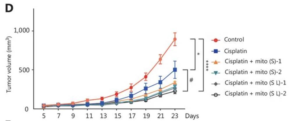
In preclinical obesity models, BGE-102 monotherapy achieved dose-dependent weight loss of up to 15%, comparable to semaglutide. When combined with semaglutide, BGE-102 produced additive effects, achieving approximately 25% weight reduction, supporting its potential application as part of an all-oral obesity regimen [link].

The Phase 1 study is a randomized, double-blind, placebo-controlled trial designed to evaluate the safety, tolerability, pharmacokinetics, and pharmacodynamics of BGE-102 in healthy participants. Part 1 will assess single ascending doses; Part 2 will evaluate multiple ascending doses administered once daily for 14 days. The study is designed to characterize BGE-102’s pharmacokinetic profile through blood sampling, assess CNS penetration through cerebrospinal fluid sampling, and evaluate pharmacodynamic effects using an ex vivo whole blood stimulation assay that measures BGE-102’s ability to inhibit the production of key inflammatory signals such as IL-1β.

“We’re excited to announce the dosing of our first participant in the Phase 1 trial for BGE-102 – a significant milestone in our mission to target the biology of aging and transform obesity treatment,” said Kristen Fortney, PhD, CEO and co-founder of BioAge. “This study was designed to deliver key data on safety, dosing, and activity. By inhibiting NLRP3-driven inflammation, a core driver of metabolic dysfunction, BGE-102 has the potential to complement existing therapies like GLP-1 agonists to enhance weight loss and curb excess inflammation. We believe that with convenient once-daily oral dosing and exceptional brain penetration, BGE-102 is positioned to tackle neuroinflammation in obesity and related conditions, offering versatility as a standalone or combination option.”

Following successful completion of the Phase 1 study, with initial SAD data expected by year-end 2025, BioAge plans to advance BGE-102 into a proof-of-concept study in obesity in 2026, with top-line data anticipated by end of year.

About BioAge Labs, Inc.

BioAge is a clinical-stage biopharmaceutical company developing therapeutic product candidates for metabolic diseases by targeting the biology of human aging. The Company’s lead product candidate, BGE-102, is a potent, orally available, brain-penetrant small-molecule NLRP3 inhibitor being developed for obesity. BGE-102 has demonstrated significant weight loss in preclinical models both as monotherapy and in combination with GLP-1 receptor agonists. A Phase 1 SAD/MAD trial of BGE-102 is underway, with initial SAD data anticipated by end of year. The Company is also developing long-acting injectable and oral small molecule APJ agonists for obesity. BioAge’s additional preclinical programs, which leverage insights from the Company’s proprietary discovery platform built on human longevity data, address key pathways involved in metabolic aging.

Forward-looking statements

This press release contains “forward-looking statements” within the meaning of, and made pursuant to the safe harbor provisions of, the Private Securities Litigation Reform Act of 1995. All statements contained in this press release that do not relate to matters of historical fact should be considered forward-looking statements, including, but not limited to, statements regarding our plans to develop and commercialize our product candidates, including BGE-102 and our APJ program, the potential for BGE-102 as a treatment for obesity and the expected timeline for data readout from our ongoing Phase 1 clinical trial, the timing and results of our clinical activities, risks associated with clinical trials, including our ability to adequately manage clinical activities, the timing of our IND filing for our APJ program, our ability to obtain and maintain regulatory approvals, the clinical utility of our product candidates or their ultimate ability to treat human disease, the expected timeline for completing proteomic analysis, anticipated analytical results and the potential for identifying novel therapeutic targets, and general economic, industry and market conditions. These forward-looking statements may be accompanied by such words as “aim,” “anticipate,” “believe,” “could,” “estimate,” “expect,” “forecast,” “goal,” “intend,” “may,” “might,” “plan,” “potential,” “possible,” “will,” “would,” and other words and terms of similar meaning. These statements involve risks and uncertainties that could cause actual results to differ materially from those reflected in such statements, including: our ability to develop, obtain regulatory approval for and commercialize our product candidates; the timing and results of preclinical studies and clinical trials; the risk that positive results in a preclinical study or clinical trial may not be replicated in subsequent trials or success in early stage clinical trials may not be predictive of results in later stage clinical trials; risks associated with clinical trials, including our ability to adequately manage clinical activities, unexpected concerns that may arise from additional data or analysis obtained during clinical trials, regulatory authorities may require additional information or further studies, or may fail to approve or may delay approval of our drug candidates; the occurrence of adverse safety events; failure to protect and enforce our intellectual property, and other proprietary rights; failure to successfully execute or realize the anticipated benefits of our strategic and growth initiatives; risks relating to technology failures or breaches; our dependence on collaborators and other third parties for the development of product candidates and other aspects of our business, which are outside of our full control; risks associated with current and potential delays, work stoppages, or supply chain disruptions, including due to the imposition of tariffs and other trade barriers; risks associated with current and potential future healthcare reforms; risks relating to attracting and retaining key personnel; changes in or failure to comply with legal and regulatory requirements, including shifting priorities within the U.S. Food and Drug Administration; risks relating to access to capital and credit markets; and the other risks and uncertainties that are detailed under the heading “Risk Factors” included in BioAge’s Quarterly Report on Form 10-Q filed with the U.S. Securities and Exchange Commission (SEC) on August 6, 2025, and BioAge’s other filings with the SEC filed from time to time. BioAge undertakes no obligation to publicly update any forward-looking statement, whether written or oral, that may be made from time to time, whether as a result of new information, future developments or otherwise.

Contacts

PR: Chris Patil, media@bioagelabs.com

IR: Dov Goldstein, ir@bioagelabs.com

Partnering: partnering@bioagelabs.com

Web: https://bioagelabs.com

We would like to ask you a small favor. We are a non-profit foundation, and unlike some other organizations, we have no shareholders and no products to sell you. All our news and educational content is free for everyone to read, but it does mean that we rely on the help of people like you. Every contribution, no matter if it’s big or small, supports independent journalism and sustains our future.

Heart disease

How Inflammation Is Linked to Heart Disease

In Cell Reports Medicine, researchers have published a detailed review on the relationship between cardiovascular disease and the age-related inflammation known as inflammaging.

The immune system itself ages

Cardiovascular Inflammaging

Inflammaging occurs in both the cells sending out signals and in the cells receiving them. The numbers of specific immune cell types become imbalanced; the bone marrow begins producing more myeloid than lymphoid cells [1], leading to an alteration in the neutrophil-to-lymphocyte ratio, which itself is associated with frailty [2].

This imbalance even occurs in the heart. The self-sustaining population of CCR2- macrophages, which promote muscle growth and fight inflammation, is gradually replaced with CCR2+ macrophages, which have a different bodily origin and promote inflammation instead [3].

The thymus is where immune cells are trained, and thymic involution is the age-related process that gradually deteriorates this organ into fat. Mitochondrial dysfunction occurs in the T cells as well, causing further immune dysfunction [4].

Immune dysfunction leads directly to cardiovascular disease

Causing mitochondrial dysfunction in the T cells of mice gave them severe heart disease [5]. The contribution of T cell dysfunction to vascular problems is strong enough that depleting CD8+ T cells in aged mice reduced their atherosclerosis [6]. Similarly, T cells are modified in heart failure, and not for the better; mice that lack CD4+ T cells have better outcomes when subjected to artificial heart attacks [7], and a different study demonstrated that taking T cells from mice subjected to these heart attacks and giving them to other mice causes heart problems in the other mice [8]. Further work confirmed that dysregulated T cells cause long-term damage in this way [9].

T cells even appear to be responsible for the damage caused by well-known inducers of heart disease. As expected, feeding mice a high-fat diet and inducing hypertension causes a form of heart failure in mice, but this does not occur if the mice had their T cells depleted [10].

Inflammation also leads to problems in the vasculature. High levels of circulating inflammatory cytokines lead to endothelial dysfunction, a core contributor to atherosclerosis [11]. This immune overactivation encourages the formation of blood clots (thrombosis) [12], thus increasing the risk of heart attack and stroke [13].

Potential solutions

Unsurprisingly, reducing inflammation is being explored as a method of decreasing the likelihood of thrombotic events [14]. In the 2000s, trials of anti-inflammatory drugs specifically for preventing heart failure did not yield good results [15], but later on, colchichine was found to be successful [16], and a meta-analysis provided enough data for its effectiveness [17] that the FDA has approved it for the prevention of cardiovascular disease in people with multiple risk factors. However, even this drug does not help immediately after a heart attack [18].

Affecting cellular senescence has also been investigated as a potential solution. The link between senescence and inflammation is well-known; the circulating cytokines that can lead to dysfunction are part of the senescence-associated secretory phenotype (SASP), the signals that senescent cells emit [19]. However, fighting senescence to reduce heart problems can carry its own risks; for example, while the combination of dasatinib and quercetin is well-known as a senolytic that destroys senescent cells, dasatinib has been linked to heart problems [20]. Navitoclax, another well-known senolytic, can cause uncontrolled bleeding [21].

The researchers suggest that other drugs that modulate rather than kill senescent cells (senomorphics) may be more promising. Metformin, for example, has been found to reduce the SASP in this way [22]. This may be due to its effects on mitochondrial dysfunction; a study with a different drug suggests that reducing mitochondrial dysfunction by increasing mitochondrial turnover (mitophagy) has beneficial effects in this regard [23].

Some inflammation comes from the gut. The researchers consider well-known interventions such as probiotics, which directly provide healthy gut bacteria [24], along with prebiotics, which feed only these beneficial bacteria [25]. Combining these approaches has demonstrated benefits in pigs with cardiometabolic syndrome [26]. Directly transferring gut bacteria through fecal microbiome transplantation has demonstrated benefits in mice with heart problems [27].

Personalized medicine

The researchers suggest that the detailed relationship between inflammaging and vasculature makes personalized medicine the most preferable approach. Not all preventatives work on everyone with cardiovascular risk factors; for example, one study found that statins don’t offer benefits for people who do not show calcium on CT scans [28]. Similarly, while reducing blood pressure is a common choice to prevent cardiovascular events, antihypertensive drugs can have negative effects on some older people [29]. Advanced imaging and more in-depth examination of biomarkers may allow for more targeted treatments that lead to better outcomes.

We would like to ask you a small favor. We are a non-profit foundation, and unlike some other organizations, we have no shareholders and no products to sell you. All our news and educational content is free for everyone to read, but it does mean that we rely on the help of people like you. Every contribution, no matter if it’s big or small, supports independent journalism and sustains our future.

Literature

[1] Shaw, A. C., Goldstein, D. R., & Montgomery, R. R. (2013). Age-dependent dysregulation of innate immunity. Nature reviews immunology, 13(12), 875-887.

[2] Dillon, K., Goodman, Z. T., Kaur, S. S., Levin, B., & McIntosh, R. (2023). Neutrophil-to-lymphocyte ratio amplifies the effects of aging on decrements in grip strength and its functional neural underpinnings. The Journals of Gerontology: Series A, 78(6), 882-889.

[3] Bajpai, G., Schneider, C., Wong, N., Bredemeyer, A., Hulsmans, M., Nahrendorf, M., … & Lavine, K. J. (2018). The human heart contains distinct macrophage subsets with divergent origins and functions. Nature medicine, 24(8), 1234-1245.

[4] Thapa, P., & Farber, D. L. (2019). The role of the thymus in the immune response. Thoracic surgery clinics, 29(2), 123-131.

[5] Desdín-Micó, G., Soto-Heredero, G., Aranda, J. F., Oller, J., Carrasco, E., Gabandé-Rodríguez, E., … & Mittelbrunn, M. (2020). T cells with dysfunctional mitochondria induce multimorbidity and premature senescence. Science, 368(6497), 1371-1376.

[6] Tyrrell, D. J., Wragg, K. M., Chen, J., Wang, H., Song, J., Blin, M. G., … & Goldstein, D. R. (2023). Clonally expanded memory CD8+ T cells accumulate in atherosclerotic plaques and are pro-atherogenic in aged mice. Nature aging, 3(12), 1576-1590.

[7] Yang, Z., Day, Y. J., Toufektsian, M. C., Xu, Y., Ramos, S. I., Marshall, M. A., … & Linden, J. (2006). Myocardial infarct–sparing effect of adenosine A2A receptor activation is due to its action on CD4+ T lymphocytes. Circulation, 114(19), 2056-2064.

[8] Maisel, A., Cesario, D., Baird, S., Rehman, J., Haghighi, P., & Carter, S. (1998). Experimental autoimmune myocarditis produced by adoptive transfer of splenocytes after myocardial infarction. Circulation research, 82(4), 458-463.

[9] Bansal, S. S., Ismahil, M. A., Goel, M., Zhou, G., Rokosh, G., Hamid, T., & Prabhu, S. D. (2019). Dysfunctional and proinflammatory regulatory T-lymphocytes are essential for adverse cardiac remodeling in ischemic cardiomyopathy. Circulation, 139(2), 206-221.

[10] Smolgovsky, S., Bayer, A. L., Kaur, K., Sanders, E., Aronovitz, M., Filipp, M. E., … & Alcaide, P. (2023). Impaired T cell IRE1α/XBP1 signaling directs inflammation in experimental heart failure with preserved ejection fraction. The Journal of clinical investigation, 133(24).

[11] Sprague, A. H., & Khalil, R. A. (2009). Inflammatory cytokines in vascular dysfunction and vascular disease. Biochemical pharmacology, 78(6), 539-552.

[12] Riegger, J., Byrne, R. A., Joner, M., Chandraratne, S., Gershlick, A. H., Ten Berg, J. M., … & Zahman, A. (2016). Histopathological evaluation of thrombus in patients presenting with stent thrombosis. A multicenter European study: a report of the prevention of late stent thrombosis by an interdisciplinary global European effort consortium. European heart journal, 37(19), 1538-1549.

[13] Liu, Y., Guan, S., Xu, H., Zhang, N., Huang, M., & Liu, Z. (2023). Inflammation biomarkers are associated with the incidence of cardiovascular disease: a meta-analysis. Frontiers in Cardiovascular Medicine, 10, 1175174.

[14] Eikelboom, J. W., Connolly, S. J., Bosch, J., Dagenais, G. R., Hart, R. G., Shestakovska, O., … & Yusuf, S. (2017). Rivaroxaban with or without aspirin in stable cardiovascular disease. New England Journal of Medicine, 377(14), 1319-1330.

[15] Chung, E. S., Packer, M., Lo, K. H., Fasanmade, A. A., & Willerson, J. T. (2003). Randomized, double-blind, placebo-controlled, pilot trial of infliximab, a chimeric monoclonal antibody to tumor necrosis factor-α, in patients with moderate-to-severe heart failure: results of the anti-TNF Therapy Against Congestive Heart Failure (ATTACH) trial. Circulation, 107(25), 3133-3140.

[16] Tardif, J. C., Kouz, S., Waters, D. D., Bertrand, O. F., Diaz, R., Maggioni, A. P., … & Roubille, F. (2019). Efficacy and safety of low-dose colchicine after myocardial infarction. New England journal of medicine, 381(26), 2497-2505.

[17] Fiolet, A. T., Poorthuis, M. H., Opstal, T. S., Amarenco, P., Boczar, K. E., Buysschaert, I., … & Kelly, P. J. (2024). Colchicine for secondary prevention of ischaemic stroke and atherosclerotic events: a meta-analysis of randomised trials. EClinicalMedicine, 76.

[18] Jolly, S. S., d’Entremont, M. A., Lee, S. F., Mian, R., Tyrwhitt, J., Kedev, S., … & Yusuf, S. (2025). Colchicine in acute myocardial infarction. New England Journal of Medicine, 392(7), 633-642.

[19] Acosta, J. C., Banito, A., Wuestefeld, T., Georgilis, A., Janich, P., Morton, J. P., … & Gil, J. (2013). A complex secretory program orchestrated by the inflammasome controls paracrine senescence. Nature cell biology, 15(8), 978-990.

[20] Xu, Z., Cang, S., Yang, T., & Liu, D. (2009). Cardiotoxicity of tyrosine kinase inhibitors in chronic myelogenous leukemia therapy. Hematology Reviews, 1(1), e4.

[21] Schoenwaelder, S. M., Jarman, K. E., Gardiner, E. E., Hua, M., Qiao, J., White, M. J., … & Jackson, S. P. (2011). Bcl-xL–inhibitory BH3 mimetics can induce a transient thrombocytopathy that undermines the hemostatic function of platelets. Blood, The Journal of the American Society of Hematology, 118(6), 1663-1674.

[22] Abdelgawad, I. Y., Agostinucci, K., Sadaf, B., Grant, M. K., & Zordoky, B. N. (2023). Metformin mitigates SASP secretion and LPS-triggered hyper-inflammation in Doxorubicin-induced senescent endothelial cells. Frontiers in Aging, 4, 1170434.

[23] Kelly, G., Kataura, T., Panek, J., Ma, G., Salmonowicz, H., Davis, A., … & Korolchuk, V. I. (2024). Suppressed basal mitophagy drives cellular aging phenotypes that can be reversed by a p62-targeting small molecule. Developmental cell, 59(15), 1924-1939.

[24] Wierzbicka, A., Mańkowska-Wierzbicka, D., Mardas, M., & Stelmach-Mardas, M. (2021). Role of probiotics in modulating human gut microbiota populations and activities in patients with colorectal cancer—a systematic review of clinical trials. Nutrients, 13(4), 1160.

[25] Yoo, S., Jung, S. C., Kwak, K., & Kim, J. S. (2024). The role of prebiotics in modulating gut microbiota: implications for human health. International Journal of Molecular Sciences, 25(9), 4834.

[26] Herisson, F. M., Cluzel, G. L., Llopis-Grimalt, M. A., O’Donovan, A. N., Koc, F., Karnik, K., … & Caplice, N. M. (2025). Targeting the gut-heart axis improves cardiac remodeling in a clinical scale model of cardiometabolic syndrome. Basic to Translational Science, 10(1), 1-15.

[27] Hatahet, J., Cook, T. M., Bonomo, R. R., Elshareif, N., Gavini, C. K., White, C. R., … & Aubert, G. (2023). Fecal microbiome transplantation and tributyrin improves early cardiac dysfunction and modifies the BCAA metabolic pathway in a diet induced pre-HFpEF mouse model. Frontiers in cardiovascular medicine, 10, 1105581.

[28] Mitchell, J. D., Fergestrom, N., Gage, B. F., Paisley, R., Moon, P., Novak, E., … & Villines, T. C. (2018). Impact of statins on cardiovascular outcomes following coronary artery calcium scoring. Journal of the American College of Cardiology, 72(25), 3233-3242.

[29] Benetos, A., Petrovic, M., & Strandberg, T. (2019). Hypertension management in older and frail older patients. Circulation research, 124(7), 1045-1060.

Cannabis

Cannabis as a Treatment for Age-Related Diseases

Researchers have recently published a review on how cannabis use among older adults impacts age-related conditions and longevity [1].

Cannabinoids and longevity

In recent years, researchers have observed an increase in cannabis use among older adults, mostly for chronic conditions, such as arthritis, pain, sleep improvement, anxiety, and depressive symptoms.

However, there is also a recent interest in using cannabis-derived cannabinoids, especially cannabidiol (CBD) and Δ9-tetrahydrocannabinol (THC), as potential anti-aging and longevity-promoting treatments.

CBD and THC interact with the endocannabinoid system (ECS), which has been linked to impacting aging and longevity [2]. While the researchers do not have a detailed understanding of how the endocannabinoid system interacts with cannabinoids to promote health and longevity, the reviewed studies suggest that cannabinoids might promote cellular homeostasis.

This review encompassed what is currently known about the cannabinoids’ anti-aging properties, as well as their limitations, drawbacks, and side effects. The reviewers excluded studies whose focus was on the acute effects of cannabis or its action in different medical conditions. They found 11 preclinical and 7 human studies that met their search criteria.

Promising animal data

Most of the early studies regarding biological questions are done in model organisms. This is also the case for cannabis, as the researchers utilized worms, fruit flies, zebrafish, and mice to analyze its connection to health and lifespan outcomes.

Worm and fruit fly studies show that CBD and THC exposure can lead to lifespan extension, improved neuronal health, delayed age-related neurodegeneration, and increased autophagy. However, the beneficial effects often depend on using a specific dose of cannabis [3-6].

Another model organism that was used for cannabis research is the zebrafish, as its endocannabinoid system is well conserved, meaning it shows high similarity to humans. A study that analyzed the impact of THC exposure during development pointed to the dose-dependent differences, with lower doses leading to increased male survival and egg production, and reduced markers of aging and inflammation in the liver. Higher doses had negative consequences for offspring, survival, and reproduction [7].

Mice’s endocannabinoid systems are also similar to those of humans. In older mice, lower doses of THC led to improvements in memory and reversal of cognitive decline; however, this was not the case for THC-treated younger animals, which showed a decline in cognitive performance [8].

Cognitive performance improvements were also seen in two different studies where older mice were either chronically (for 28 days) exposed to a low dose of THC [9] or received a single injection of an extremely low THC dose [10]. Similarly, the memory and brain health of aged mouse models of Alzheimer’s disease benefited from intranasal low-dose THC treatment [11].

A mouse study also pointed to potential interactions between THC and CBD, as a low dose of THC was beneficial for spatial learning in aged mice, but when combined with CBD, the beneficial effect was not present [12]. This study points to the importance of further investigation into the combined effects of different cannabinoids.

The human data

While the evidence from model animals is promising, it needs to be confirmed in human studies, which are mostly lacking, and existing results are inconsistent. The human studies show a difference in outcomes regarding the age at which people started to use cannabis and how long they have been taking it. Several studies that examined people who used cannabis during adolescence and/or engaged in long-term use found that they had worse executive function, reduced grey matter, poorer verbal memory, accelerated biological aging, and worse health [13-16].

The effects are different when cannabis is taken by the elderly. For example, a study that included older adults, aged 60-88, who used cannabis weekly for at least the past year, showed increased connectivity in several brain regions, suggesting improved communication and information processing [17].

However, another study of people at least 60 years old showed lower executive functions in long-term cannabis users compared to non-users or short-term users. Short-term use didn’t seem to impact cognitive performance in this study [18].

Since there is a shortage of human studies on cannabis, the researcher also examined evidence regarding cannabis-based medicinal products (CBMPs) among older people. The data suggest CBMPs’ potential in treating aging-associated conditions, such as insomnia, depression, anxiety, and chronic pain. Cannabis use can also help reduce opioid dosage [19].

In total, the data from human studies and observations show age-dependent effects of cannabis. When cannabis use is initiated early in life, it leads to cognitive impairment later in life, but initiating cannabis use later in life shows more promising outcomes; however, there is still a need for more investigation in people.

The authors believe that some of the opposing age-dependent effects of cannabis might be caused by the changes that occur to the endocannabinoid system as it ages, such as changes in receptor binding and gene expression.

While analyzing different studies, the authors noticed methodological issues in human studies investigating cannabis use. First, the dose reporting is inconsistent and reports broad terms such as “heavy” or “recreational,” but no precise measurements are given. Second, there is a generalization of age categories, such as “older adults.” Both of those make interpreting the results and drawing strong conclusions harder. However, those researchers point out that they used those studies to gain an understanding of broader trends.

The authors also point out that there are differences in cannabis use patterns among the older population, with some people being new users (people who started after 60), while others are intermittent or consistent users. Those differences make cannabis research use more complex, as the effects of cannabis in each group might be different, and optimal therapeutic approaches might differ.

Cautious optimism

While most of the reviewed results seem to be optimistic, they should be interpreted with caution, as they are scarce and have many limitations. Additionally, most human studies are observational and cannot establish a causal link.

In the future, there is a need for well-designed human trials to understand the effect of cannabis on health and longevity, the consequences of the long-term effects of cannabis use among the elderly, and the effect of different dosing and routes of administration. There is also a need to investigate other cannabinoids beyond THC and CBD along with their interactions with each other and other medications and compounds.

We would like to ask you a small favor. We are a non-profit foundation, and unlike some other organizations, we have no shareholders and no products to sell you. All our news and educational content is free for everyone to read, but it does mean that we rely on the help of people like you. Every contribution, no matter if it’s big or small, supports independent journalism and sustains our future.

Literature

[1] Nain, S., Singh, N., Schlag, A. K., & Barnes, M. (2025). The impact of cannabis use on ageing and longevity: a systematic review of research insights. Journal of cannabis research, 7(1), 52.

[2] Paradisi, A., Oddi, S., & Maccarrone, M. (2006). The endocannabinoid system in ageing: a new target for drug development. Current drug targets, 7(11), 1539–1552.

[3] Land, M. H., Toth, M. L., MacNair, L., Vanapalli, S. A., Lefever, T. W., Peters, E. N., & Bonn-Miller, M. O. (2021). Effect of Cannabidiol on the Long-Term Toxicity and Lifespan in the Preclinical Model Caenorhabditis elegans. Cannabis and cannabinoid research, 6(6), 522–527.

[4] Wang, Z., Zheng, P., Chen, X., Xie, Y., Weston-Green, K., Solowij, N., Chew, Y. L., & Huang, X. F. (2022). Cannabidiol induces autophagy and improves neuronal health associated with SIRT1 mediated longevity. GeroScience, 44(3), 1505–1524.

[5] Wang, Z., Zheng, P., Xie, Y., Chen, X., Solowij, N., Green, K., Chew, Y. L., & Huang, X. F. (2021). Cannabidiol regulates CB1-pSTAT3 signaling for neurite outgrowth, prolongs lifespan, and improves health span in Caenorhabditis elegans of Aβ pathology models. FASEB journal : official publication of the Federation of American Societies for Experimental Biology, 35(5), e21537.

[6] Candib, A., Lee, N., Sam, N., Cho, E., Rojas, J., Hastings, R., DeAlva, K., Khon, D., Gonzalez, A., Molina, B., Torabzadeh, G., Vu, J., Hasenstab, K., Sant, K., Phillips, J. A., & Finley, K. (2024). The Influence of Cannabinoids on Drosophila Behaviors, Longevity, and Traumatic Injury Responses of the Adult Nervous System. Cannabis and cannabinoid research, 9(3), e886–e896.

[7] Pandelides, Z., Thornton, C., Lovitt, K. G., Faruque, A. S., Whitehead, A. P., Willett, K. L., & Ashpole, N. M. (2020). Developmental exposure to Δ9-tetrahydrocannabinol (THC) causes biphasic effects on longevity, inflammation, and reproduction in aged zebrafish (Danio rerio). GeroScience, 42(3), 923–936.

[8] Bilkei-Gorzo, A., Albayram, O., Draffehn, A., Michel, K., Piyanova, A., Oppenheimer, H., Dvir-Ginzberg, M., Rácz, I., Ulas, T., Imbeault, S., Bab, I., Schultze, J. L., & Zimmer, A. (2017). A chronic low dose of Δ9-tetrahydrocannabinol (THC) restores cognitive function in old mice. Nature medicine, 23(6), 782–787.

[9] Komorowska-Müller, J. A., Gellner, A. K., Ravichandran, K. A., Bilkei-Gorzo, A., Zimmer, A., & Stein, V. (2023). Chronic low-dose Δ9-tetrahydrocannabinol (THC) treatment stabilizes dendritic spines in 18-month-old mice. Scientific reports, 13(1), 1390.

[10] Sarne, Y., Toledano, R., Rachmany, L., Sasson, E., & Doron, R. (2018). Reversal of age-related cognitive impairments in mice by an extremely low dose of tetrahydrocannabinol. Neurobiology of aging, 61, 177–186.

[11] Fihurka, O., Hong, Y., Yan, J., Brown, B., Lin, X., Shen, N., Wang, Y., Zhao, H., Gordon, M. N., Morgan, D., Zhou, Q., Chang, P., & Cao, C. (2022). The Memory Benefit to Aged APP/PS1 Mice from Long-Term Intranasal Treatment of Low-Dose THC. International journal of molecular sciences, 23(8), 4253.

[12] Nidadavolu, P., Bilkei-Gorzo, A., Krämer, M., Schürmann, B., Palmisano, M., Beins, E. C., Madea, B., & Zimmer, A. (2021). Efficacy of Δ9 -Tetrahydrocannabinol (THC) Alone or in Combination With a 1:1 Ratio of Cannabidiol (CBD) in Reversing the Spatial Learning Deficits in Old Mice. Frontiers in aging neuroscience, 13, 718850.

[13] Meier, M. H., Caspi, A., Ambler, A., Hariri, A. R., Harrington, H., Hogan, S., Houts, R., Knodt, A. R., Ramrakha, S., Richmond-Rakerd, L. S., Poulton, R., & Moffitt, T. E. (2022). Preparedness for healthy ageing and polysubstance use in long-term cannabis users: a population-representative longitudinal study. The lancet. Healthy longevity, 3(10), e703–e714.

[14] Thayer, R. E., YorkWilliams, S. L., Hutchison, K. E., & Bryan, A. D. (2019). Preliminary results from a pilot study examining brain structure in older adult cannabis users and nonusers. Psychiatry research. Neuroimaging, 285, 58–63.

[15] Burggren, A. C., Siddarth, P., Mahmood, Z., London, E. D., Harrison, T. M., Merrill, D. A., Small, G. W., & Bookheimer, S. Y. (2018). Subregional Hippocampal Thickness Abnormalities in Older Adults with a History of Heavy Cannabis Use. Cannabis and cannabinoid research, 3(1), 242–251.

[16] Auer, R., Vittinghoff, E., Yaffe, K., Künzi, A., Kertesz, S. G., Levine, D. A., Albanese, E., Whitmer, R. A., Jacobs, D. R., Jr, Sidney, S., Glymour, M. M., & Pletcher, M. J. (2016). Association Between Lifetime Marijuana Use and Cognitive Function in Middle Age: The Coronary Artery Risk Development in Young Adults (CARDIA) Study. JAMA internal medicine, 176(3), 352–361.

[17] Watson, K. K., Bryan, A. D., Thayer, R. E., Ellingson, J. M., Skrzynski, C. J., & Hutchison, K. E. (2022). Cannabis Use and Resting State Functional Connectivity in the Aging Brain. Frontiers in aging neuroscience, 14, 804890.

[18] Stypulkowski, K., & Thayer, R. E. (2022). Long-Term Recreational Cannabis Use Is Associated With Lower Executive Function and Processing Speed in a Pilot Sample of Older Adults. Journal of geriatric psychiatry and neurology, 35(5), 740–746.

[19] Tumati, S., Lanctôt, K. L., Wang, R., Li, A., Davis, A., & Herrmann, N. (2022). Medical Cannabis Use Among Older Adults in Canada: Self-Reported Data on Types and Amount Used, and Perceived Effects. Drugs & aging, 39(2), 153–163.

Nir Barzilai Interview

Nir Barzilai: “Positive Evidence for Metformin is Mounting”

Dr. Nir Barzilai, the director of the Institute for Aging Research at the Albert Einstein College of Medicine, among his many other titles, is one of geroscience’s most prominent figures. He is everywhere all at once, seemingly collaborating with the entire world, but is best known as a staunch proponent of metformin, the anti-diabetes drug that extends healthspan and lifespan in animal models and maybe in humans, as well as for his fascinating research into centenarians. Recently, a review paper came out that questions metformin’s reputation as a geroprotector. We thought it was the perfect moment to reach out to Dr. Barzilai for an update on his faith in metformin, the long-awaited Targeting Aging with Metformin (TAME) trial, and other exciting topics.

Let’s start with the recent metformin paper that intends to pour some cold water on the idea that metformin is a good gerotherapeutic.

There are 34,731 papers about metformin, which has been around for decades, and most of them are good; the positive evidence is accumulating. The authors of this one decided to talk about the “bad” papers, because the fact that metformin has clinically proven benefits beyond just diabetes is well known.

Metformin didn’t start as an anti-diabetic drug. In the 1950s, this extract of the French lilac was used for osteoarthritis, to prevent flu, and for a variety of remedies when it was noticed that it also lowers glucose. When they gave it to people with diabetes, they said, “Hey, what’s going on? It’s doing other things”. Aging is where it started and where it’s going.

When I do a “metformin and aging” search in PubMed, there are 1,400 papers on that, and the majority are good. In just the last year, there are 111, most of which are good, and they were not quoted in this recent paper. The reason this was brought up is because of an effort by Christensen to look at people who took metformin for diabetes in Denmark. It wasn’t a clinical study; it was just looking at data.

There’s a problem with this approach: the data showed that metformin decreased mortality initially, but not later on. But of course! When it decreases mortality initially, the people who would have died have now passed, and the people who gained lifespan will eventually match the plot. That’s a time-to-event bias. He didn’t talk about this part; he just said at the end, “We didn’t show an effect”.

This Danish study came after another foundational study from the UK, which was also observational and showed that people on metformin not only have half the mortality of people on other drugs, which is true and is good data, but they also had less mortality than people without diabetes.

Let’s explain to our readers that you’re talking about two influential populational studies of metformin. One, from a few years ago, reinforced metformin’s reputation as a gerotherapeutic, and then, a couple of years ago, another study with a somewhat comparable design came out and, according to some opinions, refuted the first one.

“Refute” is not the right word. One study was done in England, the other in Denmark. One of the differences is that the obesity rate in England is 20%, while in Denmark, it’s 5%. Obesity is one of the main reasons people are prescribed metformin in England. So, we are not talking about the same population, and it hasn’t been done the same way. I accept that you can do different studies around the world, but my point is that those were two flawed observational studies, and there are better studies out there.

The most important study that connects metformin with aging is the one that showed that if you give it during COVID, you slash mortality and the rate of long COVID in half. The critics say, “Okay, but this study didn’t reach its primary endpoint”. Their primary endpoint was hypoxia. It’s silly! It’s just a stupid endpoint because metformin isn’t working on hypoxia; it’s working on inflammation, on immunity, on the ability of the body to resist.

So, who cares? You can take any study in the world and find its limitations. That’s our profession. In every journal club, an outsider would think every paper is a bad paper. But no, every paper has some limitations, and we accept a certain idea only when data accumulates. My point is that there’s so much data on metformin. The authors of this new paper include references to all the good studies; they’re just not talking about them.

Then they do another thing that doesn’t make sense: they take the DPP, the Diabetes Prevention Program. The DPP study was concluded around the year 2000. It’s a study where you take non-diabetics who are at risk of diabetes.

Yes, it was basically a prevention study.

Exactly. And they stopped the study after four years, though it was planned for five, because both metformin and lifestyle changes clearly prevented diabetes. If people say metformin was never given to non-diabetics, that’s wrong; it was. In fact, more metformin is probably given to non-diabetics now than to diabetics.

Anyway, the study ended at four years, and the participants were followed up, but what happened? The study found that metformin was good for you, so some people in the control group started taking it. The study also said healthy lifestyle changes were good for you, so some people changed their lifestyle. Conversely, some people who were on metformin stopped taking it, and some people who were on a lifestyle plan stopped doing it. Moving forward, it wasn’t a clinical study anymore; it became an observational study, and they didn’t find much. It’s just another example of a study where all the groups have changed and mixed. To make such a big story about the DPP is ridiculous.

They’re saying this is “emerging” evidence, but it’s not. They’re just taking three studies that are not RCTs [randomized controlled studies]. Then there’s the monkey study published in Cell, which was a big deal. Aging was delayed by eight years on the transcriptomic level. There are so many other good studies on metformin.

Another thing, regarding Rich Miller from the ITP [Interventions Testing Program]. Rich believes that whatever doesn’t work in his animals doesn’t work in humans, but this is the opposite situation! The drug already showed effects in humans. What are you defending? What are you trying to say?

I guess he’s trying to say that we don’t see a lifespan effect in mice, and we also don’t have definitive lifespan data in humans.

But that is wrong. There is a lifespan effect in mice, just not as much as with rapamycin. It’s been recorded by 20-something studies, but Rich ignored something from his own ITP data. They’re missing a very important point: metformin is not for young people; it’s only for old people.

This relates to the antagonistic pleiotropy hypothesis of aging, where not everything that’s good for you when you’re young is good for you when you’re old, and it’s the other way around with drugs. Not every drug for aging is good for the young, and metformin is a perfect example. Whoever takes metformin who doesn’t have diabetes and is not at least 50 years old is making a mistake, in particular if they’re trying to build their muscle or increase their VO2max.

I’m saying this because there’s a new study from the ITP, which re-analyzes their own data and shows that metformin wasn’t good in the first half of the animals’ lives but performed significantly better in the second half of life.

There is also the time-bias issue. For example, there was a paper from China claiming metformin is associated with more Alzheimer’s. Usually, it’s the opposite, but this is what happens: if metformin prevents your mortality in an observational study, you are pushing the endpoint. People might get Alzheimer’s later. There’s a paper coming out in the Journal of Gerontology showing that people on metformin are twice as likely to reach age 90 as other people with diabetes, but if you give metformin and you have decreased mortality, it can look like metformin is bad later on.

You’re saying that if a drug prolongs lifespan, the survivors are actually more likely to eventually get Alzheimer’s simply because they are older, correct?

Right. And that makes a lot of sense.

To summarize, you’re still bullish on metformin. This brings us to the TAME trial, which was designed to answer questions about delaying aging in humans. Can you give me an update on where things stand?

TAME is designed to measure a cluster of outcomes; it doesn’t look at mortality independently. The primary endpoint is a cluster of cardiovascular disease, cognitive decline, cancer, and then mortality. So, you’re not going to get a single mortality number out of it.

Yes, I think TAME’s design is pretty ingenious: a cluster of age-related diseases serving as a proxy for aging. We are all rooting for TAME. What’s happening with it now?

Let me give you a nice update. To call something a “gerotherapeutic” from a preclinical perspective, you have to show that it hits the hallmarks of aging. By the way, metformin hits more hallmarks of aging than any other drug; rapamycin comes close, but metformin is broader.

You also want to show that your animals live healthier and longer. Clinically, you want a placebo-controlled study where you give the drug for months or years and show that although it was given for one purpose, it delayed several other age-related outcomes and decreased overall mortality. I would say that evidence is enough to call something a gerotherapeutic, particularly if it’s already FDA-approved.

It’s important to see what’s happening with SGLT2 inhibitors. These drugs were developed for diabetes, but now we have studies in non-diabetic populations with moderate renal failure. In a study of 4,000 people over three years, their primary endpoints – renal-specific, cardiovascular-specific, and all-cause mortality – were all significantly decreased by 30-40%.

In the same vein, metformin has already been repurposed for many things, just not formally by the FDA. It’s the first line of choice for PCOS, pre-diabetes, COVID, and macular degeneration. Each of these is a different disease, which shows metformin is doing something to several hallmarks, not just metabolism.

That’s a great point about the breadth of the effect. On the other hand, there’s this idea I’ve heard from many people that with all our gerotherapeutics, we are kind of running in circles around the same few pathways that control the trade-off between growth and repair. What do you say to that?

I think it’s almost the opposite. The reason we started arguing about these drugs is because somebody would say, “No, this is not only about metabolism, it does something to the immune system or to mitochondrial function”. The point is the hallmarks of aging are all associated with each other. If you target one hallmark, you’re going to affect the others, and that is the confusion. When you treat aging, you affect many things, which made us argue about the primary mechanism until we understood the hallmarks.

Metformin, from a mechanistic perspective, is doing two main things. On one hand, it has the metabolic pathway effects: activating AMPK, decreasing mTOR, and improving insulin sensitivity by blocking complex I in the mitochondria. The second thing that happens, because it blocks that complex, is there’s less oxidative stress. Because of less oxidative stress, other things happen with inflammation, senescence, and genetic instability. This is why metformin’s effects are quite global.

I want to circle back to TAME because people are very interested in it.

I cannot give you a perfect update because it’s now being handled within ARPA-H. I think there will be two major trials that come out of this. One is going to be from Eli Lilly; they’re going to do a TAME-like study but with their GLP-1 agonist. There are negotiations with the FDA about what they need to show. The investigators might want to add some resilience measurements, but I think the FDA is very determined to see if it affects diseases. This conversation is ongoing, and we’re holding everything because I would love for all four major drug classes to be tested so we can get comparisons.

I’d imagine the TAME design is generalizable, and it would indeed make a lot of sense to test GLP-1 agonists, the rising stars, in the same manner.

Yes. This administration is very good for aging research, and the FDA is engaged. The most important thing about TAME is that it’s a template for the pharmaceutical industry, and that’s probably why Lilly is interested. They see the effect on aging, and they’re saying, “Let’s just do the whole thing and get an indication for aging”.

When you say, “the whole thing,” you mean applying the TAME framework of a cluster of diseases to GLP-1 agonists?

Exactly, and they probably only need 2,000 people to show an effect, but that’s why metformin is so important. Longevity doctors have already adopted it, and it’s the cheapest drug in the formulary. We want to democratize aging, and the best way to do that is to have metformin out there. 90% of the people who should be on it will benefit, and it’s affordable. Healthcare providers will immediately see in the next two years that their healthcare expenses have decreased. Metformin might be more effective than other drugs, but I wouldn’t know unless it’s a head-to-head trial.

Once you do the calculation of the diseases prevented, you’ll be able to afford much more expensive drugs. We calculated that even at its current price, a GLP-1 agonist would be a cost-saving measure for a healthcare provider. They’ll see less Alzheimer’s, fewer strokes, less cardiovascular disease, less kidney failure. It will be so cost-effective that it’s worth the price, and the price will eventually get cheaper.

Let’s switch gears. You’re still doing your centenarian studies. Any interesting findings in the last few years?

I think the most interesting thing is that 60% of our centenarians have functional mutations that decrease the actions of growth hormone. There are many mechanisms, maybe 50 ways to get there, but the IGF-1 pathway is a really good one to target. We actually took a drug that was developed to fight cancer by inhibiting the IGF-1 receptor, gave it to animals, and not only did they live longer, but they also lived much, much healthier. We went from centenarians back to animals with a drug that has already been in humans.

Another thing I’m most excited about goes back to biomarkers. We took proteomic data, measuring 5,000 proteins in a thousand people, and 500 of them were the children of centenarians. They were, on average, eight years younger on a proteomic level than their spouses, but that’s not even the most exciting thing. A lot of those differentiating proteins are related to breakdown of tissues, collagen, and other things. With Tony Wyss-Coray, we’re trying to find which of those 5,000 proteins are specific to certain organs. They could come only from the liver or only from the brain.

We covered that paper last year. It was amazing.

We are continuing with that because we have, for example, people who are “slow agers”. Their proteome says they are younger than their age, but their liver is older than their age. What’s going on? Are they alcoholic? Do they have cancer, or maybe their brain is older? We think in the future, it’s not just about the overall biomarker, but you could find your weakness and go there first. If it’s your kidney, maybe metformin is the best drug for you. If it’s your brain, maybe it’s a GLP-1 agonist.

I just had this thought: what if some centenarians age more uniformly? What if they don’t have those weak spots, and all their organs age more or less simultaneously? That could explain some of their longevity.

That’s a good question. When we look at the children of centenarians versus controls, it looks more like their overall age is lower, rather than them having a specific decrease in unbalanced organs, but they do have a decreased incidence generally. I think it’s a good thought; I would look at it some more. We don’t really do proteomics on centenarians because they are at the end of their lives. That’s why we study their children; we’re interested in their genes. For the centenarians, 30% of them will die in the next year. Maybe the phenotype predicts their demise, or maybe it’s what brought them here, but it’s very hard to deal with.

Of course, you don’t know someone is a future centenarian before they become one, so working with their children is a great approach. Just one last question. How satisfied are you with how things have been going in the longevity field for the past four years since our last interview? Are you excited about where the field is today?

Yes. As the president of the Academy for Health & Lifespan Research, I can say that on one hand, we are so excited about the future and the progress we’re making. We are already telling doctors that there are safe drugs to think about for aging. I gave the keynote at the American Association of Physicians, and people are accepting this premise.

While we’re excited about the research, there’s also a lot of noise growing up in parallel. Sometimes, the noise is even worse than the real progress, and we’re trying to balance that. Without the noise, maybe nobody would have noticed us, but there is noise. One important thing we identified is that although we all have the same mission, we are not using the same terms. If you say “rejuvenation”, it means different things to different people. “Regeneration”, “healthspan”, “longevity”, “anti-aging”, “gerontology”, “geriatrics” – we have lots of terms.

Several organizations hired a rebranding company. They did a lot of work, interviewed people, did studies, and tested things on thousands of people. They came up with a plan based on the fact that if “anti-aging” is our enemy, the best word for us to use is “geroscience”. Why? Because we need the word “science”. To be clear, “geroscience” itself is not the best term for the public, but the plan is to use “geroscience” with something else, depending on the stakeholders, whether we’re talking to lay people, scientists, politicians, or pharma.

We have a roadmap of how we are going to present ourselves and launch a campaign. Although “geroscience” didn’t pick up on its own, with the right marketing, it can become really important and distinguish us from the noise. This is going to be a campaign that hopefully will make us look new, exciting, and innovative.

We would like to ask you a small favor. We are a non-profit foundation, and unlike some other organizations, we have no shareholders and no products to sell you. All our news and educational content is free for everyone to read, but it does mean that we rely on the help of people like you. Every contribution, no matter if it’s big or small, supports independent journalism and sustains our future.
Atrophied Muscles

How FGF21 Fights Back Against a Muscle-Wasting Disease

In Aging, researchers have reported on how an increase in FGF21, a myokine that encourages muscle growth, impacts the progression of amyotrophic lateral sclerosis (ALS).

Progressive and fatal

ALS is an age-related disease that is characterized by the degeneration of motor neurons throughout the spinal cord and in the brain, leading to death by respiratory failure three to five years after onset [1]. Last year, the authors of this study conducted a review concluding that the earliest stages of ALS can be detected in skeletal muscle [2], and other work has found evidence that the disease progresses from the muscles to the brain, not the other way around [3].

Identifying the key factors behind this progression, however, remains an uncompleted task. The transcriptome, which represens RNA gene expression, is heavily dysregulated in ALS patients [2]. Which signals represent the disease, and which signals represent a cellular attempt to mitigate the disease, however, remains an open question [4], one that has been investigated for nearly a decade [5].

For example, one of these biomarkers is FGF21, which this research team had previously investigated in this context [6]. Here, they redoubled their efforts in an effort to determine FGF21’s role in ALS and how it may impact the disease.

FGF21 is co-located with atrophied fibers

This study was carried out using muscle biopsies from patients gleaned at this team’s ALS clinic. Like the broader population, they found this disease to be more common in males than females, and the average age of patients was approximately 57.

Compared to biopsies of normal tissue, the FGF21 expression in the muscle of most, but not all, ALS patients was highly elevated as measured by an mRNA analysis. In the spinal cord, some ALS patients had levels below the norm, but others had extraordinarily high levels. While most patients that had high FGF21 in the spinal cord also had high levels in muscle, there were exceptions.

These patterns were mimicked in model mice that express a mutant version (G93A) of a particular antioxidant gene, SOD1, in skeletal muscle. These short-lived mice had much higher levels of FGF21 in both muscle tissue and spinal cord than their unmodified counterparts. While much of this came from the liver, even more originated from the muscle itself.

ALS does not cause every muscle fiber to suffer the same level of atrophy at once; rather, both atrophied and unatrophied fibers can be found within the same biopsy. In human muscle tissue, FGF21 and ALS were found to be co-located; atrophied fibers were found to have much more FGF21 than unatrophied ones.

FGF21 mitigates, not accelerates

An increase in FGF21 in blood plasma was associated with a slower progression and increased survival. Patients with low circulating FGF21 were likely to survive for only 18 months, while patients with high levels survived for an average of 75. Interestingly, a high BMI was associated with greater FGF21.

KLB is the gene that encodes β-Klotho, a co-receptor of FGF21. Its levels varied wildly in ALS patients; before the patients’ deaths, they expressed four times as much KLB as the control group, but a post-mortem examination showed that they expressed only half as much as controls, a finding that was recapitulated in G93A model mice.

Using iPSC technology to generate motor neurons from ALS patients, these findings were recapitulated in nervous tissue as well. Compared to controls, ALS motor neurons had half as much FGF21 but thrice as much KLB, a finding that appeared to be related to the effects of oxidative stress.

ALS-affected cells are much more vulnerable to oxidative stress than unaffected cells. Relatively low levels of hydrogen peroxide, which do not kill most of the control group, killed the majority of ALS motor neurons. Administering FGF21 to these cells increased their viability, although not quite to the level of controls.

FGF21 is myogenic; under normal circumstances, it generates functional tissue and increases strength. These researchers found that it indeed decreases stress in muscle tissue while increasing the number of muscle cells.

In total, the upregulation of FGF21 in ALS appears to be an attempt to mitigate the atrophy and cellular stress that characterize the disease. However, the researchers point to a problem with the FGF21-KLB axis and suggest that this dysfunction is key to the progression of ALS. Further work needs to be done to analyze this axis and determine if and how it can be effectively targeted to stop this deadly disease.

We would like to ask you a small favor. We are a non-profit foundation, and unlike some other organizations, we have no shareholders and no products to sell you. All our news and educational content is free for everyone to read, but it does mean that we rely on the help of people like you. Every contribution, no matter if it’s big or small, supports independent journalism and sustains our future.

Literature

[1] Hardiman, O., Van Den Berg, L. H., & Kiernan, M. C. (2011). Clinical diagnosis and management of amyotrophic lateral sclerosis. Nature reviews neurology, 7(11), 639-649.

[2] King, P. H. (2024). Skeletal muscle as a molecular and cellular biomarker of disease progression in amyotrophic lateral sclerosis: a narrative review. Neural Regeneration Research, 19(4), 747-753.

[3] Moloney, E. B., de Winter, F., & Verhaagen, J. (2014). ALS as a distal axonopathy: molecular mechanisms affecting neuromuscular junction stability in the presymptomatic stages of the disease. Frontiers in neuroscience, 8, 252.

[4] Verma, S., Khurana, S., Vats, A., Sahu, B., Ganguly, N. K., Chakraborti, P., … & Taneja, V. (2022). Neuromuscular junction dysfunction in amyotrophic lateral sclerosis. Molecular neurobiology, 59(3), 1502-1527.

[5] Benatar, M., Boylan, K., Jeromin, A., Rutkove, S. B., Berry, J., Atassi, N., & Bruijn, L. (2016). ALS biomarkers for therapy development: state of the field and future directions. Muscle & nerve, 53(2), 169-182.

[6] Si, Y., Cui, X., Crossman, D. K., Hao, J., Kazamel, M., Kwon, Y., & King, P. H. (2018). Muscle microRNA signatures as biomarkers of disease progression in amyotrophic lateral sclerosis. Neurobiology of disease, 114, 85-94.

Mitochondria Transplant Improves Chemotherapy in Lung Cancer

Scientists have demonstrated that injecting healthy mitochondria either systematically or directly into the tumor microenvironment boosts the efficiency of a standard anti-cancer therapy [1].

Mitochondria’s dual role in lung cancer

While not the most prevalent type of cancer, lung cancer causes more deaths than any other. Non-small cell lung cancer (NSCLC) accounts for 85% of cases. This is a less aggressive variety but is still deadly in many cases, even when caught early.

Chemotherapy remains the backbone of treatment for advanced NSCLC, but its success is often undermined by two persistent problems: tumor cells’ adaptability and the toxic impact on the immune system. Anti-cancer treatments have also been shown to accelerate aging [2]. A new study from Tongji University School of Medicine and Nantong University in China, published in Cancer Biology & Medicine, suggests a novel way to address both problems by transplanting healthy mitochondria into the tumor environment.

Cells use two major types of energy production. Oxidative phosphorylation (OXPHOS) is facilitated by mitochondria. It is a complex, multi-stage process that takes time and produces many molecules of ATP (the cell’s energy ‘currency’) for every glucose molecule. It requires oxygen and emits CO2 as a byproduct. Glycolysis occurs in the cytoplasm, does not require oxygen, and produces much smaller amounts of ATP for every glucose molecule.

Despite glycolysis being the more ancient and less effective form of energy production, many tumors reprogram cellular metabolism, including mitochondrial function, to suppress OXPHOS and rely more on glycolysis, a shift known as the Warburg effect [3]. This supports rapid growth and contributes to immune evasion by creating a more acidic environment that weakens immune cells.

Those immune cells, especially T cells and natural killer (NK) cells, also depend on mitochondria to perform their tasks. In the harsh tumor microenvironment, cancer cells can even strip mitochondria from incoming immune cells via filament-like tunneling nanotubes, further weakening the immune response. The researchers hypothesized that supplying fresh, functional mitochondria could help on both fronts, restoring metabolic balance in tumor cells to make them more sensitive to chemotherapy and revitalizing immune cells so that they can attack the tumor more effectively.

Mitochondria hurt cancer cells, boost immune cells

The team transplanted mitochondria from energy-rich human heart muscle cells (cardiomyocytes) into NSCLC models, both in vitro and in mice. In vitro, this was done by co-culturing cancer cells with mitochondria, while in vivo, the researchers used two routes: systemic delivery and local delivery via an injection directly into the tumor site.

Mitochondrial transplantation was combined with cisplatin, a DNA-damaging chemotherapy drug that is standard for NSCLC but known for its immunosuppressive side effects. The team compared three major groups: cisplatin alone, mitochondrial transplantation alone, and the combination. In in vivo experiments, subgroups varied by the type (either systemic or systemic plus local) and frequency of mitochondria delivery (either once or twice per week).

In vitro, mitochondrial transplantation by itself did not kill cancer cells. However, when paired with cisplatin, it nearly halved the concentration of cisplatin required to inhibit cell growth by 50% (IC₅₀) from about 12.9 μM to roughly 6.7 μM. Interestingly, systemic delivery exerted a similar, albeit weaker effect. The combination also shifted tumor metabolism back toward OXPHOS, counteracting the Warburg effect. Markers associated with tumor aggressiveness and therapy resistance, including HIF-1α, CD44, and CD133, were all reduced.

In mice injected with NSCLC cells, the combination treatment significantly slowed tumor growth, with the best results achieved in mice that received both local and systemic mitochondria delivery twice a week. Interestingly, systemic delivery was almost as effective.

Mitochondria tumors

With either method of delivery, tumor stemness/aggressiveness markers such as HIF-1α, CD44, and CD133 were decreased, while markers of programmed cellular death (apoptosis) in cancer cells were increased. Additionally, there was a considerable increase in reactive oxygen species (ROS) in cancer cells. These results suggest that even though the systemic immune boost due to the immune mitochondria uptake is probably a big part of the effect, the tumor cells also end up ingesting those mitochondria, which pushes them metabolically and structurally toward greater vulnerability.

Relevance for future anti-aging treatments

“This research introduces a powerful dual-action strategy,” said Dr. Liuliu Yuan, lead investigator of the study. “By replenishing immune cells with functional mitochondria, we are not just enhancing their energy but restoring their ability to fight. At the same time, tumor cells become more vulnerable to chemotherapy. It’s like rearming the immune system while disarming the tumor. This could be a promising avenue for patients who don’t respond well to conventional treatment.”

As promising as the results are, they come from early-stage research. The delivery method for mitochondrial transplantation, its durability, and its effects in the complex physiology of human cancers will all require further testing. Scaling up mitochondrial production and ensuring consistent quality will also be practical hurdles. However, if mitochondrial transplantation is mastered, it can have implications far beyond anti-cancer treatments, particularly for future anti-aging therapies.

We would like to ask you a small favor. We are a non-profit foundation, and unlike some other organizations, we have no shareholders and no products to sell you. All our news and educational content is free for everyone to read, but it does mean that we rely on the help of people like you. Every contribution, no matter if it’s big or small, supports independent journalism and sustains our future.

Literature

[1] Lin, S., Yuan, L., Chen, X., Chen, S., Wei, M., Hao, B., … & Fan, L. (2025). Mitochondrial transplantation sensitizes chemotherapy to inhibit tumor development by enhancing anti-tumor immunity. Cancer Biology & Medicine.

[2] Shafqat, S., Chicas, E. A., Shafqat, A., & Hashmi, S. K. (2022). The Achilles’ heel of cancer survivors: fundamentals of accelerated cellular senescence. The Journal of Clinical Investigation, 132(13).

[3] Potter, M., Newport, E., & Morten, K. J. (2016). The Warburg effect: 80 years on. Biochemical Society Transactions, 44(5), 1499-1505.

Lifespan Alliance

Lifespan Alliance Launch & New Leadership at LRI

Mountain View, California — Lifespan Research Institute, a nonprofit leader in longevity science and advocacy, announces the launch of the Lifespan Alliance, a sponsorship initiative uniting mission-driven companies and visionary organizations dedicated to extending healthy human lifespan.

Member organizations, including launch sponsors AgingBiotech.info, Immortal Dragons, Rejuve.bio, Ora Biomedical, and Quadrascope, have the opportunity to collaborate in a variety of initiatives that integrate science and advocacy, building a high-trust ecosystem focused on delivering real-world impact to address the diseases of aging.

More information on the Lifespan Alliance is available at Lifespan Research Institute’s website.

Lifespan Research Institute Board Members Keith Comito and Dr. Oliver Medvedik have stepped into the roles of Chief Executive Officer and Chief Scientific Officer, respectively, to lead this initiative and strengthen Lifespan Research Institute’s scientific and outreach programs. These appointments reflect LRI’s commitment to combining visionary leadership with scientific rigor, and to leverage decades of experience in ecosystem-building to create a network capable of strategically identifying and overcoming core bottlenecks in aging research.

KeithOliver

“Aging research is at a critical inflection point,” said Keith Comito. “What we do now will shape our future and that of generations to come. At Lifespan Research Institute, we’re focused on uniting the public and the field around the most promising initiatives to rapidly turn science into real-world therapies that extend healthy human life.”

As part of its commitment to advancing initiatives with the greatest potential to extend healthy life through science, innovation, and collaboration, the Institute has also revitalized its Scientific Advisory Board. Newly appointed members include distinguished researchers and science communicators such as Drs. Felipe Sierra, Irina Conboy, and Matt Kaeberlein.

“I’m excited to be part of this reinvigorated and refocused organization,” said Dr. Oliver Medvedik. “Our unified mission of research and outreach aims to equip stakeholders with accurate, actionable information in longevity biosciences, and to advance scientific understanding of the fundamental processes of aging. I believe our work is essential to guiding medicine toward a new frontier of scientifically validated anti-aging interventions.”

Backed by new leadership, a distinguished Scientific Advisory Board, and the Lifespan Alliance, Lifespan Research Institute is committed to turning bold ideas into real-world impact, advancing therapeutics that treat aging as a modifiable biological process, while also building the public trust necessary to hasten the arrival of therapies which can extend healthy human life.

To learn more, visit the redesigned website.

Media Contact:

Christie Sacco Marketing Director

Stem cells

A Better Extracellular Matrix Makes Aged Cells Act Youthful

Researchers have found that growing older cells in a youthful medium causes them to behave and function more like younger cells, suggesting a new method of creating stem cell-based therapies.

Cultivating patient-derived cells

For some time, mesenchymal stem cells (MSCs), which can be used to differentiate into multiple functional cell types, were thought to be immune privileged in that foreign (allogeneic) MSCs would not be attacked by the host’s immune system. Later experiments, however, found this to be false [1] and that their contributions were more likely to be due to their beneficial signaling effects [2], as the cells themselves were short-lived against a hostile immune system.

Ideally, cells would come from patients themselves, obviating the risks and concerns of allogeneic cells. However, cells taken from older patients are themselves affected by aging, and restoring these cells’ youthful abilities is its own challenge.

The researchers had previously done similar cellular experiments in a decellularized extracellular matrix (ECM), which boosted these older cells’ proliferation abilities [3]. This time, however, they cultivated one group of adipose-derived MSCs (AD-MSCs), derived from volunteers over the age of 65, in ECM Plus, a medium that is created by stem cells found in human amniotic fluid [4]. This cultivation medium contains various collagens, glycoproteins, and basement proteins that are part of the stem cell niche.

As youthful controls, the researchers used Wharton’s Jelly (WJ) cells, which are derived from part of the umbilical cord. They noted that younger AD-MSCs may have been preferable, but these cells’ performance varies greatly due to such factors as fat mass, sex, from where in the body they are derived, and even how they are prepared [5]. They also used tissue culture plastic (TCP) as a medium for an aged control group, as they found it impossible to create an artificial ECM based on aged tissue that could serve as a counterpart of ECM Plus.

Multiple improvements in cellular function

The difference in cultivation medium yielded strong results. Compared to the TCP group, aged AD-MSCs cultured on ECM Plus had fewer markers of cellular senescence, an increased marker of early-stage stemness, longer telomeres, reduced signs of oxidative stress, and less death by apoptosis within the first five passages.

Proliferation was also increased. The ECM Plus group generated more colony-forming units, and the colonies that were formed were more able to differentiate into other cellular types. This improvement was independent of cell type; given the proper stimuli, MSCs cultivated in ECM Plus differentiated into more cartilage-forming cells (chondrocytes), neural progenitor cells, fat cells (adipocytes), and bone-forming cells (osteoblasts). The researchers found that cultivation in this medium caused osteoblasts generated from AD-MSCs or WJ cells to generate substantially more bone, and these cells were much less likely to become adipocytes instead.

The researchers also tested how well these cells respond to an inflammatory environment. Cells generated on ECM Plus created far more anti-inflammatory factors when exposed to the well-known inflammation inducer TNF-α.

“Taken together, these results demonstrate that maintenance of AD-MSCs on ECM Plus remarkably restores the ability of the cells to self-renew, undergo differentiation into multiple cell lineages, including osteogenesis, in vivo, and produce trophic factors.”

Better mitochondria and gene expression

In another experiment using different sets of cells, the researchers took a closer look at oxidative stress and overall mitochondrial function. They found that, in this area, the AD-MSCs cultivated on ECM Plus were more like WJs than the AD-MSCs cultivated on TCP were. The ECM Plus group had less proton leakage and more youthful respiration overall, including an increase in efficiency. A supercomplex responsible for electron transport was more effective in the ECM Plus group.

The benefits were also found in gene expression. Unsurprisingly, cells cultivated in ECM Plus had significantly different gene expression relating to ECM interactions than the TCP group, and the reduced senescence was seen in this area as well. Cellular proliferation, a reduction in the senescence-associated secretory phenotype (SASP), reduced apoptosis, and differentiation potential, including differentiation into multiple lineages, were also found to be positively impacted in the realm of gene expression. Additionally, cells cultured on ECM Plus were more likely to express HLA-DR, a compound that encourages the proliferation of T cells.

While these are still aged cells, these findings suggest that a proper growth medium may be the key to effectively using patient-derived (autologous) treatments rather than potentially hazardous and short-lived allogeneic cells. If one’s own stem cells can be refined, cultivated, and properly differentiated, they can serve as treatment vectors for multiple age-related diseases. However, this was entirely a cellular study and there were no animals involved; future in vivo work will have to be conducted to determine the true abilities of cells cultivated in this way.

We would like to ask you a small favor. We are a non-profit foundation, and unlike some other organizations, we have no shareholders and no products to sell you. All our news and educational content is free for everyone to read, but it does mean that we rely on the help of people like you. Every contribution, no matter if it’s big or small, supports independent journalism and sustains our future.

Literature

[1] Ankrum, J. A., Ong, J. F., & Karp, J. M. (2014). Mesenchymal stem cells: immune evasive, not immune privileged. Nature biotechnology, 32(3), 252-260.

[2] Munoz, J. R., Stoutenger, B. R., Robinson, A. P., Spees, J. L., & Prockop, D. J. (2005). Human stem/progenitor cells from bone marrow promote neurogenesis of endogenous neural stem cells in the hippocampus of mice. Proceedings of the National Academy of Sciences, 102(50), 18171-18176.

[3] Block, T. J., Marinkovic, M., Tran, O. N., Gonzalez, A. O., Marshall, A., Dean, D. D., & Chen, X. D. (2017). Restoring the quantity and quality of elderly human mesenchymal stem cells for autologous cell-based therapies. Stem Cell Research & Therapy, 8(1), 239.

[4] Block, T., Creech, J., da Rocha, A. M., Marinkovic, M., Ponce-Balbuena, D., Jiménez-Vázquez, E. N., … & Herron, T. J. (2020). Human perinatal stem cell derived extracellular matrix enables rapid maturation of hiPSC-CM structural and functional phenotypes. Scientific reports, 10(1), 19071.

[5] Tsekouras, A., Mantas, D., Tsilimigras, D. I., Moris, D., Kontos, M., & Zografos, G. C. (2017). Comparison of the viability and yield of adipose-derived stem cells (ASCs) from different donor areas. In vivo, 31(6), 1229-1234.

Nicotine

Nicotine Consumption Improves Motor Functions in Male Mice

A recent study reported that long-term nicotine consumption had a positive impact on motor function in male mice. The beneficial effects were mediated by sphingolipid and NAD+ metabolism [1].

Two faces of tobacco smoking

Smoking tobacco is widely considered detrimental to health for multiple good reasons, as it has been associated with increased risks of cancer [2], type 2 diabetes [3], and premature mortality. [4]. However, there is also a lesser-known, different face of smoking. Epidemiological studies have reported associations between smoking and positive effects on the risk of certain inflammatory and neurodegenerative diseases [5], such as Parkinson’s disease [6] and type I diabetes [7].

Those positive associations are most likely due to the effects of nicotine and not the other components of tobacco smoke, especially since recent research has linked nicotine to increased NAD+ biosynthesis and improvements in metabolic resilience [8]. These benefits were observed at nicotine concentrations much lower than those experienced while smoking, suggesting the need for investigating dose-dependent nicotine effects.

Youthful motor functions

In this study, the researchers investigated the long-term effects of nicotine by adding nicotine to mice’s drinking water at low or high doses starting when mice were 8 weeks of age and continuing for 22 months.

In aged mice that received nicotine, the researchers observed increased locomotor and general activity, enhanced motor strength and endurance, and reduced anxiety-like behaviors compared to aged controls, especially in mice that received higher nicotine doses. The researchers reported that the behavioral patterns of aged mice that received the highest dose of nicotine were most similar to those of the young mice. Also, postural dynamics, which assess how the body maintains balance and stability, of nicotine-treated aged mice showed patterns more similar to those of young controls.

These observations suggest a protective effect of nicotine on motor functions and anxiety-like behavior. Such a protective effect was not observed for cognitive function since the researchers didn’t observe significant differences between groups in memory performance tests.

Linking metabolism with motor functions

A metabolic analysis followed the behavioral and motor function observations. The researchers observed no significant differences between nicotine-treated and untreated aged mice in glucose tolerance tests, insulin tolerance tests, or body weight. However, there were alterations in the distribution of adipose tissue, with the group that received the highest nicotine dose having an elevated visceral-to-subcutaneous fat ratio.

The researchers analyzed all the metabolites across multiple organs, which revealed changes in metabolism caused by nicotine consumption and suggested that both the low dose and the high dose of nicotine partially reversed age-associated metabolic changes. This analysis revealed that nicotine alters the levels of many energy-related metabolites, including changes in amino acid and NAD+ metabolism, suggesting to the researchers that nicotine modulates energy-related metabolic pathways.

These changes in metabolic pathways were also linked to motor functions in aged mice. An analysis of the high-dose group revealed a correlation between significantly altered metabolites and locomotor behaviors.

The researchers concluded that their “findings suggest that nicotine exerts its effects on motor function in aged mice by reshaping metabolic networks, primarily through glucose and lipid-related pathways,” with white adipose tissue being the central mediator of these nicotine-induced metabolic changes. They believe that increased energy expenditure might be responsible for the improved motor performance of nicotine-treated mice.

Behavior-Metabolome Age Score

To quantify the effects of nicotine on biological aging, the researchers developed a composite score called the Behavior-Metabolome Age Score (BMAge score). It includes results from multiple behavioral assays and metabolic profiles from multiple tissues.

The BMAge score of the aged mice treated with high nicotine doses showed the most similarities to that of the young animals. In contrast, the group that received a lower nicotine dose showed intermediate results. While BMAge is useful in these experiments, since it’s a new tool, it should be validated on different cohorts.

Shifting microbiotal composition

Due to the nicotine delivery method (drinking water), nicotine had direct contact with the intestine, and it could impact gut microbiota. Therefore, the researchers tested whether the nicotine impacted microbes in the gut by sequencing the mice’s fecal samples every 4 weeks, starting at 12 months of age. The results revealed a nicotine-induced shift in microbial communities’ structures and found that nicotine treatment was associated with the upregulation of gut microbes known for supporting gut homeostasis and anti-aging effects.

Profiling of microbiota-derived metabolites also revealed substantial nicotine-associated shifts in metabolic profiles. In particular, there were changes to metabolites of the sphingolipid pathway and a decrease in ceramide, which has been linked to age-associated metabolic disorders [9]. In the plasma of high-dose nicotine-treated mice, they also observed increased sphingomyelin and an elevated sphingomyelin/ceramide ratio, which they suggest could serve as an age-related biomarker.

Nicotine administration also substantially altered the expression of enzymes involved in sphingolipid metabolism as well as increased the levels of nicotinamide phosphoribosyltransferase (NAMPT), a key enzyme in NAD+ biosynthesis, which is in line with increased NAD+ levels in muscles. The essential role of these enzymes in nicotine-mediated sphingolipid and NAD+ metabolism remodeling was confirmed using mouse myoblast cell cultures.

Context-dependent findings

All in all, the researchers concluded that nicotine-induced NAD+ availability positively impacts energy metabolism in aged mice and impacts sphingolipid turnover. These metabolic changes were correlated with improved motor performance and molecular profiles similar to those of young mice.

Under the conditions tested, the researchers didn’t observe any organ toxicity or adverse effects resulting from long-term nicotine intake; however, they caution against extrapolating the findings to humans, since there are known risks regarding nicotine, such as its addictive nature. The researchers also investigated nicotine only in male mice and didn’t address any potential sex-dependent differences.

While this study shows a positive impact of nicotine, the researchers discuss that previous literature showed different, sometimes conflicting results regarding the effects of nicotine. They believe that the differences stem from the route of delivery, length of treatment, and the dose, making the biological effects of nicotine treatment context-dependent.

We would like to ask you a small favor. We are a non-profit foundation, and unlike some other organizations, we have no shareholders and no products to sell you. All our news and educational content is free for everyone to read, but it does mean that we rely on the help of people like you. Every contribution, no matter if it’s big or small, supports independent journalism and sustains our future.

Literature

[1] Jia, S., Jing, X., Wang, R., Su, M., Wang, P., Feng, Y., Ren, X., Tu, L., Wei, P., Lu, Z., Jia, Y., Hong, F., Mo, Z., Zou, J., Huang, K., Yan, C., Zou, Q., Wang, L., Zhong, G., Zeng, Z., … Liu, X. A. (2025). Nicotine Reprograms Aging-Related Metabolism and Protects Against Motor Decline in Mice. Advanced science (Weinheim, Baden-Wurttemberg, Germany), e15311. Advance online publication.

[2] Grando S. A. (2014). Connections of nicotine to cancer. Nature reviews. Cancer, 14(6), 419–429.

[3] Chen, Z., Liu, X. A., & Kenny, P. J. (2023). Central and peripheral actions of nicotine that influence blood glucose homeostasis and the development of diabetes. Pharmacological research, 194, 106860.

[4] GBD 2021 Tobacco Forecasting Collaborators (2024). Forecasting the effects of smoking prevalence scenarios on years of life lost and life expectancy from 2022 to 2050: a systematic analysis for the Global Burden of Disease Study 2021. The Lancet. Public health, 9(10), e729–e744.

[5] Piao, W. H., Campagnolo, D., Dayao, C., Lukas, R. J., Wu, J., & Shi, F. D. (2009). Nicotine and inflammatory neurological disorders. Acta pharmacologica Sinica, 30(6), 715–722.

[6] Ascherio, A., & Schwarzschild, M. A. (2016). The epidemiology of Parkinson’s disease: risk factors and prevention. The Lancet. Neurology, 15(12), 1257–1272.

[7] Wei, Y., Edstorp, J., Feychting, M., Andersson, T., & Carlsson, S. (2023). Prenatal and adult exposure to smoking and incidence of type 1 diabetes in children and adults-a nationwide cohort study with a family-based design. The Lancet regional health. Europe, 36, 100775.

[8] Yang, L., Shen, J., Liu, C., Kuang, Z., Tang, Y., Qian, Z., Guan, M., Yang, Y., Zhan, Y., Li, N., & Li, X. (2023). Nicotine rebalances NAD+ homeostasis and improves aging-related symptoms in male mice by enhancing NAMPT activity. Nature communications, 14(1), 900.

[9] Laurila, P. P., Wohlwend, M., Imamura de Lima, T., Luan, P., Herzig, S., Zanou, N., Crisol, B., Bou-Sleiman, M., Porcu, E., Gallart-Ayala, H., Handzlik, M. K., Wang, Q., Jain, S., D’Amico, D., Salonen, M., Metallo, C. M., Kutalik, Z., Eichmann, T. O., Place, N., Ivanisevic, J., … Auwerx, J. (2022). Sphingolipids accumulate in aged muscle, and their reduction counteracts sarcopenia. Nature aging, 2(12), 1159–1175.

Human Augmentation Summit

Augmentation Lab Announces the Human Augmentation Summit

Join us on August 23, 2025, at the MIT Media Lab in Cambridge, MA, for a full-day summit exploring the frontiers of human potential and longevity. The 2025 Human Augmentation Summit is a gathering of creators – from innovative startups and global companies to independent researchers and artists – all shaping the future of the human condition.

Expect a day packed with live demos, provocative talks, a live in-person Q&A from Stephen Wolfram, immersive exhibits, and hands-on tech experiences that push the boundaries of what it means to be human.

Speakers & Special Guests: Stephen Wolfram, Curt Jaimungal from Theories of Everything, Nataliya Kosmyna, Pat Pataranutaporn, Albert Nerenberg, and more TBA!

Register to Attend Here: https://lu.ma/qejp7644

Want to Exhibit your Project, Startup, or Company? Apply by August 15: https://forms.gle/ZwRoYKxv9v3LcYAZA

EVENT DETAILS

What: 2025 Human Augmentation Summit & Exhibition

When: 9 AM – 9 PM (+ Afterparty) | Saturday, August 23, 2025

Where: MIT Media Lab, Cambridge, MA

Who: Researchers, developers, artists, founders, futurists, and anyone curious about the augmented human future

Areas of exhibited work include:

  • Intelligence: AI & Cognitive Augmentation
  • Embodiment: Cyborgism, Sensors & Bio-Interfaces
  • Biology: Longevity, Genetics, Biotech, Biohacking
  • Networks: Digital Civilization & Collective Intelligence
  • Worldviews: Spiritual, Ethical & Civilizational Frames
  • Design & Ventures: Making it Real
  • Complexity: Planetary Intelligence & Simulated Futures
  • Governance: Power, Norms, and Control of Tech
  • Consciousness: Subjectivity, Psychedelics, and Mind Design

About Augmentation Lab:

They are a trans-disciplinary community of philosopher-builders developing technologies to enhance the human condition, led by their co-founders Dünya Baradari from MIT Media Lab and Aida Baradari from Harvard.

They’re joined by this summer’s lead Summit and Residency team: Mian Irtiza Aftab from SCAD, Yuvraj Virk from UIUC, Jared from Berkeley and WashU, Alice Cai from MIT Sloan, Parth Raghav from VibeCheck, and Addy Cha from Ekkolapto Research.

Contact, Website, and Social Media:

Webste: https://augmentationlab.org/

Email: contact@augmentationlab.org

X (Twitter): https://x.com/auglab

Instagram: https://instagram.com/augmentation.lab

YouTube: https://www.youtube.com/@augmentationlab

Intestinal wall

Study Paves Road for Oral Delivery of Proteins

Using a pathogen-originated protein and a human antibody, scientists have created a chimeric construct that can deliver protein cargo via the intestine. This technology could potentially replace cumbersome injections [1].

Getting rid of the needle

Protein-based treatments are very powerful, but they cannot yet be administered orally because the human gut breaks proteins down. Instead, such treatments, which include antibodies, certain hormones, and peptides, are administered as injections, which are much more cumbersome to deliver. A new study from the University of Bath, published in the Journal of Controlled Release, offers an ingenious potential solution.

The researchers used the ability of cholix (Chx), a toxin produced by Vibrio cholerae, the bacteria that causes cholera, to penetrate gut cells. They trimmed Chx down to its first 197 amino acids, rendering it benign while preserving its cell-penetrating ability, and linked it to human growth hormone (hGH), which is used to treat certain disorders and is being researched in the context of longevity, with mixed results [2].

To make hGH enter the bloodstream, it must be eventually detached from the Chx domain. The protease (protein-cleaving enzyme) furin can do it, so furin-cleavable sequences were introduced to the chimera. Furin is abundant in enterocytes, the absorptive cells that form the lining of the small-intestinal villi and take up nutrients. The idea was that if the chimera could travel across the entire cell to its basal region, and get cleaved by furin there, hGH would be released into the bloodstream.

Using antibodies did the trick

However, the Chx-hGH chimeras were instead cleaved by furin soon upon arrival in enterocytes, crashing bioavailability. To overcome this challenge, the researchers turned to human IgG1 antibodies, adding one of their domains, CH2, to the chimera.

When grafted onto other proteins, CH2 can sometimes act as an address tag that can direct the chimera towards a certain route inside the cell [3]. The researchers found that CH2-incorporating chimeras less often ended up being cleaved by furin at the apical end of the cell, which faces the intestinal wall. Instead, they seemed to be rerouted and successfully transported across the cell, arriving at the basal end, which faces the bloodstream, and being cleaved by furin there.

The researchers also noticed that these improved chimeras were colocalized with the protein FCRLA. It might be that, with CH2 as a ‘swipe-badge’, the chimera was allowed into FCRLA-marked back corridors, keeping the linker intact for longer. However, the researchers still do not understand the mechanism behind this effect.

With the help of this bag of tricks, the team’s best-performing chimera delivered roughly 4% of the dose into the rats’ bloodstream without any noticeable toxicity. According to the paper, this is among the highest reported in pre-clinical protein studies and more than enough to start thinking about translation into actual treatments.

“This pathway is well understood and has been derived from events in the human intestine, so we know it will work in patients,” said Professor Randy Mrsny, from the University of Bath’s Department of Life Sciences, who led the study. “Unlike previous systems, our method doesn’t damage the epithelium and can generically transport a large range of medications, including hormones and cancer treatments that can currently only be injected. This has the potential to transform the lives of patients who currently have to inject themselves daily, such as children who need to take growth hormones.”

Not exactly the oral route, for now

However, there are a few caveats. First, the delivery in the rat model was performed not orally but by a direct injection into various parts of the intestine while isolating them by clamping the rest of the gut. The researchers found that absorption in the ileum, the final section of the small intestine, was much greater than in the other parts. Compared to this “idealized” method of delivery, the conventional oral route poses significant challenges and would require a lot of tuning.

Second, no antibody-formation or immune-activation studies were performed. The authors do not discuss whether repeated exposure to a bacterial-protein carrier or the human CH2 tag might trigger neutralizing antibodies or hypersensitivity. In summary, the acute rat model that the team used cannot reveal longer-term issues such as receptor saturation in the ileum, effects on gut barrier integrity, or unintended delivery of cargo to immune cells.

The researchers, however, are quite optimistic. “While it’s not the first system to replace injections, ours is the first platform to work safely and consistently, delivering the drug at effective doses and using a well-understood pathway,” said Mrsny. “Once it’s been developed into a pill, our system would be more convenient for patients than injections, meaning no more needles.”

We would like to ask you a small favor. We are a non-profit foundation, and unlike some other organizations, we have no shareholders and no products to sell you. All our news and educational content is free for everyone to read, but it does mean that we rely on the help of people like you. Every contribution, no matter if it’s big or small, supports independent journalism and sustains our future.

Literature

[1] Taverner, A., Hunter, T., MacKay, J., Varenko, V., Gridley, K., & Mrsny, R. J. (2025). Human Fc CH2 domain modifies cholix transcytosis pathway to facilitate efficient oral therapeutic protein delivery. Journal of Controlled Release, 113964.

[2] Fernández-Garza, L. E., Guillen-Silva, F., Sotelo-Ibarra, M. A., Domínguez-Mendoza, A. E., Barrera-Barrera, S. A., & Barrera-Saldaña, H. A. (2025). Growth hormone and aging: a clinical review. Frontiers in Aging, 6, 1549453.

[3] Ying, T., Wang, Y., Feng, Y., Prabakaran, P., Gong, R., Wang, L., … & Dimitrov, D. S. (2015, September). Engineered antibody domains with significantly increased transcytosis and half-life in macaques mediated by FcRn. In MAbs (Vol. 7, No. 5, pp. 922-930). Taylor & Francis.

Brain and DNA

A Brain Clock for Finding Rejuvenating Medications

Researchers have developed a transcription-based clock that estimates brain age and used it to identify potential interventions against age-related neurodegeneration.

Deciding which -omic to use

While neurodegeneration and brain aging are not precisely the same [1], the two are tightly linked [2]. Substantial previous work has found that directly addressing brain aging in multiple forms, including the use of Yamanaka factors to facilitate epigenetic rejuvenation, leads to better outcomes in models [3]. However, finding the right approaches, particularly approaches that can be safely and effectively administered to human beings, has proven difficult.

These researchers note the distinctions between transcriptomic and proteomic approaches, which measure RNA and protein expression in a cell, to epigenetic approaches that measure DNA methylation. While they acknowledge that epigenetics are more stable and better for estimating age, this transcriptomic clock’s focus is on identifying changes in cellular function, which are directly altered by interventions and are far easier to interpret. Previously, this team created a similar clock for skin [4], but this is their first foray into creating something for the brain.

Large datasets for an accurate clock

To generate their clock, the researchers used bulk data from multiple major datasets, including an Alzheimer’s-related database, a tissue expression project, a study on traumatic brain injury and dementia, and a brain-specific gene expression study. In total, there were 778 unique people (all healthy, age range 20 to 97), 2,458 samples, and 43,840 transcriptional profiles of both neuronal progenitor cells (NPCs) and neurons. With this data, this team created a clock that uses the transcriptions of 365 genes to judge how well interventions might impact the brain.

Despite not being an epigenetic clock, it was found to be highly accurate for estimating chronological age. While their test set yielded an average error of 2.55 years, an external validation set found the average deviation to be approximately 6 years. Despite being based on bulk sequencing data, it was still found to be predictive of age when used on data derived from single-cell sequencing.

Of the 365 genes, 91 were found to be specific to brain processes. Synapse functionality was a common finding, but the strongest connection between aging and transcriptomics was found to be related to the development of the helper cells known as microglia. DNA processing was very commonly associated as well, and sterol metabolism was also noted. Interestingly, genes that had been specifically marked as relating to neuropathology had less representation than the researchers had expected.

There was, however, a significant link between neuropathology and transcriptomic brain aging. The researchers derived other samples from unhealthy donors and found that people with neurodegenerative disorders, such as Alzheimer’s and Parkinson’s, had older brains according to this clock, with extremely small p-values. There was also a highly significant correlation between disease severity and transcriptional age; people with more severe symptoms were likely to have even older brains.

Beneficial perturbations

These researchers then used both chemical and genetic perturbation datasets to identify how they impacted the transcriptome of their clock, finding 4,047 perturbations that affect neurons and 5,770 that affect NPCs. Of course, it is easier to cause accelerated aging than to rejuvenate, but the researchers found 971 perturbations that led to their clock signaling rejuvenation in NPCs and 68 in neurons.

Two of the strongest transcriptomic rejuvenators in NPCs were found to be BGT-226 and WYE-354, which inhibit mTOR and were tried but not approved as cancer drugs. Both of them have a similar mechanism of action as rapamycin and related drugs. Other rejuvenators include alvocivid, an approved leukemia drug; iloprost, an approved hypertension drug that has never been investigated for age-related benefits; and an entirely experimental compound, BRD-K48950795. In neurons, a variety of potential cancer drugs along with the approved cancer drug ponatinib were found to be rejuvenators.

Some of the beneficial perturbations were found to be directly related to known hallmarks of aging. For example, anti-inflammatory compounds were predicted to reduce transcriptomic age, and a compound that inhibits hypermethylation and thus slows epigenetic aging was also noted. A total of 23 of the identified compounds were found to extend lifespan in animal models of aging, and many of them were chemically similar to rapamycin.

Effects in mice

The researchers then selected a potentially therapeutic combination of three of these compounds: 5-azacytidine, a rejuvenating drug according to the DrugAge database; tranylcypromine, which is similar to rapamycin; and JNK-IN-5A, which influences epigenetics. Administering this combination to 18-month-old mice appeared to reduce their anxiety in an open field test, and there appeared to be a trend towards exploring a novel object.

This combination caused more profound changes at the transcriptomic level. Mice given this combination had gene expression that was more similar to that of younger animals, suggesting functional rejuvenation.

However, this combination has not been evaluated for human use, and it is unclear if stronger combinations can be found using this clock or other transcriptomic clocks. A more in-depth examination will have to be done to determine if this line of inquiry will result in the discovery of new drugs or the repurposing of existing ones to slow or reverse some aspects of brain aging.

We would like to ask you a small favor. We are a non-profit foundation, and unlike some other organizations, we have no shareholders and no products to sell you. All our news and educational content is free for everyone to read, but it does mean that we rely on the help of people like you. Every contribution, no matter if it’s big or small, supports independent journalism and sustains our future.

Literature

[1] Nelson, P. T., Head, E., Schmitt, F. A., Davis, P. R., Neltner, J. H., Jicha, G. A., … & Scheff, S. W. (2011). Alzheimer’s disease is not “brain aging”: neuropathological, genetic, and epidemiological human studies. Acta neuropathologica, 121(5), 571-587.

[2] Podtelezhnikov, A. A., Tanis, K. Q., Nebozhyn, M., Ray, W. J., Stone, D. J., & Loboda, A. P. (2011). Molecular insights into the pathogenesis of Alzheimer’s disease and its relationship to normal aging. PloS one, 6(12), e29610.

[3] Shen, Y. R., Zaballa, S., Bech, X., Sancho-Balsells, A., Rodríguez-Navarro, I., Cifuentes-Díaz, C., … & Del Toro, D. (2024). Expansion of the neocortex and protection from neurodegeneration by in vivo transient reprogramming. Cell Stem Cell, 31(12), 1741-1759.

[4] Plesa, A. M., Jung, S., Wang, H. H., Omar, F., Shadpour, M., Buentello, D. C., … & Church, G. M. (2023). Transcriptomic reprogramming screen identifies SRSF1 as rejuvenation factor. bioRxiv, 2023-11.

Human brain

Study Finds Metformin’s Action Is Regulated by the Brain

A new study has shown that, unlike many other glucose-lowering drugs, metformin is regulated by the protein Ras1 in a specific subset of neurons, and when injected into the brain, even tiny doses of metformin can do the job [1].

The brain connection

The safe and cheap anti-diabetes drug metformin has been in use for decades. In addition to its strong glucose-lowering activity, metformin also exerts metabolic effects that lead to weight loss, improved lipid profile, and enhanced insulin sensitivity [2].

Metformin is although one of the most famous small molecules in the longevity field. It has demonstrated healthspan benefits and, in some experiments, lifespan extension, in animal models, and it has been linked to improved health outcomes and survival in diabetic humans [3].

Despite metformin being around for so long, scientists were still not entirely sure what its mechanism of action is. It was thought that it works via peripheral organs, such as the liver and the gut. However, in this new study from the Baylor College of Medicine, published in Science Advances, researchers have found that metformin’s activity might be mediated by the brain. “The brain is now widely recognized as a key regulator of whole-body glucose metabolism and a potential therapeutic target for the treatment of diabetes,” the paper says. “However, whether and how the brain contributes to the antidiabetic effects of metformin have not been thoroughly explored.”

In one of their previous studies, the same team found that Ras-related protein 1 (Rap1) in the hypothalamus is a major regulator of whole-body glucose metabolism, and that activating it produces a diabetes-like phenotype in mice, while reducing its activity ameliorates hyperglycemia (high blood sugar) [4]. This time, the researchers set off to find whether Rap1 might regulate the effects of metformin.

The metformin-specific protein

The team genetically engineered brain-specific Rap1-deficient mice. These mice and their wild-type littermates that were used as controls received a high-fat diet (HFD) to recreate a diabetes-like condition. The animals were then treated with several glucose-lowering drugs, including metformin, sulfonylurea (glibenclamide), a GLP-1 receptor agonist (exendin-4), an SGLT-2 inhibitor (dapagliflozin), and insulin.

Interestingly, only metformin failed to significantly lower blood glucose levels in Rap1-deficient mice, while the other drugs worked as intended, suggesting that Rap1 affects some metformin-specific pathway. Circulating levels of metformin were similar in Rap1-deficient and control mice, but in the former, metformin failed to improve glucose tolerance. The researchers had to ramp up the dose of metformin considerably for it to start working in Rap1-deficient mice.

“It’s been widely accepted that metformin lowers blood glucose primarily by reducing glucose output in the liver. Other studies have found that it acts through the gut,” said corresponding author Dr. Makoto Fukuda, associate professor at Baylor. “We looked into the brain as it is widely recognized as a key regulator of whole-body glucose metabolism. We investigated whether and how the brain contributes to the anti-diabetic effects of metformin.”

Aim for the brain!

The researchers reasoned that if the brain regulates metformin’s effects, a direct injection of a small dose of the drug into the brain would exert a meaningful effect. Indeed, they found that tiny doses of metformin, which would have made no difference when administered systemically, recreated the known effects of the drug when injected intracerebroventricularly (into the fluid-filled spaces of the brain). Metformin injection also inhibited the activity of Rap1 protein in the hypothalamus.

The team created a new line of mice that expressed in their forebrain neurons a permanently active version of Rap1 that cannot be inhibited by metformin. As they predicted, these mice were resistant to metformin. The drug failed to improve their glucose tolerance, confirming that metformin’s ability to inhibit Rap1 is essential for its action.

Using advanced methods, the researchers found that metformin specifically activated a group of neurons (SF1 neurons) in a small part of the hypothalamus called the ventromedial hypothalamic nucleus (VMH). Metformin’s ability to excite these specific neurons is lost when the Rap1 gene is deleted from them.

To prove that this particular cell population is the true site of metformin’s action, the researchers used precise genetic tools to either delete or activate Rap1 only in the VMH neurons. Deleting Rap1 mimicked the glucose-lowering effect of metformin. Conversely, activating it was enough to block metformin’s therapeutic effect on glucose tolerance.

“This discovery changes how we think about metformin,” Fukuda said. “It’s not just working in the liver or the gut, it’s also acting in the brain. We found that while the liver and intestines need high concentrations of the drug to respond, the brain reacts to much lower levels. These findings open the door to developing new diabetes treatments that directly target this pathway in the brain.”

The possibility of tapping into metformin’s anti-aging potential was not lost on the researchers. “In addition, metformin is known for other health benefits, such as slowing brain aging,” Fukuda noted. “We plan to investigate whether this same brain Rap1 signaling is responsible for other well-documented effects of the drug on the brain.”

We would like to ask you a small favor. We are a non-profit foundation, and unlike some other organizations, we have no shareholders and no products to sell you. All our news and educational content is free for everyone to read, but it does mean that we rely on the help of people like you. Every contribution, no matter if it’s big or small, supports independent journalism and sustains our future.

Literature

[1] Lin, Y., Lu, W., He, Y., Fu, Y., Kaneko, K., Huang, P., Wang, C., Yang, Y., Li, F., Xu, Y., & Fukuda, M. (2025). Low-dose metformin requires brain Rap1 for its antidiabetic action. Science Advances.

[2] Diabetes Prevention Program Research Group. (2012). Long-term safety, tolerability, and weight loss associated with metformin in the Diabetes Prevention Program Outcomes Study. Diabetes care, 35(4), 731-737.

[3] Soukas, A. A., Hao, H., & Wu, L. (2019). Metformin as anti-aging therapy: is it for everyone? Trends in Endocrinology & Metabolism, 30(10), 745-755.

[4] Kaneko, K., Lin, H. Y., Fu, Y., Saha, P. K., De la Puente-Gomez, A. B., Xu, Y., … & Fukuda, M. (2021). Rap1 in the VMH regulates glucose homeostasis. JCI insight, 6(11), e142545.

Rejuvenation Roundup July 2025

Rejuvenation Roundup July 2025

Summer is heating things up, both in the Northern Hemisphere and in the world of rejuvenation biotechnology. Here’s what’s happened in July.

Team and activities

The 2025 Longevity Summit Dublin: The 2025 Longevity Summit Dublin was held in July, and we have the highlights from the event for you along with the latest research updates and news from the conference.

Interviews

Dr. David Furman on Inflammation and Aging: Dr. David Furman, who has been studying inflammation for many years at Stanford and the Buck Institute for Research on Aging, might be the best authority to talk to about inflammation and aging.

Gabriel Cian InterviewGabriel Cian on Investment and the 2060 Longevity Forum: In this Lifespan interview, we spoke with Gabriel Cian, founder of the 2060 Longevity Forum, about healthspan innovation, credibility, bottlenecks, and opportunities.

Advocacy and Analysis

The Tale of One Tower: SF-Based Vertical Longevity Village: In downtown San Francisco, an entire tower is becoming a hub for longevity, AI, crypto, and robotics. It just hosted its first longevity conference.

Research Roundup

Rabbit earsStudy Discovers a Mammalian Mechanism of Tissue Regeneration: Scientists have analyzed the differences between mammalian species that can regrow ear tissue after injury and those that cannot. Their findings can pave the way for novel regenerative therapies.

Healthspan Effects of an Anti-Aging Vaccine on Mice: The researchers of a recent study published in Aging Cell described their novel CD38 peptide vaccine, which improved many measurements of physical health and prevented cognitive decline in aged mice.

Healthy elderlyResearchers Connect Cellular Markers to Physical Well-Being: In Aging Cell, a team of researchers has described how the health of skin fibroblasts relates to physical and functional ability.

Fixing Sugar Metabolism Shows Promise Against Dementia: Scientists have shown that aberrant metabolism of glycogen in neurons is linked to the accumulation of harmful tau protein. Caloric restriction, genetic interventions, and small molecules might help.

DNA with cellsFive Hallmarks of Stem Cell Aging Proposed: In Cell Stem Cell, a trio of reviewers has proposed five hallmarks that are specific to the aging of stem cells.

Inflammaging Might Not Be Universal Across Populations: By comparing data from industrialized and non-industrialized societies, a new study calls into question some assumptions about the relationship between inflammation and aging.

CigarettesMolecular Similarities Between Cigarette Smoking and Aging: Researchers have analyzed molecular patterns from different tissues obtained from over 700 people and learned that smoking acts as an aging accelerator.

Senolytics May Affect Inflammation-Related Cognitive Decline: Researchers have found that inflamed, senescent microglia prune too many synapses in the hippocampus and demonstrated that a senolytic compound can ameliorate this process in Aging Cell.

Mitochondrion in cellScientists Successfully Edit Mitochondrial DNA: A new study demonstrates that novel gene-editing tools can correct disease-causing mutations in mitochondrial DNA in primary human cells.

How Blood-Brain Barrier Leaks Make Parkinson’s Worse: Researchers have discovered how α-synuclein (α-syn), a key protein in Parkinson’s disease and Lewy body dementia, leads to inflammation and disruption of the axons in the brain.

Blood vessel in boneNon-Toxic Stem Cell Transplantation Prevents Cancer in Mice: Scientists have developed a protocol for hematopoietic stem cell transplantation that reconstructs a healthy blood system and prevents blood cancers in old mice while also reducing toxicity.

A Hallucinogenic Mushroom Compound Extends Mouse Lifespan: Psilocybin, a psychedelic compound found in hallucinogenic mushrooms, extends cellular and organismal lifespan, even when administered later in life.

Lungs

Engineered Stem Cells Reduce Lung Fibrosis in Mice: In Molecular Therapy, researchers have described their creation of cells that express the regenerative factor GDF11 and found that they ameliorate fibrosis in a mouse model.

AI Reveals a Hidden Effect in a Failed Alzheimer’s Trial: Scientists have created an AI model that stratifies Alzheimer’s patients into subgroups that progress slowly or rapidly. When applied to a real-world failed trial, it revealed a robust effect in the former subgroup.

Deer antlersVesicles From Antler Cells Restore Bone in Monkeys: Researchers publishing in Nature Aging have discovered that extracellular vesicles (EVs) derived from antler blastema progenitor cells (ABPCs) restore bone mass to rhesus macaques.

FDA-Approved Drug Combo Rescues Alzheimer’s in Mice: Scientists have creatively used large databases of existing FDA-approved drugs and electronic medical records to locate candidates that are potentially effective against Alzheimer’s.

Human organsOrgan-Specific Aging Analysis Reveals Disease Connections: A recent study explored the differences in the speed of organ aging. The researchers have built models that can predict the odds of diseases and mortality risk based on organ-specific proteins found in plasma.

A Gene That Keeps Cells Under Control: Researchers publishing in Cell Stem Cell have investigated the function of the gene DNMT3A and found that it has wide-ranging effects beyond methylation.

Older people walking7,000 Steps a Day Are Enough for Most Benefits: A massive new meta-analysis confirms that 10,000 daily steps are not required for most of the health benefits of walking. Around 7,000 seems to be the sweet spot.

Fighting Osteoarthritis by Targeting Fatty Acids: In the Cell journal iScience, researchers have published their discovery of a protein that inhibits osteoarthritis in mice by diminishing fatty acid production.

Black 6 miceRejuvenating Muscles in Mice With Senomorphic Treatment: A recent study investigated senescence in mouse and human skeletal muscle tissue, demonstrating that the antiviral drug maraviroc reduces senescence and improves muscle health in aged mice.

Senescent Cells, Osteoporosis, and Alzheimer’s Are Linked: Researchers publishing in Nature Aging have discovered how Alzheimer’s-related protein aggregates are also related to senescent cells and osteoporosis.

A high-fiber diet mimics aging-related signatures of caloric restriction in mammals: These results indicate that the high-fiber diet confers promising benefits for metabolic homeostasis and represents a valuable candidate for further health and aging studies.

Optimal exercise interventions for enhancing cognitive function in older adults: a network meta-analysis: Different exercise modalities provide domain-specific cognitive benefits in healthy older adults.

Differential associations of dietary inflammatory potential, antioxidant capacity, and Mediterranean diet adherence with biological aging:This study provides robust evidence that dietary pro-inflammatory potential, antioxidant capacity, and Mediterranean diet adherence exhibit independent and differential associations with biological aging.

Linking dietary creatine to DNA methylation-based predictors of mortality in individuals aged 50 and above: These findings highlight creatine’s potential as a modifiable dietary factor promoting healthy aging and longevity.

Rapamycin Does Not Compromise Exercise-Induced Muscular Adaptations in Female Mice: The detrimental effects of rapamycin on glucose metabolism in the context of voluntary exercise may be reduced by intermittent dosing.

GrimAge and GrimAge2 Age Acceleration effectively predict mortality risk: a retrospective cohort study: These findings suggest that both GrimAge and GrimAge2 are effective epigenetic biomarkers for mortality risk prediction and may be valuable tools in future ageing-related research.

Human clinical trial of plasmapheresis effects on biomarkers of aging (efficacy and safety trial): Plasmapheresis can rapidly change the levels of pro-inflammatory and other pro-aging molecules in the circulation. However, the selected protocol has not provided conclusive data supporting benefits. Based on epigenetic clock parameters, it may accelerate epigenetic aging.

Human umbilical cord-derived mesenchymal stromal cell exosomes ameliorate aging-associated skeletal muscle atrophy and dysfunction in SAMP10 mice: These findings indicate that hucMSC-Exos treatment ameliorated skeletal muscle atrophy and dysfunction via mitochondrial biogenesis, anti-apoptosis, and protein anabolism mechanisms.

A Machine-Learning Approach Identifies Rejuvenating Interventions in the Human Brain: These results demonstrate the platform’s ability to identify brain-rejuvenating interventions, offering potential treatments for neurodegenerative diseases.

Drug combination-wide association studies of cancer: These results demonstrate the platform’s ability to identify brain-rejuvenating interventions, offering potential treatments for neurodegenerative diseases.

Comparative efficacy of topical interventions for facial photoaging: a network meta-analysis: These findings provide evidence-based guidance for clinical decision-making in anti-photoaging therapy.

Advancing Geroscience Research – A Scoping Review of Regulatory Environments for Gerotherapeutics: The researchers did not identify any geroscience specific regulatory frameworks but identified barriers to their development.

The impact of cannabis use on ageing and longevity: a systematic review of research insights: While preliminary research suggests intriguing possibilities, more studies are needed to solidify the link between cannabis use and healthy aging in humans.

Nicotine Reprograms Aging-Related Metabolism and Protects Against Motor Decline in Mice: These findings suggest that life-long oral nicotine consumption reprograms aging-associated metabolism through regulation of systemic sphingolipid homeostasis, conferring resilience against age-related motor decline.

News Nuggets

Gero logoChugai and Gero Enter Into Research and License Agreement: Chugai Pharmaceutical Co., Ltd. and Gero PTE. LTD, a Singapore-based biotechnology company, announced today that they have entered into a joint research and license agreement to develop novel therapies for age-related diseases.

Immortal Dragons Launches $40M Longevity Fund: Immortal Dragons, a purpose-driven longevity fund headquartered in Singapore, today announced its unique approach to investing in radical life extension technologies.

Coming Up

TransVision SummitMadrid Set to Become the Longevity Capital of Europe: We are thrilled to announce the second edition of the International Longevity Summit (www.TransVisionMadrid.com) in beautiful Madrid after the major success in 2024.

We would like to ask you a small favor. We are a non-profit foundation, and unlike some other organizations, we have no shareholders and no products to sell you. All our news and educational content is free for everyone to read, but it does mean that we rely on the help of people like you. Every contribution, no matter if it’s big or small, supports independent journalism and sustains our future.

Brain in skull

Senescent Cells, Osteoporosis, and Alzheimer’s Are Linked

Researchers publishing in Nature Aging have discovered how Alzheimer’s-related protein aggregates are also related to senescent cells and osteoporosis.

Beyond the brain

The amyloid tangles that result from a loss of proteostasis are very well-known in the context of Alzheimer’s and other neurodegenerative diseases. However, amyloid fibrils can appear in many other organs, including the liver, kidneys, and heart [1]. When this occurs throughout the body, it is known as systemic amyloidosis, a potentially fatal condition [2]. Like other proteostasis diseases, this can be caused by rare genetic disorders but is most often linked to aging [3].

Interestingly, the incidence of proteostasis diseases seems to be linked between entirely different organs. Compared to an average person of the same age, someone with Alzheimer’s is more likely to have been suffering from bone loss well before Alzheimer’s was diagnosed [4]. In Alzheimer’s model mice, the rate of bone loss is above that of wild-type mice [5].

Despite some work being done in intervertebral and related tissues [6], the causes and consequences of amyloid deposition in bone have been the subject of very little previous work. These researchers sought to fill that gap, discovering a relationship between these amyloids and senescent cells.

Nerve amyloids affecting bone tissue

These researchers reared two variants of Alzheimer’s model mice and compared them to wild-type mice at 9 months of age. While the two variants did not share the exact same metrics, both types had prematurely aged bones. There were significant indicators of osteoporosis, including thinner and less dense bones, and they had fat deposits throughout their bones that wild-type mice did not.

The researchers found antibodies against amyloid beta (Aβ) in the bones of the Alzheimer’s mice. Their data suggested that these amyloids may have originated from nervous tissue, which implies that Alzheimer’s itself causes some of this premature bone aging. However, older wild-type mice, which do not get Alzheimer’s, also had Aβ deposits. In both cases, these Aβ deposits formed rings around fat cells in the bone marrow, which prompted the researchers to surmise that the fat cells were stabilizing them.

These fat cells were found to have substantial markers of cellular senescence in Alzheimer’s mice, including the well-known p16, p21, and SA-β-gal. p19, a regulator of the relationship between p21 and the tumor suppressor p53, was also upregulated, as was the DNA damage marker γH2AX. The relationship between CEBPα, which drives the formation of fat cells in bone tissue, and p19 was found to be a crucial part of this accelerated senescence.

Further experiments found that it was these senescent cells that were causing the bone loss. The researchers transplanted these fat cells from Alzheimer’s model mice into 4-month-old wild-type mice alongside a control group that had transplants from other wild-type mice. The senescent fat cells derived from the Alzheimer’s mice secreted signals (the SASP) that led to significant bone loss, and removing these cells with the senolytic combination of dasatinib and quercetin ameliorated some of the damage.

A SASP factor can cause amyloid aggregation

An antibody array found that the main factor involved in this bone loss was SAP/PTX2, which was found to be largely localized to the senescent fat cells. Administering either the dasatinib and quercetin combination or ruxolitinib, a compound that inhibits the SASP, to Alzheimer’s mice was sufficient to reduce the level of SAP to that of the wild-type mice. Importantly, SAP was found to be directly related to amyloid formation itself; introducing SAP to unaggregated amyloid beta peptides caused them to aggregate.

The researchers then tested another compound, CPHPC, which directly targets SAP. This compound was found to aid against both Aβ deposition and bone loss. Osteoclasts, cells that are responsible for destroying bone, were significantly less prevalent in the CPHPC-treated Alzheimer’s mice.

This direct relationship between a SASP factor and Aβ deposition is surprising and suggests new potential therapies. While this approach does not affect the production of amyloids within cells, a SASP factor that causes these amyloids to aggregate is a clear target. However, this approach has not yet been tested in human beings, it is not clear if other amyloids are involved, and it has yet to be determined if senolytics, senomorphics, or compounds such as CPHPC may be effective against amyloid-related osteoporosis.

We would like to ask you a small favor. We are a non-profit foundation, and unlike some other organizations, we have no shareholders and no products to sell you. All our news and educational content is free for everyone to read, but it does mean that we rely on the help of people like you. Every contribution, no matter if it’s big or small, supports independent journalism and sustains our future.

Literature

[1] Gertz, M. A., Comenzo, R., Falk, R. H., Fermand, J. P., Hazenberg, B. P., Hawkins, P. N., … & Grateau, G. (2005). Definition of organ involvement and treatment response in immunoglobulin light chain amyloidosis (AL): a consensus opinion from the 10th International Symposium on Amyloid and Amyloidosis. American journal of hematology, 79(4), 319-328.

[2] Gertz, M. A., & Dispenzieri, A. (2020). Systemic amyloidosis recognition, prognosis, and therapy: a systematic review. Jama, 324(1), 79-89.

[3] Hipp, M. S., Kasturi, P., & Hartl, F. U. (2019). The proteostasis network and its decline in ageing. Nature reviews Molecular cell biology, 20(7), 421-435.

[4] Tan, Z. S., Seshadri, S., Beiser, A., Zhang, Y., Felson, D., Hannan, M. T., … & Kiel, D. P. (2005). Bone mineral density and the risk of Alzheimer disease. Archives of neurology, 62(1), 107-111.

[5] Dengler-Crish, C. M., Ball, H. C., Lin, L., Novak, K. M., & Cooper, L. N. (2018). Evidence of Wnt/β-catenin alterations in brain and bone of a tauopathy mouse model of Alzheimer’s disease. Neurobiology of aging, 67, 148-158.

[6] Mihara, S., Kawai, S., Gondo, T., & Ishihara, T. (1994). Intervertebral disc amyloidosis: histochemical, immunohistochemical and ultrastructural observations. Histopathology, 25(5), 415-420.