×

The Blog

Building a Future Free of Age-Related Disease

Phoenix Aerie logo

Phoenix Aerie: The Launchpad for Longevity Pioneers

Phoenix Aerie (P//A), the first-ever co-living house specifically dedicated to enriching, uplifting, and empowering young longevity pioneers, will be launching. Designed to catalyze breakthroughs in longevity-related biotech, implementation, and communication from a variety of angles, P//A offers a unique environment where emerging leaders live, learn, and grow together in the heart of the Bay Area’s thriving biotech ecosystem.

Why Phoenix Aerie?

As the longevity industry experiences unprecedented growth, there’s a critical need for spaces that nurture the next generation of people working for lasting innovation and implementation in the field—from myriad backgrounds and disciplines. Phoenix Aerie fills this gap by creating an intellectually vibrant, collaborative environment that accelerates innovation beyond traditional academic settings. Residents gain access to mentorship, networking opportunities, and an ecosystem that fuels both personal and professional growth.

Longevity work isn’t just about extending life—it’s about expanding the potential of human health. Phoenix Aerie exists to empower the minds that will lead this charge. We are creating a 24-7 launchpad where bold ideas and transformative collaborations will thrive.

Early Momentum and Growing Impact

Phoenix Aerie (P//A) has captured attention within the longevity community as it builds towards its inaugural cohort. P//A was recently featured in The SF Standard as part of the Bay Area’s burgeoning longevity movement and has received support from LongX, a leader in fostering youth-driven biotech initiatives.

A Call for Strategic Partnerships and Support

Phoenix Aerie is actively seeking sponsors, funders, and strategic partnerships to support its mission. Investors, nonprofits, and corporate leaders have the unique opportunity to be part of a groundbreaking initiative that cultivates future leaders in longevity science and biotech. Further, P//A’s plan for scalable growth and long-term impact makes it a pivotal force in shaping the future of health and aging.

About Phoenix Aerie

Phoenix Aerie is a co-living community for brilliant young thinkers and doers passionate about longevity, uniquely designed to foster interdisciplinary collaboration, rapid idea exchange, and direct access to the Bay Area’s vibrant biotech ecosystem. We foster and embolden the exploration of innovative ideas through curiosity and discourse. Phoenix Aerie’s core belief is that well-rounded groups make well-rounded solutions – which make lasting impacts. Phoenix Aerie isn’t just a place to live; it’s where bold ideas take root, driving innovations that will shape the future of human health and longevity.

Media Contact:

Hudson Eaton

Founder, Phoenix Aerie

Email: hudson@phoenixaeries.com

Website: www.phoenixaeries.com

We would like to ask you a small favor. We are a non-profit foundation, and unlike some other organizations, we have no shareholders and no products to sell you. All our news and educational content is free for everyone to read, but it does mean that we rely on the help of people like you. Every contribution, no matter if it’s big or small, supports independent journalism and sustains our future.

Sprague-Dawley Rat

How the Yamanaka Factors Affect Female Reproduction in Rats

The authors of a recent study reported that OSKM/Yamanaka factor gene therapy in rats results in higher fertility at an older age compared to controls and allows older rats to have regular cycles [1].

Rejuvenating Yamanaka factors

Multiple studies have suggested that gene therapy that uses the Yamanaka factors has profound regenerative and lifespan-extending potential. For example, David Sinclair’s lab has reversed glaucoma in mice using three of the four factors.

Based on this evidence, the authors of this paper designed a gene therapy that used an adenovirus to carry these four factors. The researchers chose the hypothalamus, a brain structure that is a part of the hypothalamic-pituitary-gonadal axis and is essential for many functions, including reproduction [2]. The goal of this long-term gene therapy was to slow down fertility decline.

Decreasing reproductive capacity

A human female’s reproductive system ages and fails to function correctly much earlier than other systems, with females spending a significant portion of their lives in the post-menopausal stage. A female rat’s reproductive capacity decreases in middle age, and hormonal changes lead to changes in estrous cyclicity, which is the rat’s equivalent of the menstrual cycle [3, 4].

This group of researchers had previously demonstrated that insulin-like growth factor-I (IGF-I) gene therapy, targeted to the hypothalamus and started at 8 months of age, could extend rats’ regular cycles beyond the age of 10 months, which is when untreated rats’ cycles become irregular. At 11 months, treatment with that gene therapy also “preserves the integrity of ovarian structure.” In contrast, age-matched controls mostly didn’t have cycles, and a high percentage had polycystic ovaries [5].

Extended regular cycles

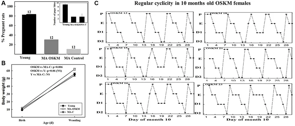
In this study, the researchers used female Sprague-Dawley rats and divided them into groups of 12 animals each. At four months of age, they injected an specifically modified virus (adenoviral vector) carrying either OSKM and green fluorescent protein (GFP) genes or only GFP as a control into the rodents’ hypothalami, spurring the production of proteins. Then, they observed how the rat’s cycles changed as the animals aged.

The control group of rats had the typical age-related changes to their estrous cycles. The young rats had regular estrous cycles, which last for 4-5 days and have four stages: proestrus, estrus, metestrus, and diestrus.

This regularity changed around 9 months of age, when cycles became more irregular. Starting at 10 months of age, the researchers observed the prevalence of constant estrus status and the presence of numerous fluid-filled sacs (follicular cysts) in the ovaries. Past 20 months of age, female rats transitioned into the constant diestrus phase.

OSKM Rat Fertility 1

OSKM treatment impacted these cycles, and the OSKM-treated animals continued to have regular cycles at 10 months old.

Impact on fertility

The animals were mated for one week with a young male rat. The mating occurred almost half a year after the gene therapy. The authors point out that even though gene expression should still occur, the expression levels are most likely lower at the time of mating compared to the time of injection.

The researchers also admit that they expected viral vectors carrying OSKM to reach only a small proportion of hypothalamic cells. However, this was enough to impact the rate of reproductive aging.

The young group had the highest pregnancy rate at 83%, and the old control group had almost exactly a tenth of that, at 8.3%. The OSKM-treated group had a pregnancy rate of 25%, which, while lower than that of the young group, was still improved compared to the age-matched control group. However, it was not statistically significantly different (p=0.06552). An increase in the size of the experimental cohort would be beneficial in obtaining statistically significant and more robust experimental proof, but these results show a very positive trend.

The younger group also had a larger litter size (mean litter size 9.1 pups), while the mean litter size of the older animals, both control and OSKM-treated, was three pups. Pups from all groups survived and showed normal behavior.

The researchers observed differences in body weight at birth. It was lower for pups born to young mothers but similar in both groups of older mothers. However, there were differences between the pups from the older mothers at the time of weaning. The pups from the OSKM-treated animals gained more weight than the old control animals’ pups, suggesting that age affects milk supply. However, OSKM treatment can remedy the lack of milk supply and/or increase milk quality.

The weight of the pups from younger mothers remained lower, which was possibly caused by the larger litter size and more animals that the mother needed to feed.

OSKM Rat Fertility 2

A long road to optimizing fertility

The researchers believe that achieving regular cycles, and a 25% fertility rate in rats that are close to the cessation of their reproductive span, shows that this OSKM gene therapy significantly benefited the animals’ reproductive system; however, the fertility rate was lower than in young animals, suggesting that “regular ovulation is a necessary but not sufficient condition for keeping the rats at optimal fertility levels.” There are also other components of the reproductive system that impact fertility.

The researchers of this study were unable to assess any changes in hypothalamic DNA methylation following OSKM gene therapy. However, they expect that, as in previous work, OSKM treatment led to the reversal of age-related epigenetic changes.

The researchers suggest that this kind of therapy might be used to help extend women’s reproductive span. However, given that this research was done in rats, the impact and side effects of such a therapy on human beings need to be extensively studied in the future.

We would like to ask you a small favor. We are a non-profit foundation, and unlike some other organizations, we have no shareholders and no products to sell you. All our news and educational content is free for everyone to read, but it does mean that we rely on the help of people like you. Every contribution, no matter if it’s big or small, supports independent journalism and sustains our future.

Literature

[1] Gallardo, M. D., Girard, M., Portiansky, E. L., & Goya, R. G. (2025). Oct4, Sox2, Klf4, c-My (OSKM) gene therapy in the hypothalamus prolongs fertility and ovulation in female rats. Aging, null, 10.18632/aging.206191. Advance online publication.

[2] Neal-Perry, G., Nejat, E., & Dicken, C. (2010). The neuroendocrine physiology of female reproductive aging: An update. Maturitas, 67(1), 34–38.

[3] Neal-Perry, G. S., Zeevalk, G. D., Santoro, N. F., & Etgen, A. M. (2005). Attenuation of preoptic area glutamate release correlates with reduced luteinizing hormone secretion in middle-aged female rats. Endocrinology, 146(10), 4331–4339.

[4] Huang, H. H., Marshall, S., & Meites, J. (1976). Capacity of old versus young female rats to secrete LH, FSH and prolactin. Biology of reproduction, 14(5), 538–543.

[5] Rodríguez, S. S., Schwerdt, J. I., Barbeito, C. G., Flamini, M. A., Han, Y., Bohn, M. C., & Goya, R. G. (2013). Hypothalamic IGF-I gene therapy prolongs estrous cyclicity and protects ovarian structure in middle-aged female rats. Endocrinology, 154(6), 2166–2173.

Down arrows

Early Adult Mortality Remains High in the US

Mortality among Americans aged 25-44 has risen substantially between 2011 and 2023, a new study has found, and it remains high even after having passed the COVID-related peak [1].

Riches to rags

Despite the US being one of the world’s richest countries, Americans’ average life expectancy has been lagging behind that of comparable countries by more than four years (even more when compared to top performers such as Spain and Japan). The gap has widened substantially starting at around 2010, when US life expectancy growth ground to a halt and later took an unusually hard hit during the COVID-19 pandemic (interestingly, it was followed by a robust rebound in 2022 and 2023).

Part of this multifaceted phenomenon is increased mortality in young adults. While much of it has been driven by an epidemic of illegal drugs, deaths of natural causes such as cancer have also skyrocketed. This trend worries researchers and policymakers and is being actively investigated.

Not just COVID

In a new study that originated from the University of Minnesota and Boston University and was published in JAMA Open Network, the researchers took an unusually long perspective to analyze the dynamics of excess deaths in American adults aged 25-44. The team used 2010 as their baseline and defined excess mortality as that exceeding extrapolation of 1999-2010 trends.

Having analyzed 3,392,364 deaths, they found that excess mortality per capita in this category began to rise well before the pandemic. In 2019, it was 34.6% higher than expected (i.e., had pre-2011 trends continued).

Excess mortality

The pandemic brought another sharp increase in mortality, two major drivers being COVID-19 and drug overdoses. However, deaths from multiple other causes, including cancer, circulatory problems, and metabolic diseases, increased as well. Some of these may be connected to COVID, as studies have linked COVID infection to various complications such as cardiovascular conditions [2], or substance abuse. As a result, in 2021, all-cause excess mortality was almost three times what it had been in 2019 (116.2 vs 41.7 deaths per 100,000 population).

A complex phenomenon

Hinting at this interconnectedness is the fact that after 2021, excess mortality began to fall in several categories, such as cardiometabolic conditions. However, even for these causes, excess mortality remains much higher than pre-pandemic levels. All-cause early adult mortality was 70.0% higher in 2023 than it would have been had pre-2011 trends continued. In absolute numbers, this excess mortality translates into 71,124 deaths annually.

This might be partially due to the long-lasting effects of COVID, which are only beginning to attract researchers’ attention (“long COVID” and other complications). Another possible explanation is that drug- and alcohol-related deaths are still very high compared to a decade ago. Substance abuse is associated with multiple health hazards and might drive other causes of death.

The top five causes of death that collectively accounted for nearly three-quarters of the excess mortality in 2023 were drug poisoning (31.8% of excess mortality), the residual natural-cause category (16.0%), transport-related deaths (14.1%), alcohol-related deaths (8.5%), and homicide (8.2%). “Additionally, the combined contribution of cardiometabolic conditions, including circulatory and endocrine, metabolic, and nutritional, was substantial (9.2%),” the paper notes.

“The rise in opiate deaths has been devastating for Americans in early and middle adulthood,” said Elizabeth Wrigley-Field, lead author and an associate professor in the University of Minnesota College of Liberal Arts and Institute for Social Research and Data Innovation. “What we didn’t expect is how many different causes of death have really grown for these early adults. It’s drug and alcohol deaths, but it’s also car collisions, it’s circulatory and metabolic diseases – causes that are very different from each other. That tells us this isn’t one simple problem to fix, but something broader.”

“Our findings underscore the urgent need for comprehensive policies to address the structural factors driving worsening health among recent generations of young adults,” said author Andrew Stokes of Boston University. “Solutions may include expanding access to nutritious foods, strengthening social services and increasing regulation of industries that affect public health.”

Cancer is on the rise

While most natural causes are declining, cancer is an outlier, with excess mortality at an all-time high. A large-scale study in The Lancet last year found that cancer prevalence has increased in young compared to older cohorts, particularly in Generation Xers and millennials, for 17 out of 34 cancer types [3]. Alarmingly, cancer is often more aggressive in younger people.

This hike in cancer incidence has happened despite a declining prevalence of smoking and some other unhealthy behaviors. On the other hand, factors such as alcohol consumption, obesity, ultra-processed food consumption, and environmental pollution remain a challenge. A recent advisory from the US Surgeon General warns that alcohol is a major risk factor for cancer.

However, early detection might also have contributed to the rates of cancer in earlier cohorts. Some slow-growing cancers might be detectable now at earlier ages, affecting the statistic.

The lost Americans

Interestingly, the US was not always behind its peers in life expectancy. In 2023, a study by the University of Boston scientists aptly named “Missing Americans: Early death in the United States” stated that “The United States had lower mortality rates than peer countries in the 1930s-1950s and similar mortality in the 1960s and 1970s. Beginning in the 1980s, however, the United States began experiencing a steady increase in the number of missing Americans, reaching 622,534 in 2019 alone.” [4] The situation got much worse during the COVID-19 pandemic.

The authors of that study attributed the growing discrepancy to the lack of large-scale public health initiatives in the US. “While COVID-19 brought new attention to public health, the backlash unleashed during the pandemic has undermined trust in government and support for expansive policies to improve population health,” said the study’s lead and corresponding author Jacob Bor, associate professor of global health and epidemiology, at the time. “This could be the most harmful long-term impact of the pandemic, because expansion of public policy to support health is exactly how our peer countries have attained higher life expectancy and better health outcomes.”

We would like to ask you a small favor. We are a non-profit foundation, and unlike some other organizations, we have no shareholders and no products to sell you. All our news and educational content is free for everyone to read, but it does mean that we rely on the help of people like you. Every contribution, no matter if it’s big or small, supports independent journalism and sustains our future.

Literature

[1] Wrigley-Field, E., Raquib, R. V., Berry, K. M., Morris, K. J., & Stokes, A. C. (2025). Mortality Trends Among Early Adults in the United States, 1999-2023. JAMA Network Open, 8(1), e2457538-e2457538.

[2] Lee, C. C., Ali, K., Connell, D., Mordi, I. R., George, J., Lang, E. M., & Lang, C. C. (2021). COVID-19-associated cardiovascular complications. Diseases, 9(3), 47.

[3] Sung, H., Jiang, C., Bandi, P., Minihan, A., Fidler-Benaoudia, M., Islami, F., … & Jemal, A. (2024). Differences in cancer rates among adults born between 1920 and 1990 in the USA: an analysis of population-based cancer registry data. The Lancet Public Health, 9(8), e583-e593.Chicago

[4] Bor, J., Stokes, A. C., Raifman, J., Venkataramani, A., Bassett, M. T., Himmelstein, D., & Woolhandler, S. (2023). Missing Americans: early death in the United States—1933–2021. PNAS nexus, 2(6), pgad173.

New Nanoparticles for Treating Arthritis

In the Journal of Nanobiotechnology, researchers have described a new method of delivering a long-lasting treatment into cartilage.

A protein that promotes autophagy

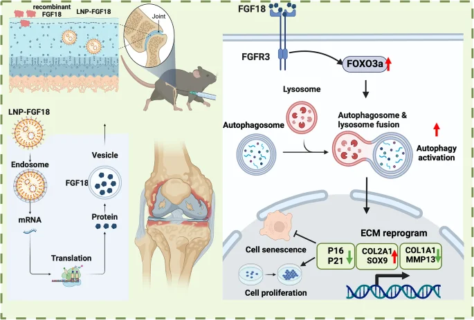
Previous work has linked expression of the FGF18 protein with healthy cartilage and joints [1]. Problems with the gene responsible for FGF18 lead to osteoarthritis [2], and it has been found to be important alongside arthritis therapies, such as hydrogels that form a lattice for cartilage growth [3]. This is because FGF18 is known to have positive effects on the FOXO3 pathway, which stimulates autophagy, the cellular self-consumption process that removes unwanted and harmful components [4].

However, using a protein as a cartilage treatment has its own problems. Recombinant proteins directly delivered into tissue don’t last very long [5], and even mRNA-based therapies are vulnerable to rapid degradation in the human body [6]. To combat this, the researchers have chosen lipid nanoparticles (LNPs), which encapsulate the mRNA in order to deliver it into cells [7].

The researchers first confirmed the existence of a link between FGF18 and osteoarthritis. A broad gene expression database has reported that elderly people have only a quarter the FGF18 of young people. Tissue samples have revealed that people who undergo total knee arthroplasty have their FGF18 reduced by half. Similarly, mice that have had arthritis artificially induced by meniscus destabilization, as well as naturally aged mice, have approximately half the FGF-18-positive cells of healthy young mice.

Exposing cartilage-generating cells (chondrocytes) to an inflammatory environment characterized by TNF-α resulted in FGF18 expression being reduced to a fourth of its normal value. Driving chondrocytes senescent by exposing them to hydrogen peroxide reduced FGF18 to two-fifths of its normal value.

Effective delivery and therapeutic effects

LNP FGF18 Action

The mRNA delivery appeared to be effective. A cellular examination showed no toxicity to chondrocytes even at high concentrations. Unlike recombinant FGF18, the LNP-encapsulated mRNA nanoparticles penetrated relatively deeply into the cartilage of both young and old mice. The nanoparticles were just small enough to fit within the pores of the mice’s collagen networks, even the dense, cross-linked collagen of older animals.

Using a bioluminescent reporter, the researchers found out that this LNP treatment stays confined to where it needs to be and does not migrate to other organs, such as the liver, in appreciable amounts. Instead, it stays within the knee joint for approximately six days, and its effects diminish far slower than mRNA without LNP encapsulation. The LNP-mRNA was found to successfully cause cells to express significant amounts of the FGF18 protein.

This approach had significant beneficial effects in a cellular culture. Cellular senescence induced by the inflammatory cytokine IL-1β was cut approximately in half, as measured by p16, p21, p53, and SA-β-gal staining. Proliferation was approximately doubled as well. LNP-mRNA for FGF18 had very similar effects to pure FGF18 in this cellular experiment.

Autophagy was similarly upregulated. FOXO3 is downregulated when chondrocytes are exposed to IL-1β, but the LNP-mRNA was found to restore it nearly to the level of the control group. Cells that were only exposed to LNP-mRNA without IL-1β had even higher levels of FOXO3. This led to an increase of cartilage-producing proteins, and further experiments confirmed that this was due to an increase in autophagy.

After confirming its effects in cells, the researchers turned to mice: a control group, a group with a destabilized meniscus and no treatment, a group treated with FGF18 protein every week, and a group treated with LNP-mRNA every week. The damaged, untreated group was hypersensitive to pain, which was partially ameliorated by FGF18 and slightly moreso by the LNP treatment, although all of the damaged mice gradually got more sensitive to pain over eight weeks.

The LNP treatment was also found to benefit the mice’s gait and physical biomarkers, in both the destabilized meniscus model and in naturally aged mice. In many of the tests, there were no significant differences between FGF18-treated and LNP-treated mice, but there were some benefits to the new approach.

Most notably, the cartilage of the LNP-treated mice was significantly thicker, restoring the cartilage of damaged mice nearly to that of undamaged mice and, most critically, restoring the cartilage of aged mice nearly to that of young mice. A closer investigation found that the LNP injection was having the same effects in the mice as in the cellular culture, restoring proliferative capacity to the mice’s chondrocytes.

This is not a human study, but it appears that human trials are the next logical step for this approach, as it appears to be both safe and effective in animal models. Time will tell whether this particular LNP approach will be tested for the clinic or if it will undergo further refinement first.

We would like to ask you a small favor. We are a non-profit foundation, and unlike some other organizations, we have no shareholders and no products to sell you. All our news and educational content is free for everyone to read, but it does mean that we rely on the help of people like you. Every contribution, no matter if it’s big or small, supports independent journalism and sustains our future.

Literature

[1] Davidson, D., Blanc, A., Filion, D., Wang, H., Plut, P., Pfeffer, G., … & Henderson, J. E. (2005). Fibroblast growth factor (FGF) 18 signals through FGF receptor 3 to promote chondrogenesis. Journal of Biological Chemistry, 280(21), 20509-20515.

[2] Boer, C. G., Hatzikotoulas, K., Southam, L., Stefánsdóttir, L., Zhang, Y., de Almeida, R. C., … & Wilkinson, J. M. (2021). Deciphering osteoarthritis genetics across 826,690 individuals from 9 populations. Cell, 184(18), 4784-4818.

[3] Gothard, D., Rotherham, M., Smith, E. L., Kanczler, J. M., Henstock, J., Wells, J. A., … & Oreffo, R. O. (2024). In vivo analysis of hybrid hydrogels containing dual growth factor combinations, and skeletal stem cells under mechanical stimulation for bone repair. Mechanobiology in Medicine, 2(4), 100096.

[4] Cinque, L., Forrester, A., Bartolomeo, R., Svelto, M., Venditti, R., Montefusco, S., … & Settembre, C. (2015). FGF signalling regulates bone growth through autophagy. Nature, 528(7581), 272-275.

[5] Evans, C. H., Kraus, V. B., & Setton, L. A. (2014). Progress in intra-articular therapy. Nature Reviews Rheumatology, 10(1), 11-22.

[6] Hajj, K. A., & Whitehead, K. A. (2017). Tools for translation: non-viral materials for therapeutic mRNA delivery. Nature Reviews Materials, 2(10), 1-17.

[7] Hou, X., Zaks, T., Langer, R., & Dong, Y. (2021). Lipid nanoparticles for mRNA delivery. Nature Reviews Materials, 6(12), 1078-1094.

Rejuvenation Roundup January 2025

Rejuvenation Roundup January 2025

As rejuvenation research advances from theory to practice, more therapies start making their way into the clinic. 2025 continues with both mouse experiments and human clinical trials.

Interviews

Matthew O'ConnorCyclarity Launches Human Trial to Cure Atherosclerosis: Recently, Cyclarity Therapeutics announced the launch of a Phase 1 human clinical trial for a drug that aims to remove the arterial plaques that lead to heart attacks and strokes. Its primary cyclodextrin drug candidate, UDP-003, focuses on 7-ketocholesterol, a type of oxidized cholesterol that increases in cells and tissues as people age.

Marco Quarta on Cellular Senescence in Aging: Dr. Marco Quarta runs one of the most interesting start-ups in the longevity field: Rubedo, which focuses on utilizing the senolytic approach to cellular senescence. This company has developed ingenious ways to cope with the notorious heterogeneity of senescent cells and is one of the first to bring its senolytic drug candidate into clinical trials.

Advocacy and Analysis

The Battle for Long Life Has Been Accomplished: What’s Next?: How long can people live? This is not just a foundational question in science. The answer has important public policy implications and is of interest to us all. Recent scientific evidence has revealed the answer, so what’s next in humanity’s never-ending battle agaiS. Jay Olshanskynst disease and the persistent ravages of aging?

Research Roundup

Keeping Stem Cells Healthy and Young: A team of researchers has outlined a new approach that uses mRNA to prevent senescence and strengthen mesenchymal stem cells (MSCs) against aging before they are transplanted into patients.

Human ear anatomyA Potential Gene Therapy for Hearing Loss: In JCI Insight, researchers have explored the possibility of using gene therapy to restore a crucial protein and repair hearing loss. The most critical finding is that the adult cochlea, which processes hearing, is in fact capable of being remodeled through changes in gene expression after birth.

Drinking and Dying: Alcohol as a Risk Factor for Cancer: A new advisory by the US Surgeon General highlights a topic that – as the document itself notes – has been flying mostly under the public’s radar: the relationship between alcohol consumption and cancer.

Caloric restrictionReceiving Caloric Restriction Benefits Without Practicing It: In a new study, researchers have found that lithocholic acid, a metabolite found in the serum of calorically restricted mice, can mimic the effects of caloric restriction. While their research was conducted on model systems, they point to a previous study that observed that this metabolte was observed to be increased in the serum of healthy humans following 36 hours of fasting.

Precision Targeting of Senescent Cells: In a journal called Small, researchers have described a new targeting mechanism for delivering senolytic compounds where they need to go. These molecules are encapsulated in tiny soap bubbles rather than silica-based nanoparticles.

Time-restricted eatingIntermittent Fasting Improves Coordination in Mice: Researchers have discovered that intermittent fasting increases myelin in aged mice, leading to better neural function and coordination. Normally, neuronal axons are coated in a protein sheath made of myelin, which is necessary for their proper function, but demyelination occurs in aging.

A Gut Metabolite Reduces Senescence and Inflammation: In a preprint study, scientists from Lifespan Research Institute and the Buck Institute for Research on Aging have published their findings that Urolithin A, a molecule that has garnered a lot of attention in the longevity field, potently reduces senescence-related markers in human fibroblasts.

Gut bacteriaThe Impact of a Human Breast Milk Probiotic on Sarcopenia: The authors of this study, citing evidence of a link between the gut microbiome, muscle health, and sarcopenia, investigated the effect of the consumption of a probiotic on the muscle health of sarcopenia patients.

Enhancing NAD+ Efficiency by Energizing Sirtuins: Researchers publishing in Physical Review X have discovered compounds that can double the efficiency of the sirtuin SIRT3 in processing NAD+. Unlike previous efforts, this drug does not rely on a substrate to function.

Genetic examinationNew Study Links Epigenetic Changes to Genetic Mutations: A new paper published in Nature Aging suggests that somatic mutations cause significant remodeling of the epigenetic landscape. The findings might be relevant to future anti-aging interventions.

Fighting Alzheimer’s by Helping Neurons Consume Proteins: Researchers have found that kinesin family member 9 (KIF9), a protein that diminishes with aging, is instrumental in allowing cells to consume harmful proteins and fights Alzheimer’s in a mouse model.

TelomeresMaintaining Telomeres Extends Lifespan in Mice: A recent study has found that the overexpression of telomerase reverse transcriptase (TERT), which is a subunit of telomerase, an enzyme essential for telomere maintenance, leads to lifespan extension in mice without significant side effects.

Maintaining Muscle by Restoring Gut Bacteria: In Aging Cell, researchers have described how different combinations of gut bacteria impact muscle strength in mice. The link between gut bacteria and health is well-documented, and multiple biomarkers have confirmed that a healthy gut leads to health elsewhere.

Targeting cancerNew Drug Eliminates Breast Cancer in Mouse Study: Researchers have discovered a small molecule that effectively kills cancer cells in the most prevalent type of breast cancer. The new drug could help against cancer recurrence and decrease the need for surgery.

Restoring Cellular Proliferation Through Exosomes: In Cell Metabolism, researchers have described how a microRNA (miRNA) derived from exosomes generated by human embryonic stem cells (hESCs) restores function and fights senescence in cell cultures and mice.

Ultrasound deviceUltrasound as a Tool to Eliminate Senescent Cells: A new study suggests that low-intensity pulsed ultrasound (LIPUS) can be beneficial in eliminating senescent cells through the recruitment and activation of immune cells. LIPUS is a technology that can be easily applied in the clinic.

Inhibiting a Fundamental Factor in Brain Inflammation: Researchers have devised a method of reducing brain inflammation by creating a long-lasting inhibitor of the inflammatory factor NF-κB. These researchers believe that it “may serve as a potent therapeutic agent against pathological age-related inflammatory processes, especially those that target macrophages and microglia.”

Printed heartArtificially Grown Tissue Repairs Heart Failure in Monkeys: German scientists have created lab-grown “patches” of heart muscle tissue derived from pluripotent stem cells. Following a success with rhesus monkeys, they have obtained approval for a human trial.

Reducing functionally defective old HSCs alleviates aging-related phenotypes in old recipient mice: This study demonstrates the presence of “younger” HSCs in old mice and that aging-associated functional decline can be mitigated by reducing dysfunctional HSCs.

Tenascin-C promotes bone regeneration via inflammatory macrophages: Taken together, this study reveals the regulation of macrophage recruitment and its function in the activation of skeletal stem cells after bone injury, providing a strategy to accelerate bone regeneration by TNC delivery.

Innovative treatment of age-related hearing loss using MSCs and EVs with Apelin: These findings highlight the regenerative capabilities of MSCs and EV-mediated therapeutic approaches for this condition.

Delivery of FGF18 using mRNA-LNP protects the cartilage against degeneration via alleviating chondrocyte senescence: In summary, this study presents a novel approach superior to recombinant protein alone and holds promise as a new therapeutic strategy for OA.

Data-driven discovery of associations between prescribed drugs and dementia risk: A systematic review: Drug repurposing for use in dementia is an urgent priority. These findings offer a basis for prioritizing candidates and exploring underlying mechanisms.

Intestine-specific disruption of mitochondrial superoxide dismutase extends longevity: Combined, these results indicate that disruption of sod-2 in neurons, intestine, germline, or muscle is not required for lifespan extension, but that decreasing sod-2 expression in just the intestine extends lifespan.

Protection of Alzheimer’s disease progression by a human-origin probiotics cocktail: These results suggest that this unique probiotics cocktail could serve as a prophylactic agent to reduce the progression of cognitive decline and AD pathology.

The role of the Mediterranean diet in reducing the risk of cognitive impairement, dementia, and Alzheimer’s disease: a meta-analysis: These findings underscore the Mediterranean diet’s potential as a central element in neuroprotective public health strategies to mitigate the global impact of cognitive decline and dementia and to promote healthier cognitive aging.

Dietary carotenoid intakes and biological aging among US adults, NHANES 1999–2018: Increased dietary intakes of various carotenoids were associated with lower biological aging indices, which was possibly and mainly driven by lutein/zeaxanthin and β-carotene.

Multi-omics characterization of improved cognitive functions in Parkinson’s disease patients after the combined metabolic activator treatment: These results show that combined metabolic activator administration leads to enhanced cognitive function and improved metabolic health in Parkinson’s disease patients as recently shown in Alzheimer’s disease patients.

Combination of rapamycin and adipose-derived mesenchymal stromal cells enhances therapeutic potential for osteoarthritis: These findings suggest that the rapamycin and AD-MSC combination enhances the therapeutic efficacy of these cells in senescence-driven degenerative diseases such as OA, notably by improving their anti-fibrotic and anti-inflammatory properties.

Long-term intake of Tamogi-take mushroom (Pleurotus cornucopiae) mitigates age-related cardiovascular dysfunction and extends healthy life expectancy: Ingestion of Tamogi-take mushrooms could serve as a dietary intervention to promote cardiovascular health, support healthy aging and slow the progression of age-related diseases.

Ergothioneine improves healthspan of aged animals by enhancing cGPDH activity through CSE-dependent persulfidation: These findings elucidate this compound’s multifaceted actions and provide insights into its therapeutic potential for combating age-related muscle decline and metabolic perturbations.

Comprehensive evaluation of lifespan-extending molecules in C. elegans: These findings confirmed robust lifespan extension by many, but not all, of the 16 tested compounds from the literature and revealed that some of them could be combined to obtain additive effects.

Oct4, Sox2, Klf4, c-Myc (OSKM) gene therapy in the hypothalamus prolongs fertility and ovulation in female rats: Long-term OSKM gene therapy in the hypothalamus is able to extend the functionality of such a complex system as the hypothalamo-pituitary-ovarian axis.

Transcriptomic signatures and network-based methods uncover new senescent cell anti-apoptotic pathways and senolytics: Identifying new antiapoptotic resistance targets and drugs with potential senolytic activity paves the way for developing new pharmacological therapies to eliminate senescent cells selectively.

NAD World 3.0: the importance of the NMN transporter and eNAMPT in mammalian aging and longevity control: This approach features multi-layered feedback loops to provide a more comprehensive understanding of NAD.

Activation of Nuclear Receptor CAR: A Pathway to Delay Aging through Enhanced Capacity for Xenobiotic Resistance: These results suggest that the longevity effects of CAR agonists may be related to the enhancement of xenobiotic resistance of animals.

Long-Term Impact of Using Mobile Phones and Playing Computer Games on the Brain Structure and the Risk of Neurodegenerative Diseases: Lengthy mobile phone use is associated with a reduced risk of neurodegenerative diseases and improved brain structure compared to minimal usage.

News Nuggets

Futuristic CityVitalia Co-Founders Announce Split-up: Vitalia co-founders Niklas Anzinger and Laurence Ion today announced that they will be leading two new, separate organizations, Viva City and Infinita City. “Together, we built Vitalia from the ground up, establishing a foundation that has led us to this exciting new chapter,” said Anzinger and Ion in a joint statement.

Cyclarity Launches Human Trial for Atherosclerosis: Cyclarity Therapeutics, a biotechnology company based at the Buck Institute in California, has launched its first human clinical trial. Its primary candidate cyclodextrin drug, UDP-003, focuses on 7-ketocholesterol, an oxidized cholesterol variant that builds up in cells as we age.

Roswell ParkCutting-Edge Facility Expands to Support Cancer Therapy: New York Gov. Kathy Hochul and leaders from The Roswell Park Comprehensive Cancer Center came together on Monday to celebrate the opening of the newly expanded Roswell Park Good Manufacturing Engineering and Cell Manufacturing Facility (GMP).

New Database Lets You Know How Processed Your Food Is: Scientists have presented GroceryDB, an open-access online database that measures the degree of processing of tens of thousands of products sold in three major US grocery chains.

Coming Up

Founders Longevity ForumFounders Longevity Forum and NUS Announce Event: Founders Longevity Forum Singapore, hosted in collaboration with the National University of Singapore (NUS) Academy for Healthy Longevity, Yong Loo Lin School of Medicine, and Longevity.Technology is set to host a pivotal two-day event on 27-28 February 2025, in Singapore.

The Global Conference on Gerophysics: Chaired by Prof Brian Kennedy, Assoc Prof Jan Gruber and Dr Maximilian Unfried, this pioneering conference will bring together leading theoretical physicists and eminent researchers in ageing and rejuvenation biology to explore a transformative new field: ‘Gerophysics’.

Longevity Med Summit FullThe 3rd Longevity Med Summit Heads to Lisbon in May 2025: The Global 3rd Longevity Med Summit, the premier global event in longevity medicine, wellness, and healthcare innovation, is set to take place in Lisbon from May 6 to 8, 2025. This year’s summit promises an expanded agenda featuring groundbreaking topics, world-renowned speakers, and an exclusive Pre-Summit Day focused on the Future of Wellness.

Hevolution Foundation Hosts Second Global Healthspan Summit: On February 4-5, 2025, Hevolution Foundation will hold its second Global Healthspan Summit (GHS) in Riyadh, Saudi Arabia. The two-day event at the Four Seasons Hotel brings together international attendees, including world leaders, policymakers, researchers & scientists, and experts from the biotechnology, pharmaceutical, healthcare, and private sectors.

We would like to ask you a small favor. We are a non-profit foundation, and unlike some other organizations, we have no shareholders and no products to sell you. All our news and educational content is free for everyone to read, but it does mean that we rely on the help of people like you. Every contribution, no matter if it’s big or small, supports independent journalism and sustains our future.
Printed heart

Artificially Grown Tissue Repairs Heart Failure in Monkeys

German scientists have created lab-grown “patches” of heart muscle tissue derived from pluripotent stem cells. Following a success with rhesus monkeys, they have obtained approval for a human trial [1].

Wear and tear

As one of the most hard-working tissues in the body, the heart muscle is subject to incessant wear and tear due to aging and various health conditions. Unsurprisingly, heart failure is one of the most common age-related causes of death.

Scientists have tried to repair damaged heart tissue by injecting healthy heart muscle cells (cardiomyocytes), but retention and rejection issues are abundant. In a new study published in Nature, a group of German researchers has reported on an exciting new technique: growing entire patches of brand-new heart tissue from scratch.

Let it grow

The process starts with induced pluripotent stem cells (iPSCs), which are cells that were de-differentiated using cellular reprogramming methods into a stem-like pluripotent state. Such cells can then be re-differentiated into many cell types. Reprogramming also makes them epigenetically younger, so they are ready to do heavy lifting.

These newly differentiated cardiomyocytes are then mixed with stromal cells that provide structural support, and a patch of something closely resembling heart muscle tissue is grown in culture. The researchers call these structures engineered heart muscle (EHM).

After a series of previous experiments in rodent models, the group decided to take a major step up and move to non-human primates. While it is possible to produce iPSCs from the patient’s own cells, the researchers decided to use existing lines of iPSC-derived cardiomyocytes. The trade-off was the need for immunosuppression.

A group of rhesus macaques was subjected to a procedure imitating heart failure, and then their injured hearts were reinforced with EHMs in two different doses: either two or five patches. The higher dose uses about 200 million cardiomyocytes.

High retention, improved function

With both doses, but more so with the higher one, the researchers achieved a sustained and significant increase in heart wall thickness. Two of the three monkeys in the high dose group also showed increased heart wall contractility, indicating improved heart function.

The engrafted tissue, which initially lacked its own blood vessels, underwent vascularization upon implantation, even though blood perfusion was not as good as in the surrounding tissue. EHM cardiomyocytes were less developed, “younger,” than their resident counterparts, which is to be expected. It remains to be seen to what degree they can eventually develop.

Importantly, graft retention was confirmed for up to six months after the procedure, when the study ended. The researchers claim that this is the best result achieved by anyone so far.

Now, to humans!

In another experiment, the group previously implanted EHMs in a human patient who was awaiting a transplant for his severely damaged heart. After the new heart was transplanted, the researchers were able to study how their EHM patches performed on the old one.

Just like in monkeys, cardiomyocyte retention was good, and a high degree of vascularization was achieved. The patient demonstrated a stable disease course. “Collectively, the obtained clinical data confirmed the translatability of heart remuscularization by EHM allograft implantation from rhesus macaques to human patients with advanced heart failure,” the paper says.

“We have shown in rhesus macaques that cardiac patch implantation can be applied to re-muscularize the failing heart. The challenge was to generate and implant enough heart muscle cells from rhesus macaque induced pluripotent stem cells to achieve sustainable heart repair without dangerous side effects such as cardiac arrhythmia or tumor growth,” said Professor Wolfram-Hubertus Zimmermann, director of the Department of Pharmacology and Toxicology at the University Medical Center Göttingen, the study’s lead author.

Based on these results, the researchers have secured approval for a first-of-its-kind trial in human patients: “Safety and Efficacy of Induced Pluripotent Stem Cell-derived Engineered Human Myocardium as Biological Ventricular Assist Tissue in Terminal Heart Failure.”

We would like to ask you a small favor. We are a non-profit foundation, and unlike some other organizations, we have no shareholders and no products to sell you. All our news and educational content is free for everyone to read, but it does mean that we rely on the help of people like you. Every contribution, no matter if it’s big or small, supports independent journalism and sustains our future.

Literature

[1] Jebran, AF., Seidler, T., Tiburcy, M. et al. (2025). Engineered heart muscle allografts for heart repair in primates and humans. Nature.

Brain inflammation

Inhibiting a Fundamental Factor in Brain Inflammation

Researchers have devised a method of reducing brain inflammation by creating a long-lasting inhibitor of the inflammatory factor NF-κB.

Targeting inflammaging at its roots

This study, published in the Nature journal Experimental & Molecular Medicine, begins with a discussion of age-related chronic inflammation (inflammaging) and its contributions to aging. Specifically, the researchers focus on neuroinflammation, which occurs when age-affected brain microglia begin sending out pro-inflammatory signals, particularly the cytokine NF-κB [1]. While considerable research has elucidated many of the fundamental reasons why this signaling occurs [2], treatments have remained elusive.

NF-κB, in particular, has been well documented; many papers on age-related diseases have pinpointed it as a problem and potential target [3]. However, while these researchers have noted that despite the existence of more than 700 NF-κB inhibitors in the laboratory, there is not a single one that has gone through the clinical trial process.

These researchers’ candidate is a variant of a known natural inhibitor, IκB. Replacing two of its amino acids prevents this protein from being degraded by cells as the natural version would be; this engineered super-repressor is termed srIκB, and it is intended to linger in the cellular cytoplasm and inhibit NF-κB in a long-lasting way.

As their delivery vector, the researchers have chosen exosomes, cellular messengers that do not stimulate the immune system [4], and the exosomes loaded with the super-repressor are called Exo-srIκB. In previous work, this research team has used Exo-srIκB to treat inflammatory diseases in animal models [5]; this, however, is their first foray into tackling brain inflammation.

Directly affecting aspects of inflammation

In the researchers’ first experiment, they examined the brains of 2- to 3-month-old mice and compared them with the brains of 21- to 22-month-old mice. As expected, the cytokines and inflammatory factors were significantly greater in the old mice, and the amount of natural IκB was lower. Leukocytes had infiltrated the brains of the old mice, and a gene expression analysis revealed a broad increase in inflammatory factor production.

The researchers then injected pairs of 2- to 3-month-old and 18- to 22-month-old mice with Exo-srIκB for three days, along with control groups receiving empty exosomes. The older mice given Exo-srIκB had considerably lower levels of key inflammatory factors, including interleukins such as IL-1α. They also had significantly lower levels of immune B cells and macrophages compared to their control group, meaning that the immune systems had reduced responses to inflammation. Genes relating to leukocyte migration and activation were downregulated as well.

Oligodendrocytes, play supportive roles in the functioning of the brain, such as myelination, and become more oriented towards inflammation with age. However, this age-related shift was largely reduced in the older mice given Exo-srIκB. Interestingly, however, the number of oligodendrocytes engaged in initial myelination was also reduced with the treatment; the researchers hypothesize that this is due to less need for it, as inflammation decreases myelination.

Astrocytes, which also play a supporting role in the brain, did not appear to change how they behaved. Concerningly, the numbers of some cells were changed in the same direction with Exo-srIκB as with aging. However, the endothelial cells appeared to move towards a more youthful phenotype, with brain permeability being decreased.

Exo-srIκB Action

Intercellular communication was also significantly affected. Pathways involved in chemokine activation, which encourage B cells to infiltrate the brain, were significantly reduced in the old mice given Exo-srIκB. However, other pathways relating to T cells seemed to be even stronger than before, which, as these researchers discuss, may explain why the T cells continued to be prevalent even after Exo-srIκB treatment.

While it is clearly not a complete solution by itself, these researchers believe that “Exo-srIκB may serve as a potent therapeutic agent against pathological age-related inflammatory processes, especially those that target macrophages and microglia.” They note that while they used high concentrations of this protein, it did not appear to have any significant side effects. However, the populations used were low, and this was only a mouse study conducted over a limited time period. Further work will need to be done to determine if this approach could work in human beings.

We would like to ask you a small favor. We are a non-profit foundation, and unlike some other organizations, we have no shareholders and no products to sell you. All our news and educational content is free for everyone to read, but it does mean that we rely on the help of people like you. Every contribution, no matter if it’s big or small, supports independent journalism and sustains our future.

Literature

[1] Rawji, K. S., Mishra, M. K., Michaels, N. J., Rivest, S., Stys, P. K., & Yong, V. W. (2016). Immunosenescence of microglia and macrophages: impact on the ageing central nervous system. Brain, 139(3), 653-661.

[2] Hammond, T. R., Dufort, C., Dissing-Olesen, L., Giera, S., Young, A., Wysoker, A., … & Stevens, B. (2019). Single-cell RNA sequencing of microglia throughout the mouse lifespan and in the injured brain reveals complex cell-state changes. Immunity, 50(1), 253-271.

[3] Barnes, P. J., & Karin, M. (1997). Nuclear factor-κB—a pivotal transcription factor in chronic inflammatory diseases. New England journal of medicine, 336(15), 1066-1071.

[4] Shiue, S. J., Rau, R. H., Shiue, H. S., Hung, Y. W., Li, Z. X., Yang, K. D., & Cheng, J. K. (2019). Mesenchymal stem cell exosomes as a cell-free therapy for nerve injury–induced pain in rats. Pain, 160(1), 210-223.

[5] Chae, J. S., Park, H., Ahn, S. H., Han, E. C., Lee, Y., Kim, Y. J., … & Kim, W. J. (2023). The effect of super-repressor IkB-Loaded Exosomes (Exo-srIκBs) in chronic post-ischemia pain (CPIP) models. Pharmaceutics, 15(2), 553.

Hevolution

Hevolution Foundation Hosts Second Global Healthspan Summit

On February 4-5, 2025, Hevolution Foundation will hold its second Global Healthspan Summit (GHS) in Riyadh, Saudi Arabia. The two-day event at the Four Seasons Hotel brings together international attendees, including world leaders, policymakers, researchers & scientists, and experts from the biotechnology, pharmaceutical, healthcare, and private sectors, to explore innovative solutions in the rapidly advancing fields of geroscience and healthspan. The event will address one of humanity’s greatest challenges: the rapidly growing aging population. Attendees will gain exclusive insights into pioneering research and emerging technologies that are shaping the future of healthspan science, presented by biotechnology founders, leaders, and researchers.

There is a significant gap between global life expectancy and healthspan—the number of years lived in good health — currently about 10 years (73.4 vs. 63.7 years, respectively). The global population aged 60 and older is expected to double by 2050, with individuals aged 65 and above projected to represent 1 in 6 people, up from 1 in 10 in 2021.

“A key part of our commitment to bringing everyone to the table is the Global Healthspan Summit. As a convener of stakeholders across sectors, GHS – the world’s largest event of its kind – provides a unique platform to kick off discussions among researchers, industry leaders, entrepreneurs, investors, and policymakers,” says Dr. Mehmood Khan, CEO, Hevolution Foundation. “Under the theme ‘Architecting the Future’, this summit not only serves as a forum for sharing insights and showcasing advancements but also as a catalyst for future collaborations.”

GHS 2025 features a diverse pool of speakers who will foster out-of-the-box thinking among all attendees. Some of the areas these sessions will focus on include:

  • The current healthspan investment landscape and perspectives on the latest market trends
  • How philanthropy can be a catalyst for advancing equity and driving policy change to lead to a sustainable, systematic transformation of our global healthcare system
  • Implementing healthspan-focused approaches within complex healthcare systems, addressing challenges such as interdisciplinary collaboration, data integration, and policy alignment.

In 2023, Hevolution hosted the first edition of the Global Healthspan Summit, bringing together leading experts for discussions on aging, healthcare innovation, and the healthspan ecosystem. The event attracted over 2,000 delegates and 120 speakers from top organizations such as Eli Lilly, GSK, Harvard, Mayo Clinic, Milken Institute, Saudi Arabia’s Ministry of Health, the World Bank, and the World Health Organization.

At the inaugural summit, Hevolution announced over $100 million in funding to accelerate healthspan research, including $40 million as the lead funder for the Hevolution XPRIZE healthspan partnership, $21 million for a multi-year partnership with the Buck Institute, $16 million for early-career researchers through the American Federation for Aging Research, and $5 million for postdoctoral fellowships.

This demographic shift makes aging a critical global issue, which will be addressed by international stakeholders at GHS 2025. The Hevolution Foundation leads efforts to tackle these changes, using its unique model to increase the number of geroscientists, expand the number of companies in the healthspan field, and attract funding. Through collaborative partnerships, the foundation is driving the shift from lifespan to healthspan, working toward solutions to the global challenge of aging.

About Hevolution Foundation

Hevolution Foundation is a global catalyst, partner, and convener dedicated to extending healthy human lifespans and advancing our understanding of aging. By treating aging as a process that can be addressed, the Foundation works to increase the availability of aging-related treatments, accelerate drug development timelines, and improve access to therapeutics that enhance healthspan — the number of years we live in good health. Headquartered in Riyadh, Saudi Arabia with a North American hub and an annual budget of up to $1 billion, Hevolution is the world’s largest philanthropic funder in healthspan and aging research. Over the past three years, the Foundation has committed $400 million to advancing research and innovation in this field. With plans to establish offices in additional locations worldwide, the Foundation is on a mission to propel and deliver breakthroughs that empower humanity to live healthier, longer lives.

Summit Website: Home – GHS – Hevolution Website

Social media links: X, LinkedIn

We would like to ask you a small favor. We are a non-profit foundation, and unlike some other organizations, we have no shareholders and no products to sell you. All our news and educational content is free for everyone to read, but it does mean that we rely on the help of people like you. Every contribution, no matter if it’s big or small, supports independent journalism and sustains our future.
Ultrasound device

Ultrasound as a Tool to Eliminate Senescent Cells

A new study suggests that low-intensity pulsed ultrasound (LIPUS) can be beneficial in eliminating senescent cells through the recruitment and activation of immune cells [1].

The double-edged sword of the SASP

One of the characteristics of an aging organism is the accumulation of senescent cells. Various approaches are being developed to remove or neutralize those cells.

Senescent cells produce the senescence-associated secretory phenotype (SASP), a cocktail of chemokines, pro-inflammatory cytokines, growth factors, and proteases [2]. While the SASP might have many deleterious effects, it can be a double-edged sword. Some of these many molecules have positive effects, such as attracting immune cells, which can eliminate senescent cells [3]. Precise modulation of the SASP can be a potent strategy for senescent cell elimination.

The authors of this study turned to ultrasound as a possible non-invasive therapeutic tool to eliminate senescent cells. Previous studies observed that LIPUS has positive effects on many types of tissues, including promoting wound healing or bone repair [4] and regulating the secretion of inflammation-associated cytokines [5].

Therefore, the authors of this study hypothesized that “LIPUS can modulate the secretion of SASP in senescent cells and thereby manipulate these cells” or aid in attracting immune cells.

Eating up the old cells

The authors cultured human male fibroblast cells as their research model. They made these cells replicatively senescent by allowing them to grow and divide multiple times. They divided the cells into two groups: ‘late cells,’ which had replicated many times but had not yet reached senescence, and ‘early cells,’ which had not replicated many times.

Following 20 minutes of stimulation with LIPUS, the researchers observed a marker of senescent cells, SA-β-gal, to be selectively increased in the ‘late cells’ but not in the ’early cells.’

When the researchers tested the impact of LIPUS on the expression of multiple SASP molecules, they learned that “LIPUS stimulation specifically increased the expression of immune cell attraction markers in the ‘late cells.’”

This increased expression led to an increased migration of immune cells, specifically monocytes and specific families of macrophages, towards these stimulated cells. Ultimately, it resulted in the ingestion and elimination of the ‘late cells’ by macrophages in a process called phagocytosis.

The molecules behind the scenes

In the next steps of their research, the authors investigated the molecular mechanism behind LIPUS’s selective stimulation of the SASP.

After excluding other possibilities, they tested the involvement of reactive oxygen species (ROS) since previous reports suggested increased ROS production following LIPUS stimulation [6]. These researchers confirmed that LIPUS stimulation increased intracellular ROS generation in the ‘late cells’ and observed that ROS production was required to increase SA-β-gal activity in LIPUS-stimulated ‘late cells.’

Further, they investigated the molecules that regulate the expression of SASP factors, focusing on two in particular: NF-κB and p38. NF-κB is a transcription factor family member that regulates gene expression, and p38 increases NF-κB activity.

Their experiments suggested that in ‘late cells,’ LIPUS stimulation leads to ROS-dependent activation of the p38-NF-κB pathway, activating immune cell-attracting SASP factors and leading to immune cell migration.

ROS plays a significant role in this process, but how did LIPUS stimulation cause the ROS generation? The researchers generated a few hypotheses. First, they learned that the LIPUS stimulation-generated production of extracellular ROS was not significant. Instead, LIPUS generated intracellular ROS via an enzyme called NOX4. NOX is a family of enzymes located in lipid rafts, special compartments on the plasma membranes that surround cells.

Further experiments showed that LIPUS stimulation led to perturbations in the structure and organization of the cellular membrane, creating transient pores and resulting in increased permeability, affecting the formation and localization of lipid rafts. This leads to NOX activation and ROS generation. This occurred only in ‘late cells’, whose membrane composition differs from that of ‘early cells.’

Reversing skin aging

At the end of their study, the researchers used an in vivo model of mouse skin aging to test whether LIPUS could be an efficient tool to remove senescent cells by regulating the SASP in a living organism.

After UVA-induced skin aging, LIPUS was applied for five days, and skin tissue was analyzed 10 days later. Neither UVA nor LIPUS treatment were found to impact body weight nor major organs of these mice. However, as expected, UVA resulted in extensive and deep wrinkles on the applied area and increased the levels of senescence markers.

LIPUS treatment increased SASP markers for immune cell attraction in a UVA-induced skin aging model. This increase translated into more attraction of immune cells than with UVA irradiation alone. At the same time, LIPUS treatment significantly reduced the number of cells with senescence markers, suggesting a decrease in these cells.

The researchers summarized that “these data suggest that senescent cells could be eliminated by macrophage infiltration via LIPUS stimulation.”

Optimizing for clinical use

LIPUS is a technology that can be easily applied in the clinic. Those researchers propose that it can be used to help remove senescent cells, possibly combined with senolytic treatment.

However, before this therapy can enter the clinic, LIPUS parameters need to be optimized and tested for side effects, as it is known that different LIPUS parameters can elicit different effects in different types of cells. Additionally, its limitations, such as penetration efficiency, and patients’ aged immune systems, which might not be as effective in clearing senescent cells, need to be considered.

We would like to ask you a small favor. We are a non-profit foundation, and unlike some other organizations, we have no shareholders and no products to sell you. All our news and educational content is free for everyone to read, but it does mean that we rely on the help of people like you. Every contribution, no matter if it’s big or small, supports independent journalism and sustains our future.
Literature

[1] Gwak, H., Hong, S., Lee, S. H., Kim, I. W., Kim, Y., Kim, H., Pahk, K. J., & Kim, S. Y. (2025). Low-Intensity Pulsed Ultrasound Treatment Selectively Stimulates Senescent Cells to Promote SASP Factors for Immune Cell Recruitment. Aging cell, e14486. Advance online publication.

[2] Watanabe, S., Kawamoto, S., Ohtani, N., & Hara, E. (2017). Impact of senescence-associated secretory phenotype and its potential as a therapeutic target for senescence-associated diseases. Cancer science, 108(4), 563–569.

[3] Burton, D. G. A., & Stolzing, A. (2018). Cellular senescence: Immunosurveillance and future immunotherapy. Ageing research reviews, 43, 17–25.

[4] Schortinghuis, J., Bronckers, A. L., Stegenga, B., Raghoebar, G. M., & de Bont, L. G. (2005). Ultrasound to stimulate early bone formation in a distraction gap: a double blind randomised clinical pilot trial in the edentulous mandible. Archives of oral biology, 50(4), 411–420.

[5] Li, J. K., Chang, W. H., Lin, J. C., Ruaan, R. C., Liu, H. C., & Sun, J. S. (2003). Cytokine release from osteoblasts in response to ultrasound stimulation. Biomaterials, 24(13), 2379–2385.

[6] Duco, W., Grosso, V., Zaccari, D., & Soltermann, A. T. (2016). Generation of ROS mediated by mechanical waves (ultrasound) and its possible applications. Methods (San Diego, Calif.), 109, 141–148.

Ultra-processed food

New Database Lets You Know How Processed Your Food Is

Scientists have presented GroceryDB, an open-access online database that measures the degree of processing of tens of thousands of products sold in three major US grocery chains [1].

What is ultra-processed food?

While there is no universally accepted definition, the NOVA food classification system is widely used, and it defines ultra-processed food as “industrially manufactured food products made up of several ingredients including sugar, oils, fats and salt (generally in combination and in higher amounts than in processed foods) and food substances of no or rare culinary use (such as high-fructose corn syrup, hydrogenated oils, modified starches and protein isolates).”

In other words, ultra-processed food involves breaking down “real food” and creating something mostly new, such as instant soup or candy, with a nutrient makeup unlike anything encountered in nature. This food equivalent of Frankenstein’s monster fools the gratification circuits developed by millions of years of evolution, tricking people into ingesting too much unhealthy and too little healthy food while generally overeating.

Even though ultra-processed food encompasses a wide range of different products, from beverages to sausages, as a category, it has been consistently linked to adverse health outcomes, such as cancer [2], cardiovascular disease [3], and obesity [4]. Alarmingly, people in developed nations consume up to 60% of their calories from ultra-processed food.

Getting to know your food

It is not always straightforward to know the processing of foods in a local grocery store. It is sometimes possible to make an educated guess, such as with sugary beverages, but other products are not as obvious. Unfortunately, minimalistic food labels don’t offer much help.

The group, which included researchers from Harvard Medical School and Northeastern University, has been studying ultra-processed food for several years. In their new paper published in Nature Food, the researchers describe GroceryDB, an open online database that contains information on more than 50,000 products offered by three major US chains: Walmart, Whole Foods Market, and Target.

Last year, the group published FPro, a food processing score that they had developed using machine learning techniques, that translates the nutritional content of a food item into its degree of processing. FPro, which powers GroceryDB, is mostly based on NOVA (it was trained to predict a NOVA category of the product from its ingredients) but can accommodate other food processing classification systems. The reliance on the list of nutrients is due to several reasons, such as that in unprocessed food, their quantities are constrained by biochemistry-determined physiological ranges.

Browsing GroceryDB, available at Truefood.tech, and comparing favorite foods to less or more processed alternatives is a captivating pastime. One of the main takeaways is that the degree of food processing can vary a lot even within a single category. The paper provides several examples, starting with bread.

The tale of the two cheesecakes

A multi-grain bread from Manna Organics, sold by Whole Foods, which mostly contains whole wheat, barley, and brown rice without any additives, salt, oil, and yeast, has an FPro of 0.314 (the index ranges from 0 to 1). The two less health-oriented chains, Walmart and Target, both carry Aunt Millie’s and Pepperidge Farmhouse breads (FPros of 0.732 and 0.997, respectively) with ingredients including soluble corn fiber, sugar, resistant corn starch, wheat gluten, and monocalcium phosphate.

The researchers saw a similar picture with yogurts: Seven Stars Farm yogurt made from grade A pasteurized organic milk has an FPro of 0.355. Siggi’s yogurt, with an FPro of 0.436, uses pasteurized skim milk, which, according to the paper, requires more food processing to eliminate fat. The two pale in comparison to Chobani Cookies and Cream yogurt with its whopping FPro of 0.918, thanks to loads of cane sugar and multiple additives such as caramel color, fruit pectin, and vanilla bean powder.

FPro 1

Some food categories are inherently highly processed, so it is unlikely to find cookies with a low FPro. However, even in those categories, the distribution of FPro scores is quite wide, and healthier alternatives are available. Unsurprisingly, the prevalence of less processed food was much higher in Whole Foods Market than in the other two chains.

The researchers highlighted one of the reasons for the abundance of ultra-processed food: processing decreases the cost per calorie. Across all GroceryDB categories, a 10% increase in FPro leads to an 8.7% decrease in the price per calorie. In some categories, the decline is much steeper, however, with milk, the relationship is reversed, probably due to more expensive plant milks also being more processed.

To illustrate the makeup of the FPro score for every product in GroceryDB, its ingredients are presented as a tree. This allows accounting for “ingredients of ingredients,” such as in this example with two cheesecakes. While both are highly processed, the one on the left has an FPro of 0.953 and the one on the right – 0.720. The former, along with many additives, contains sour cream which, in turn, contains a number of ingredients, such as modified food starch.

FPro 2

The highly processed ingredients are designated by red dots. The researchers mention, however, that this does not necessarily mean they are harmful. For example, xanthan gum, guar gum, and locust bean gum are considered generally safe. The purpose of GroceryDB is to allow people to dig into the ingredients of pantry staples and make informed decisions.

Informing the public’s choices

“GroceryDB serves as a proof of concept, demonstrating the potential of accessible, algorithm-ready data to advance nutrition research,” said Dr. Giulia Menichetti, a Principal Investigator and Junior Faculty at Harvard Medical School, and a co-author of the study. “This is especially significant in a field where much of the work still depends on labor-intensive manual curation, relying on descriptive definitions that suffer from poor inter-rater reliability and lack of reproducibility.”

“While the general population is increasingly aware of the potential health impacts of ultra-processed foods, they lack the knowledge to distinguish minimally processed foods, which have no known health consequences, from ultra-processed ones,” said Prof. Barabási. “Here, we set out to offer this resource by measuring the degree of processing for the foods that constitute a significant fraction of the US food supply. Most importantly, through this online resource, consumers are empowered to replace the ultra-processed foods they consume with brands that are less processed.”

Menichetti, too, underscored the potential societal benefits of their project. “Our vision with GroceryDB is not just to build a database, but to catalyze a global effort toward open-access, internationally comparable data that advances nutrition security and ensures equitable access to healthier food options for all,” she said.

Another nutrition scientist, Barry M. Popkin, Distinguished Professor of Nutrition at the University of North Carolina, who was not involved in this study, voiced some critique regarding its design: “Rather than doing an exact study of the ingredients list to find those colors, flavors and other additives identified in NOVA as identifying ultra-processed food, they guess on a set of foods that they were ultra-processed and then the machine identified the other ultra-processed foods.”

However, according to Menichetti, the approach suggested by Popkin “is not currently practical from an algorithmic perspective, due to the poor standardization of ingredient lists worldwide and the absence of definitions tied to robust, measurable variables across food composition databases.”

“Implementing such an approach,” she noted, “would have required significant manual curation, more than double the funding and time, and the incorporation of subjective opinions into the classification process. We see these challenges firsthand with our friends at Open Food Facts, a non-profit initiative powered by thousands of volunteers globally, which grapples with these same limitations daily.”

We would like to ask you a small favor. We are a non-profit foundation, and unlike some other organizations, we have no shareholders and no products to sell you. All our news and educational content is free for everyone to read, but it does mean that we rely on the help of people like you. Every contribution, no matter if it’s big or small, supports independent journalism and sustains our future.

Literature

[1] Ravandi, B., Ispirova, G., Sebek, M., Mehler, P., Barabási, A. L., & Menichetti, G. (2025). Prevalence of processed foods in major US grocery stores. Nature Food, 1-13.

[2] Fiolet, T., Srour, B., Sellem, L., Kesse-Guyot, E., Allès, B., Méjean, C., … & Touvier, M. (2018). Consumption of ultra-processed foods and cancer risk: results from NutriNet-Santé prospective cohort. bmj, 360.

[3] Srour, B., Fezeu, L. K., Kesse-Guyot, E., Allès, B., Méjean, C., Andrianasolo, R. M., … & Touvier, M. (2019). Ultra-processed food intake and risk of cardiovascular disease: prospective cohort study (NutriNet-Santé). bmj, 365.

[4] Hall, K. D., Ayuketah, A., Brychta, R., Cai, H., Cassimatis, T., Chen, K. Y., … & Zhou, M. (2019). Ultra-processed diets cause excess calorie intake and weight gain: an inpatient randomized controlled trial of ad libitum food intake. Cell metabolism, 30(1), 67-77.

An exosome filled with protein-based information.

Restoring Cellular Proliferation Through Exosomes

In Cell Metabolism, researchers have described how a microRNA (miRNA) derived from exosomes generated by human embryonic stem cells (hESCs) restores function and fights senescence in cell cultures and mice.

Looking for a better senomorphic

This study begins with a discussion of cellular senescence and its role in aging, focusing on the main approaches to dealing with it: senolytics, which kill senescent cells, and senomorphics, which transform them. Senolytics may have side effects because their number goes up with advanced age [1], and some of them are necessary for life. Even such techniques as directly affecting the SASP may have adverse impacts on the immune system [2].

While senomorphics appear to be a viable strategy, they are still in their infancy. One potential strategy involves exosomes: messenger particles that are regularly sent by cells. Previous work with hESC-derived exosomes (hESC-Exos) has found that their contents are instrumental in rejuvenating multiple tissues [3]. Therefore, these researchers decided to examine them as a senomorphic, discovering which of their many components is best at restoring senescent cells.

Cells regained the power to proliferate

The first experiment was a basic test of hESC-Exos on IMR-90 cells, a line of human fibroblasts. After 30 divisions, this cell line remains youthful, but at 50, it has reached replicative senescence. However, treating those senescent cells with hESC-Exos almost entirely reversed their senescent biomarkers, downregulating SASP-related genes such as those for inflammatory interleukins, restoring cellular proliferation, and sharply decreasing the senescence biomarker SA-β-gal. Other genes related to senescence were inhibited, while those relating to proliferation were enhanced.

These findings were recapitulated at the single-cell level. First, the researchers created a population of human cells that were modified to express the fluorescence protein along with the senescence marker p21. Then, they drove these cells senescent by exposing them to doxycycline, after which they exposed some of them to hESC-Exos.

Compared to an untreated control group, the treated cells had substantially less p21-related fluorescence and less SA-β-gal, and some of them regained the ability to proliferate. Just like with the replicatively senescent cells, hESC-Exos diminished senescence-related gene expression and enhanced proliferation-related expression instead.

Mice regained youthful attributes

From the 20th month to the 30th month of life, wild-type mice on a normal diet were injected with hESC-Exos. Compared to a control group, the exosome-treated mice performed better on both fixed and accelerating rotarod tests, had higher body weight, and retained normal bladder activity. In the Morris water maze test, they both found the platform more quickly and remembered its location more accurately. They retained their hair color and skin smoothness as well.

Biomarkers of senescence, just as in the cellular culture, were substantially reduced in the treated mice. Inflammatory factors, such as cytokines and TNF-α, were also substantially reduced in their circulation, and γ-H2AX, a marker of genomic damage, was notably reduced. Just as in cellular cultures, the mice’s biomarkers of cellular proliferation were improved. Overall, the researchers found the treated mice to be substantially rejuvenated as a whole.

Finding the key molecule

The researchers hypothesized that much of this rejuvenative power can be distilled down to the exosomes’ individual components. They selected one of them, miR-302b, which is abundantly found in hESC-Exos and is documented to play a role in cellular proliferation [4]. However, it had remained untested against aging.

This particular miRNA was found to directly regulate expression of the Cdkn1a gene, which is related to cellular senescence. Exposing IMR-90 cells to miR-302b recapitulated the effects of hESC-Exos, reducing senescence and promoting proliferation.

Encouraged, the researchers then turned to mice. This time, they injected 25- to 30-month-old mice with artificially transfected exosomes containing miR-302b. They found that this approach recapitulated the results found in hESC-Exos as well, reducing inflammatory factors and SA-β-gal, substantially improving the results of rotarod and Morris water maze tests, and restoring cellular proliferation.

Llifespan itself was also improved by this approach. The median lifespan of the mice treated with miR-302b was 137 days greater than that of the control group. While the difference was not significant, the effect seemed to be stronger in males.

While hESC-Exos and miR-302b were not compared directly, they appear to be largely similar in terms of their effects. Still, this is a cell and mouse study, and further work needs to be done to determine if this approach is safe for clinical use. It is also not known which of these approaches will be the most scalable and suitable for mass production.

We would like to ask you a small favor. We are a non-profit foundation, and unlike some other organizations, we have no shareholders and no products to sell you. All our news and educational content is free for everyone to read, but it does mean that we rely on the help of people like you. Every contribution, no matter if it’s big or small, supports independent journalism and sustains our future.

Lifterature

[1] Huang, W., Hickson, L. J., Eirin, A., Kirkland, J. L., & Lerman, L. O. (2022). Cellular senescence: the good, the bad and the unknown. Nature Reviews Nephrology, 18(10), 611-627.

[2] Zhang, L., Pitcher, L. E., Yousefzadeh, M. J., Niedernhofer, L. J., Robbins, P. D., & Zhu, Y. (2022). Cellular senescence: a key therapeutic target in aging and diseases. The Journal of Clinical Investigation, 132(15).

[3] Bi, Y., Qiao, X., Liu, Q., Song, S., Zhu, K., Qiu, X., … & Ji, G. (2022). Systemic proteomics and miRNA profile analysis of exosomes derived from human pluripotent stem cells. Stem Cell Research & Therapy, 13(1), 449.

[4] Subramanyam, D., Lamouille, S., Judson, R. L., Liu, J. Y., Bucay, N., Derynck, R., & Blelloch, R. (2011). Multiple targets of miR-302 and miR-372 promote reprogramming of human fibroblasts to induced pluripotent stem cells. Nature biotechnology, 29(5), 443-448.

Targeting cancer

New Drug Eliminates Breast Cancer in Mouse Study

Researchers have discovered a small molecule that effectively kills cancer cells in the most prevalent type of breast cancer. The new drug could help against cancer recurrence and decrease the need for surgery [1].

The ongoing fight against breast cancer

Medicine has made great strides in treating breast cancer, but the fight is far from over. About 70% of all cases are estrogen receptor alpha positive (ERα+), meaning that cancer cells express ERα, and tumor growth is driven by the hormone estrogen.

Current therapies already ensure a high five-year survival rate for ERα+ cancer patients. However, it hinges on early detection, surgical resection, and consequent long-term hormone therapy that in itself can cause serious side effects, including increased risk of endometrial cancer and osteoporosis [2]. Moreover, there is a high chance of recurrence: 10%-50% over 20 years, depending on the initial tumor size. When this happens, the resurrected cancer often does not respond to endocrine therapy due to mutations in ERα or other mechanisms.

In this case, there is an unmet need for treatments that would eliminate the cancer completely, preferably in a single swoop. A new study coming from the University of Illinois at Urbana-Champaign features a worthy candidate.

The researchers have been working on small molecules for treating ERα+ breast cancer for several years. Resistance in this type of cancer “occurs partly because endocrine therapies typically are cytostatic: tumor cell proliferation is inhibited, but cell death is modest,” the paper says. Hence, the researchers were looking for a drug that would kill breast cancer cells instead of just preventing their division.

The group’s previous candidate, ErSO, was effective, but it harmed ERα-negative cells as well [3]. This time, the researchers described an improved formulation: ErSO-TFPy. In previous studies, it showed high potency at low concentrations and tolerability at high concentrations. ErSO-TFPy targets the protein TRPM4, which is involved in cation transport and is upregulated in some cancers, including breast cancer.

First, the researchers tested ErSO-TFPy versus a string of current state-of-the-art treatments in several ERα+ breast cancer lines. The current drugs were less effective and, as expected, mostly cytostatic, causing the cells to stop dividing, while ErSO-TFPy effectively induced cell death.

Similar results were demonstrated in vivo: while fulvestrant, a currently used drug that served as a positive control, was only able to halt tumor growth, ErSO-TFPy achieved full tumor regression in concentrations well within the therapeutic window.

One of the models used was a xenograft derived from a patient who had developed drug-resistant cancer due to a mutation in ESR1, the gene coding for ERα+. In this setting, fulvestrant proved mostly ineffective, while ErSO-TFPy again eliminated the tumor completely.

In these experiments, the drug was given weekly as an intravenous injection. “This quantitative tumor regression is highly unusual for single-agent breast cancer therapeutics and may be the result of the unique, necrotic mechanism of action for this class of small molecules,” the paper says.

A single dose – even for large tumors

Given the surprising efficacy of the weekly regimen, the researchers wanted to see whether a single dose of their drug would do the trick – and it did. “If recapitulated in humans, such a minimal dosing regimen would revolutionize ERα+ breast cancer therapeutic management through improved treatment compliance, quality-of-life, and long-term outcomes for breast cancer patients,” the authors note.

“It is very rare for a compound to shrink tumors in mouse models of breast cancer, let alone completely eradicate those tumors with a single dose, so we are eager for ErSO-TFPy to advance for treatment of breast cancer,” said Paul Hergenrother, Kenneth L. Rinehart Jr. Endowed Chair in Natural Products Chemistry and the leading author of the study.

The efficacy of the current treatments for ERα+, as well as the chance of recurrence, is highly dependent on the stage at which the disease was diagnosed, so the researchers tested their drug in the extreme conditions of well-developed, extra-large tumors. Even in this challenging setting, a single IV dose of ErSO-TFPy (albeit at a higher concentration) was enough to shrink the tumors by more than 80%. This indicates the exciting possibility of a drug that can tackle late-stage breast cancer.

ErSO-TFPy results

Interestingly, ErSO-TFPy gets washed out of circulation quickly. The researchers were pleasantly surprised and somewhat baffled by the prolonged effect of their drug and are looking for possible explanations.

“The ability of ErSO-TFPy to induce complete regressions after a single dose is surprising given ErSO-TFPy serum levels peak within 10 min of administration in mice and are undetectable after 16 h when dosed at 15 mg/kg IV,” the paper says. “The xenograft experiments show that tumor regression occurs over a period of weeks, long after the compound is eliminated.”

We would like to ask you a small favor. We are a non-profit foundation, and unlike some other organizations, we have no shareholders and no products to sell you. All our news and educational content is free for everyone to read, but it does mean that we rely on the help of people like you. Every contribution, no matter if it’s big or small, supports independent journalism and sustains our future.

Literature

[1] Mulligan, M. P., Boudreau, M. W., Bouwens, B. A., Lee, Y., Carrell, H. W., Zhu, J., Mousses, S., Shapiro, D. J., Nelson, E. R., Fan, T. M., & Hergenrother, P. J. (2025). Single dose of a small molecule leads to complete regressions of large breast tumors in mice. ACS Central Science. Advance online publication.

[2] Goss, P. E., Ingle, J. N., Martino, S., Robert, N. J., Muss, H. B., Piccart, M. J., … & Pater, J. L. (2005). Randomized trial of letrozole following tamoxifen as extended adjuvant therapy in receptor-positive breast cancer: updated findings from NCIC CTG MA. 17. Journal of the National Cancer Institute, 97(17), 1262-1271.

[3] Boudreau, M. W., Mulligan, M. P., Shapiro, D. J., Fan, T. M., & Hergenrother, P. J. (2022). Activators of the anticipatory unfolded protein response with enhanced selectivity for estrogen receptor positive breast cancer. Journal of medicinal chemistry, 65(5), 3894-3912.

Marco Quarta

Marco Quarta on Cellular Senescence in Aging

Dr. Marco Quarta runs one of the most interesting start-ups in the longevity field: Rubedo, which focuses on utilizing the senolytic approach to cellular senescence. This company has developed ingenious ways to cope with the notorious heterogeneity of senescent cells and is one of the first to bring its senolytic drug candidate into clinical trials. Marco also co-founded Turn Biotechnologies, a company with a different vision based on partial cellular reprogramming, as he sees the future of anti-aging interventions as combinations of various drugs and therapies that tackle different aspects of aging.

Let’s start with how you got to where you are today.

My passion for the longevity field began when I was around five years old: I had this sudden inspiration that I wanted to become a scientist. With scientists and physicians in my family, it was natural inspiration, but what drove me was wanting to understand why different organisms live in different ways.

I had this realization that since we’re all made of the same matter, we should be able to change how long we live. It was naive, but that really inspired me. I never stopped; by age six, I had set up my first lab, playing with a chemistry kit and microscope with my family’s support.

I’m still exploring these questions today, perhaps less naively and more sophisticatedly. My studies and readings, not just in science but in philosophy, mythology, and history, were all about trying to understand this quest that humanity has pursued for millennia: how to live longer, healthier lives. The alchemist’s quest exists across all cultures and populations.

I think the difference now is that we now have real science. We’ve hit a tipping point where we have enough understanding, though there’s still much to learn, to move to Medicine 3.0 and develop longevity medicine.

As a quick side note, I actually wanted to ask you why your company Rubedo and its drug discovery platform ALEMBIC are named after alchemy terms.

I’ve long been interested in alchemy as a broader view of science and nature, a source of inspiration, essentially. Major scientists like Isaac Newton, the father of modern physics, and Robert Boyle, the father of modern chemistry, spent most of their careers as alchemists, trying to understand the language of nature and how it manifests in both animate and inanimate worlds.

There’s almost a spiritual dimension to science that has become disconnected from modern scientific practice, but philosophy and science have always been intertwined. Reading Persian, Arabic, Egyptian, Greek, Medieval, and Renaissance works, you find observational insights that remain inspiring for modern scientific approaches.

The names indeed come from alchemy. Rubedo is the final phase in creating the universal medicine, the elixir of long life meant to cure all diseases and extend longevity. Alembic was the apparatus alchemists used to distill and prepare this medicine, but ALEMBIC is also an acronym describing what our platform actually does: Algorithms for Life-Extending Medicine with Biology, Informatics, and Chemistry.

Okay, let’s go back to your story.

When choosing my university studies, I took this direction: aging biology. I did my undergraduate work in a lab under the patronage of the Nobel Laureate Rita Levi-Montalcini, who discovered nerve growth factor. I studied brain aging and regeneration, along with acute neuropathic pain. Then, I continued with a PhD in neuroscience, studying aging, physiology, and bioengineering. I wanted to understand aging from all angles, spending time on bioengineering, regenerative medicine, and stem cell biology.

I came to Stanford to work with Thomas Rando, a pioneer in the field. It was a great environment to develop as a scientist. I went on to direct the research center for Tissue Regeneration, Restoration and Repair at Veteran Medical Center of Palo Alto and Stanford Medicine, leading a team working on regenerative medicine, bioengineering, stem cells, aging, and rejuvenation in different areas. Our work on epigenetic reprogramming along with the lab of Vittorio Sebastiano, also at Stanford, became the foundation of Turn Bio.

Yes, it is interesting that you’re also a co-founder in a cellular reprogramming company

Aging is complex, but what we could do immediately is remove pathologic cells, like we have been doing for cancer for the past 50-60 years. You can’t live with them. Now we know there are other pathologic cells, more age-related, such as senescent cells, but, like cancer cells, they are highly heterogeneous. Also, they’re not all bad, but rather pleiotropic, they have different states.

The field is exciting because it reminds me of oncology in the 60s-70s. Back then, people naively wanted to develop chemotherapies to kill all cancer cells. After 50 years, we have cures for some cancers, we’re still working on others, and we understand how complex it is – we need multiple therapies and modalities.

A lot of it is the same for cellular senescence, which is why I started Rubedo: we needed to build tools first to understand the differences in cells. The idea is to remove the pathologic cells that emerge with aging biology. The other approach, with Turn Bio, is to preserve or restore useful biology in aging cells that are becoming dysfunctional – turning back the clock using cellular reprogramming.

I see strong synergy between these two strategies and platforms. My hope is that in the future, these therapies can work together for a truly comprehensive longevity approach, rather than just treating diseases of aging as we’re doing now. The field is changing and advancing quickly.

I founded the Phaedon Institute a couple of years ago with several key opinion leaders from academia and industry. We have members from leaders in academics, industry. The goal is to raise awareness, improve scientific rigor, help the field advance, create winners. I see the field made of collaborators, not competitors. If someone succeeds, everybody will benefit. So, it’s important that we create this ecosystem. It’s a complex mission but advancing very quickly.

Since you mentioned the Phaedon Institute, tell me more about it. I think it’s an interesting organization that’s been largely flying under the radar.

We’ve been working more behind the scenes. This year, we’re organizing the new Senotherapeutic Summit. The last one was in 2023 – we hold it every two years, as we believe annual meetings don’t allow enough progress. This year, it will be combined with the International Cellular Senescence Society for a longer meeting combining academic work, industry, and clinical perspectives. It will be held in Rome in September.

We’ve been creating a framework to help advance the field and coordinate different aspects from education to research to investments. Our goal is to have focused activities rather than constant PR. We’re planning another event focused on epigenetic reprogramming and other aspects of aging. I’m not a fan of the “hallmarks of aging” framework, but we want to understand different areas separately rather than mixing them all together, having different communities work on different aspects.

Let’s move on to Rubedo. The year 2024 was really busy for you, right? You closed funding series for 40 million dollars, you signed a lucrative partnership, you’re gearing up for your first human trial, and you also have a new CEO. Tell me all about it.

We actually ended up raising 46 million after the announcement as more investors wanted to join. Part of this came from CDP Ventures, a major Italian investment fund – essentially a government sovereign fund that drives innovation. This supported our expansion into Italy, where we opened an office in Milan. We chose Milan as our European headquarters not just for our upcoming clinical trial in the Netherlands, but with the goal to eventually expand our R&D operations across Europe. We announced the target of the first program going to the clinic now.

And you chose skin indications – atopic dermatitis, psoriasis – correct?

Yes, among other chronic inflammatory skin diseases and skin aging, we chose the skin as the tip of the spear, an entry point. While we’ve been investigating many therapeutic areas – muscle, brain, liver, lung – we found skin to be the most accessible starting point. It allows us to move quickly in multiple directions.

This trial gives us a unique opportunity to study aging in a clinical setting through a basket trial approach, starting immediately with patients in a Phase 1. Skin is ideal because you can directly observe and assess it. We received approval for a Phase 1 trial primarily testing safety, but we’ll be treating multiple patients with chronic inflammatory skin conditions that we’ve carefully profiled in parallel with skin aging. We’ve analyzed clinical samples using our ALEMBIC platform, incorporating single-cell RNA sequencing, multi-omics, spatial transcriptomics, and AI integration to identify senescent cell types and potential targets.

These are great market opportunities – some are still unmet medical needs predicted to become blockbuster markets. Guggenheim’s end-of-year forecast surprisingly placed dermatology at the top of therapeutic areas, which aligns with our pipeline indications. Strategy& flagged an analysis that forecast senotherapeutics to be at $127B market by 2030.

We’ll be studying target engagement, biomarkers, and various changes. Beyond examining biopsies of diseased tissue, we’ll also analyze normal aged skin to look not just at disease biomarkers but at changes in skin aging biology – including epigenetic clock measurements and specific aging markers. This makes it a true dual-purpose trial examining both therapeutic effect and aging processes.

As you said, we also closed a major deal with Beiersdorf, one of the largest cosmetics companies globally, known for brands like Nivea, Coppertone, and Eucerin. We’re collaboratively developing anti-aging skincare that actually rejuvenates the skin – not just marketing, but creating genuinely healthier, younger skin.

As we move into the clinical stage, I wanted to focus more on innovation and R&D for our new programs, so I looked for someone to complement my work. We found Frederick Beddingfield, who was an ideal fit. He’s a dermatologist with a PhD who became an executive, serving as CMO at Allergan where he launched several drugs including Botox for medical aesthetics and therapeutics. He’s had successful CEO roles in several companies, including one he sold back to Allergan for 2.2 billion, and has led multiple IPOs.

The last two years, Beddingfield worked as a venture capitalist at Apollo Health Ventures, focusing on longevity therapeutics. When he saw our work, his words were, “I can’t say no here.” He brings the experience to build our clinical strategy with the understanding that we’re not just a dermatology company – we’re a longevity company, and skin is just our starting point.

Tell me more about your target.

We just announced our target during the JP Morgan conference: it’s GPX4. Our compound RLS1496 is a proprietary GPX4 modulator. We developed a molecule that can modulate GPX4 and target vulnerabilities in senescent cells while sparing healthy cells, and its effects extend beyond skin.

GPX4 is central to ferroptosis, a distinct form of cell death different from apoptosis or necroptosis. Though this pathway was only discovered about ten years ago, it’s generating a lot of interest. Major companies like Takeda, Bayer, and Calico (in collaboration with AbbVie) are working on it.

This target has been studied mostly in the context of oncology so far. Now, people are looking at cardiovascular conditions, inflammation, and fibrosis. Our own next step will be systemic applications targeting inflammation and metabolic disorders. We also have other programs with different targets – for instance, our lung interstitial disease program, supported by the California Institute for Regenerative Medicine (CIRM), targets lung stem cells that become senescent. These cells trigger a cascade leading to fibrosis as in IPF, and tissue degeneration leading to COPD or pulmonary hypertension. We’ll start with lung fibrosis before expanding to other indications.

In oncology, ferroptosis has been explored as a therapeutic opportunity studying aggressive cancer cells that resist traditional treatments. Researchers are trying to use synthetic lethality approaches to sensitize treatment-resistant cancer cells to ferroptosis, with GPX4 as a target. This presents challenges because cancer cells proliferate rapidly, develop resistance, and require carefully engineered synthetic lethality.

What we discovered is that certain senescent cells are naturally vulnerable to ferroptosis. But senescent cells have an advantage over cancer cells – they don’t divide or grow. This means we can use more flexible dosing schedules and don’t need to eliminate every single cell immediately. We can gradually reduce their population over time.

In the case of ferroptosis, researchers found they could make cancer cells more sensitive to this type of cell death by inhibiting CDK4/6 with drugs like palbociclib. What makes senescent cells particularly interesting is that they have a natural predisposition to this sensitivity. When cells become senescent, they can elevate levels of p16 and/or p21, which are natural inhibitors of CDK4/6, creating an almost built-in synthetic lethality mechanism.

We’ve found that by modulating GPX4 in specific ways, we can trigger ferroptosis in senescent cells while sparing healthy cells, giving us a therapeutic window. Our compound, RLS1496, is a potent GPX4 modulator that can achieve this effect at single-digit nanomolar concentrations.

Studies have shown that reducing GPX4 levels throughout life in mice (not completely removing it, which is lethal at birth) increases lifespan by 7-10%, and these mice develop fewer tumors and are generally healthier. While this suggests a broader role in longevity, we’re currently focusing on targeting specific pathological senescent cell populations.

This brings up an interesting question about the notorious heterogeneity of senescent cells. Are you targeting specific subsets of senescent cells, and is your ALEMBIC platform designed to address this heterogeneity problem?

Yes, exactly. Senescent cells come in different types and flavors, but unlike cancer cells that keep dividing and developing resistance, senescent cells maintain relatively stable states once established. The type of senescence depends on the initial trigger: different stressors lead to different senescent states. Cell types also acquire different senescent states depending on their tissue of origin and the disease context. Once you identify these distinct populations, you can target them specifically, and you don’t need to eliminate them all at once.

The challenge was how to identify and characterize these different populations. When we started Rubedo, the tools to address this complexity didn’t exist. That’s why we developed ALEMBIC, which is built on single-cell multi-omics technology. This is a relatively new frontier – even ten years ago these technologies didn’t exist, but now they’re becoming mainstream.

We began with single-cell RNA sequencing and have since incorporated ATAC sequencing, genomics, and spatial multi-omics, continuously adding layers of data. We source our samples directly from patients – such as skin biopsies, lung tissue from IPF patients at Cedars Sinai Medical Center in LA, and samples from COVID patients with interstitial lung diseases.

These patient samples are our starting point for data generation. To make sense of this complex data, we use deep neural networks and machine learning algorithms that we’ve developed in-house. Just as we create our own molecules rather than repurposing existing drugs, we build our own computational tools.

The discovery component of ALEMBIC uses algorithms trained on our large in-house datasets. These can analyze samples and triangulate across different cell types to identify senescent states, their various subtypes, and potential biomarkers or signatures. We’re now developing different modalities that take advantage of vulnerabilities identified with ALEMBIC. An example of such targeted modalities are prodrugs based on this understanding – creating inactive senolytics with specific modifications that are only activated by enzymes present in senescent cells. This improves our safety profile.

Through ALEMBIC, we’re discovering novel enzymes, mechanisms, and pathways specific to certain senescent cell populations. This requires understanding cellular states at single-cell resolution across healthy and diseased tissue, different ages, and how various cell types interact. This forms the foundation of ALEMBIC. We then leverage a chemistry platform that translates these insights into molecules, using both in silico simulation and generative chemistry approaches specifically designed for targeting senescent cells or pathologically aging cells. Using these approaches to support our medicinal chemistry to optimize lead molecules, we accelerate and inform our drug discovery and development, derisking the process.

We’ve seen a couple of early failures in senolytic clinical trials. Does this concern you? Everyone was excited about senolytics as a low-hanging fruit, but it’s proved more complex than anticipated.

I always look back to the early days of oncology and how many therapies failed before we started seeing success. Now, we have cures for certain cancers. Clinical trials are experiments in humans – as a scientist, I know you design them as carefully as possible but like in any experiment, failure is part of learning how to redesign and recalibrate.

The field of senolytics started with great excitement but perhaps moved too quickly. You’re probably talking about UNITY’s first trial, followed by another in 2023. I believe they chose an interesting target but in a challenging indication as their starting point. Their trial design was also complicated – a single intra-joint dose followed by a 12-week wait to assess pain scores, which are inherently noisy measurements. Additionally, the trial coincided with COVID’s onset, leading to significant patient dropout and loss of statistical power.

The results were ultimately inconclusive. One limitation was their choice of a BCL-2/XL inhibitor, which comes with toxicity issues from its oncology origins. This first-generation senolytic has systemic limitations like thrombocytopenia that need addressing for systemic use. They’ve since shifted to localized delivery, first in joints for arthritis and now in the eye.

Their recent trials in diabetic macular edema are actually quite exciting. A single intraocular injection showed functional improvements in visual acuity over twelve months. Compare this to standard VEGF inhibitors that require injection every three to four months – I’ve seen how difficult this is, as my father had this condition. Their results show that removing senescent endothelial cells from the eye with a single treatment could provide superior and potentially long-lasting results to current standard of care.

On the other side, we’re seeing promising results from academic trials, like those at Mayo Clinic. While they’re using less selective senolytics like dasatinib plus quercetin – essentially repurposed chemotherapy – they’re showing effects. Their studies in IPF patients, kidney diseases, preeclampsia, diabetes, and cancer are demonstrating reduced senescence, decreased inflammation, and functional improvements. Ongoing trials are assessing senolytic interventions for frailty, Alzheimer’s, chronic kidney diseases, and other neurodegenerative conditions, to name a few.

The Mayo Clinic’s IPF open-label pilot trial is particularly noteworthy. Despite being a small trial with a challenging patient population, they showed improved function, quality of life, such as six-minute walk distance and four-minute gait speed. Current approved drugs for IPF – nintedanib and pirfenidone – slow down fibrosis and extend survival for some patients, but at the cost of quality of life. They’re so toxic that patients often choose to discontinue treatment despite having only 2-3 years life expectancy at diagnosis. Even the relatively crude combination of dasatinib plus quercetin improved quality of life in these patients. This suggests we’re on the right track: we just need to learn from the biology and refine our therapeutic approaches.

You’ve said that senescent cells are key drivers of chronic diseases, from cancer to cardiovascular disease, dementia, and diabetes. How fundamental is cellular senescence as a cause of aging?

Aging is a complex systems problem – that’s why I don’t like the “hallmarks of aging” framework. All components are interconnected; when one thing changes, everything responds. But senescence is a particularly interesting integration point for cellular dysfunction. It can arise from DNA damage, mitochondrial dysfunction, telomere attrition, epigenetic changes, and other causes. It’s fundamentally a defensive response – when something’s wrong, cells stop dividing and start secreting signaling factors (SASP).

This process is actually beneficial in certain contexts: during development, embryogenesis, and wound healing. These factors help recruit immune cells and modulate tissue responses, supporting regeneration when working properly. It’s a pleiotropic biology with two faces. Problems arise when these cells escape immune surveillance and accumulate, sending aberrant signals to immune cells and stem cells. They create a maladaptive response where inflammation becomes chronic and self-perpetuating.

So, senescence is a response to damage and stress. It’s part of aging, because over decades we accumulate damage and lose our capacity to buffer these responses, but this isn’t just about chronological aging. Consider children with leukemia undergoing chemotherapy – they show very high levels of inflammation and senescence markers. Cancer survivors, including pediatric patients, often develop age-related diseases decades earlier than average. It’s an acceleration of the normal aging process.

COVID-19 provided another example: SARS-CoV-2 causes senescence in epithelial cells. At Cedars, we’re seeing people in their 40s in need of lung transplants, with lung tissue aging phenotypes equivalent to typical 70-year-old IPF patients. Senescent cells both accelerate aging and are produced by aging, creating an amplifying cycle.

Removing senescent cells breaks this cycle and clears tissues of factors that drive chronic inflammation. They’re not the only source, but they’re a persistent one. Consider current treatments for severe dermatological conditions like psoriasis or atopic dermatitis – these biologics target single factors like TNF-alpha, IL-17, IL-23, or IL-6, which are all SASP components. Instead of targeting individual factors, we can remove their source upstream and let immune cells and stem cells to restore normal function.

This process happens in disease but also in healthy aging, with gradual degeneration and loss of tissue function. Removing senescent cells isn’t the only solution, but it’s an important part of the longevity medicine toolkit. After removing these cells, you can apply other interventions: epigenetic reprogramming, improving mitochondrial function, remodeling the extracellular matrix. These therapies could eventually be personalized and combined.

The importance of senescent cells in driving premature aging has been demonstrated experimentally – we’ve shown this too. Transplanting senescent cells, whether from humans into mice or between mice, causes premature aging. Even a small number of these cells can drive frailty and age-related symptoms. Moreover, Campisi and Conboy’s lab’s elegant work showed that exposing young mice to even a single exchange of blood from aged mice accelerates aging and induces cellular senescence prematurely. Conversely, this induction of cellular senescence is abrogated if the old mice are pre-treated with senolytic drugs, suggesting that cellular senescence is neither a simple stress and damage response nor a chronological cell-intrinsic phenomenon.

This transition to human applications is just beginning. In conditions like IPF, the role of senescence is becoming clear. Evidence from 15 years ago showed that the primary problem isn’t fibroblasts causing fibrosis – it’s upstream, where epithelial progenitors and stem cells lose their healing capacity, especially in patients with genetic predispositions like telomere mutations.

When you add environmental stress – smoking, pollution, COVID – this triggers a strong and persistent cellular senescent response. You lose functional progenitors and stem cells, which are replaced by senescent cells that perpetuate the cycle and activate aberrant immune responses. The resulting pro-inflammatory, pro-fibrotic environment activates fibroblasts to become myofibroblasts, leading to collagen deposition and scarring. Breaking this cycle by removing senescent cells could lead to disease-modifying treatments.

We’re about to publish work in an important peer-reviewed scientific journal with Stanford collaborators showing how cellular senescence influences chronic pain development: neurons become senescent after injury, spread senescence to surrounding neurons, and drive the transition from acute to chronic pain. Removing senescent cells can reverse this process.

So, you believe that senolytic therapies should be customized to specific disease conditions, perhaps even individual patients, and that longevity medicine will eventually involve multiple simultaneous approaches. Can you elaborate on this vision?

The future of longevity medicine doesn’t necessarily require customization to individual patients for senolytics. Rather, we need to understand how single therapies can be applied across multiple diseases and patient groups. We also need to identify the best responders and how to stratify patients effectively. That’s for treating existing diseases.

The next frontier is prevention: understanding how to remove specific senescent cell populations before disease develops. This isn’t possible yet because we need validated biomarkers, which we’re working to develop in our trials.

I envision a future where you visit your doctor for routine tests, and just as they now tell you when your glucose or HbA1c levels indicate pre-diabetes, they’ll check a panel of senescence markers. They might tell you that you have concerning senescence levels in your liver and recommend specific interventions to prevent conditions like NASH from developing 10-20 years later.

As we learn more, we’ll understand which treatments work best for particular marker patterns, whether used alone or in combination. But first, we need to validate these therapies and understand how they work. Look at GLP-1 drugs: there’s tremendous excitement, but we’re still learning about different patient responses and side effects. That’s just how medicine and drug discovery work.

We can accelerate this process with modern tools like AI, de-risk development, and speed up pipelines, but we still need to follow the scientific journey. Unfortunately, the current drug development process isn’t well-suited to preventive medicine. Drug development faces multiple challenges – market penetration, payer acceptance, physician and patient adoption. The marketing aspect isn’t just about sales but creating awareness and changing habits.

We often see improved medicines fail simply because patients stick with familiar treatments, missing potentially life-changing opportunities. These become sleeping assets in pharmaceutical companies, perhaps to be rediscovered later for different purposes.

That’s why advocacy is crucial, creating awareness about longevity medicine, educating physicians and policymakers, and preparing society for this medical paradigm shift. As we develop new therapies, we need a society ready to understand and implement them.

We would like to ask you a small favor. We are a non-profit foundation, and unlike some other organizations, we have no shareholders and no products to sell you. All our news and educational content is free for everyone to read, but it does mean that we rely on the help of people like you. Every contribution, no matter if it’s big or small, supports independent journalism and sustains our future.
Longevity Med Summit Full

The 3rd Longevity Med Summit Heads to Lisbon in May 2025

Lisbon, Portugal – The Global 3rd Longevity Med Summit, the premier global event in longevity medicine, wellness, and healthcare innovation, is set to take place in Lisbon from May 6 to 8, 2025. This year’s summit promises an expanded agenda featuring groundbreaking topics, world-renowned speakers, and an exclusive Pre-Summit Day focused on the Future of Wellness Spaces: Longevity, Hospitality, and Clinics on May 6.

With over 60+ exhibitors and 70+ speakers, the Longevity Med Summit 2025 is a must-attend event for professionals in healthcare, wellness, and longevity. Attendees will gain access to cutting-edge innovations, insights from leading experts, and the opportunity to connect with global leaders shaping the future of preventive medicine.

Key Highlights of the Global 3rd Longevity Med Summit 2025

  • Cutting-Edge Topics: Explore advancements in regenerative medicine, AI-driven diagnostics, longevity clinics, and wellness innovation.

World-Class Speakers: Featuring notable leaders such as:

  • Dr. Robert Hariri, Founder and CEO of Celularity, on cellular therapies for human performance and longevity.
  • Anna Bjurstam, Senior Strategic Advisor at Six Senses, on integrating clinical practice with wellness services.
  • Simone Gibertoni, CEO of Clinique La Prairie, moderating a panel on investing in longevity startups.
  • Gordan Lauc, Founder of GlycanAge, exploring the use of glycans in personalized health.
  • Elizabeth Yurth, MD, Co-Founder of Boulder Longevity Institute, on optimizing NAD therapies.
  • Ernst Kuipers, Former Minister of Health, Netherlands, on rethinking healthcare systems for longevity.
  • Pre-Summit Day Focus: Discussions on integrating longevity, wellness, and hospitality into real estate projects, creating environments that support health and well-being.
  • LMS App for Networking: The Summit introduces the Longevity Med Summit App, a dedicated platform for seamless 1:1 Matchmaking and networking.

Attendees can connect with industry leaders, schedule meetings, and engage with peers through this innovative digital tool.

  • Global Expertise: Featuring 70+ speakers, including top-tier researchers, clinicians, and thought leaders driving innovation in longevity and wellness.
  • Exhibitor Showcase: With 60+ exhibitors, the Summit’s exhibition hall will highlight cutting-edge solutions, products, and technologies transforming the healthcare and wellness sectors.
  • Networking & Collaboration: A vibrant platform to connect with clinicians, innovators, investors, and thought leaders from around the world.

Why in Lisbon?

Lisbon, with its rich history, vibrant culture, and status as a growing hub for innovation, provides the perfect backdrop for exploring the future of health, wellness, and longevity.

The 3rd Longevity Med Summit is not just an event; it’s a call to action for professionals and organizations dedicated to improving healthspan and advancing longevity science. With its comprehensive program, focus on collaboration, and the innovative LMS App, the Summit is the must-attend event of 2025 for those at the forefront of healthcare and longevity medicine.

For more information and registration, visit the summit’s website www.longevitymedsummit.com or contact us at register@longevitymedsummit.com.

About Longevity Med Summit

The 3rd Longevity Med Summit brings together global leaders, researchers, and innovators to explore and advance the science of longevity and preventive medicine. With a mission to transform the future of healthcare, the Summit offers a platform for collaboration, innovation, and actionable insights.

Media Contact:

press@longevitymedsummit.com

Gut bacteria inside

Maintaining Muscle by Restoring Gut Bacteria

In Aging Cell, researchers have described how different combinations of gut bacteria impact muscle strength in mice.

Expanding upon a known link

The link between gut bacteria and health is well-documented, and multiple biomarkers have confirmed that a healthy gut leads to health elsewhere [1]. This is not just due to inflammation caused by pathogenic bacteria: previous work in mice without existing gut bacterial populations has found that introducing beneficial bacteria leads to better muscle health [2]. The biochemical links have also also been found; for example, beneficial bacteria create short-chain fatty acids (SCFAs) that were demonstrated to benefit muscle health in mice [3].

Research in this area is ongoing; for example, we reported on a paper on a probiotic derived from breast milk earlier this month. These researchers took a different approach to the subject: using bacteria derived from older people with and without sarcopenia, they sought to push towards an effective clinical therapy that uses gut bacterial populations to alleviate frailty.

People with sarcopenia have different gut bacteria

This experiment recruited 51 people with an average age of 74.5 years, and roughly three-fourths were women. 28 of the participants had sarcopenia, and 23 did not.

Sarcopenia was associated with lower levels of acetic acid and butyric acid; this is unsurprising, as butyrate has been documented to have physical benefits. They also trended towards having less SCFAs, although this finding did not meet statistical significance. A total of 37 metabolites were found to be different between the two groups, particularly purine.

People with sarcopenia also had less of Clostridiales and Lachnospira species while having more Butyricimonas virosa, a species that, despite producing butyric acid, has been found to be pathogenic [4]. An evaluation of 16 known probiotics found that one was related to muscle mass and two more were related to physical performance.

The effects of these bacteria were analyzed in mice. There were four groups used in this experiment: mice that were given gut bacteria from people with sarcopenia, mice given gut bacteria from people without it, mice that had their gut bacteria removed through antibiotics, and a pure control group of unaffected mice.

Two weeks later, the antibiotic-treated mice, as expected, had poorer physical metrics than the control group. Their grip strength, interestingly, was on par with the mice given human non-sarcopenic bacteria; the mice given sarcopenic bacteria fared even worse. However, the mice given non-sarcopenic bacteria had greater twitch force than any of the other groups. There were no significant differences in body weight between the four groups. Force induced by repeated (tetanic) contractions was significantly lower in the mice given sarcopenic bacteria, which, unsurprisingly, had the lowest muscle mass.

The gut health of the mice was also affected. The gut mucus of the mice given sarcopenic bacteria was significantly thinner than that of the ones given non-sarcopenic bacteria. They also had more of the inflammatory biomarker Il-1β.

A probiotic solution

In the next part of their study, the researchers looked into probiotics, specifically Lacticaseibacillus rhamnosus (LR), which is correlated with muscle function, and Faecalibacterium prausnitzii (FP), which is correlated with muscle mass. The researchers also tested a combination of the two (LF). Beginning at 20-21 months of age, mice were given one of these treatments alongside a control group for three months.

Only some muscle sizes were improved by the treatments; the quadriceps and gastrocnemius muscle sizes were improved in all treatment groups, and other muscle sizes were only improved in the FP and LF groups. All of the treatments improved grip strength and both twitch and tetanic forces compared to the control group; however, there were no improvements over the baseline, meaning that these probiotic treatments were found to delay but not reverse sarcopenia. Muscle fiber cross-sections were improved by all three treatments.

Metabolism was also found to be positively affected: multiple proteins related to mitochondrial fusion and fission were upregulated, and biochemical cycles that occur in the mitochondrial matrix were upregulated as well. The LF and FP groups had more NRF1, a protein that encourages the creation of mitochondria. Interestingly, either of the bacteria alone improved the NAD+/NADH ratio, but the combination of the two did not. Additionally, a few gene expressions related to muscle atrophy were by from the treatments, although most were unaffected.

There were also improvements in gut health. The gut barrier was improved by all three treatments, but only the LR and LF groups enjoyed increased immunological biomarkers. LR was found to improve amino acid and lipid metabolism, LR and FP separately were found to improve vitamin metabolism, and FP and LF had fewer metabolic diseases than the control group.

While this study was performed with bacteria taken from humans, it was not performed on humans. While substantial work has been done in this overall area, these particular probiotics need clinical verification to determine if they are in fact valuable for fighting sarcopenia.

We would like to ask you a small favor. We are a non-profit foundation, and unlike some other organizations, we have no shareholders and no products to sell you. All our news and educational content is free for everyone to read, but it does mean that we rely on the help of people like you. Every contribution, no matter if it’s big or small, supports independent journalism and sustains our future.

Literature

[1] Hou, K., Wu, Z. X., Chen, X. Y., Wang, J. Q., Zhang, D., Xiao, C., … & Li, J. Microbiota in health and diseases., 2022, 7.

[2] Lahiri, S., Kim, H., Garcia-Perez, I., Reza, M. M., Martin, K. A., Kundu, P., … & Pettersson, S. (2019). The gut microbiota influences skeletal muscle mass and function in mice. Science translational medicine, 11(502), eaan5662.

[3] Liu, C., Wong, P. Y., Wang, Q., Wong, H. Y., Huang, T., Cui, C., … & Wong, R. M. Y. (2024). Short‐chain fatty acids enhance muscle mass and function through the activation of mTOR signalling pathways in sarcopenic mice. Journal of Cachexia, Sarcopenia and Muscle, 15(6), 2387-2401.

[4] García-Agudo, L., & Nilsen, E. (2018). Butyricimonas virosa: a rare cause of bacteremia. Anaerobe, 54, 121-123.