×

The Blog

Building a Future Free of Age-Related Disease

Cells and DNA

How Extracellular Vesicles From Stem Cells Fight Senescence

Researchers have discovered a cocktail of micro-RNA strands that make some extracellular vesicles (EVs) effective in reducing cellular senescence and published their findings in Aging Cell.

A new approach to senescence

The researchers began their paper discussing the well-trodden ground of senolytics, which kill senescent cells, and senomorphics, which alter them instead. They noted that cellular senescence is often targeted in aging because it is presumably easier to drug than other aspects are, and they listed various senolytics and senomorphics, such as fisetin and the well-known combination of dasatinib and quercetin. They even noted rapamycin’s senomorphic qualities [1].

However, they have chosen a different, and possibly more effective, approach. EVs are signaling molecules sent from one cell to another, and they contain various molecules that control how cells behave. We have reported on researchers finding EVs to be effective against a variety of conditions, such as enlarged hearts, and even extending lifespan in mice. With an eye towards how they affect senescent cells, these researchers took a look at EVs in depth, attempting to find which of their components, specifically micro-RNA strands (miRNAs), lead to such benefits.

A variety of EVs demonstrate benefits

This study began by driving fibroblasts senescent through etoposide for 48 hours, then removing it and waiting for six days. The researchers then confirmed that EVs derived from stem cells, including embryonic stem cell-derived vesicles (AC83) known to be effective in mice, reduce the senescence of these cells. EVs from endothelial stem cells, different embryonic progenitor stem cells, and human liver stem cells were all compared to AC83 and a control group.

All of these cells had some degree of statistically significant effect, although AC83 appeared to be slightly more effective than the other types in rapidly reducing the proportion of senescent cells, from 100% to approximately 70%, as measured by SA-β-gal. Other senescence biomarkers, including p16, p21, and the inflammatory interleukins IL-1β and IL-6, were also reduced, although AC83 was the only one to reduce p21 and the human liver stem cells were the least effective overall.

Critically, they were all found to be senomorphic rather than senolytic; the number of total cells did not significantly decrease due to EVs derived from any source.

Looking for the right combination

The next step was to take a look at the specific miRNAs involved. Using the miRNA Enrichment Analysis and Annotation Tool (miEAA) and an age-related genetic database called GenAge, the researchers sought to determine which of the miRNAs they extracted from their EVs might be having these beneficial effects, intentionally filtering out miRNAs that also appear in EVs derived from non-stem cell fibroblasts. They found eight different ones that scored highly based on this metric.

Interestingly, none of these miRNAs had complete benefits on their own. One compound decreased SA-β-gal but significantly increased inflammatory interleukins. Another compound did nothing to SA-β-gal but decreased these interleukins. One miRNA that decreased senescence also decreased p16 but provided no benefits elsewhere.

Intrigued by these results, the researchers began testing various combinations and screening them for effectiveness. After multiple attempts, they found that a combination of four of these miRNAs (E5) was sufficient to have broad, positive effects on senescent fibroblasts, reducing the senescent proportion even more than AC83 and having broad benefits against inflammation and other senescence markers.

A genetic investigation found a potential reason why this is the case. Specifically, the genes PCAF and HIPK2 work together to activate p21 in response to genetic damage, beginning a senescence response. These genes were downregulated by E5 at different times, apparently blocking this source of senescence. E5 was also found to affect multiple other pathways, including those related to inflammation, mTOR, and the cell cycle.

Effectiveness in mice

Unsuprisingly, the expression of all four of the E5 miRNAs was downregulated in old mice compared to young mice. To determine its possible therapeutic effectiveness, the researchers injected two-year-old mice with E5 three times over two weeks. They found that, in liver tissue, there was a reduction of both senescence and a DNA damage marker along with reductions in other senescence markers. Inflammation markers trended toward reduction, and the difference was barely outside of statistical significance.

There were several aspects of these miRNAs that went unexplored; for example, it is not known what precise effects they had on other types of cells, and this paper did not include a lifespan study. It may be that miRNA cocktails that specifically instruct cells not to become senescent, delivered through nanoparticles or genetated EVs, may be more potent than senolytic or senomorphic small molecules. Significant work needs to be done to determine the extent of miRNAs’ potential effectiveness.

We would like to ask you a small favor. We are a non-profit foundation, and unlike some other organizations, we have no shareholders and no products to sell you. All our news and educational content is free for everyone to read, but it does mean that we rely on the help of people like you. Every contribution, no matter if it’s big or small, supports independent journalism and sustains our future.

Literature

[1] Selvarani, R., Mohammed, S., & Richardson, A. (2021). Effect of rapamycin on aging and age-related diseases—past and future. Geroscience, 43, 1135-1158.

Skin aging

OS-01 Peptide Tested in Skin Aging Pilot Study

A recent study featured in the Journal of Cosmetic Dermatology has analyzed the impact of a topical product containing OS-01. This is a senotherapeutic peptide that targets senescence, affecting the skin’s barrier function and multiple aging biomarkers [1].

Conducted by a team from OneSkin alongside academic partners, this initial trial aimed to determine if skin treatment could yield significant results both at the epidermal layer and throughout the body, particularly regarding inflammation and signs of biological aging.

Destroy or modify?

As people age, senescent cells build up in various tissues, such as the skin. These old, damaged cells release persistent inflammatory signals, which harm tissue integrity and function. These inflammatory signals are collectively called the senescence-associated secretory phenotype (SASP).

Senotherapeutics have shown promise in targeting aging cells and improving overall health. These are therapies that target senescent cells, which have stopped dividing and contribute to aging and age-related diseases.

By eliminating or altering these cells to be less harmful, senotherapeutics aim to improve overall health, reduce inflammation, and potentially extend lifespan.

The elimination of these cells using senolytics has emerged as the primary approach in research. A number of companies are actively developing senolytic drugs to remove these potentially harmful cells from the body.

While some researchers initially believed that all senescent cells were a problem and that it would be better to remove them all, the situation appears to be nuanced. Some scientists warn that targeting all senescent cells may result in reduced functions, including impaired wound healing [2].

The late Dr. Judith Campisi, a pioneer in senescent cell research, cautioned that there are many kinds of senescent cells in tissues and that their roles are not fully understood. This is part of why there are ongoing efforts to better understand the different kinds of senescent cells in tissues and what they do.

While interest in senolytics continues apace, some researchers hold that senomorphics, which prevent, modify, or reverse senescence, might be a better approach [3]. OS-01, or Pep 14, can be considered a senomorphic because rather than actively seeking and destroying senescent cells, it instead reduces SASP markers. In other words, it does not kill old, damaged cells; it modifies the harmful signals they secrete.

OS-01 and other senomorphics have previously shown promise in various studies, particularly in relation to skin aging and systemic inflammation. We reported back in 2023 how the OS-01 peptide puts the brakes on cellular senescence.

Anti-inflammatory results

As the skin is the body’s biggest organ, it can potentially generate a large number of senescent cells as it ages. Therefore, these cells’ contribution to whole-body inflammation may be highly significant. Any therapy that can remove them, slow down their accumulation, or modify their signals to be less harmful could have a significant impact on inflammation.

This study appears to suggest that the use of OS-01 correlates with a reduction of systemic inflammatory biomarkers. The data also appears to show an improvement in skin function.

The study looked at cytokines and how they changed following OS-01 treatment. The activity of IL-8, a pro-inflammatory cytokine, was reduced. Interestingly, IL-10, another cytokine that normally has an anti-inflammatory function, was also reduced. The study’s authors suggest that this drop in IL-10, which still remained within its typical range, may be due to a balancing of the wider cytokine landscape.

The researchers reported that TNF-α and IFN-γ both significantly increased in the control group. TNF-α is a master regulator of inflammatory responses and is involved in some age-related and autoimmune diseases. IFN-γ is a cytokine that has many functions, mainly in the immune system. It helps activate macrophages and boosts their ability to eat and kill germs. This cytokine coordinates both innate and adaptive immune responses against viruses, bacteria, and tumors.

The team used mass spectrometry to analyze blood components. They confirmed that the OS-01 peptide was not present in the circulatory system before initial treatment and 12 weeks following application. This confirms that OS-01 remains localized within the skin area where it is administered.

However, even though it was not present in the bloodstream, test group participants showed a reduction in inflammatory markers. This suggests that simply improving the skin’s integrity and function may reduce systemic inflammation. This does make sense given that the skin is the first line of defense for keeping invading pathogens out.

Finally, instrumental evaluations indicated that the OS-01 cohort demonstrated more significant enhancements in skin moisture levels and transepidermal water loss than the control group. Participants also reported that their skin quality had improved, including skin elasticity, hydration, and visual appearance.

An interesting study, but there are issues

Unfortunately, the paper isn’t too specific on the size of the skin area that was treated, so it is hard to understand the scale of the treatment and the quantity of the peptide that was used.

Another issue was that the control group was given another commercial skin product. It would have been useful to have opted for a control group given DMSO or even the OneSkin product with the OS-01 peptide removed. Because they opted to use another skin product, it makes a comparison challenging.

The study group was also quite small. The smaller the study group, the more outliers can skew the results, which may have happened here. The authors have acknowledged some of these limitations, and there is certainly enough evidence to justify further, larger-scale studies.

Finally, and perhaps most obviously, many of the study’s authors have a commercial interest in selling products containing OS-01. OneSkin at the end of the day is a company that needs to sell products and while being directly involved in the research is not a deal breaker, it is something to keep in mind when evaluating the published data.

The skin is an ideal target for testing senotherapeutics due to ease of access and ability to take measurements. Hopefully, we will see more studies using OS-01 or other senolytics or senomorphics in the near future.

We would like to ask you a small favor. We are a non-profit foundation, and unlike some other organizations, we have no shareholders and no products to sell you. All our news and educational content is free for everyone to read, but it does mean that we rely on the help of people like you. Every contribution, no matter if it’s big or small, supports independent journalism and sustains our future.

Literature

[1] Zonari, A., Brace, L. E., Buhrer, L. B., Harder, N. H., Harker, C., Aronson, A. B., … & Carvalho, J. L. (2025). OS‐01 Peptide Topical Formulation Improves Skin Barrier Function and Reduces Systemic Inflammation Markers: A Pilot 12‐Week Clinical Trial. Journal of Cosmetic Dermatology, 24(4), e70169.

[2] Demaria, M., Ohtani, N., Youssef, S. A., Rodier, F., Toussaint, W., Mitchell, J. R., … & Campisi, J. (2014). An essential role for senescent cells in optimal wound healing through secretion of PDGF-AA. Developmental cell, 31(6), 722-733.

[3] Kim, E. C., & Kim, J. R. (2019). Senotherapeutics: emerging strategy for healthy aging and age-related disease. BMB reports, 52(1), 47.

Strawberry basket

How Fisetin May Fight Blood Vessel Calcification

In Aging, researchers have described how the well-known supplement fisetin may fight calcification of the blood vessels, seeing significant successes in both cellular and mouse models.

When calcium goes where it doesn’t belong

Calcification is not the same as ‘hardening’ of blood vessel walls (atherosclerosis), which occurs due to plaque deposits. Calcification occurs when phosphates in the blood cause calcium to precipitate, forming crystals; normally, regulatory processes prevent this from happening, but conditions such as chronic kidney disease [1] and systemic inflammation [2] can disrupt them, leading to stiff, dangerously narrow arteries.

Senescence of the smooth muscle cells of the vasculature (VSMCs) has been found to play a part. Exposing these cells to excessive phosphates, or excessive glucose, drives them senescent [3], and suppressing phosphate has been found to be beneficial in a rat model of kidney disease [4]. The p38/MAPK pathway also plays a significant role in this process, and previous work has found that activating it leads directly to additional calcification [5] and that inhibiting it prevents calcification [6].

As senolytics have been found to potentially alleviate this problem [7], these researchers took a close look at fisetin, which was not previously examined in vascular calcification, and its relationship to p38/MAPK.

Establishing a chain of causation

The researchers first took a population of human aortic cells and exposed them to both calcium and a phosphate donor. Under these conditions, as expected, the cells quickly began to express two calcification markers well above those of the control group. However, introducing even a single micromole of fisetin reduced both of these markers nearly to control-group levels, with increasing doses having no beneficial effects.

The fisetin was only effective when administered under the pro-calcium conditions; pre-treatment had no effect. Similarly, fisetin did not affect cells that were not exposed to pro-calcium conditions. However, in a cellular model of uremic conditions meant to reflect chronic kidney disease, fisetin reduced senescence- and calcification-related markers.

The researchers also investigated the role of p38/MAPK in these effects, focusing on four core RNA markers: the calcification markers BMP2, CBFA1, and ALPL along with the senescence marker CDKN1A.

They found that fisetin increases DUSP1, a negative regulator of the p38/MAPK pathway. Inhibiting this effect through another compound neutralized the effects of fisetin. Similarly, silencing or knocking down DUSP1 made calcification significantly worse and stopped fisetin from having any benefit. However, directly affecting p38 in these DUSP1-silenced cells was able to provide the same benefits as fisetin did in the unsilenced cells. Therefore, the causal chain is clear: fisetin affects DUSP1, which affects p38.

Effective on mouse models

The next experiment involved explanted mouse aortae, which were subjected to a pro-calcium environment. Fisetin reduced markers of both senescence and calcification, just as it had in the cellular experiments.

In living mice that were given cholecalciferol in order to induce calcification, fisetin had similar beneficial effects. While the anti-senescence and anti-calcification marker effects were not quite as strong as in the cellular and explant studies, there was still a very strong effect on actual calcification: the mice given both cholecalciferol and this supplement had arteries that looked much like those of the control group.

While these results are strongly positive, the researchers urge caution, as they did not have a model that perfectly recapitulates chronic kidney disease and its characteristic depletion of vitamin D. They note that while fisetin appears to be strongly effective against calcification itself, there may also be sex-dependent effects or pecularities that prevent it from having such benefits in actual people. Further work needs to be done to determine whether or not fisetin is effective in real-world situations involving calcification. However, fisetin is sold as a supplement, so it may be relatively inexpensive to conduct a clinical trial.

We would like to ask you a small favor. We are a non-profit foundation, and unlike some other organizations, we have no shareholders and no products to sell you. All our news and educational content is free for everyone to read, but it does mean that we rely on the help of people like you. Every contribution, no matter if it’s big or small, supports independent journalism and sustains our future.

Literature

[1] Voelkl, J., Cejka, D., & Alesutan, I. (2019). An overview of the mechanisms in vascular calcification during chronic kidney disease. Current opinion in nephrology and hypertension, 28(4), 289-296.

[2] Voelkl, J., Egli-Spichtig, D., Alesutan, I., & Wagner, C. A. (2021). Inflammation: a putative link between phosphate metabolism and cardiovascular disease. Clinical Science, 135(1), 201-227.

[3] Zhang, M., Li, T., Tu, Z., Zhang, Y., Wang, X., Zang, D., … & Zhou, H. (2022). Both high glucose and phosphate overload promote senescence-associated calcification of vascular muscle cells. International Urology and Nephrology, 54(10), 2719-2731.

[4] Yamada, S., Tatsumoto, N., Tokumoto, M., Noguchi, H., Ooboshi, H., Kitazono, T., & Tsuruya, K. (2015). Phosphate binders prevent phosphate-induced cellular senescence of vascular smooth muscle cells and vascular calcification in a modified, adenine-based uremic rat model. Calcified Tissue International, 96, 347-358.

[5] Yang, Y., Sun, Y., Chen, J., Bradley, W. E., Dell’Italia, L. J., Wu, H., & Chen, Y. (2018). AKT-independent activation of p38 MAP kinase promotes vascular calcification. Redox biology, 16, 97-103.

[6] Kang, J. H., Toita, R., Asai, D., Yamaoka, T., & Murata, M. (2014). Reduction of inorganic phosphate-induced human smooth muscle cells calcification by inhibition of protein kinase A and p38 mitogen-activated protein kinase. Heart and vessels, 29, 718-722.

[7] Ceccherini, E., Gisone, I., Persiani, E., Ippolito, C., Falleni, A., Cecchettini, A., & Vozzi, F. (2024). Novel in vitro evidence on the beneficial effect of quercetin treatment in vascular calcification. Frontiers in Pharmacology, 15, 1330374.

Older man using laptop

Technology Use Associated With Reduced Cognitive Impairment

New research suggests that digital technologies provide beneficial effects on cognition in middle-aged and older adults who did not grow up with such technologies [1].

Childhood without computers

For younger people, it is difficult to imagine living in a world without the internet and digital technologies. However, older people grew up in a world without computers, smartphones, and the internet, and they only started using these technologies after they reached adulthood. Such ‘digital pioneers’ are also old enough to have increased risks of dementia.

A tale of two theories

The authors of this study aimed to determine whether technology use increased or decreased the risk of dementia in the ‘digital pioneer’ population, given the two opposing schools of thought regarding this topic.

“You can flip on the news on just about any day, and you’ll see people talking about how technologies are harming us,” said study co-author Michael K. Scullin, Ph.D., associate professor of psychology and neuroscience at Baylor University. “People often use the terms ‘brain drain’ and ‘brain rot,’ and now ‘digital dementia’ is an emerging phrase. As researchers, we wanted to know if this was true.”

The first hypothesis is that that the daily use of digital technologies worsens cognitive abilities. This has its roots in studies conducted primarily on children and adolescents, linking passive screen time to decreased cognitive abilities [2]. In adults, the researchers point to such observations as the ‘Google effect’, which is the tendency to forget pieces of information that can be easily found in a quick search or offloading the need to remember information to digital technologies.

This passive screen time and cognitive offloading, combined with the distraction found in the digital world, are three components of the digital dementia hypothesis [3] that link them to an increase in “rates of dementia in older adults, especially those with greater lifetime exposures to these technologies.”

However, there is also a competing theory that states that the use of digital technologies has a protective effect against cognitive decline, also known as the technological reserve hypothesis.

Previous studies defined the reserve as “a property of the brain that allows for cognitive performance that is better than expected given the degree of life-course-related brain changes and brain injury or disease” [4]. In this hypothesis, digital technology helps build such a reserve.

One way to do this is by engaging in complex mental activities using digital devices. Such an approach fosters more dynamic interaction than analog engagement, and the user, apart from engaging with the task, must also learn about software and hardware, troubleshoot device or internet connectivity issues, and filter distractions such as ads.

“One of the first things that middle-aged and older adults were saying is that ‘I’m so frustrated by this computer. This is hard to learn.’ That’s actually a reflection of the cognitive challenge, which may be beneficial for the brain even if it doesn’t feel great in the moment,” Scullin said. “If you’re doing that for years and you’re really engaging with it, even though you might experience frustration, that may be a sign of you exercising your brain,” he adds.

Using digital technologies also allows us to stay connected with family and friends, which has a positive impact on cognitive health.

“Now you can connect with families across generations,” Scullin said. “You not only can talk to them, you can see them. You can share pictures. You can exchange emails, and it’s all within a second or less. That means there’s a greater opportunity for decreasing loneliness.”

Some research also suggests that cognitive offloading may not be as detrimental as the related theory suggests. Emerging data indicate that using digital devices for cognitive offloading can be beneficial for the elderly, compensating for cognitive shortcomings that often accompany aging, such as setting reminders to take medications or pay bills on time, thereby allowing for independent functioning for longer [5].

Studying ‘digital pioneers’

The study’s authors conducted a meta-analysis and systematic review of 136 observational or cohort studies that examined the impact of general digital technology use on cognitive decline and dementia. These studies contained a total of 411,430 participants who were at least 50 years old. This age cutoff was to establish a population of ‘digital pioneers’: adults without internet access during childhood.

More technology, better cognition

The researchers concluded that the “greater use of everyday digital technologies was associated with reduced odds for cognitive decline” in this population.

The researchers broke down the analysis into different types of technology. Computer use, internet use, smartphone use, and mixed uses of digital technologies were all associated with a reduced risk of cognitive impairment, but social media use showed inconsistent results among the analyzed studies.

The observed positive effects persisted even when the researchers controlled for demographic, socioeconomic, social support, health factors, reading, playing games or puzzles, or similar lifestyle activities, suggesting that it’s the digital technologies that positively impact cognition.

When compared to previous reports on cognitive decline risks, the use of technology had a comparable or even more beneficial effect than blood pressure reduction, physical activity, increased years of education, and other cognitively stimulating activities.

“If you have a parent or grandparent who’s just staying away from technology, maybe revisit that. Could they learn to use photo, messaging, or calendar apps on a smartphone or tablet? Start simple and be very patient while they learn,” Scullin suggested.

Association doesn’t equal causation

The researchers acknowledge that observing an association between two factors does not necessarily mean that one is causing the other. They elaborate on the possibility of causal and non-causal relationships between the use of digital technologies and cognitive decline.

First, people who use technology tend to have higher incomes, more education, better physical health, social support, and more mentally active lifestyles. However, the studies analyzed in this review controlled for those factors and showed that digital technology use is associated with reduced cognitive decline independently of those factors.

Therefore, the researchers elaborated on the causal direction: whether it is better cognition that leads to increased technology use with age or technology use fosters better cognitive outcomes.

The scientific literature provides evidence to support both possibilities, and it is possible that there is a bidirectional relationship between technology use and cognition. Such relationships are common in cases of dementia; for example, cognitive decline is often preceded by a decrease in walking speed, while regular walking can improve cognitive performance. However, if this is the case, further research is required to understand this relationship.

Two faces of technology

The researchers summarize that, while their analysis shows positive results, the connection between technology and cognition is more complex, and the use of technology cannot be labeled as always positive or negative.

The dose is frequently a crucial measurement, with moderate use having a positive impact, while excessive use is detrimental; one study suggests a U-shaped interaction between technology and cognition in older adults [6]. The impact of digital technology may also differ for younger generations who have been exposed to the internet since childhood, in contrast to the ‘digital pioneers’ described in this research.

The authors point out that further research is needed to understand the mechanisms that connect technology use and cognition.

We would like to ask you a small favor. We are a non-profit foundation, and unlike some other organizations, we have no shareholders and no products to sell you. All our news and educational content is free for everyone to read, but it does mean that we rely on the help of people like you. Every contribution, no matter if it’s big or small, supports independent journalism and sustains our future.

Literature

[1] Benge, J. F., & Scullin, M. K. (2025). A meta-analysis of technology use and cognitive aging. Nature human behaviour, 10.1038/s41562-025-02159-9. Advance online publication.

[2] Madigan, S., Browne, D., Racine, N., Mori, C., & Tough, S. (2019). Association Between Screen Time and Children’s Performance on a Developmental Screening Test. JAMA pediatrics, 173(3), 244–250.

[3] Manwell, L. A., Tadros, M., Ciccarelli, T. M., & Eikelboom, R. (2022). Digital dementia in the internet generation: excessive screen time during brain development will increase the risk of Alzheimer’s disease and related dementias in adulthood. Journal of integrative neuroscience, 21(1), 28.

[4] Stern, Y., Albert, M., Barnes, C. A., Cabeza, R., Pascual-Leone, A., & Rapp, P. R. (2023). A framework for concepts of reserve and resilience in aging. Neurobiology of aging, 124, 100–103.

[5] Scullin, M. K., Jones, W. E., Phenis, R., Beevers, S., Rosen, S., Dinh, K., Kiselica, A., Keefe, F. J., & Benge, J. F. (2022). Using smartphone technology to improve prospective memory functioning: A randomized controlled trial. Journal of the American Geriatrics Society, 70(2), 459–469.

[6] Cho, G., Betensky, R. A., & Chang, V. W. (2023). Internet usage and the prospective risk of dementia: A population-based cohort study. Journal of the American Geriatrics Society, 71(8), 2419–2429.

Cellular division

Researchers Fight Some Mutations by Targeting Mitochondria

Clonal hematopoiesis, a condition linked to numerous age-related disorders, can be caused by overachieving mitochondria, and it may be susceptible to drugs such as MitoQ and metformin [1].

The attack of the clones

The human body constantly produces vast numbers of blood cells from hematopoietic stem and progenitor cells (HSPCs). Over time, some HSPCs may acquire mutations that make them better at reproduction. These cells’ progeny then overwhelm the blood cell population in a phenomenon known as clonal hematopoiesis (CH). CH is rare in people under 40, but its prevalence rises steadily with age, reaching about 50% in 80-year-olds. Most centenarians are thought to be affected.

CH has been tied to elevated risks of blood cancers, cardiovascular disease, and immune exhaustion [2]. The same mutations that drive CH usually cause HSPCs to produce more myeloid cells, which include most innate immune cells, and fewer lymphocytes, which are predominantly B and T cells that power adaptive immunity.

An immune system affected by myeloid skewing tends to produce excessive inflammatory responses yet is less competent at actually fighting pathogens. Many geroscientists believe that CH plays a major role in age-related immunosenescence and chronic low-grade inflammation (inflammaging). It might even be one of the factors limiting human lifespan to about 120 years.

Supercharged mitochondria

In this new study published in Nature Communications, scientists from the Jackson Laboratory (JAX) focused on the most prevalent CH-related mutation. It is located in the gene DNMT3A, which encodes DNA methyltransferase. The researchers’ goal was to understand why this mutation confers a competitive advantage.

The researchers used a mouse model that mimicked the aged bone marrow microenvironment by downregulating insulin-like growth factor 1 (IGF-1), which supports HSPC maintenance. After depleting the original immune cells in those mice, HSPCs were transplanted from wild-type mice and from mice carrying the DNMT3A mutation. This created a competition between the two cell types, which the mutation-carrying cells easily won.

Looking for reasons behind this superior performance, the researchers found that the mutation improved mitochondrial efficiency by causing DNA hypomethylation and overexpression of genes related to oxidative phosphorylation, the primary form of cellular energy production. Indeed, mitochondria in mutated cells had increased membrane potential, suggesting enhanced function. The mutation essentially doubled the cells’ energy output.

“This was really unexpected,” said Jennifer Trowbridge, professor and Dattels Family Chair at JAX. “This gene [DNMT3A] was not previously known to impact metabolism or mitochondria.”

MitoQ and metformin calm things down

This advantage, however, turned out to be the mutated cells’ Achilles’ heel. Their elevated membrane potential made them especially vulnerable to inhibition of the electron transport chain, the heart of oxidative phosphorylation, by molecules such as MitoQ.

MitoQ is better known as an antioxidant that supposedly boosts mitochondrial function. However, in this context, the increased mitochondrial membrane potential caused excessive accumulation of MitoQ in the mitochondrial matrix, reducing function instead. In mutated cells, MitoQ also upregulated genes related to apoptosis (programmed cell death). The treatment caused about half of the mutated cells to die off and restored normal respiration in the rest.

“In contrast,” the paper notes, “transcriptional changes induced by MitoQ in control HSCs reflected reduced reactive oxygen species and increased mitochondrial function, consistent with MitoQ being an antioxidant that has a beneficial effect on metabolism and function of aged wild-type HSCs.”

Essentially, the treatment nullified the competitive advantage of mutated cells while also improving the health of wild-type cells: a win-win situation. “Seeing this selective vulnerability where mutated cells were weakened, but normal stem cells are fine, was really exciting,” said Trowbridge.

The researchers extended their findings to human cells. Here, too, the addition of MitoQ significantly reduced the competitive growth advantage of cells with DNMT3A knocked down.

In a separate paper published in Nature, the team reported a similar effect for metformin, an anti-diabetes drug that has gained fame as a potential geroprotector [3]. While scientists are still not entirely sure how it works, this study found that it inhibits mitochondrial complex I of the electron transport chain. The resulting metabolic stress hit DNMT3A-mutant HSPCs harder, as they rely more heavily on oxidative phosphorylation than wild-type cells.

“This work gives us a new window into how and why blood stem cells change with age and how that sets up an increased risk of diseases like cancer, diabetes, and heart disease,” Trowbridge said. “It also points toward a new opportunity to intervene and potentially prevent age-associated conditions not only in the blood but everywhere the blood touches.”

We would like to ask you a small favor. We are a non-profit foundation, and unlike some other organizations, we have no shareholders and no products to sell you. All our news and educational content is free for everyone to read, but it does mean that we rely on the help of people like you. Every contribution, no matter if it’s big or small, supports independent journalism and sustains our future.

Literature

[1] Young, K. A., Hosseini, M., Mistry, J. J., Morganti, C., Mills, T. S., Cai, X., … & Trowbridge, J. J. (2025). Elevated mitochondrial membrane potential is a therapeutic vulnerability in Dnmt3a-mutant clonal hematopoiesis. Nature Communications, 16(1), 3306.

[2] Jaiswal, S., Fontanillas, P., Flannick, J., Manning, A., Grauman, P. V., Mar, B. G., … & Ebert, B. L. (2014). Age-related clonal hematopoiesis associated with adverse outcomes. New England Journal of Medicine, 371(26), 2488-2498.

[3] Hosseini, M., Voisin, V., Chegini, A., Varesi, A., Cathelin, S., Ayyathan, D. M., … & Chan, S. M. (2025). Metformin reduces the competitive advantage of Dnmt3a R878H HSPCs. Nature, 1-10.

Blood vessels

A Senescence-Related Target for Blood Vessel Formation

In Aging Cell, researchers have linked macrophage senescence to the failure of new blood vessel formation, finding a key target that might make it easier to treat arterial clogs.

Macrophages can impair blood vessel formation

Heart attack and stroke are not the only problems caused by clogged and unusable blood vessels; when this occurs in the extremities, it is known as peripheral arterial disease (PAD), which affects roughly 113 million people around the world [1]. It is possible to fix some cases of this through surgery, but conducting this kind of surgery in older people can be a dicey prospect [2]. Ideally, it would be possible to encourage the body to restore these blood vessels itself [3], but this approach has seen little success, as it is hampered by the processes of aging [4].

Previous research has pointed out clues as to why. Macrophages normally encourage the formation of new blood vessels (angiogenesis) [5], but these, like other cells, gradually become senescent [6], leading to a wide variety of other downstream disorders. PAD is characterized by changes in vascular endothelial growth factor A (VEGF-A), specifically a decrease in one of its isoforms, VEGF-A165A, and an increase in another isoform, VEGF-A165B [7]. As macrophages are responsible for this molecule, these researchers decided to determine if macrophage senescence was causing this shift.

When one cell’s senescence harms another

The researchers’ first experiment was a basic analysis of macrophage senescence. Here, everything was entirely as expected: the macrophages in the skeletal muscle of old mice were less able to proliferate, expressed more inflammatory factors, and had more senescence biomarkers, including p16, p21, and SA-β-gal.

These senescent macrophages were then tested on young mice. Three days after a hindlimb injury, young mice were injected with a control serum, non-senescent macrophages, or senescent macrophages. There were no differences between the angiogenesis of the first two groups. However, the mice in the third group were more like old mice subjected to the same injury: their toes were more likely to become necrotic, their muscles became more fibrotic, and there were fewer capillaries in the tissue after healing. Even a pre-existing artery that had not been part of the injury had its diameter reduced by the presence of senescent macrophages.

This was accompanied by negative effects on the endothelial cells that line blood vessel walls. The proliferation ability of these cells was harmed, and a crucial pathway required for angiogenesis was impaired. These findings were confirmed by a cellular experiment: driving mouse macrophages senescent through hydrogen peroxide exposure, then exposing endothelial cells to them, led to significant decreases in the endothelial cells’ angiogenesis-related protein expression and abilities.

Pinpointing the cause

As the researchers expected, these changes were found to be directly related to VEGF-A165B. In one group of macrophages, the researchers knocked down the gene responsible for producing this protein, and in another culture, they introduced an antibody against it. Both methods were sufficient to prevent senescent macrophages from harming endothelial cells’ abilities.

With this knowledge in hand, the researchers then returned to mice. Mice were given senescent macrophages that were modified not to produce VEGF-A165B, and their angiogenesis and tissue necrosis were practically identical to mice that were given non-senescent macrophages. Additionally, giving mice non-senescent macrophages that also could not produce VEGF-A165B appeared to be beneficial.

Finally, the researchers took a look at human beings. Older people have more VEGF-A165B, and more total VEGF-A, than younger people do, and this was directly and significantly correlated with smaller blood vessels.

The researchers note some of the limitations with this work; for example, they were not able to stratify their results by sex, which has an impact on PAD [8], and inflammatory molecules secreted by senescent macrophages might also be having an effect alongside VEGF-A165B. Still, this work presents a clear potential target and opportunities for future clinical work involving either macrophage-targeting senolytics or anti-VEGF-A165B drugs.

We would like to ask you a small favor. We are a non-profit foundation, and unlike some other organizations, we have no shareholders and no products to sell you. All our news and educational content is free for everyone to read, but it does mean that we rely on the help of people like you. Every contribution, no matter if it’s big or small, supports independent journalism and sustains our future.

Literature

[1] Kim, M. S., Hwang, J., Yon, D. K., Lee, S. W., Jung, S. Y., Park, S., … & Seylani, A. (2023). Global burden of peripheral artery disease and its risk factors, 1990–2019: a systematic analysis for the Global Burden of Disease Study 2019. The Lancet Global Health, 11(10), e1553-e1565.

[2] Gornik, H. L., Aronow, H. D., Goodney, P. P., Arya, S., Brewster, L. P., Byrd, L., … & Wilkins, L. R. (2024). 2024 ACC/AHA/AACVPR/APMA/ABC/SCAI/SVM/SVN/SVS/SIR/VESS guideline for the management of lower extremity peripheral artery disease: a report of the American College of Cardiology/American Heart Association Joint Committee on Clinical Practice Guidelines. Journal of the American College of Cardiology, 83(24), 2497-2604.

[3] Annex, B. H., & Cooke, J. P. (2021). New directions in therapeutic angiogenesis and arteriogenesis in peripheral arterial disease. Circulation research, 128(12), 1944-1957.

[4] Cooke, J. P., & Losordo, D. W. (2015). Modulating the vascular response to limb ischemia: angiogenic and cell therapies. Circulation research, 116(9), 1561-1578.

[5] Takeda, Y., Costa, S., Delamarre, E., Roncal, C., Leite de Oliveira, R., Squadrito, M. L., … & Mazzone, M. (2011). Macrophage skewing by Phd2 haplodeficiency prevents ischaemia by inducing arteriogenesis. Nature, 479(7371), 122-126.

[6] Lin, J. B., Sene, A., Santeford, A., Fujiwara, H., Sidhu, R., Ligon, M. M., … & Apte, R. S. (2018). Oxysterol signatures distinguish age-related macular degeneration from physiologic aging. EBioMedicine, 32, 9-20.

[7] Kikuchi, R., Nakamura, K., MacLauchlan, S., Ngo, D. T. M., Shimizu, I., Fuster, J. J., … & Walsh, K. (2014). An antiangiogenic isoform of VEGF-A contributes to impaired vascularization in peripheral artery disease. Nature medicine, 20(12), 1464-1471.

[8] Pabon, M., Cheng, S., Altin, S. E., Sethi, S. S., Nelson, M. D., Moreau, K. L., … & Hess, C. N. (2022). Sex differences in peripheral artery disease. Circulation research, 130(4), 496-511.

Genetic data

Researchers Use Big Data to Find a Longevity Target

Researchers publishing in Aging Cell have used large databases to discover a causal relationship between multiple genes and overall mortality risk, finding a new potential target in the process.

Putting -omics databases to work

These researchers introduce their study by discussing genetic databases, which have previously been used to determine associations of specific genes with longevity [1], particularly in cases of extreme longevity [2]. Using molecular quantitative trait loci (QTLs) cements this understanding, translating the genes into their expressed proteins and pathways in a way that gives researchers some understanding as to how and why certain genes lead to longer or shorter lifespans [3]. These databases have been used to identify druggable targets for specific conditions, such as stroke [4].

These researchers aimed to build on that work by combining multiple -omics sources into a cohesive whole, using advanced statistical techniques and a close look at protein interactions in order to discover potential druggable targets for longevity. They found several proteins that appear to extend lifespan, but they also found the reverse.

This study used three metrics: parental lifespan, being in the top 1% of longevity, and being in the top 10% of longevity. The latter two groups had tends of thousands of data points, and the first had over a million. As expected, the researchers found strong genetic correlations between overall lifespan and extreme longevity.

Multiple needles in a large haystack

Because of the sheer number of genes and related proteins that were tested, the standard p value of 0.05 was insufficient. Instead, the researchers began looking at over 500 proteins with that low of a p value, but after filtration, co-localization, and comparison between different databases, they found 14 proteins with extremely small p values, meaning that they are nearly certain to have longevity-related effects.

The researchers looked at plasma expression and found that many of the related genetic pathways have been documented to severely amplify the likelihood of common causes of death. For example, HYKK is related to lung cancer, NRG1 is related to stroke, NTN5, ADD1, and BECN1 are all related to metabolic issues, and SRFBP1 is related to blood pressure. Other genes showed their effects when their proteins were expressed in specific tissues. These included LPA in the liver and DNAJA4 in the thyroid and skin.

A protein worthy of suppression

One gene in particular, however, stood out as being particularly dangerous: PDAP1. In every statistical model that the researchers used, having high expression of PDAP1 was correlated with a higher likelihood of mortality according to UK Biobank data. People over the age of 60 in that database with high levels of the PDAP1 protein were statistically likely to live nearly a year shorter than people with low levels.

Epigenetic clocks, including PhenoAge and the mortality predictor GrimAge, confirmed this finding: people with more PDAP1 expression get older faster. The protein PDAP1 has been previously linked to cancer, but these researchers connected it to a host of other issues, including waist circumference, high blood pressure, hormone imbalances, and heart failure.

The researchers then decided to directly examine PDAP1 in the cellular context, and they found that it has bidirectional causality with senescence. First, they caused lung fibroblasts to enter senescence through multiple replications; PDAP1 expression increased in these cells as they entered a pre-senescent state. Inducing senescence through radiation or chemicals yielded similar results.

Similarly, introducing PDAP1 into these fibroblasts drove them senescent in a dose-dependent manner, as measured by well-known biomarkers, including p16, p21, and SA-β-gal. By silencing PDAP1 through RNA from the 60th passage onward in these cells, the researchers were able to decrease the rate at which these cells became senescent, allowing them to survive for more passages before eventually being unable to replicate further: knocking down PDAP1 extended their Hayflick limit.

This study was based on broad genetic databases and cells, and there were no animals involved. However, even with those limitations, it is clear that PDAP1 is a protein worth further exploring as a druggable target. If this protein can be safely downregulated in living people, it may decrease the rate of senescence, aid in metabolism, and allow people to live longer. Preclinical models and clinical trials can determine if this is the case.

We would like to ask you a small favor. We are a non-profit foundation, and unlike some other organizations, we have no shareholders and no products to sell you. All our news and educational content is free for everyone to read, but it does mean that we rely on the help of people like you. Every contribution, no matter if it’s big or small, supports independent journalism and sustains our future.

Literature

[1] Deelen, J., Evans, D. S., Arking, D. E., Tesi, N., Nygaard, M., Liu, X., … & Murabito, J. M. (2019). A meta-analysis of genome-wide association studies identifies multiple longevity genes. Nature communications, 10(1), 3669.

[2] Sebastiani, P., Solovieff, N., DeWan, A. T., Walsh, K. M., Puca, A., Hartley, S. W., … & Perls, T. T. (2012). Genetic signatures of exceptional longevity in humans. PloS one, 7(1), e29848.

[3] Tyshkovskiy, A., Ma, S., Shindyapina, A. V., Tikhonov, S., Lee, S. G., Bozaykut, P., … & Gladyshev, V. N. (2023). Distinct longevity mechanisms across and within species and their association with aging. Cell, 186(13), 2929-2949.

[4] Chen, L., Peters, J. E., Prins, B., Persyn, E., Traylor, M., Surendran, P., … & Howson, J. M. (2022). Systematic Mendelian randomization using the human plasma proteome to discover potential therapeutic targets for stroke. Nature communications, 13(1), 6143.

Low back pain

Senolytics Decrease Low Back Pain in Mice

Researchers have tested a synthetic and natural senolytic combination of RG-7112 and o-vanillin in mice with early-onset low back pain and disc degeneration. They observed reduced signs of back pain; decreased senescence and disc degeneration; and improvements in vertebral bone quality [1].

A painful global problem

Globally, low back pain is responsible for the most time that people spend living with some form of disability [2], reducing quality of life and losing billions of dollars a year due to its economic and health care costs [3, 4].

Low back pain is often caused by intervertebral disc degeneration (IVD), which, on the molecular level, is linked with the accumulation of senescent cells that produce the senescence-associated secretory phenotype (SASP). This creates a pro-inflammatory environment that further worsens IVD. Removing senescent cells, and thus ameliorating the SASP, may be a viable remedy for this.

In this study, the researchers used two senolytic drugs, the synthetic compound RG-7112 and the natural compound o-vanillin. This group has previously reported that RG-7112 and o-vanillin reduce senescence and SASP factors in intervertebral discs [5, 6]. In this study, they continued the exploration of these compounds’ anti-senescence and pain-reducing effects.

Mice with back pain

In human patients with low back pain, sparc gene expression and protein levels are reduced in degenerating intervertebral discs [7, 8]. Therefore, this study used a mouse model with a removed sparc gene. Those mice develop early-onset low back pain and disc degeneration, mainly in the lower back area, and they accumulate senescent cells in their intervertebral discs in a similar way as humans.

Gene expression analysis and a SASP factor release assay revealed many differentially expressed genes between the discs of wild-type mice and mice lacking the sparc gene. Many of those genes involved SASP factors or regulated senescence. Out of the 15 SASP factors that the researchers tested, 10 were increased in these modified mice. The researchers hypothesized that senolytics could be used to address these molecular changes, potentially leading to back pain reduction.

Relieving the pain

Reducing senescence is essential, but molecular changes must translate into actual pain reduction for the treatment to succeed. Therefore, the researchers tested pain reduction after 8 weeks of treatment.

Unlike wild-type mice, the animals without a functional sparc gene experienced low back pain, which worsened with time. Another sparc-less group received weekly oral doses of o-vanillin, RG-7112, or a combination at full or half doses.

Treatment improved low back discomfort, cold sensitivity, and radiating pain after just four weeks of treatment, and even more significant improvement was observed after eight weeks. The effects were enhanced in animals receiving drug combinations when at least one senotherapeutic was was administered at the full dose.

Reducing SASP and senescence

All of the 10 factors that were increased by the lack of a sparc gene were decreased by o-vanillin and RG-7112 treatment. The combination treatment of both senotherapeutics showed additive effects, but only when the senotherapeutics were used at the full, not half, dose.

The treatment also reduced the number of cells with senescence markers in intervertebral discs. The researchers observed around a 40% reduction in senescent cells for single-drug treatment. Combining drugs, with at least one taken at the full dose, increased the effect by around 25%.

Fixing bones and discs

Low back pain is complex, and several underlying problems might lead to it. One such possibility is low bone density (osteopenia). To assess bone health, the researchers focused on analyzing the trabecular part of the vertebrae, a segment at the end of the bone. Its structure is not solid; rather, it has many pores connected by rods and plates of bone tissue.

In the 9-month-old mice lacking the sparc gene, the bone density and thickness in the measured area was worse than in the wild-type mice. Single and combination of senotherapeutic treatments improved most measurements, suggesting that senolytic treatment enhances bone health, especially with combined drugs.

The researchers also assessed the health of intervertebral discs. The intervertebral disc volume of the 9-month-old mice lacking the sparc gene was around 34% lower than that of the wild-type mice. While single-compound treatment showed a trend toward improvements, combining the two at the full dose showed a significant improvement (~27%) in disc volume.

Treatment with o-vanillin and RG-7112 also improved the histological degeneration score of the discs, something that no drug had achieved before. Combining the drugs at their full doses showed an additive effect.

Pain mediators

The researchers also investigated the mediators of the pain sensation in the nervous system. They analyzed senescence markers in the spinal cord dorsal horn, which is the spinal cord area responsible for receiving sensory information, including pain, from the body and transmitting it to the brain. The dorsal horns of the mice lacking the sparc gene had significantly larger areas with senescence markers than the wild-type mice did.

Treatment with o-vanillin and RG-7112 led to a significant reduction (by 24 to 30%) of the senescent area, and the drug combination was even more effective (64% reduction compared to untreated mice without the sparc gene).

Previous research had also pointed to increased markers of spinal neuron activity and signalling in the dorsal horn of the spinal cord of mice lacking the sparc gene, possibly contributing to the pain. The treatment reduced the biomarker levels significantly, with the full-dose combination having an even higher effect, but drug combinations with lower doses varied in effect.

“These results suggest that senotherapeutics could be contributing to reducing back pain by decreasing pain-related neuroplasticity,” the researchers state, along with the fact that it could impact the activity of cells in the central nervous system.

Safe and effective, but needs testing in humans

This study has found that the senolytic drugs o-vanillin and RG- 7112 show promise as a treatment for low back pain or other painful disorders that involve senescent cells, especially when used in combination. The mouse results suggested “that o-vanillin, RG-7112, and their combination can slow or prevent IVD degeneration.”

The natural compound o-vanillin offers anti-inflammatory properties and a strong safety profile [9, 10]. RG-7112, when used in high doses for cancer treatment, has severe adverse effects [11]. However, the researchers used lower concentrations in this study and did not observe adverse effects. They point out that further studies should optimize their dosing and drug delivery strategies to minimize side effects.

We would like to ask you a small favor. We are a non-profit foundation, and unlike some other organizations, we have no shareholders and no products to sell you. All our news and educational content is free for everyone to read, but it does mean that we rely on the help of people like you. Every contribution, no matter if it’s big or small, supports independent journalism and sustains our future.

Literature

[1] Mannarino, M., Cherif, H., Ghazizadeh, S., Martinez, O. W., Sheng, K., Cousineau, E., Lee, S., Millecamps, M., Gao, C., Gilbert, A., Peirs, C., Naeini, R. S., Ouellet, J. A., S Stone, L., & Haglund, L. (2025). Senolytic treatment for low back pain. Science advances, 11(11), eadr1719.

[2] Vos, T., Flaxman, A. D., Naghavi, M., Lozano, R., Michaud, C., Ezzati, M., Shibuya, K., Salomon, J. A., Abdalla, S., Aboyans, V., Abraham, J., Ackerman, I., Aggarwal, R., Ahn, S. Y., Ali, M. K., Alvarado, M., Anderson, H. R., Anderson, L. M., Andrews, K. G., Atkinson, C., … Memish, Z. A. (2012). Years lived with disability (YLDs) for 1160 sequelae of 289 diseases and injuries 1990-2010: a systematic analysis for the Global Burden of Disease Study 2010. Lancet (London, England), 380(9859), 2163–2196.

[3] Relieving Pain in America: A Blueprint for Transforming Prevention, Care, Education, and Research. (2016). Military medicine, 181(5), 397–399.

[4] Crow, W. T., & Willis, D. R. (2009). Estimating cost of care for patients with acute low back pain: a retrospective review of patient records. The Journal of the American Osteopathic Association, 109(4), 229–233.

[5] Cherif, H., Bisson, D. G., Jarzem, P., Weber, M., Ouellet, J. A., & Haglund, L. (2019). Curcumin and o-Vanillin Exhibit Evidence of Senolytic Activity in Human IVD Cells In Vitro. Journal of clinical medicine, 8(4), 433.

[6] Cherif, H., Bisson, D. G., Mannarino, M., Rabau, O., Ouellet, J. A., & Haglund, L. (2020). Senotherapeutic drugs for human intervertebral disc degeneration and low back pain. eLife, 9, e54693.

[7] Cherif, H., Mannarino, M., Pacis, A. S., Ragoussis, J., Rabau, O., Ouellet, J. A., & Haglund, L. (2022). Single-Cell RNA-Seq Analysis of Cells from Degenerating and Non-Degenerating Intervertebral Discs from the Same Individual Reveals New Biomarkers for Intervertebral Disc Degeneration. International journal of molecular sciences, 23(7), 3993.

[8] Grégoire, S., Cheishvili, D., Salmon-Divon, M., Dymov, S., Topham, L., Calderon, V., Shir, Y., Szyf, M., & Stone, L. S. (2021). Epigenetic signature of chronic low back pain in human T cells. Pain reports, 6(4), e960.

[9] Mantzorou, M., Pavlidou, E., Vasios, G., Tsagalioti, E., & Giaginis, C. (2018). Effects of curcumin consumption on human chronic diseases: A narrative review of the most recent clinical data. Phytotherapy research : PTR, 32(6), 957–975.

[10] Oliveira, A. S., Sousa, E., Vasconcelos, M. H., & Pinto, M. (2015). Curcumin: A Natural Lead for Potential New Drug Candidates. Current medicinal chemistry, 22(36), 4196–4232.

[11] Ray-Coquard, I., Blay, J. Y., Italiano, A., Le Cesne, A., Penel, N., Zhi, J., Heil, F., Rueger, R., Graves, B., Ding, M., Geho, D., Middleton, S. A., Vassilev, L. T., Nichols, G. L., & Bui, B. N. (2012). Effect of the MDM2 antagonist RG7112 on the P53 pathway in patients with MDM2-amplified, well-differentiated or dedifferentiated liposarcoma: an exploratory proof-of-mechanism study. The Lancet. Oncology, 13(11), 1133–1140.

PEARL logo

Results of a Crowdfunded One-Year Human Rapamycin Trial

In Aging, Dr. Sajid Zalzala and his team have published the results of Participatory Evaluation of Aging with Rapamycin for Longevity (PEARL), a randomized, controlled human clinical trial that was crowdfunded by Lifespan.io.

Crowdfunded research bears fruit

Four years ago, in collaboration with AgelessRx, Lifespan.io crowdfunded the PEARL trial, raising $182,838 and significantly overshooting its donation goal of $75,000. This support was greatly appreciated, but in a way, it was unsurprising: rapamycin is a very popular subject in longevity, being one of the most well-known and most thoroughly researched compounds in this space. In fact, this compound’s principal target is known as the mammalian target of rapamycin (mTOR) [1]. Its life-extending benefits are related to Complex 1 of this target (mTORC1) [2]; mTORC2, which is a useful target for cancer but is not a desirable target in aging, is less sensitive to rapamycin.

Most of the previous work on rapamycin’s potential for life extension was conducted on animals; mTOR inhibition has been found to extend lifespan in mice [3] and monkeys [4]. However, despite that research, and the fact that rapamycin is already an FDA-approved drug for immunosuppression, there has been relatively little research into whether or not it can truly extend human lifespan or healthspan. Most previous work in this area was done in short durations and focused on specific aspects of aging, such as the immune system [5].

These researchers, therefore, sought to fill this gap by devising PEARL, which they described as the first long-running randomized controlled trial of rapamycin in humans. Over a period of 48 weeks, they administered rapamycin at 5 and 10 milligrams to two different groups along with a placebo control; these are the doses that are normally taken off-label by longevity enthusiasts and are much lower than the immunosuppressive dose. Importantly, in order to better facilitate the existence of a control group, they used compounded rapamycin, but they paused the trial and found it to be only a third as effective as introducing rapamycin into the blood than the commercial variety, sirolimus [6].

Generally safe with mixed benefits

In total, 114 people with an average age of approximately 60 years completed this study: 35 in the 10 mg group, 40 in the 5 mg group, and 39 in the control group. Only 40 of the participants were women. Adverse events, including significant adverse events, were not different between the groups, suggesting that these doses of rapamycin are generally safe. Most metrics of body composition were unaffected over the 48 weeks, including visceral fat mass, which was the researchers’ primary endpoint.

Red blood cell counts did increase in the 5 mg group, and blood carbon dioxide decreased in the 10 mg group. Men in the 10 mg group had decreased blood calcium and an increase in blood urea nitrogen (BUN), potentially suggesting a kidney-related concern, and men in the 5 mg group had slightly increased A1C, a potential marker of diabetes.

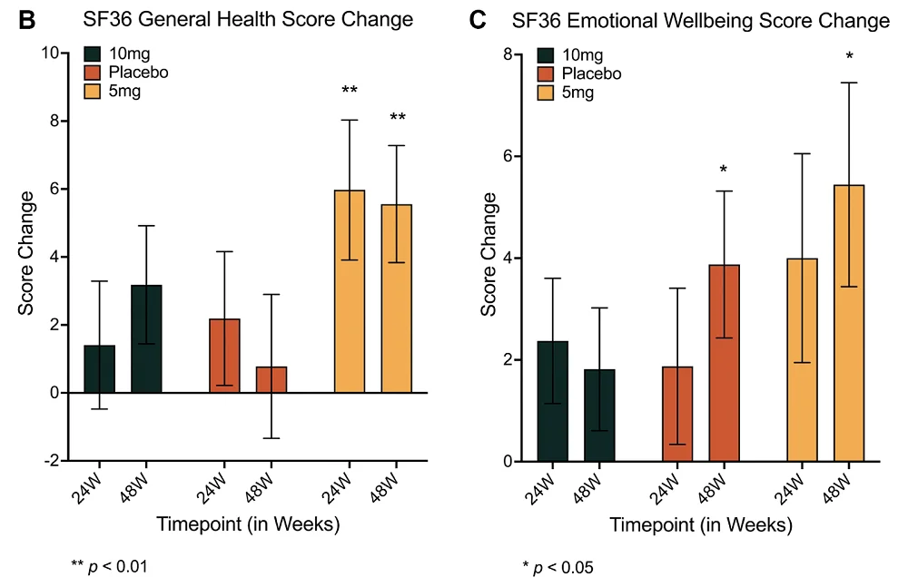
Only women experienced most of the benefits, despite there being fewer women in this study than men. Women in the 10 mg group who took rapamycin had significantly increased lean tissue mass and reported reduced amounts of pain. Interestingly, only the 5 mg group saw benefits for general health, but this included both women and men. Emotional well-being was improved in the 5 mg group after 48 weeks, but it was improved in the placebo group as well.

PEARL Results

“In general, the PEARL trial showed that female participants had the greatest benefits overall, with women in the higher-dose group seeing significant improvements in lean muscle mass and improvements in self-reported pain,” said Dr. Sajad Zalzala, CEO of AgelessRX and lead author of this study. “This outcome was particularly meaningful, as the study was powered primarily to assess safety and tolerability in a small pilot cohort rather than efficacy.”

Rapamycin showed no benefits over placebo in other self-reported metrics, such as physical function and social interactions. Osteoarthritis, as measured by WOMAC, was also unaffected.

This study showed a potential side effect of gut problems. A subgroup of participants (50 females, 31 males) had their gut microbiomes tested: in males, markers of gut dysbiosis increased in the 10 mg group, and in females, there was a trend towards increased permeability of the intestines.

A subgroup of 15 male and 9 female participants also underwent epigenetic aging analysis, performed using the commercially available test TruAge from the company TruDiagnostics. Unfortunately, there were no statistically significant effects in this area.

“This study adds to a growing body of evidence indicating that rapamycin can be used safely in relatively healthy individuals,” said Matt Kaeberlein, former professor at the University of Washington, CEO of Optispan, and co-founder of Ora Biomedical. “That alone is an important step forward. I’d say we’re seeing consistent anecdotal signals – supported now by the PEARL data – that rapamycin may help preserve lean mass and potentially improve quality of life, particularly in women. That’s promising, but due to low dosing in this study and limited sample size, not yet definitive.”

Future studies

These researchers note that this study was conducted on a mixed group of participants who are largely longevity-conscious, and they suggest that rapamycin may have stronger effects on less healthy people. Despite the low efficacy of compounded rapamycin compared to sirolimus, there were still significant effects on lean tissue mass and pain in women. The researchers hold that these statistically significant benefits, despite the low number of people involved, suggest that rapamycin does indeed have significant longevity benefits.

This trial relied largely on self-reporting and did not provide direct evidence that rapamycin use extends the lives of humans, and most of its effects on long-term health were limited. To directly make the claim that rapamycin improves lifespan or healthspan, a much larger and much longer trial would have to be conducted. Such a trial may use a compound that only affects mTORC1 (a rapalog) or different doses of rapamycin.

Still, PEARL was a significant step forward in studying the use of rapamycin for staving off aging in humans. “We are delighted to have supported the PEARL clinical trial back in 2021 and are excited to see the data from it appearing in academic publications,” said Keith Comito, founder of Lifespan.io, which has since merged with SENS Research Foundation to become the Lifespan Research Institute. “Trials like PEARL are an important step towards a deeper understanding of what aging is and what drives it. Ultimately, through crowdfunding and conducting research directly, we can all do our part in overcoming age-related diseases, together.”

We would like to ask you a small favor. We are a non-profit foundation, and unlike some other organizations, we have no shareholders and no products to sell you. All our news and educational content is free for everyone to read, but it does mean that we rely on the help of people like you. Every contribution, no matter if it’s big or small, supports independent journalism and sustains our future.

Literature

[1] Mannick, J. B., & Lamming, D. W. (2023). Targeting the biology of aging with mTOR inhibitors. Nature Aging, 3(6), 642-660.

[2] Castilho, R. M., Squarize, C. H., Chodosh, L. A., Williams, B. O., & Gutkind, J. S. (2009). mTOR mediates Wnt-induced epidermal stem cell exhaustion and aging. Cell stem cell, 5(3), 279-289.

[3] Harrison, D. E., Strong, R., Sharp, Z. D., Nelson, J. F., Astle, C. M., Flurkey, K., … & Miller, R. A. (2009). Rapamycin fed late in life extends lifespan in genetically heterogeneous mice. nature, 460(7253), 392-395.

[4] Colman, R. J., Anderson, R. M., Johnson, S. C., Kastman, E. K., Kosmatka, K. J., Beasley, T. M., … & Weindruch, R. (2009). Caloric restriction delays disease onset and mortality in rhesus monkeys. Science, 325(5937), 201-204.

[5] Mannick, J. B., Morris, M., Hockey, H. U. P., Roma, G., Beibel, M., Kulmatycki, K., … & Klickstein, L. B. (2018). TORC1 inhibition enhances immune function and reduces infections in the elderly. Science translational medicine, 10(449), eaaq1564.

[6] Harinath, G., Lee, V., Nyquist, A., Moel, M., Wouters, M., Hagemeier, J., … & Zalzala, S. (2025). The bioavailability and blood levels of low-dose rapamycin for longevity in real-world cohorts of normative aging individuals. GeroScience, 1-14.

Dopaminergic neuron

Neurons Hidden to Immune Cells Improve Parkinson’s in Rats

By tweaking genes used by placental and cancerous cells to evade immune detection, scientists have created non-immunogenic neuronal grafts that may help Parkinson’s patients [1].

Sneakiness needed

In recent years, scientists have learned to produce cells via cellular reprogramming, a process in which differentiated cells are driven back to a pluripotent, stem-like state. These are known as induced pluripotent stem cells (iPSCs). The cells can then be re-differentiated into other types of cells and benefit from substantial rejuvenation in the process [2].

This approach has the potential to solve many medical challenges, but the problem of graft rejection remains. When cells come from a genetically different donor (an allogeneic graft), they typically trigger an immune response. Immunosuppressants can reduce this reaction, but they come with significant side effects.

However, evolution has provided some inspiration. In many cases, cells have naturally evolved to become invisible to the immune system, such as placental cells, certain parasites and pathogens, and cancer cells.

The idea of borrowing these cloaking mechanisms to create universally compatible, off-the-shelf donor cells has been around for years [3]. Multiple teams have made progress, and in a new study published in Cell Stem Cell, a group of Australian researchers reports a major advance in rodent models of Parkinson’s disease.

The invisibility cloak

In Parkinson’s disease, dopamine-producing neurons in a brain region called the substantia nigra progressively die off. Dopamine is essential for controlling movement and coordination. As levels fall, the result is the hallmark motor symptoms of Parkinson’s: tremors, stiffness, slowness, and balance issues.

Using cellular reprogramming, the researchers generated neuronal progenitor cells (NPCs) capable of giving rise to new, rejuvenated neurons. These cells were genetically engineered to overexpress eight genes that placental and cancer cells use to evade immune detection. The combination was designed to neutralize nearly all major arms of the immune system: T cells, natural killer cells, macrophages/microglia, and dendritic cells.

However, making cells invisible to the immune system introduces a new risk: if one turns cancerous, the body won’t catch it. To mitigate this, the researchers included a “kill switch” by inserting the gene for herpes simplex virus thymidine kinase (HSV-TK) – an enzyme not naturally found in humans. It was placed under the control of a promoter active only in dividing cells so that any cell that starts to proliferate uncontrollably, which mature neurons typically don’t do, could be selectively eliminated using an antiviral drug.

When co-cultured with immune cells from three human blood donors, the cloaked dopaminergic neurons triggered significantly less immune activation, proliferation, and cytokine release. The team then moved to a humanized mouse model with functional human T and B cells.

In this model, uncloaked grafts provoked a strong immune response, while cloaked grafts caused little to no T cell expansion and no rise in pro-inflammatory cytokines. As a result, cloaked grafts grew to about twice the size of uncloaked ones and contained more dopamine-producing neurons.

Functional improvement

To test whether the cloaked neurons could functionally reverse Parkinsonian symptoms, the researchers turned to an immunodeficient rat model of the disease. These rats do not reject either cloaked or uncloaked grafts, so the comparison focused on function, not immune evasion.

Both types of grafts performed comparably: they survived, integrated into the host brain, and reversed motor symptoms by 20 weeks after transplantation. This showed that the cloaking did not compromise the cells’ ability to function.

Finally, the team tested the kill switch in nude mice: animals that lack a functional immune system, making it easier to isolate the effects of the suicide gene. They chose a time point when about half of the NPCs had matured into non-dividing neurons, while the rest were still proliferating. After administering the antiviral drug ganciclovir, the number of proliferating cells dropped significantly while the mature neurons remained unaffected.

“The researchers’ ability to engineer a graftable cell line that can become dopaminergic neurons and evade immune cell targeting fills a gap in the field,” said Dr. Kate Joyce, a researcher at the Lifespan Research Institute. “Not only is the physical process of grafting cells disruptive to the immune system in the brain but in Parkinson’s disease and other neurodegenerative diseases, there is an issue of increased immune surveillance due to a leaky blood brain barrier that further compounds the problem. This method also avoids generating cells from the patients that might harbor harmful mutations and cause more problems later.”

We would like to ask you a small favor. We are a non-profit foundation, and unlike some other organizations, we have no shareholders and no products to sell you. All our news and educational content is free for everyone to read, but it does mean that we rely on the help of people like you. Every contribution, no matter if it’s big or small, supports independent journalism and sustains our future.

Literature

[1] Pavan, C., Davidson, K. C., Payne, N., Frausin, S., Hunt, C. P., Moriarty, N., Berrocal Rubio, M. Á., Elahi, Z., Quattrocchi, A. T., Abu-Bonsrah, K. D., Wang, L., Clow, W., Yang, H., Pellegrini, M., Wells, C. A., Thompson, L. H., Nagy, A., & Parish, C. L. (2025). A cloaked human stem-cell-derived neural graft capable of functional integration and immune evasion in rodent models. Cell Stem Cell.

[2] Simpson, D. J., Olova, N. N., & Chandra, T. (2021). Cellular reprogramming and epigenetic rejuvenation. Clinical Epigenetics, 13, 1-10.

[3] Lanza, R., Russell, D. W., & Nagy, A. (2019). Engineering universal cells that evade immune detection. Nature Reviews Immunology, 19(12), 723-733.

Parkinson's symptom

How Exercise May Fight Parkinson’s Disease

Experimenting on rodents, researchers have found a pathway through which exercise could fight Parkinson’s disease and a molecule that might recapitulate this effect.

Fighting inflammation is crucial in Parkinson’s

Parkinson’s disease is characterized by Lewy bodies and the loss of dopaminergic neurons [1]. Recent work has found that this degeneration is related to neuroinflammation: the increase of microglial activity and inflammatory compounds within the brain [2]. The accumulated α-synuclein in the hippocampus has been found to be a key driver of this inflammation, and reducing neuroinflammation slows the rate of deterioriation [3], suggesting that treatments that suppress this process may be at least somewhat effective in treating the symptoms of Parkinson’s.

Previous work has found that exercise naturally reduces inflammation, including in the brain, and that it may be effective against Parkinson’s disease symptoms [4]. Deriving plasma from exercising rodents, and injecting it into rodents suffering from Parkinson’s symptoms, has also been previously found to have beneficial effects [5].

However, as these researchers note, that previous work did not fully elucidate the biochemical mechanisms involved. They specifically focus on irisin, an exercise-related compound that has been found to have benefits against neuroinflammation [6]. However, the connections between irisin, inflammation, and Parkinson’s have never been fully explored; these researchers, therefore, sought to close that gap.

Exercise affects more than just inflammation

The first experiment involved treating mice with MPTP, a compound that causes Parkinson’s-like symptoms. As expected, this caused both an increase in Parkinson’s pathology and a decrease in the hippocampus’ ability to create new neurons (neurogenesis). Similarly, the MPTP-treated mice performed worse on the Morris water maze test.

However, exercise seemed to partially ameliorate most of these effects. Following 10 weeks of regularly running on treadmills, neurogenesis was largely restored, and performance on the Morris water maze test was improved compared to unexercised MPTP-treated mice. Most interestingly, and most encouragingly, exercise was found to decrease α-synuclein itself: one of the core pathologies of Parkinson’s disease.

In these mice, exercise also decreased cellular death by apoptosis in the hippocampus. MPTP treatment causes an increase in a marker of apoptosis and a decrease in a marker of anti-apoptosis; both of these markers were brought closer to their normal levels by the exercise program. Key, well-known markers of inflammation, including TNF-α, NFκB, and IL-1β, were affected in the same way. NLRP3, an inflammatory protein that increases in Parkinson’s disease, leading to neuroinflammation and mitochondrial dysfunction [7], was also decreased by exercise, while irisin was increased.

A closer look at irisin

The researchers began their irisin experiments in cellular culture. First, they took microglia and cultured them in the presence of α-synuclein. As expected, these cells began to show signs of inflammatory activity, which could be largely reduced by exposing them to MCC950, a compound that suppresses NLRP3. Crucially, irisin was found to accomplish similar results, reducing NLRP3 expression and its downstream effects.

The researchers then subjected rats to four weeks of treadmill running and harvested their serum. When microglia were exposed to both α-synuclein and runner serum, they had reduced NLRP3 and inflammatory markers along with an increase in irisin, as compared to microglia exposed to α-synuclein alone. Further work involving NLRP3 agonists and the irisin blocker cRGDyk confirmed that these beneficial effects were indeed related to irisin’s suppression of NLRP3. Similarly, administering cRGDyk to mice prevented exercise from having beneficial effects.

Most importantly, administering irisin directly to mice recapitulated many of the benefits of exercise. Inflammatory and apoptosis markers were reduced, and signs of neurogenesis were increased. Performance on the Morris water maze test was better as well. Overall, irisin was found to recapitulate many of the benefits of exercise.

These encouraging findings provide hope to people suffering from Parkinson’s disease. Given its significantly deleterious effects on motor function, this particular disease often precludes exercise as a treatment. Therefore, an exercise mimetic, which irisin appears to be, may be a key part of future treatments. However, these findings were in model mice, not people, and a clinical trial would have to be conducted to determine if irisin works in human beings as it does in these rodents.

We would like to ask you a small favor. We are a non-profit foundation, and unlike some other organizations, we have no shareholders and no products to sell you. All our news and educational content is free for everyone to read, but it does mean that we rely on the help of people like you. Every contribution, no matter if it’s big or small, supports independent journalism and sustains our future.

Literature

[1] Zaman, V., Shields, D. C., Shams, R., Drasites, K. P., Matzelle, D., Haque, A., & Banik, N. L. (2021). Cellular and molecular pathophysiology in the progression of Parkinson’s disease. Metabolic brain disease, 36, 815-827.

[2] Han, Q. Q., & Le, W. (2023). NLRP3 inflammasome-mediated neuroinflammation and related mitochondrial impairment in Parkinson’s disease. Neuroscience Bulletin, 39(5), 832-844.

[3] Kouli, A., Camacho, M., Allinson, K., & Williams-Gray, C. H. (2020). Neuroinflammation and protein pathology in Parkinson’s disease dementia. Acta neuropathologica communications, 8, 1-19.

[4] Wang, R., Ren, H., Kaznacheyeva, E., Lu, X., & Wang, G. (2023). Association of glial activation and α-synuclein pathology in Parkinson’s disease. Neuroscience bulletin, 39(3), 479-490.

[5] De Miguel, Z., Khoury, N., Betley, M. J., Lehallier, B., Willoughby, D., Olsson, N., … & Wyss-Coray, T. (2021). Exercise plasma boosts memory and dampens brain inflammation via clusterin. Nature, 600(7889), 494-499.

[6] Zhao, R. (2022). Irisin at the crossroads of inter-organ communications: Challenge and implications. Frontiers in Endocrinology, 13, 989135.

[7] Khot, M., Sood, A., Tryphena, K. P., Khan, S., Srivastava, S., Singh, S. B., & Khatri, D. K. (2022). NLRP3 inflammasomes: A potential target to improve mitochondrial biogenesis in Parkinson’s disease. European Journal of Pharmacology, 934, 175300.

Elderly couple eating food

Dietary Patterns Associated With Healthy Aging

A recent study investigated the impact of eight different mid-life dietary patterns on the odds of healthy aging, including cognitive, mental, and physical health [1].

You are what you eat

Diet is an easily modifiable intervention in aging, as what we eat has a tremendous impact on our health. There is a wealth of evidence that good diets can be beneficial in preventing cardiovascular diseases [2], type 2 diabetes [3], and premature mortality [4]. Diet also impacts age-related cognitive decline and physical performance [5, 6]. Apart from being used as a tool to prevent diseases, a proper diet can help to achieve healthier aging and a better quality of life for the elderly.

In this study, the researchers aimed to determine which of the many dietary approaches and nutritional recommendations is the best. They compared the associations between various measurements of aging and long-term adherence to eight healthy dietary patterns.

“Studies have previously investigated dietary patterns in the context of specific diseases or how long people live. Ours takes a multifaceted view, asking, how does diet impact people’s ability to live independently and enjoy a good quality of life as they age?” said the study’s co-corresponding author Frank Hu, Fredrick J. Stare Professor of Nutrition and Epidemiology and chair of the Department of Nutrition at Harvard Chan School.

Eight healthy diets

The dietary approaches included in the study were the Alternative Healthy Eating Index (AHEI), te Alternative Mediterranean Index (aMED), Dietary Approaches to Stop Hypertension (DASH), the Mediterranean-DASH Intervention for Neurodegenerative Delay (MIND), a healthful plant-based diet (hPDI), the Planetary Health Diet Index (PHDI), an empirically dietary inflammatory pattern (EDIP), the empirical dietary index for hyperinsulinemia (EDIH), and ultraprocessed food (UPF) consumption.

Those approaches aim to promote good health by prioritizing healthy foods, such as fruits, vegetables, and whole grains, while reducing red and processed meats; however, there are differences between the diets.

For example, the aMED emphasizes olive oil, fish, and nuts. In the MIND, berries are an essential diet component. The hPDI focuses on plant-based foods, the PHDI focuses on foods that generate low levels of greenhouse gases, and DASH focuses on sodium restriction.

To investigate the impact of those diets on healthy aging, the researchers used the data of 105,015 participants: 70,091 women and 34,924 men from the Nurses’ Health Study (1986-2016) and the Health Professionals Follow-Up Study (1986-2016). They noted that since the study participants were health professionals, it limits the generalizability of results, and similar studies should be conducted on a more diverse population.

In 1986, at the beginning of the data acquisition step, the participants filled out the Food Frequency Questionnaire, which was repeated periodically over 14 years. At the end of the study, the participants’ health in aging was assessed with self-reported questionnaires that suggested that, after up to 30 years of follow-up, 9,771 (9.3%) of the study participants experienced healthy aging.

Healthy diet, healthy aging

The researchers concluded that “higher adherence to all dietary patterns was associated with greater odds of healthy aging.” However, there were differences between dietary patterns.

The strongest association between healthy aging and diet was observed for the AHEI, followed by the reverse EDIH, while the association for the hPDI was the weakest.

When the researchers compared the participants in the lowest 20% of AHEI adherence to those who were in the highest 20%, they reported “86% greater odds of achieving healthy aging using an age cutoff of 70 years and 2.24 times greater odds using an age cutoff of 75 years” that was independent of other lifestyle factors, including physical activity level, smoking, and BMI.

The AHEI was created by the Harvard T.H. Chan School of Public Health researchers. It’s similar to the US Department of Agriculture’s Healthy Eating Index, a measure of adherence to the federal Dietary Guidelines for Americans. The AHEI focuses on foods that help to reduce the risk of chronic disease.

Different diets for different domains of aging

Aging is not uniform; not everyone goes through it the same way, and its various aspects are affected to different degrees in different people. That’s why the researchers analyzed the impact of various dietary patterns on healthy aging in a few different domains. As previously, they noted that higher adherence to all of the tested diets was associated with healthy aging in all tested domains, but there were some differences between the extent of the effects.

Just as in overall healthy aging, the AHEI showed the strongest association with intact physical function and intact mental health. The strongest association for being free from chronic diseases was observed for the reversed EDIH. Intact cognitive health and surviving to 70 are strongly associated with the PHDI.

Diet effects 1

An apple a day keeps the doctor away

Analysis of individual dietary factors’ impact on healthy aging suggested that healthy aging was associated with higher consumption of fruits, whole grains, vegetables, unsaturated fats, nuts, legumes, and low-fat dairy. Meanwhile, consuming ultraprocessed food, trans fats, sodium, total meats, and red and processed meats decreased the odds of healthy aging. Similar results were observed when different aging domains were analyzed.

“Our findings suggest that dietary patterns rich in plant-based foods, with moderate inclusion of healthy animal-based foods, may promote overall healthy aging and help shape future dietary guidelines,” said co-corresponding author Marta Guasch-Ferré, associate professor in the Department of Public Health at the University of Copenhagen and adjunct associate professor of nutrition at Harvard Chan School.

Diet effects 2

Different people, different approaches

The researchers also analyzed different subgroups. The authors found that for both men and women, there was an association between all dietary patterns and healthy aging; however, that association was stronger for women for most diets.

For most diets, the associations were also stronger for smokers, participants with a BMI above 25, and those whose physical activity was below the median.

The researchers also reported significant interactions between socioeconomic status and two dietary patterns, reversed EDIH and reversed EDIP. They did not observe any statistically significant interactions between these diets and having European or non-European heritage.

Diet effects 3

One size doesn’t fit all

While all diets showed benefits, this research has found that different diets have different effects between the sexes and in people with different health concerns. Future studies could address more individualized approaches in detail.

“Our findings also show that there is no one-size-fits-all diet. Healthy diets can be adapted to fit individual needs and preferences,” summarized lead author Anne-Julie Tessier, assistant professor in the Department of Nutrition at the University of Montreal, researcher at the Montreal Heart Institute, and visiting scientist at Harvard Chan School.

We would like to ask you a small favor. We are a non-profit foundation, and unlike some other organizations, we have no shareholders and no products to sell you. All our news and educational content is free for everyone to read, but it does mean that we rely on the help of people like you. Every contribution, no matter if it’s big or small, supports independent journalism and sustains our future.

Literature

[1] Tessier, A. J., Wang, F., Korat, A. A., Eliassen, A. H., Chavarro, J., Grodstein, F., Li, J., Liang, L., Willett, W. C., Sun, Q., Stampfer, M. J., Hu, F. B., & Guasch-Ferré, M. (2025). Optimal dietary patterns for healthy aging. Nature medicine, 10.1038/s41591-025-03570-5. Advance online publication.

[2] Shan, Z., Li, Y., Baden, M. Y., Bhupathiraju, S. N., Wang, D. D., Sun, Q., Rexrode, K. M., Rimm, E. B., Qi, L., Willett, W. C., Manson, J. E., Qi, Q., & Hu, F. B. (2020). Association Between Healthy Eating Patterns and Risk of Cardiovascular Disease. JAMA internal medicine, 180(8), 1090–1100.

[3] Forouhi, N. G., Misra, A., Mohan, V., Taylor, R., & Yancy, W. (2018). Dietary and nutritional approaches for prevention and management of type 2 diabetes. BMJ (Clinical research ed.), 361, k2234.

[4] English, L. K., Ard, J. D., Bailey, R. L., Bates, M., Bazzano, L. A., Boushey, C. J., Brown, C., Butera, G., Callahan, E. H., de Jesus, J., Mattes, R. D., Mayer-Davis, E. J., Novotny, R., Obbagy, J. E., Rahavi, E. B., Sabate, J., Snetselaar, L. G., Stoody, E. E., Van Horn, L. V., Venkatramanan, S., … Heymsfield, S. B. (2021). Evaluation of Dietary Patterns and All-Cause Mortality: A Systematic Review. JAMA network open, 4(8), e2122277.

[5] Scarmeas, N., Anastasiou, C. A., & Yannakoulia, M. (2018). Nutrition and prevention of cognitive impairment. The Lancet. Neurology, 17(11), 1006–1015.

[6] Tessier, A. J., & Chevalier, S. (2018). An Update on Protein, Leucine, Omega-3 Fatty Acids, and Vitamin D in the Prevention and Treatment of Sarcopenia and Functional Decline. Nutrients, 10(8), 1099.

Brain and food

Fasting Affects the Immune System via the Brain

A new study has found that the immune remodeling associated with fasting can be recapitulated by activating a subset of neurons in the hypothalamus. The findings could be important in the context of fasting mimicking, metabolic disorders, and cancer [1].

Fasting and the immune system

Decades after the modern field of geroscience was born, caloric restriction still holds the crown as the very first and arguably still the most effective intervention. While fasting, a form of caloric restriction, offers numerous benefits [2], there are also many caveats and the practice should be undertaken with caution. For instance, several recent studies tie intermittent fasting to a higher risk of cardiovascular mortality [3].

Fasting can have a profound effect on the immune system. It is known to lower inflammation and reduce the number of circulating pro-inflammatory monocytes – marrow-derived cells that can infiltrate tissue and become macrophages or dendritic cells, part of the innate immune system. However, the mechanisms that drive this immune reorganization are not entirely understood.

In this new study published in Science Immunology, researchers from the University of Manchester posed an intriguing question: does this regulation happen via direct sensing of nutrient levels by the immune system, or does it require signaling from the brain? If the latter is the case, can these effects be induced without fasting, solely by neuronal stimulation?

Mind over body

Hunger and satiety are known to be regulated by two distinct subsets of hypothalamic neurons. The first subpopulation, called Agouti-related peptide (AgRP) neurons, promotes the feeling of hunger, while the second one, pro-opiomelanocortin (POMC) neurons, signals satiety.

The researchers genetically engineered mice in order to allow transient activation of these two groups of neurons. When AgRP neurons were activated in mice that had not fasted, the levels of circulating pro-inflammatory monocytes decreased, mirroring the effect of a 20-hour fast (a 4-hour fast did not have that effect). However, unlike in fasting, no drop in blood glucose was detected.

When fasted mice were re-fed, concurrent artificial activation of AgRP neurons prevented the expected monocyte increase, even though feeding restored normal blood glucose levels. This suggests that the brain’s perceived hunger state, driven by AgRP neurons’ activity, can override the systemic nutrient signal in regulating monocyte abundance.

In another experiment, the researchers activated satiety-promoting POMC neurons in fasted mice. This effectively reversed the fasting effect, increasing the number of monocytes despite the continuing absence of food.

“Our perceptions can shape our bodies in ways we don’t always notice. It’s easy to see how thoughts guide our actions, but this study reminds us that even our internal body adjustments that are not under conscious control respond to the brain’s signals,” said the lead senior researcher and coordinator of the study, Dr. Giuseppe D’Agostino. “This study underlines how important the brain is in regulating the immune system. But if internal or external factors alter the brain’s perception, these processes can go awry, reminding us how deeply the mind and body are – and should remain – connected.”

Collaborator and Manchester immunologist Professor Matt Hepworth added, “This work challenges the long-standing view that fasting’s immunological impact is driven purely by nutrient levels. It highlights the nervous system’s profound influence on how the immune system adapts during fasting.”

The mTOR connection

Activating AgRP neurons mimicked fasting in yet another aspect: by reducing the levels of CCL2, a cytokine known to recruit monocytes from the bone marrow into circulation. Adding exogenous CCL2 partially counteracted this effect.

Since the liver is known to regulate CCL2 levels, the researchers investigated this organ’s potential role. Interestingly, key genes that are usually upregulated in the liver during fasting were not upregulated by activating AgRP neurons. However, the researchers noticed reduced levels of hepatic mTOR activity in what resembled natural fasting. This protein, the mammalian target of rapamycin, is recognized as a potent regulator of longevity in various animal models [4].

Preventing the inhibition of mTOR in the liver blunted the effect of AgRP neuron activation on CCL2 levels and monocyte numbers. “Together, these findings suggest that hepatic mTOR activity is under direct neuronal control via hypothalamic AgRP and POMC neurons and that this control is not exclusively dependent on local nutrient availability,” the paper says.

The mechanism the researchers discovered resembles “fly-by-wire,” as opposed to a direct mechanical connection: a signal goes into the nervous system, which sends outgoing signals that eventually affect the immune system. The authors suggest that their findings might be important in the context of infections, malnutrition, and muscle wasting in cancer (cachexia).

The study’s lead author, Dr. Cavalcanti de Albuquerque, said, “By showing how the brain exerts top-down control over immune cells, we can further explore when and how fasting might deliver health benefits. It also opens up potential ways to treat infectious, inflammatory, metabolic, and psychiatric conditions.”

We would like to ask you a small favor. We are a non-profit foundation, and unlike some other organizations, we have no shareholders and no products to sell you. All our news and educational content is free for everyone to read, but it does mean that we rely on the help of people like you. Every contribution, no matter if it’s big or small, supports independent journalism and sustains our future.

Literature

[1] Hunter, J., Domingues, R. G., Harno, E., Worth, A. A., Liguori, F. M., Aviello, G., Bechtold, D., White, A., Luckman, S. M., & Hepworth, M. R. (2025). Brain sensing of metabolic state regulates circulating monocytes. Science Immunology.

[2] Waziry, R., Ryan, C. P., Corcoran, D. L., Huffman, K. M., Kobor, M. S., Kothari, M., … & Belsky, D. W. (2023). Effect of long-term caloric restriction on DNA methylation measures of biological aging in healthy adults from the CALERIE trial. Nature Aging, 3(3), 248-257.

[3] Sebastian, S. A., Shah, Y., & Arsene, C. (2024). Intermittent fasting and cardiovascular disease: A scoping review of the evidence. Disease-a-Month, 101778.

[4] Miller, R. A., Harrison, D. E., Astle, C. M., Fernandez, E., Flurkey, K., Han, M., … & Strong, R. (2014). Rapamycin‐mediated lifespan increase in mice is dose and sex dependent and metabolically distinct from dietary restriction. Aging cell, 13(3), 468-477.

Ora Rapamycin Project

The mTOR Inhibitors Lifespan Project Enters Next Phase

Ora Biomedical, in partnership with Rapamycin Longevity Lab, announces the successful funding of the first subproject under its ambitious initiative to conduct a rapid lifespan analysis of 601 mTOR inhibitors in roundworms.

With $50,000 secured, Ora Biomedical will now commence the next phase of the first subproject. This will be a high-throughput screening of 301 mTOR inhibitors using its cutting-edge WormBot-AI technology. This milestone marks an important step toward identifying next-generation compounds that could be more effective than rapamycin, which is currently seen as the golden standard because of its good longevity effects in multiple species.

Mitchell Lee, CEO of Ora Biomedical, emphasized the importance of this research by stating: “The potential of targeting aging to broadly improve healthy lifespan is clear from decades of studies with compounds like rapamycin. However, even for well-validated molecular targets like mTOR, we still don’t know the best interventions. We at Ora Biomedical are proud to partner with Rapamycin Longevity Lab to advance our understanding around targeting mTOR and related kinases for maximizing healthy lifespan. None of this work is possible without support from visionary donors and organizations like the Lifespan Research Institute, the nonprofit behind Lifespan.io, with whom we have partnered to create pathways for donations to advance longevity science. To all those involved, thank you again, and we are excited to get to work!”

“At Lifespan Research Institute, we believe that rigorous, scalable science is essential to advancing interventions that can combat age-related decline and extend healthspan. We are proud to help catalyze Ora Biomedical’s high-throughput screening of mTOR inhibitors, and deeply appreciate the donors making it possible,” said Keith Comito, Board President of Lifespan Research Institute.

Fundraising for the second subproject

While the first subproject moves forward, fundraising continues for the remaining 300 inhibitors, which requires an additional $40,000. “We’re thrilled to begin screening, but completing the full dataset is crucial. One of the big problems for really advancing the longevity field is that we do not have a large and publicly available dataset of essential basic longevity data. We as a field need to get basic data in place so that we can start to take things to the next level.” said Krister Kauppi, the project leader at Rapamycin Longevity Lab. “We invite organizations and individuals passionate about longevity to join us in this effort to enable research to progress from simple and basic studies to much more advanced and impactful longevity research.”

Those interested in learning more or contributing can reach out to Krister Kauppi at Rapamycin Longevity Lab via email: krister@masteronething.com

Rapamycin Longevity Lab is a research organization focused on advancing the use of mTOR inhibitors as the core component in longevity combinational therapies. Through partnerships with companies like Ora Biomedical, Rapamycin Longevity Lab efficiently screens and tests new combinational therapies, with a long-term goal of translating this research into human clinical trials.

Ora Biomedical, Inc. is a longevity biotechnology company developing therapeutics that target the biological mechanisms of aging to improve health, resilience, and performance. Using proprietary AI-driven robotic screening platforms, Ora identifies and advances small molecule interventions that extend lifespan, fight age-associated disease, and protect against stress-induced physiological decline. The company’s lead programs focus on dual-use applications in defense, aerospace, and preventive health. Ora also leads the Million Molecule Challenge—a groundbreaking citizen science initiative to create the world’s largest open-access longevity interventions database through broad public participation.

We would like to ask you a small favor. We are a non-profit foundation, and unlike some other organizations, we have no shareholders and no products to sell you. All our news and educational content is free for everyone to read, but it does mean that we rely on the help of people like you. Every contribution, no matter if it’s big or small, supports independent journalism and sustains our future.
Spinal pain

Reprogramming Epigenetics to Fight Back Pain

A new paper published in the Nature journal Bone Research has found that reprogramming the epigenetics of spinal disc cells reduces senescence and alleviates pain in a rat model.

The soft tissue degrades

Nucleus pulposus cells, which maintain the discs in the spine [1], are prone to the same age-related deterioration that other cells are. They become senescent, losing the ability to renew themselves [2], and they fail to perform their fundamental functions of extracellular matrix maintenance, thus leading to disc shrinkage and many of the back problems that people experience as they age.

We have reported on studies in this space, including a recent study that focused on targeting a specific pathway to fight senescence and restore function. These researchers, however, took an entirely different approach, using epigenetic reprogramming in an attempt to restore these cells to a youthful state.

These researchers chose a combination of the first three factors, OSK, for this experiment; c-Myc, the M in OSKM, is often omitted because it has been linked to cancer.

One of the key difficulties that they noted in these reprogramming experiments is in simply delivering the necessary factors to cells; using virus-related vectors has been found to be more difficult than expected at the clinical level [3]. Therefore, these researchers chose exosomes, intercellular signaling molecules that researchers frequently derive from youthful cells in order to benefit older tissues [4]. Instead of simply containing the common signals sent by youthful cells; however, they contained a plasmid that codes for OSK.

An engineered exosome seems to bear fruit

These exosomes went through a carefully designed process. They originated from bone marrow stem cells that had their exosomes modified with Cavin2, a protein that enhances the ability of these vesicles to be taken up into other cells. These exosomes were then filled with the OSK plasmid.

In the first experiment, the researchers verified that their OSK plasmid would benefit nucleus pulposus cells as it does other cells. Directly giving the cells plasmids did indeed encourage the production of OSK within the cells, and this led to downregulation of the senescence-related p53 tumor suppressor pathway along with a decrease in DNA damage as measured by the marker γH2AX. With aging, the epigenetic methylation marker H4K20me3 increases while the related marker H3K9me3 decreases; with the OSK plasmid, this was reversed in these cells. There were also benefits for the nuclear envelope, which maintains the cellular nucleus’ structure.

OSK also restored these cells’ fundamental abilities. Compared to a control group of untreated aging cells, markers of proliferation were increased, and critically, it also decreased markers of tissue destruction (anabolism) while increasing markers of tissue creation (catabolism). This is particularly promising, as it suggests that these cells are better able to maintain vertebral discs. There was also a reduction in inflammatory biomarkers.

The researchers then tested the effectiveness of their Cavin2 exosome modification. This change didn’t benefit its ability to enter into young cells; instead, it helped it enter older cells, which, as these researchers found, accept fewer unmodified exosomes than younger cells do. This is because Cavin2 encourages phagocytosis: the ability of cells to consume material near to them.

These modified exosomes were found to have even stronger effects than the initial way in which they had delivered OSK plasmids into the cells. Senescence biomarkers were even further reduced, DNA damage was also reduced, and there was more H3K9me3 than in the original OSK group as well. Inflammatory biomarkers were even more strongly reduced, and there were positive effects on the maintenance-related anabolism/catabolism balance.

Complete restoration in a rat model

Encouraged by these results, the researchers then turned to a rat model. Puncturing rats’ vertebral discs with a needle leads to intervertebral disc degeneration and many of the same problems seen in older people, including both pain and cellular senescence. Two weeks after this injury, the rats were given one of a variety of injections into the nucleus pulposus, including unmodified exosomes, OKS plasmids, and their modified plasmids, along with a sham-injured group and an untreated control group.

Neither unmodified exosomes nor OSK plasmids reached statistical significance on key metrics, including visual analysis of disk degeneration. It appeare that these two treatments may have helped somewhat, particularly eight weeks after treatment, when the severity of their disc degeneration was judged to be lower.

On the other hand, rats treated with Cavin2-modified, OSK-containing exosomes had immediate benefits only four weeks after injection. Looking at disc morphology, there were practically no differences between this group and the sham-injured group at all; it was if they had never been injured in the first place.

RNA sequencing suggested an anti-aging effect. In an examination of genes whose expressions change with aging, rats injected with these exosomes were found to have many of the modifications found in the cellular studies, including a decrease in the p53 pathway and in other pathways related to cellular senescence, compared to the untreated control group.

While this research is promising, it is still in its early stages and needs verification. These were not naturally aged rats, and there may be unforeseen problems with delivering these sorts of exosomes to humans. Still, this work warrants further investigation to determine if these modified exosomes can indeed be a vector for effective epigenetic reprogramming therapies in nucleus pulposus cells or other tissues.

We would like to ask you a small favor. We are a non-profit foundation, and unlike some other organizations, we have no shareholders and no products to sell you. All our news and educational content is free for everyone to read, but it does mean that we rely on the help of people like you. Every contribution, no matter if it’s big or small, supports independent journalism and sustains our future.

Literature

[1] Fine, N., Lively, S., Séguin, C. A., Perruccio, A. V., Kapoor, M., & Rampersaud, R. (2023). Intervertebral disc degeneration and osteoarthritis: a common molecular disease spectrum. Nature Reviews Rheumatology, 19(3), 136-152.

[2] Roberts, S., Evans, E. H., Kletsas, D., Jaffray, D. C., & Eisenstein, S. M. (2006). Senescence in human intervertebral discs. European Spine Journal, 15, 312-316.

[3] Pan, X., Veroniaina, H., Su, N., Sha, K., Jiang, F., Wu, Z., & Qi, X. (2021). Applications and developments of gene therapy drug delivery systems for genetic diseases. Asian journal of pharmaceutical sciences, 16(6), 687-703.

[4] Möller, A., & Lobb, R. J. (2020). The evolving translational potential of small extracellular vesicles in cancer. Nature Reviews Cancer, 20(12), 697-709.