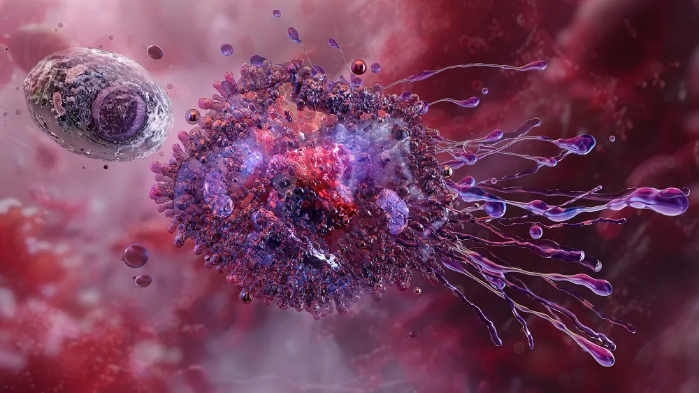
In Nature Aging, researchers have identified and categorized several macrophage subtypes, including a subtype that appears with aging and another that manages nerve function.
Not all macrophages follow the same rules

Read More
Macrophages declining with age is a well-known problem, and many proposed solutions involve changing their polarity from M1, which favors inflammation, to M2, which favors regeneration. However, this does not apply to adipose tissue macrophages (ATMs), which do not polarize themselves in this way [1]. These tissue-macrophages have subsets, including lipid-associated macrophages (LAMs) [2] and vasculature-associated macrophages (VAMs) [3]. These researchers note that little work has been done on relating these various subtypes of ATMs to aging.
This paper addresses several of these subtypes, including nerve-associated macrophages (NAMs), which not only police the nerves themselves [4] but are linked to the functions of the tissues where the nerves are located, such as the gut [5]. In the fat tissue, NAM behavior has been linked to obesity [6].
Macrophage subtypes can be clustered
The experiments began by deriving visceral white adipose tissue (VAT) from mice and isolating immune cells that were positive for both F4/80 and CD11b+. The mice were either 2 months or 22 months of age, and both male and female mice were used in this experiment.
About three-quarters of these cells were found to be tissue resident, with the other quarter flowing in the vasculature. Principal component analysis found that there were substantial differences between circulating and resident cells, and there were also found to be differences between the cells found in brown fat and white fat.
The researchers then clustered these cells based on single-cell RNA sequencing. This method allowed them to find and discard contaminating cells as well as identify cellular commonalities and differentiation into subtypes. Interestingly, the most numerous cluster was sharply different between sexes; a cluster that was very common in male mice, Cluster 0, was sharply reduced in females, and an exceptionally common female cluster, Cluster 5, was much less common in males.
Cluster 5 was found to be highly enriched for Maoa, which breaks down catecholamines such as neurotransmitters. This and other evidence led the researchers to conclude that these are NAMs. A similar gene expression analysis revealed that Cluster 0 is composed of VAMs.
Age changes the landscape
These clusters were significantly changed with aging, according to a flow cytometry analysis focusing on surface markers, and how they did so was largely dependent on sex. Between the 2-month-old and 22-month-old mice, Cluster 0 decreased with aging in males but not females, Cluster 5 decreased in females but not males, and cluster 13 increased in females but not males. Clusters 4 and 10 increased in both sexes, and Cluster 4 was recognized solely as being associated with aging rather than tissue function; the researchers called these aging-associated macrophages (AAMs), which express pro-inflammatory compounds and are characterized by the age-related marker CD38.
Interestingly, these flow cytometry results did not entirely agree with the RNA sequencing analysis, which suggested that the number of Cluster 5 NAMs remained unchanged in female mice. It is plausible that cell surface marker expression changed with aging.
NAMs were found to be almost uniquely associated with aging. While the AAMs in Cluster 4 were also found to have senescent-like features, these features were common in Cluster 5 NAMs. This was based on gene expression analysis and was relative to the behavior of aged cells rather than directly related to the known cellular markers of senescence.
A link to obesity
However, as the researchers discovered, these cells perform critical tasks. Specifically, adipose-derived NAMs were found to help maintain myelin, the cellular sheaths that protect neuronal axons. Depleting the adipose-derived NAMs of 3-month-old mice led to dysregulation of catecholamines as well as obesity; freely fed mice that had adipose-derived NAMs depleted were considerably more likely to gain weight. Further experimentation with older mice found that NAM depletion led to significant increases in inflammation.
This research highlights a small part of the heterogeneity found in biology, details its changes with aging, and suggests that it may be possible to specifically handle macrophage subtypes; for example, future experiments may find that it is beneficial to reduce the population of aging-associated macrophages as identified here. This focus on macrophages may also be valuable in examining the fundamental causes of obesity and analyzing the effects of existing anti-obesity drugs.
Literature
[1] Camell, C. D., Sander, J., Spadaro, O., Lee, A., Nguyen, K. Y., Wing, A., … & Dixit, V. D. (2017). Inflammasome-driven catecholamine catabolism in macrophages blunts lipolysis during ageing. Nature, 550(7674), 119-123.
[2] Jaitin, D. A., Adlung, L., Thaiss, C. A., Weiner, A., Li, B., Descamps, H., … & Amit, I. (2019). Lipid-associated macrophages control metabolic homeostasis in a Trem2-dependent manner. Cell, 178(3), 686-698.
[3] Silva, H. M., Báfica, A., Rodrigues-Luiz, G. F., Chi, J., Santos, P. D. E. A., Reis, B. S., … & Lafaille, J. J. (2019). Vasculature-associated fat macrophages readily adapt to inflammatory and metabolic challenges. Journal of Experimental Medicine, 216(4), 786-806.
[4] Kolter, J., Feuerstein, R., Zeis, P., Hagemeyer, N., Paterson, N., d’Errico, P., … & Henneke, P. (2019). A subset of skin macrophages contributes to the surveillance and regeneration of local nerves. Immunity, 50(6), 1482-1497.
[5] Muller, P. A., Koscsó, B., Rajani, G. M., Stevanovic, K., Berres, M. L., Hashimoto, D., … & Bogunovic, M. (2014). Crosstalk between muscularis macrophages and enteric neurons regulates gastrointestinal motility. Cell, 158(2), 300-313.
[6] Pirzgalska, R. M., Seixas, E., Seidman, J. S., Link, V. M., Sánchez, N. M., Mahú, I., … & Domingos, A. I. (2017). Sympathetic neuron–associated macrophages contribute to obesity by importing and metabolizing norepinephrine. Nature medicine, 23(11), 1309-1318.


