×

The Blog

Building a Future Free of Age-Related Disease

Asian Pacific Longevity Summit

2025 Asian Pacific Longevity Medicine International Summit

Longevity medicine is rapidly emerging as a credible, fast-growing discipline in modern healthcare, powered by rigorous science and significant global investment. On October 4–5, 2025, the Asia-Pacific Longevity Society will host the inaugural Asian Pacific Longevity Medicine International Summit (APAC-LMIS) at the SkyCity Marriott Hotel in Hong Kong, in partnership with several leading international longevity organizations.

This landmark gathering will bring together leading scientists, clinicians, biotech startups, and venture investors not only from Asia-Pacific countries, but also from around the world, to translate cutting-edge longevity research into ethical, evidence-based clinical practice across both longevity clinics and mainstream hospital settings.

In an era when online “biohacking” trends and commercially driven claims can overshadow genuine geroscience, APAC-LMIS sets itself apart by focusing exclusively on peer-reviewed research and proven approaches. As the co-organizers note:

“We’re cutting through the noise and putting credible, data-driven professional education directly into the hands of physicians and healthcare providers. Our aim is to cultivate well-trained longevity doctors who can deliver safe, effective care that truly benefits patients.”

Our mission: Live Longer, Live Healthier, Embrace the Best Lifestyle.

Why APAC-LMIS is Unique:

  1. First in APAC: Over 30 countries represented, covering 53% of the world’s population, including super-aging societies such as Japan, Hong Kong, and Singapore.
  2. Science-Based Solutions: Strong engagement from physician-scientists.
  3. Training Programs: Intensive courses for healthcare providers and physicians.
  4. Best Practices: Showcasing successful longevity clinic and healthy longevity center models across public and private sectors.
  5. Living Lab: Testing and validating non-medical longevity and wellbeing solutions.
  6. Top 10 Longevity Breakthrough Awards: Advisory committee highlights the most promising innovations for clinical and home use.

The Summit’s goal is clear: to establish standards, uphold scientific rigor, and solidify longevity medicine as a trusted medical specialty—not a fleeting trend. This Summit marks the beginning of an ongoing series of international meetings designed to shape the future of medicine and improve healthspan globally.

We warmly invite you to join us in Hong Kong this October to take part in this vital conversation and help advance the global practice of longevity medicine.

Official sites: https://www.apaclongevitysociety.org and https://longevityawards.org/

Media and partnership inquiries

For press credentials, sponsorship opportunities, speaking proposals, and delegate registration, please contact the APAC-LMIS organizing committee at Admin@GlobalMD.org

Loose Chromatin, Senescent Inflammation, and Cancer

In Aging Cell, researchers have reported that chromatin demethylation allows SASP compounds to be more easily expressed.

Chromatin and gene expression

The authors of this study begin by discussing two concepts that appear only loosely related: senescence and gene expression. Both of these concepts are very well-known in aging research, but putting them directly together is not frequently seen.

However, this work does not focus on epigenetic clocks nor on epigenetic alterations more generally. Instead, these researchers are specifically focused on chromatin, the tightly wound DNA whose configuration determines what is, and isn’t, accessible for transcription. They note that a decrease in histone methylation, which makes this DNA more accessible, is associated with premature aging [1].

Specifically, the researchers focused on lysine methylation, a common form of histone methylation that is reduced by the KDM4 subfamily of demethylases. KDM4 has been heavily implicated in cancer, and KDM4 inhibitors, alongside other epigenetic-related drugs, have been investigated in the treatment of breast cancer [2]. Little previous work, however, has been done to investigate its role in cellular senescence.

Loose chromatin allows the SASP to be produced

KDM4

These researchers used bleomycin to induce senescence in human prostate stromal cells, which consist largely of fibroblasts, and compared their protein profiles to those of non-senescent cells. Of the 447 proteins they were able to quantify, they found that 87 were more abundant in senescent cells and 29 were less abundant. Most notably, they found that this protein expression was related to methylation; key histone signals, including H3K9 and H3K36 methylation markers, were diminished.

Further work found that this relationship between histone methylation and cellular senescence was strong. SASP expression was directly related to decreased methylation along with an increase in KDM4 and an increase in the DNA damage marker γH2AX. The researchers found that these specific changes were only relevant to cellular senescence and did not appear to apply to cancers.

Two variants, KDM4A and KDM4B, were found to drive SASP expression rather than senescence itself. Silencing both of them through short hairpin RNA (shRNA) significantly decreased core SASP factors but did not affect whether or not cells became senescent. Similarly, using other compounds to maintain histone methylation did not affect the progression of senescence; it only affected the production of inflammatory SASP factors. Furthermore, these two KDM variants were not solely responsible for the SASP; blocking NF-κB, the SASP’s master regulator, attenuated the SASP even while KDM4A and B were being promoted.

Of course, KDM4A and B, having such broad effects on methylation, do not affect only compounds related to senescence. Suppressing their effects through the compound ML324 led to the downregulation of many proteins, not just those relating to the SASP. The researchers recommend further work on determining what exactly happens to cells that are suppressed in this way. The researchers did confirm, however, that senescent cells differ from normal cells in which genes are more accessible through chromatin, which appears to explain why histone methylation has such strong effects on the SASP.

Relationship to cancer

Much of this work was focused on cancer rather than senescence. Crucially, the researchers found that upregulated KDM4A and B, and the decreased methylation that they represent, are correlated with a reduced chance of disease-free survival. These compounds did not appear to have relevant effects on the cancer cells themselves but rather the cancer stroma: the noncancerous portions of a tumor that support its growth.

Utilizing a mouse model with diabetes and immunodeficiency, the researchers investigated ML324 and the genotoxin MIT as a potential treatment for cancer. Two weeks after being injected with cancer, the treatment groups of mice were given one or both of these compounds for eight weeks.

Mirroring the cellular study, growths that originated from pure cancer cells were not affected by ML324. However, when these cancer cells were accompanied by stromal cells, administering ML324 alongside MIT substantially decreased the size of the tumors compared to MIT alone. This lends weight to the idea that affecting KDM4, or perhaps the SASP itself, may be effective in prolonging survival and helping people fight cancer when used alongside more common tools. However, much more work will need to be done to determine if this approach can be used to fight prostate or any other type of cancer.

Of note is that this work bears the name of the late Judith Campisi. This is because it was originally published in Nature Aging in 2021 but was later retracted due to concerns regarding its images. This Aging Cell article was submitted after addressing these issues.

We would like to ask you a small favor. We are a non-profit foundation, and unlike some other organizations, we have no shareholders and no products to sell you. All our news and educational content is free for everyone to read, but it does mean that we rely on the help of people like you. Every contribution, no matter if it’s big or small, supports independent journalism and sustains our future.

Literature

[1] Gruenbaum, Y., & Foisner, R. (2015). Lamins: nuclear intermediate filament proteins with fundamental functions in nuclear mechanics and genome regulation. Annual review of biochemistry, 84(1), 131-164.

[2] Metzger, E., Stepputtis, S. S., Strietz, J., Preca, B. T., Urban, S., Willmann, D., … & Schüle, R. (2017). KDM4 inhibition targets breast cancer stem–like cells. Cancer research, 77(21), 5900-5912.

Rejuvenating Atherosclerotic Foam Cells

According to a study published by Cyclarity Therapeutics, its drug UDP-003 shows benefits in reversing the root cause of atherosclerotic plaques [1].

Fighting the root cause

Many available medications address only disease symptoms in an effort to mitigate them. However, the real challenge is to identify the root cause of a problem and find ways to treat it. This is the approach that Cyclarity Therapeutics is taking with its drug, UDP-003, which targets the cause of atherosclerosis, a cardiovascular disease that is a globally leading cause of mortality and consists of a buildup of plaque in and on the arterial wall.

“UDP-003 aims to address a root cause of plaque formation by removing the oxidized cholesterol that accumulates in macrophages,” said Dr. Prerna Bhargava, the lead author of this study. “This buildup transforms macrophages into foam cells. By selectively removing oxidized cholesterol, UDP-003 reverses the foam cell formation and restores the diseased macrophages into healthy cells.” Dr. Bhargava continued.

This is different from current approaches. As Dr. Bhargava explained, “Current treatments- such as statins, PCSK9 inhibitors, ezetimibe, and anti-inflammatory drugs- primarily focus on lowering circulating lipids or reducing systemic inflammation. While these approaches effectively slow the disease progression, they do not resolve the residual vascular inflammation or address the underlying driver of the disease by targeting the buildup of toxic oxysterols.”

From lipids to plaques

Atherosclerotic plaque is built from oxysterols, oxidised low-density lipoproteins (LDL), including the most abundant, highly toxic, and proinflammatory 7-ketocholesterol (7KC).

The pro-inflammatory response is initiated by the accumulation of oxysterols in the arterial walls. This leads to the recruitment of macrophages, which engulf oxysterols, including 7KC; this leads to the transformation of those cells from macrophages into foam cells, which participate in creating atherosclerotic plaques.

Targeting 7KC in foam cells

UDP-003 is intended to stop this chain of events by targeting the buildup of 7KC. UDP-003 is a cyclodextrin, a cyclic glucose oligomer with a hydrophobic cavity and a hydrophilic shell. This family of molecules can form complexes with hydrophobic substances. UDP-003 showed specificity for 7KC, which was ~1000-fold higher than for cholesterol.

The researchers used two experimental treatment modalities: preventative and additive. In the preventative treatment modality, they simultaneously added 7KC and UDP-003 to macrophages for 48 hours. Here, UDP-003 minimized the detrimental effects of 7KC by encapsulating it before it even reaches the cells.

In the additive treatment modality, they first incubated the cells with 7KC for 24 hours. After that, they added UDP-003, and the cells were kept for another 24 hours with both additives. This modality resembles a clinical context in which the treatment would be administered after the arteries were exposed to 7KC and show signs of disease.

Preventable and reversible

In their initial experiments, the researchers used one of the easiest features of foam cells to capture: lipid droplet accumulation. During the treatment of mouse macrophage cells, the addition of 7KC induced foam cell formation.

Simultaneous addition of 7KC and UDP-003 to the cells resulted in over 20% reduction of lipid droplet marker compared to the cells treated only with 7KC, which, in itself, led to an increase in the lipid droplet accumulation in macrophages. This suggests that UDP-003 can prevent 7KC from entering the cells and causing cellular damage.

In the additive modality experiment, the researchers observed almost a 30% reduction of lipid droplet marker, suggesting that UDP-003 has the ability to reverse the lipid accumulation in cultured macrophages.

UDP-003 1

Improving cellular functions

When macrophages turn into foam cells, their cellular functioning suffers. Macrophages engulf pathogens and debris (phagocytosis) and clear apoptotic cells (efferocytosis). Becoming foam cells impairs these functions.

The researchers observed decreased phagocytosis and efferocytosis in 7KC-treated human monocytes and mouse macrophages in both treatment modalities. UDP-003 treatment led to around a 15-20% increase in phagocytosis compared to the cells treated only with 7KC and prevented the loss of efferocytosis caused by 7KC in UDP-003-treated cells in the preventative modality.

UDP-003 2

Eventually, foam cells undergo cell death by apoptosis or necrosis, with necrosis leading to further tissue damage and contributing to plaque instability by increasing the necrotic core of arteriosclerotic plaque, which consists of dead cells and debris.

Exposing mouse macrophage cells and human monocytes to 7KC led to a substantial increase in the levels of apoptosis and necrosis. UDP-003 treatment reversed and prevented further apoptosis and necrosis by around 40-60%, depending on the treatment modality and cell line.

UDP-003 3

The malfunctioning of foam cells’ molecular processes causes broader systemic issues, such as disruptions in reverse cholesterol transport (RCT), since foam cells are unable to participate in it due to lysosomal dysfunction. This disrupts the first step of RCT: the removal of cholesterol from cells (cholesterol efflux).

Measuring the cholesterol efflux in murine macrophage cells exposed to 7KC showed that while 7KC reduced cholesterol efflux, UDP-003 reversed the reduction caused by 7KC, suggesting atheroprotective functions.

Fighting inflammation

Inflammatory responses were explored at the levels of gene expression in mouse macrophage foam cells. 7KC led to an increase in the expression of genes playing a role in immune cell recruitment and inflammation along with Nf-κB, which plays an essential role in the modulation of immunoregulatory gene expression. UDP-003 treatment in the additive modality decreased the Nf-κb and some of the inflammation-related gene expression. While this effect was not statistically significant in the preventative modality, a similar trend was observed.

UDP-003 treatment also significantly reduced ROS levels induced by the 7KC treatment in both treatment modalities in mouse macrophage cells. The researchers suggest that decreasing levels of ROS can help to close the inflammation feedback loop driven by 7KC, as lower ROS levels could reduce the amount of cholesterol that is being oxidised into 7KC.

Putting it into context

While cell culture experiments can provide a plethora of valuable information, atherosclerotic foam cells are located in the plaques within the arterial wall, and it’s essential to know whether UDP-003 treatment can be effective in such a context. To test this, the researchers used human arterial tissue containing atherosclerotic plaque and treated it with UDP-003. Those experiments have shown the UDP-003 potential for 7KC solubilization and removal from the human plaque.

Limited animal models

Cell culture experiments were followed by experiments in living organisms. Experiments in rats showed that UDP-003 treatment leads to 7KC being excreted in the urine during the first four hours of UDP-003 administration.

However, the results of the experiments in three different atherosclerotic mouse models were less satisfactory. The results indicated a trend towards lower 7KC levels in plasma and whole blood in female but not male mice. However, these results were mostly not statistically significant. Similarly, there was no visible impact of UDP-003 treatment on the lesions in the part of the aorta that connects to the heart (the aortic root) and minimal impact of the treatment on plaque reduction in the aortic root.

Dr. Bhargava shed some light on those results and the animal models’ limitations: “Unfortunately, there are currently no animal models that fully replicate human atherosclerosis. Most available models primarily evaluate hypercholesterolemia rather than the complex pathology of plaque development and progression. While LDLR-/- and ApoE-/- knockout mice are commonly used as potential models for atherosclerosis because they promote plaque accumulation in mice, these models have limitations. In humans, LDLR and ApoE receptors remain functional even in the presence of significant plaque buildup. Therefore, drawing parallels between the two is a poor comparison. Although high cholesterol can contribute to the development of plaque, the prevailing models are not a complete representation of atherosclerosis.“

Dr. Bhargava adds that “genetically modified animal models provide partial insights and cannot fully predict human outcomes.” Still, this study used multiple experimental approaches and models to evaluate the drug’s efficacy, and mouse models are just part of the picture.

Clinical trials underway

This is not the first time we have reported on Cyclarity’s research into UDP-003. Earlier this year, we reported that this drug had already received regulatory approval to begin its first in-human clinical trial. “This study is part of the package submitted for clinical trials approval,” Dr. Bhargava explained.

Given that things will go as planned, the quickest time we can expect the full approval would be 2030, according to what Dr. Matthew O’Connor, CEO of Scientific Affairs at Cyclarity Therapeutics, told us in a recent interview. The company is interested in pursuing accelerated and adaptive approaches “to bring our therapy to people as soon as it’s ready.”

Beyond atherosclerosis

Dr. Bhargava summarized, “Our research has demonstrated that UDP-003 removes oxidized cholesterol from dysfunctional macrophages, restoring them to a healthier phenotype, and improving their cellular function.”

UDP-003 4

“These cumulative findings provide strong evidence that our candidate drug has the potential of targeting a root cause of atherosclerosis, in contrast to current approaches that primarily manage disease symptoms rather than addressing the underlying pathology. UDP-003 is a first-of-its-kind drug with the potential to prevent atherosclerotic disease progression by selectively targeting oxidized cholesterol.”

“Oxidized cholesterol toxicity has been implicated in numerous diseases including Alzheimer’s disease, Niemann Pick disease, age-related macular degeneration, and non-alcoholic fatty liver disease,” Dr. Bhargava informed us. “We aim to expand our work to evaluate UDP-003’s potential against these diseases where oxidized cholesterol toxicity can play a prominent role.”

Disclaimer: Dr. Matthew O’ Connor, who is the corresponding author of the study, was previously the Vice President of Research at the SENS Research Foundation for nine years (now LRI, of which Lifespan.io is part). The initial research into the UDP-003 compound was conducted while Dr. O’Connor worked for SENS.

We would like to ask you a small favor. We are a non-profit foundation, and unlike some other organizations, we have no shareholders and no products to sell you. All our news and educational content is free for everyone to read, but it does mean that we rely on the help of people like you. Every contribution, no matter if it’s big or small, supports independent journalism and sustains our future.

Literature

[1] Bhargava, P., Dinh, D., Teramayi, F., Silberg, A., Petler, N., Anderson, A. M., Clemens, D. M., & O’Connor, M. S. (2023). Selective Removal of 7KC by a Novel Atherosclerosis Therapeutic Candidate Reverts Foam Cells to a Macrophage-like Phenotype. bioRxiv : the preprint server for biology, 2023.10.23.563623.

Blood vessel in brain

Impaired Brain Blood Flow Might Be Important in Alzheimer’s

Scientists have developed a potent diagnostic tool based on the vascular hypothesis for Alzheimer’s. It outperformed three current techniques and might offer clues to the mechanism behind the disease [1].

The blood flow hypothesis

Traditionally, Alzheimer’s disease has been associated with the presence of amyloid plaques and tau tangles in the brain: the amyloid cascade hypothesis. However, recent studies have engendered doubts about it [2]. For instance, researchers have noted that many people with significant amyloid buildup never develop dementia [3] and that cerebrovascular disease is a common feature of Alzheimer’s [4]. This has led to the vascular hypothesis, which suggests that problems with the brain’s blood supply are a key part of this disease.

In a new study from the University of Southern California, scientists propose that a breakdown in cerebral perfusion dynamics leads to reduced blood flow (hypoperfusion) and hypoxia. Hypoxia can, in turn, increase the production of amyloid, creating a vicious cycle in which vascular damage and amyloid pathology reinforce each other and lead to cognitive decline.

Building on this idea, they set out to create a superior diagnostic tool for Alzheimer’s. Today, early diagnostics involve either a spinal tap or a PET scan to measure amyloid content. “Physicians take the emissions from that PET radioactive tracer as an approximate measure of how much amyloid or tau the person has in their brain,” said Vasilis Marmarelis, Dean’s Professor in the Alfred E. Mann Department of Biomedical Engineering. “Speaking from experience, after having seen data in my own study, I can tell you that it’s very inadequate. But it’s the gold standard, although most physicians don’t do it because it’s very expensive.”

Building the new diagnostic tool

Another popular option involves cognitive assessment tests, such as the Mini-Mental State Examination (MMSE) and the Montreal Cognitive Assessment (MoCA). However, they have their own flaws. “The MoCA does give you an idea about the cognitive abilities of a person,” Marmarelis explained. “But these are not biological or physiological tests. These are just behavioral tests that are subject to all kinds of biases, imperfections and errors.”

The researchers recruited 167 participants and categorized them into three groups: cognitively normal, mild cognitive impairment (MCI), or mild Alzheimer’s disease. Each participant then underwent a non-invasive procedure in which researchers recorded several signals simultaneously. This included using transcranial Doppler, an ultrasound to measure blood flow speed in major brain arteries; near-infrared spectroscopy, which uses light to measure oxygen levels in the prefrontal cortex;, and continuous monitoring of arterial blood pressure and end-tidal CO₂.

“When we exert cognitive effort, we generate CO₂ from the metabolism in our cerebral cells, which obviously has to be taken away by our blood to avoid acidosis,” Marmarelis said. “Our body is endowed with this regulatory mechanism called vasomotor reactivity, which dilates our cerebral vessels when CO₂ goes up in the blood, so that more blood can go through and the excess CO₂ can be washed out.”

However, in Alzheimer’s patients, this regulatory mechanism begins to fail. “They cannot dilate the cerebral vessels to bring more blood in and provide adequate blood perfusion to the brain,” Marmarelis expalins. “This means they don’t get the oxygen, nutrients and glucose that we need for cognition in a timely manner.”

CDI beats the rest

Given the importance of early diagnostics of Alzheimer’s, the team’s goal was to develop and test a novel marker that they called the Cerebrovascular Dynamics Index (CDI). This is a single, composite score that counts how well blood vessels react to changes in CO₂, how well the brain maintains stable blood flow when blood pressure changes, and, finally, how well oxygen levels in the cortex are maintained during changes in both blood pressure and CO₂.

CDI performed extremely well compared to the conventional methods used as positive controls. The study measured performance using “area under the curve” (AUC), a statistical measure in which 1.0 represents a perfect test and 0.5 is no better than chance.

In diagnosing MCI/AD patients vs. healthy controls, CDI achieved an AUC of 0.96. This was much higher than the other physiological test used, the amyloid PET scan, which had an AUC of 0.78. CDI also narrowly outdid the two cognitive tests, MoCA (0.92) and MMSE (0.91).

CDI was also the strongest performer for determining disease severity (differentiating MCI from AD). It achieved AUC of 0.98, higher than MMSE (0.83) and MoCA (0.78). It crushed the amyloid PET scan (0.61). In fact, this last result was not even statistically significant, indicating that amyloid levels alone were not useful for this task.

AUC Alzheimer's diagnostics

“What we have that others didn’t have before is a methodology to quantify these dynamic relations that’s extremely robust and accurate,” Marmarelis said. “We can now differentiate patients with mild cognitive impairment and Alzheimer’s from cognitively normal controls far better than the PET measurement and even better than the MoCA neurocognitive test.”

However, this study’s importance potentially goes beyond diagnostics. It might help uncover hidden truths about Alzheimer’s and influence the development of therapies. According to Marmarelis, the results indicate “that the particular aspect of dysregulation of cerebral perfusion regulation may be the critical aspect in the pathogenesis of this disease, probably in conjunction with other factors, including amyloid accumulation.”

We would like to ask you a small favor. We are a non-profit foundation, and unlike some other organizations, we have no shareholders and no products to sell you. All our news and educational content is free for everyone to read, but it does mean that we rely on the help of people like you. Every contribution, no matter if it’s big or small, supports independent journalism and sustains our future.

Literature

[1] Marmarelis, V., Billinger, S., Joe, E., Shin, D., Hashem, S., Rizko, J., … & Chui, H. C. (2025). Dysregulation of cerebral perfusion dynamics is associated with Alzheimer’s disease. Alzheimer’s & Dementia: Diagnosis, Assessment & Disease Monitoring, 17(3), e70134.

[2] Kepp, K. P., Robakis, N. K., Høilund-Carlsen, P. F., Sensi, S. L., & Vissel, B. (2023). The amyloid cascade hypothesis: an updated critical review. Brain, 146(10), 3969-3990.

[3] Aizenstein, H. J., Nebes, R. D., Saxton, J. A., Price, J. C., Mathis, C. A., Tsopelas, N. D., … & Klunk, W. E. (2008). Frequent amyloid deposition without significant cognitive impairment among the elderly. Archives of neurology, 65(11), 1509-1517.

[4] De la Torre, J. C. (1994). Impaired brain microcirculation may trigger Alzheimer’s disease. Neuroscience & Biobehavioral Reviews, 18(3), 397-401.

Revive ME

Revive ME to Spotlight Healthy Longevity in Abu Dhabi

Abu Dhabi is set to host the Middle East’s first-of-its-kind conference and exhibition dedicated to longevity science, biotechnology, regenerative medicine, ultra-personalised wellness, and AI-powered healthcare innovation, Revive ME. Taking place on 26 – 27 November 2025 at Beach Rotana, Abu Dhabi, this transformative event will bring together global pioneers, scientific leaders, and healthcare futurists, positioning the UAE capital as an emerging global hub for the longevity and healthtech revolution.

With an aim to convene 1500+ attendees, 70+ sponsors & exhibitors, 85+ speakers and 700+ global & regional companies, the event will unite the entire ecosystem shaping the future of how we live, heal, and thrive.Featuring a massive expo and content-laden conference programme, Revive ME offers a comprehensive platform to witness thought provoking discussions & gain invaluable insights, explore breakthrough technologies, radical therapies & future-ready wellness solutions, while catalysing meaningful collaborations that advance the global healthy longevity agenda. From bio-startups to multinational companies, researchers to policymakers, and investors to clinical experts, the event unites the entire ecosystem shaping the future of how we live, heal, and thrive.

Over a course of two days, the event will feature a curated showcase of cutting-edge technologies and visionary companies in biotech, regenerative medicine, AI-driven diagnostics, and preventative health. Attendees will experience interactive demos, product launches, longevity startup showcases, and immersive health innovation zones offering an unparalleled platform for discovery and partnerships.

In tandem with cutting edge expo, Revive ME’s curated conference will host a series of high-impact keynotes, panel discussions, fireside chats, and innovation pitches across major themes led by world-renowned experts and industry thought leaders.

The programme will spotlight global scientific breakthroughs, regional public health perspectives, and visionary ideas redefining the future of healthcare and human lifespan. Some of the renowned speakers who will take it to the stage include

  • Dr. Ramadan AlBlooshi – Senior Advisor to the Director General, Dubai Health Authority (DHA)
  • Christian Schuhmacher – Executive Chairman, Emirates Hospital Group
  • Dr. Hinda Daggag – Director of Healthcare, PwC Middle East
  • Dr. Shaikha Almazrouei – Chairman & Co-Founder, UAE Stem Cell Society; Co-Founder, Cell Lab7
  • Philippe Gerwill – Global Digital Health Futurist and Innovation Advisor
  • Alex Aliper – Co-Founder & President, Insilico Medicine … and many more.

Strategically located in the capital city of Abu Dhabi, Revive ME offers a dedicated networking zone, startup pitch area, and hosted buyer programme designed to enable partnerships between innovators, companies, regulators, and investors. Whether you’re a startup seeking funding or an established brand entering new markets, Revive ME facilitates business opportunities that matter.

Organised by Resolute Market Intelligence, Revive ME is poised to become the premier longevity and health innovation event in the MENA region. Register now to take advantage of our early bird offer, available until 31 August 2025 and save up to 20% across all packages. Stay informed on the latest speaker announcements, exhibitor reveals, and sponsorship opportunities.

For more information please contact us at info@reviveme.expo.com

For press enquiries, please contact

Areeba Bhat

PR & Comms Executive

media@resolutemarkets.com

+971522269761 | +442039292043

Brain proteins

Reducing an Iron-Associated Protein Fights Cognitive Decline

In Nature Aging, researchers have described how an increase in the iron-associated protein ferritin light chain 1 (FTL1) is related to age-related cognitive impairment.

Impairment without cellular death

While neuronal loss is always a concern, age-related cognitive decline has been found to be primarily driven by other factors, such as synapse function [1]. Only a few factors have been identified as drivers of this process, most notably in the hippocampus, the region where new memories are formed. This region is particularly vulnerable to aging [2].

In order to discover these factors, the researchers performed a transcriptomic analysis on the neurons of 3-month-old and 18-month-old mice. Here, the researchers noted 28 genes that significantly increased in expression and 81 that significantly decreased. Unsurprisingly, many of these genes were related to synapse function.

Similarly, the researchers found 27 proteins that were upregulated in these mice’s brains with age and 19 that were decreased. One of these was FTL1, which has been found in previous work to be associated with cognitive decline [3].

Significant effects in both cells and mice

To test the impact of FTL1 on cognition, the researchers performed a variety of memory-related tests on both young and old mice. While the correlation was not perfect, mice of any age with more FTL1 were significantly more likely to perform worse on these tests.

The researchers then turned to causing neurons to express FTL1 by exposing them to a lentivirus. In vitro, mouse neurons that received this lentivirus had significantly fewer, and significantly shorter, synapses compared to the control group. This only changed how the cells function; there was no decrease in cellular viability from this exposure.

These findings were recapitulated in mice. Injecting the hippocampi of young mice with an FTL1-promoting lentivirus caused them to accumulate ferric iron in their neurons. There were decreases in molecules such as NR2A, which has been found to be necessary for synaptic plasticity [4], the ability of neurons to alter their states based on exposure to stimuli.

These changes were, as expected, mirrored by a change in behavior. For the control group, new objects were more interesting than familiar objects; for the mice overexpressing FTL1, there was no difference between the two. Similarly, in a related Y maze test, the FTL1-overexpressing mice did not have any increased interest in the novel path.

Targeting FTL1 provides benefits

Intrigued, the researchers then tried the reverse, using a lentivirus to interfere with the RNA that produces FTL1 first in vitro and then in vivo. In vitro, the results were the opposite as the FTL1-promoting lentivirus; neurons that expressed less FTL1 had significantly longer synapses in addition to increases in plasticity promoters. There was no evidence of cellular toxicity.

In vivo, when this anti-FTL1 lentivirus was introduced into the hippocampus, the results were striking; these older mice, which normally have little preference for novel objects and novel routes in the Y maze, regained some of their youthful cognitive abilities, significantly favoring the novel object more and trending towards favoring the novel Y maze route. Like in the cellular experiment, these mice had significant increases in molecules related to synaptic plasticity.

Metabolic underpinnings

An in-depth gene expression analysis found that these changes were strongly related to fundamental changes in metabolism, including mitochondrial dysfunction, as an increase in FTL1 led to a decrease in the mitochondrial energy currency ATP. Introducing NADH, a key molecule in energy generation, to these cells appeared to entirely mitigate the effects of increased FTL1 in vitro. These effects were recapitulated in vivo, with FTL1-overexpressing mice having significantly restored neuroplasticity and better cognitive performance when given NADH.

This is a mouse study, and the approaches used to test mice are normally not applicable for the treatment of human beings. However, as the researchers noted, “our data raise the exciting possibility that the beneficial effects of targeting neuronal FTL1 at old age may extend more broadly, beyond cognitive aging, to neurodegenerative disease conditions in older people.”

We would like to ask you a small favor. We are a non-profit foundation, and unlike some other organizations, we have no shareholders and no products to sell you. All our news and educational content is free for everyone to read, but it does mean that we rely on the help of people like you. Every contribution, no matter if it’s big or small, supports independent journalism and sustains our future.

Literature

[1] Morrison, J. H., & Baxter, M. G. (2012). The ageing cortical synapse: hallmarks and implications for cognitive decline. Nature Reviews Neuroscience, 13(4), 240-250.

[2] Fan, X., Wheatley, E. G., & Villeda, S. A. (2017). Mechanisms of hippocampal aging and the potential for rejuvenation. Annual review of neuroscience, 40(1), 251-272.

[3] Zhang, N., Yu, X., Xie, J., & Xu, H. (2021). New insights into the role of ferritin in iron homeostasis and neurodegenerative diseases. Molecular neurobiology, 58(6), 2812-2823.

[4] Wheatley, E. G., Albarran, E., White, C. W., Bieri, G., Sanchez-Diaz, C., Pratt, K., … & Villeda, S. A. (2019). Neuronal O-GlcNAcylation improves cognitive function in the aged mouse brain. Current Biology, 29(20), 3359-3369.

Exercise clock

Transient Epigenetic Rejuvenation Recorded in Athletes

A new study has found that professional soccer players experience a drop in their biological age after a match, as measured by biomarkers assessed with state-of-the-art methylation clocks [1]. We asked Dr. Steve Horvath, the study’s co-author, to comment.

Clocks, stress, and exercise

Epigenetic clocks have become extremely popular in the longevity field, both as endpoints in studies and as commercial diagnostics marketed to longevity-conscious customers at a hefty price. These clocks, trained to predict chronological age, mortality, and/or disease, are based on some aspects of DNA methylation that closely track aging-associated states.

While the development of such clocks is among the most important breakthroughs in longevity in the last couple of decades, they have their drawbacks, such as susceptibility to external factors. For instance, a 2023 Harvard study showed that stress, such as a major surgery, a severe case of COVID-19, or pregnancy, can cause a transient increase in methylation age [2].

Physical activity is associated with huge health benefits. However, strenuous physical activity, such as that undertaken by elite athletes, can also be considered stressful. How, then, does it affect epigenetic clock readings?

Soccer meets science

This question is the focus of a new study, which involved taking hundreds of readings from about 20 professional soccer players from the elite German Bundesliga. Supporting personnel were also tested. “Experimental evidence suggests that exercise acts as a significant stressor, driving various physiological adaptations in the body, including changes in epigenetic mechanisms,” the authors write, explaining the study’s rationale.

The researchers used the advanced methylation-based clocks DNAmGrimAge2, a predictor of health and mortality, and DNAmFitAge, a predictor of physical fitness. Here, these methylation clocks were used to estimate the levels of various proteins in the blood.

The clocks showed transient decreases in biological age immediately after a match in players but not in supporting staff. The readings returned to baseline after a period of rest. The 90-minute strenuous exercise caused some interesting immunological changes, with one inflammation-associated protein, CRP, decreasing by 50%, and another one, the cytokine IL-6, increasing by 684% on average, as measured by their methylation proxies. Immune cell composition changed as well, with a 68% post-match decline in CD4 T-cells.

Steve Horvath explains

For context, we turned to one of the study’s authors, Dr. Steve Horvath, the name probably most associated with methylation clocks. Horvath is a co-founder of the Epigenetic Clock Development Foundation and a senior investigator at Altos Labs. This is what he had to say about the study:

The study showed that a single bout of very vigorous exercise, a 90-minute Bundesliga soccer match, shifted several epigenetic aging markers measured in saliva. Right after exercise, the GrimAge methylation clock dropped by about 31%. Because saliva DNA comes mostly from white blood cells, some of this shift likely reflects short-term changes in immune cell composition.

The big takeaway is that methylation clocks are dynamic and timing matters: a transient effect after intense activity can move these measures. It remains to be seen whether the same can be observed in blood, muscle or other tissues. In short, our epigenome is responsive on short timescales exciting for measurement science and crucial for designing longevity studies. Methylation age is one of several indicators of biological age and one needs to be careful about when and how to measure it.

We’ve learned that methylation clocks can be affected by multiple factors such as stress and (according to this paper) intense physical activity. Considering these effects, why are epigenetic clocks still so predictive of age/mortality?

Great question because it goes to the core of what aging clocks measure. A few facts are rock-solid: second-generation clocks like GrimAge are highly validated predictors of mortality across ages, sexes, and ancestries in both relatively healthy and clinically unwell cohorts with predictive performance comparable to many routine clinical biomarkers.

So how can these epigenetic clocks be predictive despite sensitivity to intense exercise? I have several thoughts on the subject. First, trait versus state. Clocks contain a stable, trait-like signal that reflects long-term biology (innate aging processes, chronic inflammation, harmful exposures such as metabolic stress, smoking, immune remodeling). Acute stressors add a short-lived, state-like wobble. Cohorts and trials average over the wobble; the stable signal carries the risk information.

Second, integration over time. GrimAge aggregates DNA-methylation surrogates of plasma proteins and smoking pack-years, i.e. features tied to cumulative risk. A single workout doesn’t erase years of biology, just as one salty meal doesn’t invalidate blood pressure.

Third, representation is not essence. It reminds me of Magritte’s painting “Ceci n’est pas une pipe.” A clock is a representation of biological age, not biological age itself. It can decrease briefly after a soccer match without meaning you got rejuvenated in 90 minutes.

I’d say that the practical takeaway for researchers is: standardize timing and pre-analytics. Avoid sampling immediately after vigorous activity (ideally allow about 24 hours), and collect at a consistent time of day. Decide a priori whether to adjust for estimated blood cell composition based on your causal question: if you want the cell-intrinsic signal, adjust; if you want the immune/physiological component that may carry mortality risk information, don’t. Otherwise, you may throw out valuable signal with the noise. Report sensitivity analyses both with and without cell-composition adjustment in your reports.

Keep blood tubes, methylation measurement platform, and bioinformatics pipeline consistent; apply batch correction with a locked statistical analysis plan. Consider adjusting for cell composition (deconvolution) or measure cell counts where feasible.

In general, use before and after treatment models. This allows for baseline-adjusted change and/or repeated measures models (linear mixed models) to dampen within-person noise.

How can these post-exercise shifts affect the potential usability/approval of epigenetic clocks in future clinical trials?

I don’t think this undermines their use in trials. Let me use a familiar analogy. Blood pressure spikes with exercise but blood pressure is an accepted surrogate endpoint for cardiovascular events and is used widely in trials. No one discards blood pressure because it’s variable, they standardize measurement. On a per-reading basis, GrimAge is less variable than blood pressure; the same logic applies.

For the me key question is why the shift after vigorous physical exercise? I think it likely is immune mobilization. Intense exercise demarginates leukocytes and transiently alters cell-type proportions which can nudge methylation-based estimates. By the way, many clocks are intentionally sensitive to immune biology; that sensitivity is part of why they predict mortality and morbidity outcomes.

The bottom line is, vigorous exercise can transiently shift some epigenetic age estimates, probably via immune dynamics but with routine standardization (similar as with blood pressure), GrimAge and related clocks are highly informative for risk stratification and as potential endpoints in clinical research.

We would like to ask you a small favor. We are a non-profit foundation, and unlike some other organizations, we have no shareholders and no products to sell you. All our news and educational content is free for everyone to read, but it does mean that we rely on the help of people like you. Every contribution, no matter if it’s big or small, supports independent journalism and sustains our future.

Literature

[1] Brooke, R. T., Kocher, T., Zauner, R., Gordevicius, J., Milčiūtė, M., Nowakowski, M., Haser, C., Blobel, T., Sieland, J., Langhoff, D., Banzer, W., Horvath, S., & Pfab, F. Epigenetic Age Monitoring in Professional Soccer Players for Tracking Recovery and the Effects of Strenuous Exercise. Aging Cell, e70182. https://doi.org/10.1111/acel.70182

[2] Poganik, J. R., Zhang, B., Baht, G. S., Tyshkovskiy, A., Deik, A., Kerepesi, C., … & Gladyshev, V. N. (2023). Biological age is increased by stress and restored upon recovery. Cell metabolism, 35(5), 807-820.

Cellular secretions

Researchers Identify a Key Senescence-Spreading Factor

In Metabolism Clinical & Experimental, researchers have described how the reduced isoform of the SASP factor HMGB1 causes senescence to spread.

An alarm that doesn’t turn off

HMGB1 is an alarmin protein, which, as its name suggests, is released as a response to damage, making it a damage-associated molecular pattern (DAMP). It is also part of the SASP and a key factor in the spread of inflammation [1]. Inside the nucleus, it regulates gene expression [2], but damaged cells release it into the extracellular space, where it is picked up by other cells’ receptors and can drive them senescent [3].

Secreted factors driving other cells senescent is known as paracrine senescence. Dr. Amit Sharma, Principal Investigator of the Sharma Lab at Lifespan Research Institute, noted that this kind of senescence is “one of the least understood senescence types.”

HMGB1 has three forms, which are distinguished by the oxidation-reduction (redox) state of the cysteine residues of this protein. The terminally oxidized form, OxHMGB1, is associated with both stress and inflammation but is not an inflammatory signaling molecule [4]. Its disulfide form, DsHMGB1, triggers the release of well-known infammatory factors such as IL-6 and TNF-α [2]. Its reduced form, ReHMGB1, the form in which it is secreted, is the form that binds to receptors such as RAGE to promote inflammation [5].

HMGB1 was previously determined to promote local inflammation and senescence [3], but that work did not determine the extent of any systemic effect. This paper, therefore, focuses on what it might be doing throughout the body, focusing on the different effects of its redox types.

ReHMGB1, but not OxHMGB1, drives senescence

In their first experiment, the researchers examined WI-38 lung fibroblasts, which are commonly used to study senesence. They cultivated these cells in a medium that was derived from cells driven senescent through ionizing radation, thus containing HMGB1 [2]. One group of these cells was also dosed with an antibody against HMGB1. Compared to control groups that received the HMGB1 but not the antibody, the antibody group had greater proliferation; less of the senescence biomarkers SA-β-gal, p21, and p53; and decreased SASP expression, including inflammatory cytokines. This antibody may have reduced the contagious spread of HMGB1-related senescence: the anti-HMGB1 treated group actually had more HMGB1 in their nuclei, which suggests that they excreted less of it into the medium.

The researchers then treated two groups of cells with ReHMGB1 and OxHMGB1. While the ReHMGB1 cells behaved as expected, displaying increased senescence according to multiple biomarkers along with decreased proliferation, the OxHMGB1 group did not change at all according to any of the metrics used. This discovery, which Dr. Sharma found to be surprising, was replicated in multiple cell types.

Substantial gene expression changes

The OxHMGB1 group of treated cells had no senescence-related upregulation of genes. Instead, its 619 changes compared to controls had different effects, including an increase in interferons and a diminishment of the senescence-related TGF-β signaling pathway. Any downstream effects that these changes might have induced were not found in this study.

In the ReHMGB1 group, there were 1,087 changes to gene expression compared to controls. Overall, these changes strongly promoted senescence, with key senescence-related pathways being activated in a very similar way to irradiation-induced senesence. A further pathway analysis confirmed these findings, identifying the molecular methods by which ReHMGB1 drives cells senescent, including a positive feedback mechanism that encourages the secretion of more ReHMGB1 [6]. According to Dr. Sharma, this study shows that “ReHMGB1, secreted by senescent cells, can induce senescence in neighboring and distant cells through the RAGE–NF-κB–JAK/STAT signaling axis, effectively propagating the senescent phenotype.”

ReHMGB1 spurs senescence in mice

These findings were replicated in mice. Unsurprisingly, 24-month-old mice have significantly more circulating ReHMGB1 than 3-month-old mice. This is also true of human serum samples; despite only having a non-significant trend in total HMGB1, 70- to 80-year-old people have significantly more ReHMGB1 than 40-year-olds.

Administering ReHMGB1 to 3-month-old mice increased markers of senescence one week after injection. Compared to controls, the mice given ReHMGB1 had significantly higher levels of the inflammatory cytokines IL-6 and IL1β. While most markers of inflammation were not increased in most tissues, ReHMGB1 induced an increase in the SASP factor TIMP1 along with the senescence biomarkers p16 and p21. Mirroring the in vitro study, administering ReHMGB1 actually decreased the amount of HMGB1 in the nuclei of these mice’s cells.

The researchers then investigated if it was possible to target HMGB1 in order to promote faster healing. A day before being injured by an injection of barium chloride, 15-month-old mice were given an anti-HMGB1 antibody. Compared to a group given only an injury, the anti-HMGB1 group had reduced senescence markers, better grip strength, and better performance in rotarod and treadmill tests.

While these findings are encouraging, further work will have to be done to determine whether or not targeting HMGB1 is effective in treating muscle injuries or age-related diseases in people. Dr. Sharma commented that “it would be interesting to see how SASP and DAMP are qualitatively or quantitatively different” and that “we are only beginning to understand the consequences of DAMPS in driving aging and inflammation; it does present attack vectors for novel, more selective therapeutic strategies to limit the spread of senescence and its deleterious effects.”

We would like to ask you a small favor. We are a non-profit foundation, and unlike some other organizations, we have no shareholders and no products to sell you. All our news and educational content is free for everyone to read, but it does mean that we rely on the help of people like you. Every contribution, no matter if it’s big or small, supports independent journalism and sustains our future.

Literature

[1] Sofiadis, K., Josipovic, N., Nikolic, M., Kargapolova, Y., Übelmesser, N., Varamogianni‐Mamatsi, V., … & Papantonis, A. (2021). HMGB1 coordinates SASP‐related chromatin folding and RNA homeostasis on the path to senescence. Molecular systems biology, 17(6), e9760.

[2] Davalos, A. R., Kawahara, M., Malhotra, G. K., Schaum, N., Huang, J., Ved, U., … & Campisi, J. (2013). p53-dependent release of Alarmin HMGB1 is a central mediator of senescent phenotypes. Journal of Cell Biology, 201(4), 613-629.

[3] Venereau, E., Casalgrandi, M., Schiraldi, M., Antoine, D. J., Cattaneo, A., De Marchis, F., … & Bianchi, M. E. (2012). Mutually exclusive redox forms of HMGB1 promote cell recruitment or proinflammatory cytokine release. Journal of Experimental Medicine, 209(9), 1519-1528.

[4] Kwak, M. S., Kim, H. S., Lee, B., Kim, Y. H., Son, M., & Shin, J. S. (2020). Immunological significance of HMGB1 post-translational modification and redox biology. Frontiers in immunology, 11, 1189.

[5] Kim, S. Y., Son, M., Lee, S. E., Park, I. H., Kwak, M. S., Han, M., … & Shin, J. S. (2018). High-mobility group box 1-induced complement activation causes sterile inflammation. Frontiers in Immunology, 9, 705.

[6] Gacaferi, H., Mimpen, J. Y., Baldwin, M. J., Snelling, S. J., Nelissen, R. G., Carr, A. J., & Dakin, S. G. (2020). The potential roles of high mobility group box 1 (HMGB1) in musculoskeletal disease: A systematic review. Translational Sports Medicine, 3(6), 536-564.

Lithium pill

Low-Dose Lithium Reverses Features of Alzheimer’s in Mice

In a recent study, researchers identified the critical role that lithium plays in brain health and the development of mild cognitive impairment and Alzheimer’s disease. Supplementing with a lithium salt called lithium orotate can reverse many of its cognitive decline-related changes on the molecular and cellular levels [1].

The road less traveled

Understanding the underlying causes and factors related to Alzheimer’s disease is essential to developing effective therapies, which do not yet exist. The researchers of this paper focused on less explored factors such as metals, which play essential roles in the brain’s functioning and have not been deeply studied in the context of Alzheimer’s disease.

They started by assessing 27 metals in the brain and blood of aged people with different levels of cognitive abilities, including no cognitive impairment, mild cognitive impairment, and Alzheimer’s disease. They specifically focused on the prefrontal cortex, which is usually affected in Alzheimer’s disease, and used the cerebellum, the brain region that is not usually affected, for comparison.

Lithium and cognition

They identified that the levels of one metal, lithium, were significantly reduced in the prefrontal cortex of people with mild cognitive impairment and Alzheimer’s disease but not in the cerebellum. Lithium was also present in the amyloid beta (Aβ) plaques, and Alzheimer’s patients had higher concentrations of lithium in Aβ plaques compared to those with mild cognitive impairment.

In the next step, the researchers divided prefrontal cortex samples into two fractions: a plaque-enriched fraction and a fraction not containing amyloid plaques. Compared to people without cognitive impairment, there was less lithium in the prefrontal cortex non-plaque fraction of Alzheimer’s patients. They also noted a correlation between some cognitive abilities and lithium levels in the non-plaque cortical fraction.

Further data from older mouse models showed lower lithium levels in the non-plaque cortical fractions and lithium concentrations in the Aβ deposits, suggesting the isolation of lithium in the Aβ deposits that result in lower bioavailability.

Restricting lithium

Since previous experiments suggested decreased lithium bioavailability, the researchers imitated that state through restricting the lithium in the mouse diet by 92%, which led to a 89% drop in mean serum lithium and a 47–52% decrease in mean cortical lithium in the non-plaque fraction, suggesting the dietary approach was working in lowering lithium levels.

In mouse models prone to forming Aβ deposits, mice with reduced dietary lithium had increases in Aβ deposition and phospho-tau isoforms, Alzheimer’s disease-associated proteins, in the hippocampus compared to the age-matched, normally fed controls, which started to appear in relatively young mice and continued to accumulate with age.

A similar trend was observed in the wild-type mice; however, here the researchers observed an increase specifically in cortical and hippocampal Aβ42, the primary pathogenic Aβ form. Together, these results suggest that lithium deficiency accelerates Aβ deposits and phospho-tau accumulation.

A lithium-deficient diet also affected cognition in the mouse models prone to forming Aβ deposits and in the aging wild-type mice. It impaired learning, long-term memory, and novel-object recognition memory but didn’t impact spatial learning, locomotor activity, and exploratory behavior.

Lithium deficiency and Alzheimer’s similarities

Analysis of gene expression in the hippocampus, a part of the brain that is one of the first to be affected by mild cognitive impairment and Alzheimer’s disease, indicated many cell-type-specific changes in lithium-deficient mice that were prone to forming Aβ deposits.

Many of those changes overlapped with changes observed incortical biopsies obtained from people showing early-stage Aβ deposition, and even higher levels of overlap were observed when the researchers analyzed cortical biopsies from patients who were diagnosed with Alzheimer’s disease before or within one year of biopsy and who showed both Aβ and phospho-tau pathology.

Similarly, an analysis of microglia, the immune cells of the central nervous system, significantly overlapped the gene expression patterns of microglia in Alzheimer’s disease, including similarities to a reactive pro-inflammatory microglial state specific to Alzheimer’s disease and impaired Aβ clearance. This occured when either Alzheimer’s-prone mice or wild-type mice were deprived of lithium.

Lithium restriction also led to decreases in gene expression and proteins that relate to synaptic signaling and structure along with myelin, which forms a protective sheath around nerve fibers, in the aging mouse brain. This led to a loss of myelin itself, creating thinner myelin sheaths and a reduced number of certain types of cells in the nervous system.

The mediator of changes

The researchers analyzed differentially expressed genes to identify signaling pathways that lead to the outcomes of lithium deficiency. They identified a molecular target of lithium, serine-threonine kinase GSK3β, as a protein that regulated some of the affected signalling pathways and has a direct connection to Alzheimer’s disease, as tau is phosphorylated by GSK3β in Alzheimer’s disease. Activated GSK3β levels were increased in the hippocampal cells of lithium-deficient mice [2, 3].

When the researchers inhibited GSK3β in lithium-deficient animals or cell cultures, many of the lithium deficiency-related features were reversed, “including Aβ deposition, phospho-tau accumulation, myelination and microglial pro-inflammatory activation, as well as restoring the ability of microglia to clear Aβ.”

Reversing the decline

The researchers reasoned that since lithium is being sequestered by amyloid plaques, using lithium salts with reduced amyloid binding might have therapeutic potential. They identified lithium orotate as having the highest therapeutic potential and compared it with the clinical standard, lithium carbonate.

Compared to mice receiving lithium carbonate, the Alzheimer’s-prone mice that received low doses of lithium orotate had lower concentrations of lithium in Aβ plaques, more lithium in the non-plaque fraction, an almost complete absence of Aβ plaque deposition and phospho-tau accumulation, a reversed expression of lithium deficiency-related genes, a nearly complete reversal of memory loss, and improved learning and spatial memory.

The effect of low-dose lithium was further investigated with a focus on normal brain aging in wild-type mice. The researchers noted the positive impact of lithium orotate on brain age-related conditions, specifically, a reduction in pro-inflammatory cytokines, a restoration of the ability of microglia to degrade Aβ, synapse maintenance, and a reversal of learning and memory decline without any toxic effects.

A new and promising idea

“The idea that lithium deficiency could be a cause of Alzheimer’s disease is new and suggests a different therapeutic approach,” said senior author Bruce Yankner, professor of genetics and neurology in the Blavatnik Institute at HMS. “What impresses me the most about lithium is the widespread effect it has on the various manifestations of Alzheimer’s. I really have not seen anything quite like it all my years of working on this disease,” Yankner adds.

“One of the most galvanizing findings for us was that there were profound effects at this exquisitely low dose,” Yankner adds. This is especially important since higher doses of lithium could lead to kidney and thyroid toxicity in aged individuals. Still, such toxicity was not detected in mouse models treated with low lithium doses [4].

He also adds that lithium treatment is much different than current Alzheimer’s approaches: “My hope is that lithium will do something more fundamental than anti-amyloid or anti-tau therapies, not just lessening but reversing cognitive decline and improving patients’ lives,” he said.

However, Yankner also cautions and reminds people that human trials are imperative to ensure this approach is a viable treatment: “You have to be careful about extrapolating from mouse models, and you never know until you try it in a controlled human clinical trial,” Yankner said. “But, so far, the results are very encouraging.”

We would like to ask you a small favor. We are a non-profit foundation, and unlike some other organizations, we have no shareholders and no products to sell you. All our news and educational content is free for everyone to read, but it does mean that we rely on the help of people like you. Every contribution, no matter if it’s big or small, supports independent journalism and sustains our future.

Literature

[1] Aron, L., Ngian, Z. K., Qiu, C., Choi, J., Liang, M., Drake, D. M., Hamplova, S. E., Lacey, E. K., Roche, P., Yuan, M., Hazaveh, S. S., Lee, E. A., Bennett, D. A., & Yankner, B. A. (2025). Lithium deficiency and the onset of Alzheimer’s disease. Nature, 10.1038/s41586-025-09335-x. Advance online publication.

[2] Folke, J., Pakkenberg, B., & Brudek, T. (2019). Impaired Wnt Signaling in the Prefrontal Cortex of Alzheimer’s Disease. Molecular neurobiology, 56(2), 873–891.

[3] Leroy, K., Yilmaz, Z., & Brion, J. P. (2007). Increased level of active GSK-3beta in Alzheimer’s disease and accumulation in argyrophilic grains and in neurones at different stages of neurofibrillary degeneration. Neuropathology and applied neurobiology, 33(1), 43–55.

[4] Kakhki, S., & Ahmadi-Soleimani, S. M. (2022). Experimental data on lithium salts: From neuroprotection to multi-organ complications. Life sciences, 306, 120811.

B cell releasing antibodies

An Overly Youthful Immune System Might Cause Autoimmunity

Scientists have proposed a hypothesis that immune aging might be necessary to shield people from autoimmune effects, as the repertoire of autoantigens expands with age [1].

A double-edged sword?

The decline of the immune system is a major driver of aging, making us more susceptible to infections and cancer. Autoimmunity, on the other hand, is rarely mentioned in the context of aging, despite recent epidemiological data suggesting that many autoimmune diseases emerge primarily in later life, with type 1 diabetes being a notable exception.

A research team at Mayo Clinic, led by Cornelia Weyand, M.D., Ph.D., has been studying one such disease: giant cell arteritis (GCA), an autoimmune disorder that affects the arteries, and discovered an intriguing connection to aging. Having studied more than 100 older patients, the researchers saw that their immune systems were often surprisingly youthful.

In a new perspective paper published in Nature Aging, the scientists report that a special population of stem-like T cells persists in the bodies of GCA patients at youthful levels. Residing in protected niches within the blood vessel walls, these cells constantly supply fresh, aggressive effector T cells that attack the vessel tissue. Normally, this factory of stem-like T cells is supposed to slow down with age, but in these patients, it keeps running at full capacity.

“We are studying why some individuals have a ‘fountain of youth’ in their immune systems. We want to learn from them,” says Weyand. “We observed that these patients have very young immune systems despite being in their 60s and 70s, but the price they pay for that is autoimmunity.”

Young and aggressive

A healthy immune system has built-in safety brakes. When T cells are activated for too long, like in a chronic disease, antigen-presenting cells (APCs) produce inhibitory signals that cause T cell exhaustion. In GCA patients, APCs fail to display the proper “stop” signals on their surface. As a result, the aggressive T cells never get the message to stand down. They remain relentlessly active, causing continuous inflammation and damage.

While this and other safety mechanisms prevent collateral damage from immune responses, they might also be contributing to the age-related decline in immune function. The researchers hypothesize that the gradual weakening of the immune system with age may be a beneficial adaptation.

At a young age, the immune system is still learning, both to fight pathogens and to not harm the body it protects. This process of creating self-tolerance occurs as T cells and B cells develop in the thymus and bone marrow, respectively. There, they are tested against the body’s own antigens. If a cell is self-reactive, it is either destroyed or rendered harmless, ensuring that it cannot cause autoimmune disease.

However, aging causes an increase in the repertoire of self-antigens, as many more molecules arise that the immune system is not trained to recognize. The researchers discuss various ways in which this happens: for instance, proteins in the extracellular matrix undergo chemical changes such as glycation and carbamylation. Other processes might include DNA mutations, proteostasis failures, chronic tissue-damaging infections, and pollutant exposure, all of which expand the pool of potential targets.

The paper suggests that the age-related decline in immune function, while increasing our vulnerability to infections and cancer, might also be helpful for calming auto-immune responses. Conversely, late-life autoimmunity can arise when selected components of the immune system remain “too youthful” and interact with aged tissues that generate new self-like targets, similarly to what the researchers found in GCA patients.

“Contrary to what one may think, there are benefits to having an immune system that ages in tandem with the body,” says Jörg Goronzy, M.D., Ph.D., a Mayo Clinic researcher on aging and a co-lead author of the paper. “We need to consider the price to pay for immune youthfulness. That price can be autoimmune disease.”

Rethinking immune system rejuvenation

If this hypothesis holds true, its implications can be profound. It suggests that simplistic attempts to rejuvenate the immune systems of older adults could have the unintended consequence of unleashing autoimmune diseases that a properly “aged” immune system would naturally hold in check.

Some anti-cancer therapies are already associated with autoimmune effects [3]. Future interventions may therefore need to be more nuanced, perhaps by promoting tolerance to new self-antigens or dismantling the specific cellular niches that allow for this “age-inappropriate” immunity.

Interestingly, according to previous research, centenarians and supercentenarians are often endowed with youthful immune systems. Perhaps some mechanism, or even pure luck, allows them to enjoy the benefits of a young immune system without this autoimmunity tradeoff.

We would like to ask you a small favor. We are a non-profit foundation, and unlike some other organizations, we have no shareholders and no products to sell you. All our news and educational content is free for everyone to read, but it does mean that we rely on the help of people like you. Every contribution, no matter if it’s big or small, supports independent journalism and sustains our future.

Literature

[1] Weyand, C. M., & Goronzy, J. J. (2025). Sustained immune youth risks autoimmune disease in the aging host. Nature Aging, 5(8), 1404-1414.

[2] Conrad, N., Misra, S., Verbakel, J. Y., Verbeke, G., Molenberghs, G., Taylor, P. N., … & Cambridge, G. (2023). Incidence, prevalence, and co-occurrence of autoimmune disorders over time and by age, sex, and socioeconomic status: a population-based cohort study of 22 million individuals in the UK. The Lancet, 401(10391), 1878-1890.

[3] Wang, S. J., Dougan, S. K., & Dougan, M. (2023). Immune mechanisms of toxicity from checkpoint inhibitors. Trends in cancer, 9(7), 543-553.

Lab mouse on cage

Exosomes Reduce Sarcopenia in a Mouse Model

Researchers have discovered that exosomes secreted by mesenchymal stem cells derived from human umbilical cord tissue (hucMSC-Exos) restore muscle function in a mouse model of sarcopenia.

Useful messengers

Exosomes are vesicles that carry molecular messages from cell to cell. When derived from youthful sources, they have been repeatedly found to have benefits in multiple animal models. For example, they restore bone in monkeys, restore function to heart tissue in mice, and have even been reported to extend lifespan in a different mouse experiment.

Exosomes used in such experiments are normally derived from stem cells, particularly mesenchymal stromal cells (MSCs). In many cases, MSCs themselves are used as the therapy, and this approach has been used to regenerate muscles in pigs [1]. As these cells proliferate particularly quickly when derived from human umbilical cord tissue, hucMSCs have become preferable in newer experiments [2].

Directly applying stem cells, however, is a practice that carries its own immunological concerns when the cells are derived from sources outside the treated patient. Exosomes do not trigger the same immune reaction. This work published in Stem Cell Research & Therapy, therefore, looked to see if muscles can be restored in a model of sarcopenia through exosomes alone.

Encouraging muscle cells to form muscle tissue

The researchers first evaluated the hucMSC-Exos themselves. Unlike other experiments, this work used exosomes as a whole rather than a subset based on size. The majority of these exosomes were between 100 to 200 nanometers in diameter, with others being somewhat larger.

These exosomes were found to have benefits in C2C12 cells, immortalized muscle-generating cells (myoblasts) derived from mice. As expected, exposing these cells to 400 micromoles of hydrogen peroxide decreased their viability, causing death by apoptosis. Exposing these cells to exosomes alongside the hydrogen peroxide, however, restored a significant portion of their viability. Similar results were found when exposing these cells to doxorubicin, which induces senescence; fewer cells became senescent when also exposed to hucMSC-Exos.

Additionally, as the researchers expected, exposing C2C12 cells to hucMSC-Exos encouraged differentiation into myotubes, suggesting that these exosomes encourage these cells to fulfill their function of muscle generation. These results were accompanied by an increase in mTOR and other compounds related to the mTOR pathway along with an increase in myogenin and other factors related to muscle differentiation and growth.

Interestingly, however, the well-known longevity drug rapamycin, which suppresses mTOR, is in opposition to at least some of these exosomes’ effects. Administering rapamycin alongside hucMSC-Exos effectively nullified the increase in mTOR that they would have caused. This paper did not include a direct test of whether or not rapamycin has negative effects on myogenin or related compounds, although previous work suggests that mTOR activity and muscle growth are strongly related [3] and rapamycin has been found to prevent exercise-induced muscle growth in people [4].

Strength increases in model mice

Further experiments used SAMP10 mice, which get sarcopenia-like symptoms relatively early in life. Ten of these mice were given 20 micrograms of hucMSC-Exos, and another ten were left as controls. At 24 weeks of age, when the researchers began measurements, the two groups were similar in body weight and in endurance capability; two months later, however, they strongly diverged, with the treated group having significant advantages in muscle mass, endurance, and grip strength compared to the control group. Despite the muscle mass of the treated group being over a quarter larger than the control group, there were no differences in body weight between the two groups.

These benefits were confirmed at the molecular level. As in the cellular experiment, the treated mice had higher levels of Sirt1 along with p-mTOR. The numbers of damaged mitochondria were significantly reduced in the treated mice.

This was a relatively limited study, and the researchers acknowledge its limitations. In particular, they could not use a fluorescent protein to determine how much of the hucMSC-Exos were being taken up by the muscle tissue, and they could not directly observe the exosomes’ effects on the mice’s stem cells. Additionally, this study used a mouse model rather than naturally aged mice. Further work will have to be done to determine if hucMSC-Exos, or any components of these exosomes, are effective against sarcopenia in other organisms.

We would like to ask you a small favor. We are a non-profit foundation, and unlike some other organizations, we have no shareholders and no products to sell you. All our news and educational content is free for everyone to read, but it does mean that we rely on the help of people like you. Every contribution, no matter if it’s big or small, supports independent journalism and sustains our future.

Literature

[1] Linard, C., Brachet, M., L’homme, B., Strup-Perrot, C., Busson, E., Bonneau, M., … & Benderitter, M. (2018). Long-term effectiveness of local BM-MSCs for skeletal muscle regeneration: a proof of concept obtained on a pig model of severe radiation burn. Stem cell research & therapy, 9(1), 299.

[2] Hori, A., Takahashi, A., Miharu, Y., Yamaguchi, S., Sugita, M., Mukai, T., … & Nagamura-Inoue, T. (2024). Superior migration ability of umbilical cord-derived mesenchymal stromal cells (MSCs) toward activated lymphocytes in comparison with those of bone marrow and adipose-derived MSCs. Frontiers in Cell and Developmental Biology, 12, 1329218.

[3] Meng, X., Huang, Z., Inoue, A., Wang, H., Wan, Y., Yue, X., … & Cheng, X. W. (2022). Cathepsin K activity controls cachexia‐induced muscle atrophy via the modulation of IRS1 ubiquitination. Journal of Cachexia, Sarcopenia and Muscle, 13(2), 1197-1209.

[4] Drummond, M. J., Fry, C. S., Glynn, E. L., Dreyer, H. C., Dhanani, S., Timmerman, K. L., … & Rasmussen, B. B. (2009). Rapamycin administration in humans blocks the contraction‐induced increase in skeletal muscle protein synthesis. The Journal of physiology, 587(7), 1535-1546.

Moving to a More Walkable City Increases Step Count

Working with data from a smartphone app, scientists have shown for the first time that relocating to a more walkable city is linked to increased daily step counts [1]. Most of the increase consists of moderate-to-vigorous physical activity (MVPA), which is particularly healthy.

A natural experiment in walking

Recent research suggests that even modest increases in physical activity can result in significant health benefits. For instance, walking 7,000 steps per day is associated with a 47% lower risk of all-cause and cardiovascular mortality, a 37% lower risk of cancer mortality, and a 38% lower risk of dementia [2]. However, even hitting this goal might be a problem in a non-walkable environment, which is typical for many US cities.

Some cities are much more walkable than others, but it was previously difficult to get data about how their walkability actually affects the physical activity of their residents. Today, smartphone apps and wearables that provide troves of anonymized real-world data can help researchers determine this.

In a new study from the University of Washington, published in Nature, the researchers asked whether relocation to a more or less walkable city affects the daily number of steps a person takes. They used a large dataset of “248,266 days of minute-by-minute step recordings from 5,424 users of the Azumio Argus smartphone application who relocated at least once within a 3-year observation period.”

In total, the participants relocated 7,447 times among 1,609 US cities, “forming a countrywide natural experiment,” the paper said. The study measured their physical activity longitudinally for up to 90 days before and after relocation, but they also included enough non-relocating participants as controls. Age, gender, and body mass index (BMI) were all accounted for.

More steps and MVPA

The researchers found that moving to a more walkable city was significantly associated with increased daily number of steps. For example, 178 people who relocated to New York City (which sports a particularly high walkability score of 89) from significantly less walkable locations increased their daily step count by 1,400 on average, from 5,600 to 7,000.

Of course, this was not specific to NYC. “Difference in average daily steps aggregated across all relocations. We find that significantly more walkable locations are associated with increases of about 1,100 daily steps, and significantly less walkable locations are associated with similar decreases (for 49-80 point Walk Score increase or decrease),” the authors wrote.

Cities and step counts

“Some of our prior work suggested that our physical, built environment makes a big difference in how much we move, but we couldn’t produce particularly strong evidence showing that was the case,” said lead author Tim Althoff, a UW associate professor at the Paul G. Allen School of Computer Science & Engineering. “The large data set we worked with for this new study gave us a unique opportunity to produce this strong, compelling evidence that our built environments do indeed causally impact how much we walk.”

The team also investigated whether the higher number of steps resulted in an increase in MVPA, which is considered to be particularly healthy [3]. Indeed, those extra steps were composed mostly of MVPA, corresponding, as the researchers suggest, to brisk walking. They estimated that a large jump in walkability (about 50 points or higher) was associated with one additional hour of MVPA per week.

Walking more was not a choice

The researchers were aware of possible selection effects; for instance, when a person’s move to a more walkable city is in part motivated specifically by its walkability. However, the team reports finding “robust evidence” that their estimates are unlikely to be significantly influenced by such effects. In particular, if participants relocated to higher-walkability areas due to their walkability itself, which would indicate residential self-selection, greater increases in physical activity would be expected compared to decreases observed when moving to lower-activity locations. Yet, the changes observed were symmetric. Additionally, census data indicate that between 77% and 98% of participants who relocate do so for reasons related to family, employment, or housing rather than for walkability considerations.

“Our study shows that how much you walk is not just a question of motivation,” Althoff said. “There are many things that affect daily steps, and the built environment is clearly one of them. There’s tremendous value to shared public infrastructure that can really make healthy behaviors like walking available to almost everybody, and it’s worth investing in that infrastructure.”

This intriguing study highlights the value of big data harvested from mobile and wearable devices, which is playing an increasingly important role in research. It also suggests that walkability should be an important criterion for choosing a place to live and that increasing the walkability of a city is a great way to nudge its inhabitants towards healthier lifestyles.

We would like to ask you a small favor. We are a non-profit foundation, and unlike some other organizations, we have no shareholders and no products to sell you. All our news and educational content is free for everyone to read, but it does mean that we rely on the help of people like you. Every contribution, no matter if it’s big or small, supports independent journalism and sustains our future.

Literature

[1] Althoff, T., Ivanovic, B., King, A. C., Hicks, J. L., Delp, S. L., & Leskovec, J. (2025). Countrywide natural experiment links built environment to physical activity. Nature, 1-7.

[2] Ding, D., Nguyen, B., Nau, T., Luo, M., del Pozo Cruz, B., Dempsey, P. C., … Owen, K. (n.d.). Daily steps and health outcomes in adults: a systematic review and dose-response meta-analysis. The Lancet Public Health.

[3] Bakker, E. A., Lee, D. C., Hopman, M. T., Oymans, E. J., Watson, P. M., Thompson, P. D., … & Eijsvogels, T. M. (2021). Dose–response association between moderate to vigorous physical activity and incident morbidity and mortality for individuals with a different cardiovascular health status: A cohort study among 142,493 adults from the Netherlands. PLoS medicine, 18(12), e1003845.

BioAge Labs

BioAge Labs: First Person Dosed in Phase 1 BGE-102 Trial

BioAge Labs, Inc. (Nasdaq: BIOA) (“BioAge”, “the Company”), a clinical-stage biotechnology company developing therapeutic product candidates for metabolic diseases by targeting the biology of human aging, today announced that the first participant has been dosed in a Phase 1 clinical trial evaluating BGE-102, a structurally novel, orally available small molecule NLRP3 inhibitor with high potency and brain penetration being developed initially for the treatment of obesity.

BGE-102 represents a structurally novel class of NLRP3 inhibitors developed by BioAge. NLRP3 is a key driver of age-related inflammation that has been implicated in a broad range of diseases, including neurodegenerative conditions and cardiovascular disease as well as metabolic disorders such as obesity. BioAge’s discovery platform identified NLRP3 as a therapeutic target based on analysis of human aging cohorts, which revealed that reduced NLRP3 activity is associated with greater longevity.

The Company’s research has shown that the new molecules inhibit the NLRP3 inflammasome through a unique binding site and mechanism distinct from other NLRP3 inhibitors in development [linklink]. The compound has demonstrated high potency consistent with once-daily oral human dosing along with high brain penetration, supporting its potential to address both neuroinflammation—which disrupts appetite regulation in the brain—and systemic inflammation associated with obesity and cardiovascular risk. BGE-102 has shown a strong safety profile in GLP toxicology studies that revealed no adverse findings [link].

In preclinical obesity models, BGE-102 monotherapy achieved dose-dependent weight loss of up to 15%, comparable to semaglutide. When combined with semaglutide, BGE-102 produced additive effects, achieving approximately 25% weight reduction, supporting its potential application as part of an all-oral obesity regimen [link].

The Phase 1 study is a randomized, double-blind, placebo-controlled trial designed to evaluate the safety, tolerability, pharmacokinetics, and pharmacodynamics of BGE-102 in healthy participants. Part 1 will assess single ascending doses; Part 2 will evaluate multiple ascending doses administered once daily for 14 days. The study is designed to characterize BGE-102’s pharmacokinetic profile through blood sampling, assess CNS penetration through cerebrospinal fluid sampling, and evaluate pharmacodynamic effects using an ex vivo whole blood stimulation assay that measures BGE-102’s ability to inhibit the production of key inflammatory signals such as IL-1β.

“We’re excited to announce the dosing of our first participant in the Phase 1 trial for BGE-102 – a significant milestone in our mission to target the biology of aging and transform obesity treatment,” said Kristen Fortney, PhD, CEO and co-founder of BioAge. “This study was designed to deliver key data on safety, dosing, and activity. By inhibiting NLRP3-driven inflammation, a core driver of metabolic dysfunction, BGE-102 has the potential to complement existing therapies like GLP-1 agonists to enhance weight loss and curb excess inflammation. We believe that with convenient once-daily oral dosing and exceptional brain penetration, BGE-102 is positioned to tackle neuroinflammation in obesity and related conditions, offering versatility as a standalone or combination option.”

Following successful completion of the Phase 1 study, with initial SAD data expected by year-end 2025, BioAge plans to advance BGE-102 into a proof-of-concept study in obesity in 2026, with top-line data anticipated by end of year.

About BioAge Labs, Inc.

BioAge is a clinical-stage biopharmaceutical company developing therapeutic product candidates for metabolic diseases by targeting the biology of human aging. The Company’s lead product candidate, BGE-102, is a potent, orally available, brain-penetrant small-molecule NLRP3 inhibitor being developed for obesity. BGE-102 has demonstrated significant weight loss in preclinical models both as monotherapy and in combination with GLP-1 receptor agonists. A Phase 1 SAD/MAD trial of BGE-102 is underway, with initial SAD data anticipated by end of year. The Company is also developing long-acting injectable and oral small molecule APJ agonists for obesity. BioAge’s additional preclinical programs, which leverage insights from the Company’s proprietary discovery platform built on human longevity data, address key pathways involved in metabolic aging.

Forward-looking statements

This press release contains “forward-looking statements” within the meaning of, and made pursuant to the safe harbor provisions of, the Private Securities Litigation Reform Act of 1995. All statements contained in this press release that do not relate to matters of historical fact should be considered forward-looking statements, including, but not limited to, statements regarding our plans to develop and commercialize our product candidates, including BGE-102 and our APJ program, the potential for BGE-102 as a treatment for obesity and the expected timeline for data readout from our ongoing Phase 1 clinical trial, the timing and results of our clinical activities, risks associated with clinical trials, including our ability to adequately manage clinical activities, the timing of our IND filing for our APJ program, our ability to obtain and maintain regulatory approvals, the clinical utility of our product candidates or their ultimate ability to treat human disease, the expected timeline for completing proteomic analysis, anticipated analytical results and the potential for identifying novel therapeutic targets, and general economic, industry and market conditions. These forward-looking statements may be accompanied by such words as “aim,” “anticipate,” “believe,” “could,” “estimate,” “expect,” “forecast,” “goal,” “intend,” “may,” “might,” “plan,” “potential,” “possible,” “will,” “would,” and other words and terms of similar meaning. These statements involve risks and uncertainties that could cause actual results to differ materially from those reflected in such statements, including: our ability to develop, obtain regulatory approval for and commercialize our product candidates; the timing and results of preclinical studies and clinical trials; the risk that positive results in a preclinical study or clinical trial may not be replicated in subsequent trials or success in early stage clinical trials may not be predictive of results in later stage clinical trials; risks associated with clinical trials, including our ability to adequately manage clinical activities, unexpected concerns that may arise from additional data or analysis obtained during clinical trials, regulatory authorities may require additional information or further studies, or may fail to approve or may delay approval of our drug candidates; the occurrence of adverse safety events; failure to protect and enforce our intellectual property, and other proprietary rights; failure to successfully execute or realize the anticipated benefits of our strategic and growth initiatives; risks relating to technology failures or breaches; our dependence on collaborators and other third parties for the development of product candidates and other aspects of our business, which are outside of our full control; risks associated with current and potential delays, work stoppages, or supply chain disruptions, including due to the imposition of tariffs and other trade barriers; risks associated with current and potential future healthcare reforms; risks relating to attracting and retaining key personnel; changes in or failure to comply with legal and regulatory requirements, including shifting priorities within the U.S. Food and Drug Administration; risks relating to access to capital and credit markets; and the other risks and uncertainties that are detailed under the heading “Risk Factors” included in BioAge’s Quarterly Report on Form 10-Q filed with the U.S. Securities and Exchange Commission (SEC) on August 6, 2025, and BioAge’s other filings with the SEC filed from time to time. BioAge undertakes no obligation to publicly update any forward-looking statement, whether written or oral, that may be made from time to time, whether as a result of new information, future developments or otherwise.

Contacts

PR: Chris Patil, media@bioagelabs.com

IR: Dov Goldstein, ir@bioagelabs.com

Partnering: partnering@bioagelabs.com

Web: https://bioagelabs.com

We would like to ask you a small favor. We are a non-profit foundation, and unlike some other organizations, we have no shareholders and no products to sell you. All our news and educational content is free for everyone to read, but it does mean that we rely on the help of people like you. Every contribution, no matter if it’s big or small, supports independent journalism and sustains our future.

Heart disease

How Inflammation Is Linked to Heart Disease

In Cell Reports Medicine, researchers have published a detailed review on the relationship between cardiovascular disease and the age-related inflammation known as inflammaging.

The immune system itself ages

Cardiovascular Inflammaging

Inflammaging occurs in both the cells sending out signals and in the cells receiving them. The numbers of specific immune cell types become imbalanced; the bone marrow begins producing more myeloid than lymphoid cells [1], leading to an alteration in the neutrophil-to-lymphocyte ratio, which itself is associated with frailty [2].

This imbalance even occurs in the heart. The self-sustaining population of CCR2- macrophages, which promote muscle growth and fight inflammation, is gradually replaced with CCR2+ macrophages, which have a different bodily origin and promote inflammation instead [3].

The thymus is where immune cells are trained, and thymic involution is the age-related process that gradually deteriorates this organ into fat. Mitochondrial dysfunction occurs in the T cells as well, causing further immune dysfunction [4].

Immune dysfunction leads directly to cardiovascular disease

Causing mitochondrial dysfunction in the T cells of mice gave them severe heart disease [5]. The contribution of T cell dysfunction to vascular problems is strong enough that depleting CD8+ T cells in aged mice reduced their atherosclerosis [6]. Similarly, T cells are modified in heart failure, and not for the better; mice that lack CD4+ T cells have better outcomes when subjected to artificial heart attacks [7], and a different study demonstrated that taking T cells from mice subjected to these heart attacks and giving them to other mice causes heart problems in the other mice [8]. Further work confirmed that dysregulated T cells cause long-term damage in this way [9].

T cells even appear to be responsible for the damage caused by well-known inducers of heart disease. As expected, feeding mice a high-fat diet and inducing hypertension causes a form of heart failure in mice, but this does not occur if the mice had their T cells depleted [10].

Inflammation also leads to problems in the vasculature. High levels of circulating inflammatory cytokines lead to endothelial dysfunction, a core contributor to atherosclerosis [11]. This immune overactivation encourages the formation of blood clots (thrombosis) [12], thus increasing the risk of heart attack and stroke [13].

Potential solutions

Unsurprisingly, reducing inflammation is being explored as a method of decreasing the likelihood of thrombotic events [14]. In the 2000s, trials of anti-inflammatory drugs specifically for preventing heart failure did not yield good results [15], but later on, colchichine was found to be successful [16], and a meta-analysis provided enough data for its effectiveness [17] that the FDA has approved it for the prevention of cardiovascular disease in people with multiple risk factors. However, even this drug does not help immediately after a heart attack [18].

Affecting cellular senescence has also been investigated as a potential solution. The link between senescence and inflammation is well-known; the circulating cytokines that can lead to dysfunction are part of the senescence-associated secretory phenotype (SASP), the signals that senescent cells emit [19]. However, fighting senescence to reduce heart problems can carry its own risks; for example, while the combination of dasatinib and quercetin is well-known as a senolytic that destroys senescent cells, dasatinib has been linked to heart problems [20]. Navitoclax, another well-known senolytic, can cause uncontrolled bleeding [21].

The researchers suggest that other drugs that modulate rather than kill senescent cells (senomorphics) may be more promising. Metformin, for example, has been found to reduce the SASP in this way [22]. This may be due to its effects on mitochondrial dysfunction; a study with a different drug suggests that reducing mitochondrial dysfunction by increasing mitochondrial turnover (mitophagy) has beneficial effects in this regard [23].

Some inflammation comes from the gut. The researchers consider well-known interventions such as probiotics, which directly provide healthy gut bacteria [24], along with prebiotics, which feed only these beneficial bacteria [25]. Combining these approaches has demonstrated benefits in pigs with cardiometabolic syndrome [26]. Directly transferring gut bacteria through fecal microbiome transplantation has demonstrated benefits in mice with heart problems [27].

Personalized medicine

The researchers suggest that the detailed relationship between inflammaging and vasculature makes personalized medicine the most preferable approach. Not all preventatives work on everyone with cardiovascular risk factors; for example, one study found that statins don’t offer benefits for people who do not show calcium on CT scans [28]. Similarly, while reducing blood pressure is a common choice to prevent cardiovascular events, antihypertensive drugs can have negative effects on some older people [29]. Advanced imaging and more in-depth examination of biomarkers may allow for more targeted treatments that lead to better outcomes.

We would like to ask you a small favor. We are a non-profit foundation, and unlike some other organizations, we have no shareholders and no products to sell you. All our news and educational content is free for everyone to read, but it does mean that we rely on the help of people like you. Every contribution, no matter if it’s big or small, supports independent journalism and sustains our future.

Literature

[1] Shaw, A. C., Goldstein, D. R., & Montgomery, R. R. (2013). Age-dependent dysregulation of innate immunity. Nature reviews immunology, 13(12), 875-887.

[2] Dillon, K., Goodman, Z. T., Kaur, S. S., Levin, B., & McIntosh, R. (2023). Neutrophil-to-lymphocyte ratio amplifies the effects of aging on decrements in grip strength and its functional neural underpinnings. The Journals of Gerontology: Series A, 78(6), 882-889.

[3] Bajpai, G., Schneider, C., Wong, N., Bredemeyer, A., Hulsmans, M., Nahrendorf, M., … & Lavine, K. J. (2018). The human heart contains distinct macrophage subsets with divergent origins and functions. Nature medicine, 24(8), 1234-1245.

[4] Thapa, P., & Farber, D. L. (2019). The role of the thymus in the immune response. Thoracic surgery clinics, 29(2), 123-131.

[5] Desdín-Micó, G., Soto-Heredero, G., Aranda, J. F., Oller, J., Carrasco, E., Gabandé-Rodríguez, E., … & Mittelbrunn, M. (2020). T cells with dysfunctional mitochondria induce multimorbidity and premature senescence. Science, 368(6497), 1371-1376.

[6] Tyrrell, D. J., Wragg, K. M., Chen, J., Wang, H., Song, J., Blin, M. G., … & Goldstein, D. R. (2023). Clonally expanded memory CD8+ T cells accumulate in atherosclerotic plaques and are pro-atherogenic in aged mice. Nature aging, 3(12), 1576-1590.

[7] Yang, Z., Day, Y. J., Toufektsian, M. C., Xu, Y., Ramos, S. I., Marshall, M. A., … & Linden, J. (2006). Myocardial infarct–sparing effect of adenosine A2A receptor activation is due to its action on CD4+ T lymphocytes. Circulation, 114(19), 2056-2064.

[8] Maisel, A., Cesario, D., Baird, S., Rehman, J., Haghighi, P., & Carter, S. (1998). Experimental autoimmune myocarditis produced by adoptive transfer of splenocytes after myocardial infarction. Circulation research, 82(4), 458-463.

[9] Bansal, S. S., Ismahil, M. A., Goel, M., Zhou, G., Rokosh, G., Hamid, T., & Prabhu, S. D. (2019). Dysfunctional and proinflammatory regulatory T-lymphocytes are essential for adverse cardiac remodeling in ischemic cardiomyopathy. Circulation, 139(2), 206-221.

[10] Smolgovsky, S., Bayer, A. L., Kaur, K., Sanders, E., Aronovitz, M., Filipp, M. E., … & Alcaide, P. (2023). Impaired T cell IRE1α/XBP1 signaling directs inflammation in experimental heart failure with preserved ejection fraction. The Journal of clinical investigation, 133(24).

[11] Sprague, A. H., & Khalil, R. A. (2009). Inflammatory cytokines in vascular dysfunction and vascular disease. Biochemical pharmacology, 78(6), 539-552.

[12] Riegger, J., Byrne, R. A., Joner, M., Chandraratne, S., Gershlick, A. H., Ten Berg, J. M., … & Zahman, A. (2016). Histopathological evaluation of thrombus in patients presenting with stent thrombosis. A multicenter European study: a report of the prevention of late stent thrombosis by an interdisciplinary global European effort consortium. European heart journal, 37(19), 1538-1549.

[13] Liu, Y., Guan, S., Xu, H., Zhang, N., Huang, M., & Liu, Z. (2023). Inflammation biomarkers are associated with the incidence of cardiovascular disease: a meta-analysis. Frontiers in Cardiovascular Medicine, 10, 1175174.

[14] Eikelboom, J. W., Connolly, S. J., Bosch, J., Dagenais, G. R., Hart, R. G., Shestakovska, O., … & Yusuf, S. (2017). Rivaroxaban with or without aspirin in stable cardiovascular disease. New England Journal of Medicine, 377(14), 1319-1330.

[15] Chung, E. S., Packer, M., Lo, K. H., Fasanmade, A. A., & Willerson, J. T. (2003). Randomized, double-blind, placebo-controlled, pilot trial of infliximab, a chimeric monoclonal antibody to tumor necrosis factor-α, in patients with moderate-to-severe heart failure: results of the anti-TNF Therapy Against Congestive Heart Failure (ATTACH) trial. Circulation, 107(25), 3133-3140.

[16] Tardif, J. C., Kouz, S., Waters, D. D., Bertrand, O. F., Diaz, R., Maggioni, A. P., … & Roubille, F. (2019). Efficacy and safety of low-dose colchicine after myocardial infarction. New England journal of medicine, 381(26), 2497-2505.

[17] Fiolet, A. T., Poorthuis, M. H., Opstal, T. S., Amarenco, P., Boczar, K. E., Buysschaert, I., … & Kelly, P. J. (2024). Colchicine for secondary prevention of ischaemic stroke and atherosclerotic events: a meta-analysis of randomised trials. EClinicalMedicine, 76.

[18] Jolly, S. S., d’Entremont, M. A., Lee, S. F., Mian, R., Tyrwhitt, J., Kedev, S., … & Yusuf, S. (2025). Colchicine in acute myocardial infarction. New England Journal of Medicine, 392(7), 633-642.

[19] Acosta, J. C., Banito, A., Wuestefeld, T., Georgilis, A., Janich, P., Morton, J. P., … & Gil, J. (2013). A complex secretory program orchestrated by the inflammasome controls paracrine senescence. Nature cell biology, 15(8), 978-990.

[20] Xu, Z., Cang, S., Yang, T., & Liu, D. (2009). Cardiotoxicity of tyrosine kinase inhibitors in chronic myelogenous leukemia therapy. Hematology Reviews, 1(1), e4.

[21] Schoenwaelder, S. M., Jarman, K. E., Gardiner, E. E., Hua, M., Qiao, J., White, M. J., … & Jackson, S. P. (2011). Bcl-xL–inhibitory BH3 mimetics can induce a transient thrombocytopathy that undermines the hemostatic function of platelets. Blood, The Journal of the American Society of Hematology, 118(6), 1663-1674.

[22] Abdelgawad, I. Y., Agostinucci, K., Sadaf, B., Grant, M. K., & Zordoky, B. N. (2023). Metformin mitigates SASP secretion and LPS-triggered hyper-inflammation in Doxorubicin-induced senescent endothelial cells. Frontiers in Aging, 4, 1170434.

[23] Kelly, G., Kataura, T., Panek, J., Ma, G., Salmonowicz, H., Davis, A., … & Korolchuk, V. I. (2024). Suppressed basal mitophagy drives cellular aging phenotypes that can be reversed by a p62-targeting small molecule. Developmental cell, 59(15), 1924-1939.

[24] Wierzbicka, A., Mańkowska-Wierzbicka, D., Mardas, M., & Stelmach-Mardas, M. (2021). Role of probiotics in modulating human gut microbiota populations and activities in patients with colorectal cancer—a systematic review of clinical trials. Nutrients, 13(4), 1160.

[25] Yoo, S., Jung, S. C., Kwak, K., & Kim, J. S. (2024). The role of prebiotics in modulating gut microbiota: implications for human health. International Journal of Molecular Sciences, 25(9), 4834.

[26] Herisson, F. M., Cluzel, G. L., Llopis-Grimalt, M. A., O’Donovan, A. N., Koc, F., Karnik, K., … & Caplice, N. M. (2025). Targeting the gut-heart axis improves cardiac remodeling in a clinical scale model of cardiometabolic syndrome. Basic to Translational Science, 10(1), 1-15.

[27] Hatahet, J., Cook, T. M., Bonomo, R. R., Elshareif, N., Gavini, C. K., White, C. R., … & Aubert, G. (2023). Fecal microbiome transplantation and tributyrin improves early cardiac dysfunction and modifies the BCAA metabolic pathway in a diet induced pre-HFpEF mouse model. Frontiers in cardiovascular medicine, 10, 1105581.

[28] Mitchell, J. D., Fergestrom, N., Gage, B. F., Paisley, R., Moon, P., Novak, E., … & Villines, T. C. (2018). Impact of statins on cardiovascular outcomes following coronary artery calcium scoring. Journal of the American College of Cardiology, 72(25), 3233-3242.

[29] Benetos, A., Petrovic, M., & Strandberg, T. (2019). Hypertension management in older and frail older patients. Circulation research, 124(7), 1045-1060.

Cannabis

Cannabis as a Treatment for Age-Related Diseases

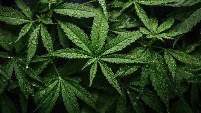
Researchers have recently published a review on how cannabis use among older adults impacts age-related conditions and longevity [1].

Cannabinoids and longevity

In recent years, researchers have observed an increase in cannabis use among older adults, mostly for chronic conditions, such as arthritis, pain, sleep improvement, anxiety, and depressive symptoms.

However, there is also a recent interest in using cannabis-derived cannabinoids, especially cannabidiol (CBD) and Δ9-tetrahydrocannabinol (THC), as potential anti-aging and longevity-promoting treatments.

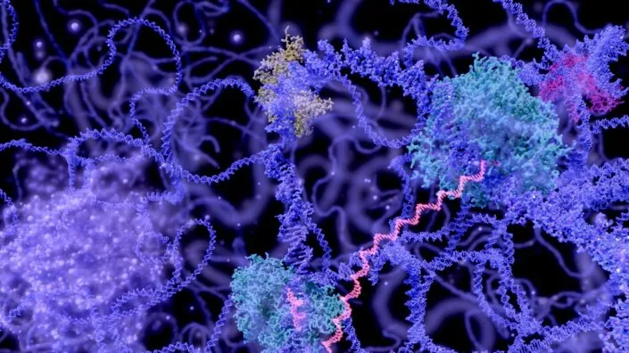
CBD and THC interact with the endocannabinoid system (ECS), which has been linked to impacting aging and longevity [2]. While the researchers do not have a detailed understanding of how the endocannabinoid system interacts with cannabinoids to promote health and longevity, the reviewed studies suggest that cannabinoids might promote cellular homeostasis.

This review encompassed what is currently known about the cannabinoids’ anti-aging properties, as well as their limitations, drawbacks, and side effects. The reviewers excluded studies whose focus was on the acute effects of cannabis or its action in different medical conditions. They found 11 preclinical and 7 human studies that met their search criteria.

Promising animal data

Most of the early studies regarding biological questions are done in model organisms. This is also the case for cannabis, as the researchers utilized worms, fruit flies, zebrafish, and mice to analyze its connection to health and lifespan outcomes.

Worm and fruit fly studies show that CBD and THC exposure can lead to lifespan extension, improved neuronal health, delayed age-related neurodegeneration, and increased autophagy. However, the beneficial effects often depend on using a specific dose of cannabis [3-6].

Another model organism that was used for cannabis research is the zebrafish, as its endocannabinoid system is well conserved, meaning it shows high similarity to humans. A study that analyzed the impact of THC exposure during development pointed to the dose-dependent differences, with lower doses leading to increased male survival and egg production, and reduced markers of aging and inflammation in the liver. Higher doses had negative consequences for offspring, survival, and reproduction [7].

Mice’s endocannabinoid systems are also similar to those of humans. In older mice, lower doses of THC led to improvements in memory and reversal of cognitive decline; however, this was not the case for THC-treated younger animals, which showed a decline in cognitive performance [8].

Cognitive performance improvements were also seen in two different studies where older mice were either chronically (for 28 days) exposed to a low dose of THC [9] or received a single injection of an extremely low THC dose [10]. Similarly, the memory and brain health of aged mouse models of Alzheimer’s disease benefited from intranasal low-dose THC treatment [11].

A mouse study also pointed to potential interactions between THC and CBD, as a low dose of THC was beneficial for spatial learning in aged mice, but when combined with CBD, the beneficial effect was not present [12]. This study points to the importance of further investigation into the combined effects of different cannabinoids.

The human data

While the evidence from model animals is promising, it needs to be confirmed in human studies, which are mostly lacking, and existing results are inconsistent. The human studies show a difference in outcomes regarding the age at which people started to use cannabis and how long they have been taking it. Several studies that examined people who used cannabis during adolescence and/or engaged in long-term use found that they had worse executive function, reduced grey matter, poorer verbal memory, accelerated biological aging, and worse health [13-16].

The effects are different when cannabis is taken by the elderly. For example, a study that included older adults, aged 60-88, who used cannabis weekly for at least the past year, showed increased connectivity in several brain regions, suggesting improved communication and information processing [17].

However, another study of people at least 60 years old showed lower executive functions in long-term cannabis users compared to non-users or short-term users. Short-term use didn’t seem to impact cognitive performance in this study [18].

Since there is a shortage of human studies on cannabis, the researcher also examined evidence regarding cannabis-based medicinal products (CBMPs) among older people. The data suggest CBMPs’ potential in treating aging-associated conditions, such as insomnia, depression, anxiety, and chronic pain. Cannabis use can also help reduce opioid dosage [19].

In total, the data from human studies and observations show age-dependent effects of cannabis. When cannabis use is initiated early in life, it leads to cognitive impairment later in life, but initiating cannabis use later in life shows more promising outcomes; however, there is still a need for more investigation in people.

The authors believe that some of the opposing age-dependent effects of cannabis might be caused by the changes that occur to the endocannabinoid system as it ages, such as changes in receptor binding and gene expression.

While analyzing different studies, the authors noticed methodological issues in human studies investigating cannabis use. First, the dose reporting is inconsistent and reports broad terms such as “heavy” or “recreational,” but no precise measurements are given. Second, there is a generalization of age categories, such as “older adults.” Both of those make interpreting the results and drawing strong conclusions harder. However, those researchers point out that they used those studies to gain an understanding of broader trends.

The authors also point out that there are differences in cannabis use patterns among the older population, with some people being new users (people who started after 60), while others are intermittent or consistent users. Those differences make cannabis research use more complex, as the effects of cannabis in each group might be different, and optimal therapeutic approaches might differ.

Cautious optimism

While most of the reviewed results seem to be optimistic, they should be interpreted with caution, as they are scarce and have many limitations. Additionally, most human studies are observational and cannot establish a causal link.

In the future, there is a need for well-designed human trials to understand the effect of cannabis on health and longevity, the consequences of the long-term effects of cannabis use among the elderly, and the effect of different dosing and routes of administration. There is also a need to investigate other cannabinoids beyond THC and CBD along with their interactions with each other and other medications and compounds.

We would like to ask you a small favor. We are a non-profit foundation, and unlike some other organizations, we have no shareholders and no products to sell you. All our news and educational content is free for everyone to read, but it does mean that we rely on the help of people like you. Every contribution, no matter if it’s big or small, supports independent journalism and sustains our future.

Literature

[1] Nain, S., Singh, N., Schlag, A. K., & Barnes, M. (2025). The impact of cannabis use on ageing and longevity: a systematic review of research insights. Journal of cannabis research, 7(1), 52.

[2] Paradisi, A., Oddi, S., & Maccarrone, M. (2006). The endocannabinoid system in ageing: a new target for drug development. Current drug targets, 7(11), 1539–1552.

[3] Land, M. H., Toth, M. L., MacNair, L., Vanapalli, S. A., Lefever, T. W., Peters, E. N., & Bonn-Miller, M. O. (2021). Effect of Cannabidiol on the Long-Term Toxicity and Lifespan in the Preclinical Model Caenorhabditis elegans. Cannabis and cannabinoid research, 6(6), 522–527.

[4] Wang, Z., Zheng, P., Chen, X., Xie, Y., Weston-Green, K., Solowij, N., Chew, Y. L., & Huang, X. F. (2022). Cannabidiol induces autophagy and improves neuronal health associated with SIRT1 mediated longevity. GeroScience, 44(3), 1505–1524.

[5] Wang, Z., Zheng, P., Xie, Y., Chen, X., Solowij, N., Green, K., Chew, Y. L., & Huang, X. F. (2021). Cannabidiol regulates CB1-pSTAT3 signaling for neurite outgrowth, prolongs lifespan, and improves health span in Caenorhabditis elegans of Aβ pathology models. FASEB journal : official publication of the Federation of American Societies for Experimental Biology, 35(5), e21537.

[6] Candib, A., Lee, N., Sam, N., Cho, E., Rojas, J., Hastings, R., DeAlva, K., Khon, D., Gonzalez, A., Molina, B., Torabzadeh, G., Vu, J., Hasenstab, K., Sant, K., Phillips, J. A., & Finley, K. (2024). The Influence of Cannabinoids on Drosophila Behaviors, Longevity, and Traumatic Injury Responses of the Adult Nervous System. Cannabis and cannabinoid research, 9(3), e886–e896.

[7] Pandelides, Z., Thornton, C., Lovitt, K. G., Faruque, A. S., Whitehead, A. P., Willett, K. L., & Ashpole, N. M. (2020). Developmental exposure to Δ9-tetrahydrocannabinol (THC) causes biphasic effects on longevity, inflammation, and reproduction in aged zebrafish (Danio rerio). GeroScience, 42(3), 923–936.

[8] Bilkei-Gorzo, A., Albayram, O., Draffehn, A., Michel, K., Piyanova, A., Oppenheimer, H., Dvir-Ginzberg, M., Rácz, I., Ulas, T., Imbeault, S., Bab, I., Schultze, J. L., & Zimmer, A. (2017). A chronic low dose of Δ9-tetrahydrocannabinol (THC) restores cognitive function in old mice. Nature medicine, 23(6), 782–787.

[9] Komorowska-Müller, J. A., Gellner, A. K., Ravichandran, K. A., Bilkei-Gorzo, A., Zimmer, A., & Stein, V. (2023). Chronic low-dose Δ9-tetrahydrocannabinol (THC) treatment stabilizes dendritic spines in 18-month-old mice. Scientific reports, 13(1), 1390.

[10] Sarne, Y., Toledano, R., Rachmany, L., Sasson, E., & Doron, R. (2018). Reversal of age-related cognitive impairments in mice by an extremely low dose of tetrahydrocannabinol. Neurobiology of aging, 61, 177–186.

[11] Fihurka, O., Hong, Y., Yan, J., Brown, B., Lin, X., Shen, N., Wang, Y., Zhao, H., Gordon, M. N., Morgan, D., Zhou, Q., Chang, P., & Cao, C. (2022). The Memory Benefit to Aged APP/PS1 Mice from Long-Term Intranasal Treatment of Low-Dose THC. International journal of molecular sciences, 23(8), 4253.

[12] Nidadavolu, P., Bilkei-Gorzo, A., Krämer, M., Schürmann, B., Palmisano, M., Beins, E. C., Madea, B., & Zimmer, A. (2021). Efficacy of Δ9 -Tetrahydrocannabinol (THC) Alone or in Combination With a 1:1 Ratio of Cannabidiol (CBD) in Reversing the Spatial Learning Deficits in Old Mice. Frontiers in aging neuroscience, 13, 718850.

[13] Meier, M. H., Caspi, A., Ambler, A., Hariri, A. R., Harrington, H., Hogan, S., Houts, R., Knodt, A. R., Ramrakha, S., Richmond-Rakerd, L. S., Poulton, R., & Moffitt, T. E. (2022). Preparedness for healthy ageing and polysubstance use in long-term cannabis users: a population-representative longitudinal study. The lancet. Healthy longevity, 3(10), e703–e714.

[14] Thayer, R. E., YorkWilliams, S. L., Hutchison, K. E., & Bryan, A. D. (2019). Preliminary results from a pilot study examining brain structure in older adult cannabis users and nonusers. Psychiatry research. Neuroimaging, 285, 58–63.

[15] Burggren, A. C., Siddarth, P., Mahmood, Z., London, E. D., Harrison, T. M., Merrill, D. A., Small, G. W., & Bookheimer, S. Y. (2018). Subregional Hippocampal Thickness Abnormalities in Older Adults with a History of Heavy Cannabis Use. Cannabis and cannabinoid research, 3(1), 242–251.

[16] Auer, R., Vittinghoff, E., Yaffe, K., Künzi, A., Kertesz, S. G., Levine, D. A., Albanese, E., Whitmer, R. A., Jacobs, D. R., Jr, Sidney, S., Glymour, M. M., & Pletcher, M. J. (2016). Association Between Lifetime Marijuana Use and Cognitive Function in Middle Age: The Coronary Artery Risk Development in Young Adults (CARDIA) Study. JAMA internal medicine, 176(3), 352–361.

[17] Watson, K. K., Bryan, A. D., Thayer, R. E., Ellingson, J. M., Skrzynski, C. J., & Hutchison, K. E. (2022). Cannabis Use and Resting State Functional Connectivity in the Aging Brain. Frontiers in aging neuroscience, 14, 804890.

[18] Stypulkowski, K., & Thayer, R. E. (2022). Long-Term Recreational Cannabis Use Is Associated With Lower Executive Function and Processing Speed in a Pilot Sample of Older Adults. Journal of geriatric psychiatry and neurology, 35(5), 740–746.

[19] Tumati, S., Lanctôt, K. L., Wang, R., Li, A., Davis, A., & Herrmann, N. (2022). Medical Cannabis Use Among Older Adults in Canada: Self-Reported Data on Types and Amount Used, and Perceived Effects. Drugs & aging, 39(2), 153–163.