×

The Blog

Building a Future Free of Age-Related Disease

Pharmaceutical shelf

Repurposing Drugs to Lower Dementia Risk

The authors of a recent review analyzed the results of 14 studies that included 139 million people to identify patterns that connect dementia risk to commonly used medications [1].

Finding a new purpose

Despite its prevalence in the elderly population, there is a lack of effective clinical treatments for dementia, and there is ongoing research to find new medications and therapies that can cure or slow down dementia. While this is essential, there is also another approach that can be simultaneously applied: repurposing existing drugs to slow down the progress of dementia. The increased availability of routinely collected medical data makes it possible to conduct studies involving millions of patients and hundreds of drugs.

The researchers point out that some drugs that are currently prescribed for different conditions are already known to affect dementia risk. For example, some diabetes drugs have been linked to dementia risk reduction [2].

Driven by data

The authors of this study conducted a systematic review of studies that researched the association between prescribed medications and dementia risk. They limited their analysis to the data-driven, rather than hypothesis-driven, approach. They define data-driven as “an exploratory approach that analyzes large datasets to extract insights and patterns by applying analytical techniques and modes of reasoning.”

Such an approach has its advantages and disadvantages. While it can exclude some high-quality studies that are hypothesis-driven, which limits the study to only a subset of data that pertains to a given hypothesis, it also has some advantages. Since the data-driven approach uses all available data of all currently prescribed drugs, the researchers can identify previously unidentified associations missed by hypothesis-based approaches.

In their analysis, they included fourteen studies from the USA, Japan, South Korea, Germany, and Wales, including 139 million people and 1 million cases of dementia, and investigated an estimated 200 pharmacological subgroups, including more than 2000 ingredients.

Some drugs reduce dementia risk

In general, the researchers found that inconsistencies between the studies made it difficult to analyze the impact of individual drugs on dementia risk. Nevertheless, they found some general trends in different classes of medicines. For example, antimicrobials, vaccines, and anti-inflammatories were linked to reduced dementia risk. Antimicrobials and vaccines may protect against dementia because they address viral and bacterial infections, which have been linked to an increase in dementia risk [3-5].

The inclusion of anti-inflammatories is not surprising, since inflammation is a widely known pathogenic pathway in aging. Suppressing inflammation has already been investigated as a possible target for Alzheimer’s disease [6]. Even though clinical trials of these agents have not been successful so far [7], the authors believe that “using the right agent at the right time point in disease progression, perhaps prior to manifestation of cognitive decline, may be crucial.”

Other drugs increase dementia risk

On the other hand, antipsychotics and drugs for diabetes were linked to increased dementia risk.

The authors believe that for antipsychotics and antidepressants, some of the associations can be explained by reverse causation. For example, antidepressants can be prescribed in the early stages of dementia to treat mood disorders. Including these patients in the dataset means that antidepressants become associated with an increased likelihood of dementia, even though dementia can lead to antidepressant use.

However, other drugs may have dementia as a potential side effect, and in these cases, it is crucial to investigate the mechanisms of action in order to better avoid prescribing such drugs to people who are at risk for dementia.

Conflicting results

There were also classes of medications including antihypertensives, antidepressants, and, to a lesser extent, drugs to manage blood glucose levels that showed conflicting results, as some data suggests that they increase dementia risk, while other data suggests that they decrease it.

The authors discuss a few possible explanations for the conflicting results. One drawback to this line of research is that it analyzes classes of drugs and not specific drugs by themselves. Different members of the same class of drugs might act on various molecular targets and exert distinct effects on organisms.

For example, some antihypertensives lead to the upregulation of autophagy, a biological process linked to longevity [8]. However, this is not the case for all antihypertensives. Since all antihypertensives are analyzed together, such beneficial effects of single drugs might be missed, leading to false negative results. Nevertheless, the authors believe that in their research, false negatives are unlikely since their data includes large numbers of both people and drugs.

The authors also bring up an important caveat: suboptimal datasets used for research. The datasets used in the study were created for clinical purposes, not research, and they may not include important information, such as drug administration. Those drugs were prescribed to patients, but we cannot be confident that patients took them.

On the other hand, there might be people who take some over-the-counter medications without a prescription. Such data will be missing. There is also no information about dose responses or analysis of taking single or multiple medications simultaneously, and there is often missing data regarding confounding factors, such as socioeconomic status, genotyping, and biomarker information. There is also a question of the accuracy of dementia diagnoses, since dementia is often under- or misdiagnosed.

Setting priorities

The researchers believe that their work can aid in prioritizing which drugs should be explored in further studies for potential repurposing to treat dementia. Those studies might involve a single drug or drug combinations since dementia is a complex condition, and to treat such a condition, it might be necessary to target it with multiple drugs that address numerous molecular pathways involved in its pathology.

We would like to ask you a small favor. We are a non-profit foundation, and unlike some other organizations, we have no shareholders and no products to sell you. All our news and educational content is free for everyone to read, but it does mean that we rely on the help of people like you. Every contribution, no matter if it’s big or small, supports independent journalism and sustains our future.

Literature

[1] Underwood, B. R., Lourida, I., Gong, J., Tamburin, S., Tang, E. Y. H., Sidhom, E., Tai, X. Y., Betts, M. J., Ranson, J. M., Zachariou, M., Olaleye, O. E., Das, S., Oxtoby, N. P., Chen, S., Llewellyn, D. J., & Deep Dementia Phenotyping (DEMON) Network (2025). Data-driven discovery of associations between prescribed drugs and dementia risk: A systematic review. Alzheimer’s & dementia (New York, N. Y.), 11(1), e70037.

[2] Nørgaard, C. H., Friedrich, S., Hansen, C. T., Gerds, T., Ballard, C., Møller, D. V., Knudsen, L. B., Kvist, K., Zinman, B., Holm, E., Torp-Pedersen, C., & Mørch, L. S. (2022). Treatment with glucagon-like peptide-1 receptor agonists and incidence of dementia: Data from pooled double-blind randomized controlled trials and nationwide disease and prescription registers. Alzheimer’s & dementia (New York, N. Y.), 8(1), e12268.

[3] Muzambi, R., Bhaskaran, K., Brayne, C., Davidson, J. A., Smeeth, L., & Warren-Gash, C. (2020). Common Bacterial Infections and Risk of Dementia or Cognitive Decline: A Systematic Review. Journal of Alzheimer’s disease : JAD, 76(4), 1609–1626.

[4] Levine, K. S., Leonard, H. L., Blauwendraat, C., Iwaki, H., Johnson, N., Bandres-Ciga, S., Ferrucci, L., Faghri, F., Singleton, A. B., & Nalls, M. A. (2023). Virus exposure and neurodegenerative disease risk across national biobanks. Neuron, 111(7), 1086–1093.e2.

[5] Ballard, C., Aarsland, D., Cummings, J., O’Brien, J., Mills, R., Molinuevo, J. L., Fladby, T., Williams, G., Doherty, P., Corbett, A., & Sultana, J. (2020). Drug repositioning and repurposing for Alzheimer disease. Nature reviews. Neurology, 16(12), 661–673.

[6] Cummings, J., Zhou, Y., Lee, G., Zhong, K., Fonseca, J., & Cheng, F. (2024). Alzheimer’s disease drug development pipeline: 2024. Alzheimer’s & dementia (New York, N. Y.), 10(2), e12465.

[7] Meyer, P. F., Tremblay-Mercier, J., Leoutsakos, J., Madjar, C., Lafaille-Magnan, M. E., Savard, M., Rosa-Neto, P., Poirier, J., Etienne, P., Breitner, J., & PREVENT-AD Research Group (2019). INTREPAD: A randomized trial of naproxen to slow progress of presymptomatic Alzheimer disease. Neurology, 92(18), e2070–e2080.

[8] Siddiqi, F. H., Menzies, F. M., Lopez, A., Stamatakou, E., Karabiyik, C., Ureshino, R., Ricketts, T., Jimenez-Sanchez, M., Esteban, M. A., Lai, L., Tortorella, M. D., Luo, Z., Liu, H., Metzakopian, E., Fernandes, H. J. R., Bassett, A., Karran, E., Miller, B. L., Fleming, A., & Rubinsztein, D. C. (2019). Felodipine induces autophagy in mouse brains with pharmacokinetics amenable to repurposing. Nature communications, 10(1), 1817.

Hevolution GHS 2025

Impressions from Hevolution’s Global Healthspan Summit 2025

These are some of the best talks from the largest healthspan conference in the world, which was held for the third time in Riyadh by the Hevolution Foundation.

Young and restless

The Hevolution Foundation has only been around for three years. Before that, Saudi Arabia, its main sponsor, was not considered a serious player in the longevity field, but a lot of money and a top-notch team can do wonders.

Hevolution, the best-funded non-profit in the field, has its hands in sponsoring breakthrough research and investing in longevity biotech. It also organizes the Global Healthspan Summit in Riyadh.

Here, too, the foundation has achieved a lot in a short amount of time. The GHS, held earlier this month for only the second time, attracted over three thousand attendees and cemented its place as the biggest longevity conference in the world, by far.

Hevolution attendees

Perhaps it can be called a healthspan conference instead. In his opening remarks, just like in his recent interview with Lifespan.io, Hevolution CEO Dr. Mehmood Khan made the distinction between longevity and healthspan one of his central points.

“A discussion in most of this field has been about longevity,” he said. “We at Hevolution don’t like to speak about longevity. Most of the people we’ve surveyed don’t like to live longer just for the sake of living longer. They want to be independent, functional, mentally and physically. They want mobility, they want to contribute. What they’re asking is ‘can I live healthy as long as possible?’ We like to use the word healthspan far more than lifespan, because this is what’s important to humanity.”

Khan credited Hevolution for shifting the discourse from longevity and lifespan towards healthspan. This shift, however, was already happening, and it probably will never become complete, as the realization grows that lifespan and healthspan – that is, the part of life lived in good health – are tightly linked.

Hevolution, Khan said, is about to publish a report showing that today, one in two physicians regularly gets asked by their patients about lifespan or healthspan. “It’s not a discussion for experts only anymore, but for patients,” he noted.

He then added that he actually prefers the word “consumers” over “patients.” According to Hevolution’s philosophy, the entire world population consists of consumers of our field. The idea is that when a person becomes a patient, it’s already too late. Geroscience should be able to intervene earlier to prevent people from developing age-related diseases in the first place.  “The best job we can do is to keep people healthy,” Khan summarized.

He then went over Hevolution’s milestones, starting with acknowledging the role of the Saudi Royal family in the non-profit’s birth. “A moment of pride for us,” he said, “is that this is not just an organization but a global movement that was launched from Saudi Arabia. I have to acknowledge first and foremost His Royal Highness Prince Mohammed bin Salman, whose vision led to the creation of Hevolution.”

“We only started funding and investing two years ago,” he added. “Today, Hevolution is the second-largest funder of geroscience on the planet, and the biggest philanthropy, with over 400 million dollars in research funding and investment, and many more to come.”

According to Khan, today, over 250 scientists in more than 200 labs are Hevolution’s partners and grant recipients. Its impact on longevity biotech has been more modest, with only four companies funded, but Khan promised that several more investments will be announced soon.

“In 2024,” Khan said, “venture capital funding in this field more than doubled to over 75 billion dollars. The size of each investment went up by 77%, which shows confidence, as investors are willing to write bigger checks. However, that’s not enough. Investments in fighting the consequences of aging are 10-100 times greater. We must close that gap.”

He concluded with a request aimed at the audience: “What do I ask from anybody here? Our goal was to bring you together, to give you the opportunity to communicate, to figure out how to collaborate, to push the boundaries of science, to create new policies, regulations, sources of funding, businesses. There is no other business in the world that’s going to affect all 8 billion humans.”

Expanding beyond the Hallmarks of Aging

Dr. Felipe Sierra, a famed geroscientist and Hevolution’s Chief Scientific Officer, expanded on Khan’s vision in his talk titled “Science beyond the biomarkers of aging”.

As someone who’s been in this field for a very long time, Sierra thinks that “the last ten years have been amazing, the explosion of things has happened.” He noted that “one of our own,” Dr. Gary Ruvkun, who dedicated a considerable part of his career to studying aging, received a Nobel prize last year – a sign of geroscience becoming widely accepted and respected.

Two events, Sierra said, contributed a lot to this change: publication of the original paper on the Hallmarks of Aging, and the first summit on geroscience: “They galvanized the field, but this happened 12 years ago. It’s time for us to reconsider where we are with hallmarks and geroscience.”

Sierra lent his support to Khan’s healthspan vision: “We’re switching more towards health as opposed to diseases. Now, it’s about keeping you young and healthy as you age.”

While the original Hallmarks paper got a facelift in 2022, Sierra thinks that the entire approach is still insufficient, albeit “useful because it focuses the field.”

“In the words of Leonard Guarente,” he said, referencing another veteran geroscientist, “it’s not the hallmarks of aging but the hallmarks of life, because every molecular pathway needed for maintenance of life will affect aging, and aging will affect that pathway as well. So, we will end up adding all of biology. How do we connect these hallmarks to the actual outcome which is health?”

There has been some advancement on this, he said: “There’s a publication from WHO on intrinsic capacities. We no more define healthy aging as absence of disease, but as the ability to function. How do we correlate molecular processes with the age-related loss of intrinsic capacity? The thing joining the two together is molecular resilience – the ability of every cell in your body to react to a challenge and come back to homeostasis.”

Hevolution’s vision, Sierra said, is that “the hallmarks of aging, either defined or not, converge on the loss of resilience, and that’s because all the hallmarks interact with each other. Loss of resilience is what leads to a decrease in intrinsic capacities and loss of health.”

Any insult, such as chemotherapy, a high-fat diet, and even exercise, which is, after all, a stress, elicits a reaction, but the ability to properly react is lost with age. Why do we lose it? Is it just wear and tear? This is a key question in today’s geroscience, according to Sierra.

With time, our ability to respond to challenges declines to a point “where even a small challenge – a cold, a flu, or a fall – presents an insurmountable challenge.” This is a central feature of aging that researchers should be focusing on, he said.

However, measuring resilience in response to challenges can be tricky due to both technical and ethical problems. Sierra predicted that this will soon become a major subfield in geroscience and suggested that continuous measurements using wearable devices might help a lot. “That’s part of the field’s future and one of the areas Hevolution is focusing on,” he concluded.

Bioelectricity – not just for eels

Dr. Elena Sergeeva works with Michael Levin at Tufts University and Harvard’s Wyss Institute. Levin is a daring pioneer in studying bioelectricity in the context of aging. Sergeeva gave a fascinating presentation of this unorthodox approach, titled “Bioelectricity, Morphogenesis and Aging.”

Like many others at the conference, Sergeeva pointed out that no widely accepted scientific definition of aging exists. In her team’s approach, aging is a defect of morphogenesis, which is the creation of forms and structure, and of morphostasis, which is the maintenance of this form. “Aging is when cell collectives fail to maintain patterns, homeostasis,” she explained.

How are those patterns created? How do we get our highly differentiated organs and tissues that disintegrate with aging? “DNA only specifies proteins, the building blocks, but where does anatomy come from?” Sergeeva mused. “How do cells know what to build and, most importantly, where to stop? Why, for instance, do we end with five fingers and not six? Why do cells sometime deflect from the target morphology, like in cancer?”

Levin’s team views DNA as the “hardware.” What is the “software” then? Many would say it’s epigenetics, but according to Sergeeva, bioelectric signals play a central role in employing this hardware to achieve the desired body morphology.

When we think about bioelectric signals, we usually envision neurons. However, the body uses bioelectric coordination between cells on a much grander scale.

The team works with model organisms such as planaria worms and frogs. “When a frog embryo develops,” Sergeeva said, “it is changing its electrical patterns, and they correspond to the developing organs, the body scheme” – something her team can visualize. Conversely, when anatomy deflects from its original form, such as in cancer, it causes changes in those bioelectrical patterns.

“We can also manipulate that,” Sergeeva added. “The patterns are properties of membrane voltage, which is formed with ion channels. Those channels can be manipulated by drugs and expressed in cells. Can we then change formation of body structure by changing bioelectrical patterns?”

Apparently, we can. Levin’s lab went as far as inducing the formation of an eye on a tadpole’s back using solely bioelectric signals.

“If you want to create an eye on the back of a frog embryo, as a molecular biologist, you’d come up with a long list of genes responsible for eye formation and try to change them,” Sergeeva said. “It’s virtually impossible.” What her team does instead is inject RNA that expresses certain ion channels. The affected cells start to form an eye and recruit other, non-modified cells. The team has grown fins, ectopic limbs, and even complete beating hearts just by manipulating certain ion channels.

Using this bioelectric system, the team can make a planaria worm, a relatively complex organism, grow two heads. However, this is not all. Planaria worms are known for their regenerative abilities. When cut in half, both halves develop into complete worms. Amazingly, when you cut a two-headed planaria in half, both halves develop two heads each. According to Sergeeva, this shows the researchers “can rewrite anatomic pattern memory.”

Sergeeva thinks that their technology has potential uses in birth defects, traumatic injury, cancer, and, of course, aging. The researchers can already see how bioelectric patterns change with age and build bioelectric aging clocks. According to Sergeeva, they found that these patterns “are corrupted with age, become fuzzier, cell communication is not as sharp and precise anymore.” Such loss of information has been demonstrated time and again in various aspects of aging.

Showcasing her team’s progress, Sergeeva mentioned two spin-out companies. Morphoceuticals works on regenerating limbs in mammals, while Astonishing Labs uses bioelectric patterns, rather than genes and proteins, for drug discovery.

Is there intrinsic value in intrinsic health?

Alan A. Cohen, Associate Professor at Columbia University’s Aging Center, picked up the note struck earlier by Mehmood Khan and Filipe Sierra in his talk “Shifting from Aging to Intrinsic Health as a Target for Interventions.”

After acknowledging that defining and measuring aging remains a monumental challenge, Cohen suggested that “the best way to target aging is to target health itself.”

Geroscientists agree that aging is multidimensional and heterogeneous both within and across individuals, Cohen said. This means that with age, individual organisms become less well-orchestrated, faultier, and they also drift away from each other.

“Aging involves trade-offs, and we need to find ways to deal with them,” Cohen said. “For instance, many hallmarks of aging, such as cellular senescence, telomere shortening, and stem cell exhaustion, play a role in anti-cancer protection. Changes in immune function also are reactions to risks. This means that, when discussing aging, we need to distinguish between damage and compensatory or adaptive mechanisms.”

There are many questions about measuring aging, Cohen noted. Do we have to map all the dimensions of aging? Do we have to know how important each one is? Distinguishing between causes and effects of aging as well as giving them proper weights is hard.

Instead, Cohen argues for using the notion of intrinsic health as a more tractable target for interventions. “We are complex dynamic systems, but it’s hard to incorporate this into the way we think about biology,” he said. “But we’re also highly optimized by evolution, which makes us different from economy or weather.”

This means that while humans are immensely complex, they are also at a long-term equilibrium. We are controlled chaos, in a sense. This equilibrium that enables our survival and reproduction, which is what evolution is all about, can be called “health.”

There are innumerable ways in which the organism moves away from this equilibrium as a result of disease or aging. Consequently, “it is much harder to track all the ways we lose health in than health itself,” Cohen said. Instead, we should figure out how to measure this “central state.”

Cohen views aging is a decline in intrinsic health, and measuring it requires going back to first principles. “What does it take to make an organism?” Cohen asked. “You need energy, communication and structure. It’s like a magnetic field that emerges from the energy of the battery, the communication with the wire, and the structure of the solenoid. When all the aspects work together just right, the organism as a whole functions well.”

Cohen also suggested a formal definition of intrinsic health: “A field-like state emerging from the dynamic interplay of energy, communication, and structure within the organism, which gives rise to robustness, resilience, plasticity, performance, and sustainability.” The elements relate to various functions the body must perform well: for instance, robustness and resilience is when you have to keep your current state, while plasticity is the opposite: when you need to change your current state.

These elements can be measured by using various biological proxies. Cohen’s team is working on assembling what he called “a basket of proxies” and statistically integrating them. “More proxies – more signal,” he said, “but there are also diminishing marginal returns. We don’t need a thousand proxies. 10, 20, 30 of the right ones, and we’ll get close to measuring the signal.”

Some of the currently known proxies include heart rate variability (HRV), cardiac function, and levels of certain proteins such as GDF15, which is linked to energetic stress. “Greater communication between the signals is associated with health,” Cohen noted.

“The vision is that intrinsic health will become a single integrative measure, measured from a single blood sample and a wearable,” he said. “It could be an outcome for many health and aging interventions and a metric of populational health. By targeting this, we should also be able to prevent many diseases.”

Biomarkers, biomarkers everywhere

Two titans of geroscience, Harvard professor Vadim Gladyshev and Dr. Steve Horvath, currently a principal investigator at Altos Labs, gave back-to-back talks on the important issue of biomarkers of aging.

“Biomarkers of aging are the most important development of this century in the field,” Gladyshev said at the beginning of his talk. However, while finding metrics that correlate with and can predict chronological age and mortality was indeed transformative, many problems remain.

Like several other speakers, Gladyshev stressed the pressing need to develop better definitions of aging: “It begins with ‘what is aging’? People generally disagree. It can be increased mortality rate, damage accumulation, functional decline, or something else. There are different ways to quantify it, and no current consensus on which one is better.”

To solve this problem, Gladyshev and dozens of other geroscientists formed the Biomarkers of Aging Consortium about two years ago. According to Gladyshev, the last meeting in November was attended by 450 people from 30 countries.

This meeting included a challenge in which teams had to develop methylation clocks that were then tested on a dataset of 500 blood samples. 37 teams competed, and several of them were able to develop clocks more accurate than the existing ones. “The average error is now approaching two years even with a one-modality clock (epigenetic),” Gladyshev said. “If we combine multiple modalities, we’ll get even better results.”

Gladyshev then mentioned several other recent developments in this field. Jesse Poganik from his lab published a paper on how severe stress, such as from major surgery, pregnancy, or sleep deprivation, reversibly increases methylation age. Another lab member, Albert Ying, developed the first causal epigenetic clock, which separates changes into damaging and adaptive. Andrei Tarkhov found the stochastic component of aging by comparing single-cell to bulk methylation.

A yet unpublished study on organ-specific methylation showed that in every case, it yielded a very interesting observation, Gladyshev said. The diseased tissue (brain affected by Alzheimer’s disease, heart affected by heart disease, kidney affected by kidney disease, and so on) was older than controls.

Organ-specific aging is a recent and important development in the field. Using UK Biobank data, Gladyshev’s team is analyzing proteins that originated in different organs, developing organ-specific mortality clocks.

“These clocks can also predict diseases,” he explained. “The lung clock best predicts COPD, which is a lung disease, while the liver model predicts cirrhosis. People with dementia are older in the brain, and so on.”

The researchers can now determine how organs are affected by stressors and interventions. Predictably, the lung is the organ most affected by smoking. However, with alcohol, the picture is more complex. Kidney and the intestines are most negatively affected, but arteries and lungs are rather “rejuvenated” by alcohol. According to Gladyshev, this means we need different interventions for different people.

Personalization of treatment was a major topic at the conference, and this talk was part of the trend. “Perhaps it makes sense to target ‘weak links’ – organs with accelerated aging,” Gladyshev suggested.

“So, is aging a disease?” he concluded. “It’s a bad question. Aging is neither a disease nor not a disease. But, the question can be asked as whether diseases are aging. Is a chronic disease a manifestation of aging? Yes, chronic diseases may be viewed as accelerated aging of a subset of organs and tissues, and disease can be viewed as pockets of non-autonomous aging.”

Methylation clocks’ coming of age

Dr. Steve Horvath, the “father of methylation clocks,” currently leads a large team at Altos Labs, the insanely well-funded company created by the entrepreneurs Jeff Bezos and Yuri Milner.

While most clock are based on cytosine methylation, epigenetics relate to many types of chemical modifications, such as histone modifications, Horvath said. Some species, including aging models such as C. elegans and drosophila, don’t have cytosine methylation, and yet clocks based on histone modifications work well.

While first-generation clocks were built to predict chronological age, newer ones, such as GrimAge, developed by Horvath in collaboration with other scientists, predict mortality (time to death). Moreover, GrimAge is based on interpretable covariates: methylation proxies of various parameters, from smoking pack/years to GDF15 levels. This means that it is possible to estimate these parameters based on cytosine methylation patterns instead of measuring them directly.

Horvath showcased GrimAge’s ability to predict healthspan. Apparently, the clock can estimate the time that someone will get coronary heart disease, cancer, physical and cognitive decline, and other conditions. GrimAge’s methylation proxy for smoking is so precise that it beats self-reported smoking history.

“The factors that GrimAge is based on, like plasma protein levels, have clear biological relevance, potentially offering insights into the aging process itself,” he said.

Horvath stressed the importance of longitudinal studies, where methylation patterns are measured more than once. Such studies can establish causality between conditions/treatments and the clocks’ readings, including by estimating the rate of epigenetic aging. Horvath mentioned a recent longitudinal study by Luigi Ferrucci’s team that showed, in a 700-strong cohort, that methylation clocks’ change rate is predictive of mortality.

However, randomized control trials (RCTs) are the best approach to establishing causality, he said, and we see more and more RCTs with epigenetic clock readings as endpoints. Horvath himself just recently published such a study in Nature Aging, in collaboration with Heike Bischoff-Ferrari.

The study, which he called “a very rigorous RCT,” looked at 777 people who took one gram of omega-3 per day for three years. The results show that the treatment “quantitatively rejuvenates you by a few months,” Horvath said. Among other interventions tested in the same study were vitamin D and exercise, and Horvath reported that “there was some additive effect.”

“This study is interesting as a prototype study. If someone has an intervention, we can measure methylation age but also ‘tease out’ which proteins might be affected by the intervention,” he explained.

More than ten years after the introduction of the first epigenetic clock, the field is going strong, becoming more diverse and sophisticated day by day. “We now have a good understanding of what stresses affect the clocks,” Horvath said. “This includes cell division, DNA repair and damage, somatic mutations, epigenetic maintenance, among other things.”

Among other recent developments are deep learning-based clocks, which can be both more accurate and more interpretable, clocks for different species, such as the axolotl, and also multi-species clocks, where a single algorithm can predict aging in several even distantly related species. “These clocks are not great,” Horvath admitted, “but it’s a new frontier. This suggests shared, conserved biology, the universality of aging.”

Learning from birds and whales

One of the most interesting and star-studded panels at the conference was devoted to mammalian longevity. Moderated by Vadim Gladyshev, it included Steve Horvath, professor at the University of Rochester Vera Gorbunova, professor at the University of Birmingham, UK, João Pedro de Magalhães, and CEO of Fauna Bio Dr. Ashley Zehnder.

Having announced the panel as “the best session at the conference,” Gladyshev started by pointing out the huge variety of lifespan across mammals, which is over 100-fold. “Nature has conducted innumerable experiments for us,” he said, “and we as scientists have to learn from these experiments, trying to unlock mechanisms that would allow radical changes in lifespan.” Admittedly, it was refreshing to hear a mainstream scientist bringing up radical life extension at a conference that was all about healthspan.

Asked about the comparative biology approaches her team used, Gorbunova responded: “We study long-lived and resistant species, such as the naked mole rat. We find genes responsible for this and transfer them into mice, making them live longer. We compare many species to identify common pathways.”

De Magalhães continued: “Mice and humans have similar biology/physiology, and yet mice age 20-plus times faster than humans. The reason must be in the genome. Evolution has selected for genomes that determine vast differences in lifespan and aging. To unravel the mechanisms responsible for this, our team does evolutionary comparative genomics. Genome is the common language between species.”

Zehnder: “We study species that have evolved resistance and ability to heal many tissues, namely hibernating species capable of rapid rewarming. We determine the genes that are activated to repair the damage from rapid rewarming and then map them to small molecules. Our current indication is heart failure. We are seeing a lot of overlap between geroprotective and hibernating pathways.”

Gladyshev, answering his own question: “We are doing comparative analysis of about 40 mammalian species, analyzing gene expression patterns associated with longevity. These patterns can be directly used to identify interventions. There’s a potential to use this comparison across species to achieve radical change in lifespan. Many labs are now working on achieving radical life extension, rather than 10-15%.”

Gladyshev then asked Gorbunova what long-lived species can teach us that other models can’t. She said that wild animals need to stay extremely healthy for their entire life to be able to survive: “Conventional model organisms are short-lived but easy to analyze. If you tweak them to live longer, this often makes them less fit. But in long-lived species, there’s no such problem, their adaptations, tested by evolution, give them both lifespan and healthspan. This is the main advantage of comparative biology. As research objects, long-lived species are directly targeted to improved healthspan.”

Asked about the challenges in this subfield, Horvath responded, “The main conceptual question is, can we learn something from long-lived species, given that we and them inhabit very different ecological niches? Vera and others in the field make a very strong case that it’s possible. The second question is, should we go beyond mammals – to reptiles, fish, birds, which all have amazing adaptations? Finally, what kind of omics we need to look at?”

Echoing Gorbunova, de Magalhães added: “Traditional biomedical models are models of disease, short lifespan, cancer. Long-lived species, on the other hand, are models of disease resistance, of healthspan. The potential effect is much larger than with the current models, but the road there is harder. Basically, this is a high-risk, high-reward approach. These species can help us understand why we, humans, are the way we are, why we are susceptible to certain diseases at certain ages.”

As the only person on the panel who runs a company in this subfield, Zehnder was asked about her team’s approaches to translating the findings of comparative biology. “Translation is key when we’re speaking about very diverse species,” she said. “Hibernation exists across a surprisingly wide variety of species, so it’s like tapping into our own hidden potential, into some highly conserved pathways. When we see the process of repair, we can map it to humans and test in human cells lines. Then we look for humans with mutations in these pathways.”

Tendon

A Key Protein for Tendon Health

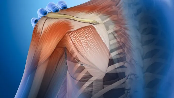
Researchers publishing in Matrix Biology Plus have discovered that cochlin, a protein that decreases with age, is vital for the health of tendons.

Tendons require a healthy extracellular matrix

Previous work has found that tendon tissues, which link muscle to bone, require a healthy extracellular matrix (ECM) to function; if the ECM is degraded, the tendon has problems with handling the forces put on it and in healing from injury [1]. Tenocytes, cells that maintain tendon tissue, respond to external pressures when building and removing the ECM and thus maintain tendon homeostasis [2].

These researchers have previously found that aging depletes the amount of Scleraxis-lineage tenocytes and that this depletion is directly responsible for the loss of tendon function, as demonstrated in a mouse model [3]. That work found that cochlin appears to be a crucial part of regulating tendon health.

While cochlin is found in the ECM, it is not part of structural collagen, and previous literature discussing it mostly focused on its effects in the inner ear. Its effects on tendon development have not been well documented. Therefore, this paper follows the team’s previous line of experiments, this time focusing specifically on cochlin.

Cochlin impacts how tendons mature

For their first experiment, the researchers created a strain of mice that do not produce cochlin and analyzed their tendons at 3, 6, and 9 months of age. At all three ages, the cochlin-less mice had significantly wider collagen fibrils than wild-type controls. While there were trends, there did not appear to be significant effects on how these tendons matured, except for a substantially reduced stiffness at 6 months. Peak load, however, was significantly affected: the tendons of 6-month-old cochlin-less mice could not withstand as much load as their wild-type counterparts, and this difference was only increased at 9 months.

There were also substantial alterations to tendon homeostasis, as revealed by a gene expression analysis. In line with previous research, the cochlin-less mice had signs of hearing damage, but they also had significant alterations to genes involved in protein conversion, RNA metabolism, lysosomal function, and cellular proliferation. The researchers hold that the loss of this single protein creates a broad impact to many aspects of cellular biology.

While the loss of cochlin impaired the tendons’ ability to handle stress, it did not seem to affect their ability to heal. One month after the researchers had surgically injured the flexor tendons of wild-type and cochlin-less mice at 10 to 12 weeks of age, the mice had healed in the same way. While there were trends towards decreased stiffness and decreased loading ability in the cochlin-less mice, it is unlikely that they were due to differences in how they healed. The researchers, therefore, posit that cochlin is necessary for proper tendon maturation but not healing.

Important but one of many

The researchers use the discussion section of this paper to analyze why this protein might have such wide-ranging effects. They note that it binds to collagen [4] and may have direct impacts on how its structure functions. Clearly, cochlin has impacts on maturation, but they did not attempt to analyze its effects on aging per se; as they also note, this would be particularly difficult to isolate in naturally aging animals, as a great many other things are going wrong with the ECM as well.

This work primarily notes the differences between young and middle-aged animals. Tendons, in mice, take a while to mature properly, and this paper concluded its work at almost half the animals’ natural lifespan; future work that analyzes changes with aging must therefore differentiate the positive effects of maturation versus the negative effects of age-related degeneration.

Further work that upregulates cochlin, and perhaps other ECM-related proteins that are downregulated with age, would be necessary to isolate their effects and determine whether the depletion of this protein with age is a cause or a consequence of other forms of age-related degeneration.

Collectively, these data identify Cochlin as a critical regulatory component of proper tendon structure and future work will define the therapeutic potential of conservation or restoration of Cochlin to facilitate continued tendon health through the lifespan.

We would like to ask you a small favor. We are a non-profit foundation, and unlike some other organizations, we have no shareholders and no products to sell you. All our news and educational content is free for everyone to read, but it does mean that we rely on the help of people like you. Every contribution, no matter if it’s big or small, supports independent journalism and sustains our future.

Literature

[1] Di, X., Gao, X., Peng, L., Ai, J., Jin, X., Qi, S., … & Luo, D. (2023). Cellular mechanotransduction in health and diseases: from molecular mechanism to therapeutic targets. Signal transduction and targeted therapy, 8(1), 282.

[2] Galloway, M. T., Lalley, A. L., & Shearn, J. T. (2013). The role of mechanical loading in tendon development, maintenance, injury, and repair. JBJS, 95(17), 1620-1628.

[3] Korcari, A., Nichols, A. E., Buckley, M. R., & Loiselle, A. E. (2023). Scleraxis-lineage cells are required for tendon homeostasis and their depletion induces an accelerated extracellular matrix aging phenotype. Elife, 12, e84194.

[4] Verdoodt, D., Van Camp, G., Ponsaerts, P., & Van Rompaey, V. (2021). On the pathophysiology of DFNA9: Effect of pathogenic variants in the COCH gene on inner ear functioning in human and transgenic mice. Hearing research, 401, 108162.

Junevity logo

Junevity Launches to Develop Cell Reset Therapeutics

Junevity, a biotechnology company on a mission to extend lifespan and healthspan by resetting cell damage from age-related diseases, today announced $10 million in seed funding led by Goldcrest Capital and Godfrey Capital.

The Junevity RESET platform is based on exclusively licensed research by co-founder Dr. Janine Sengstack at the University of California at San Francisco. RESET uses large-scale human data and AI to identify genes – or transcription factors – that can regulate cell damage. Then, it develops siRNA therapeutics against these targets to return cells to health. Junevity will use this seed funding to enhance the RESET platform and develop its first therapeutic candidates in Type 2 diabetes, obesity and frailty.

“My research at UCSF showed the power of targeting transcription factors to restore aged human cells back to health,” said Janine Sengstack, Ph.D., co-founder and Chief Scientific Officer at Junevity. “Based on these discoveries, we are bringing forward a new class of cell reset therapeutics for diseases, with the ultimate goal of greater human longevity.”

Diseases like obesity, diabetes, frailty, neurodegeneration and many others shorten human lifespan and are associated with complex cell damage at the transcriptional level. RESET uses billions of data points from human diseases and AI to rank and evaluate potential targets. Together, the platform outputs the Cell RESET Atlas, a collection of promising transcription factor targets by cell type and by disease for therapeutic targeting. Junevity then develops novel silencing RNA (siRNA) therapeutics to restore cellular transcription back to a healthy state.

Junevity’s preclinical data demonstrates the power of the RESET platform. In Type 2 diabetes, Junevity’s first siRNA therapeutic candidate improved glucose control and insulin sensitivity in diabetic mice without causing weight gain or other side effects associated with insulin sensitizers. In obesity, Junevity’s second siRNA candidate improved adipose tissue metabolism and reduced food intake, leading to 30% weight loss versus controls. Importantly, this weight loss was driven by fat loss with retention of lean mass. Both drug candidates are siRNA, meaning dosing once every 3-12 months is possible. This approach is patient-friendly and could increase compliance and satisfaction for diabetes and obesity treatments.

“Junevity’s RESET platform is a big idea that could broadly impact human health by addressing aging at the cellular level,” said John Hoekman, Ph.D., co-founder and Chief Executive Officer at Junevity. “We plan to advance multiple clinical programs, both directly and with partners, to make progress against diseases of aging.”

Junevity’s team includes world-class operators and advisors driven to extend human longevity, with an “outlier culture”based on mission, excellence, teamwork and intensity/pace. Junevity’s founding executive team includes:

  • Dr. John Hoekman, Ph.D. – Co-founder, CEO – Created the technology for Impel Pharmaceuticals’ Trudhesa® nasal spray during his Ph.D. and led it to FDA approval in 2021
  • Dr. Janine Sengstack, Ph.D. – Co-founder, CSO – Inventor of the RESET platform during her Ph.D. in Cellular Aging at UCSF
  • Rob Cahill – Co-founder, COO – Previously machine learning researcher at UCSF and co-founder and CEO at Jhana, which was acquired by FranklinCovey (NYSE: FC)

“The Junevity team has a novel approach, incredible early data and tremendous potential to treat metabolic and age-related diseases,” said Brent Saunders, CEO and chairman of Bausch + Lomb, and an advisor to Junevity. “I’m excited to see how Junevity will advance this innovative platform.”

Junevity has exclusively licensed relevant technology from UCSF through its Office of Technology Management and Advancement. Junevity has since filed multiple composition-of-matter patents for its siRNA therapeutic candidates.

About Junevity

Junevity is a biotechnology company developing cell reset therapeutics for longevity. The Junevity RESET platform is the first to use large-scale human data and AI to identify transcription factor targets and repress them with siRNA therapeutics. The company is creating siRNA therapeutics to address diseases collectively impacting billions of people worldwide, including Type 2 diabetes, obesity, frailty and more. Based in San Francisco and founded out of UCSF in 2023, Junevity’s mission is to bring cell reset therapeutics to the world for longer lifespan and healthspan. Learn more at junevity.com.

We would like to ask you a small favor. We are a non-profit foundation, and unlike some other organizations, we have no shareholders and no products to sell you. All our news and educational content is free for everyone to read, but it does mean that we rely on the help of people like you. Every contribution, no matter if it’s big or small, supports independent journalism and sustains our future.
Brain aging

A Potential New Target for Normal Brain Aging

Yesterday in Aging Cell, researchers published their findings that using gene therapy to overexpress a synaptic promoter increases cognitive ability in ordinary, middle-aged mice.

Hevin vs. SPARC

Astrocytes are general-purpose helper cells of the brain, and one of their tasks is to maintain synapse structure [1]. They secrete synapse-modifying molecules, including members of the SPARC family, including SPARC-like 1 (Hevin) and SPARC itself [2].

Despite being closely related, these two molecules perform opposing tasks. Hevin spurs the development of new synapses, while SPARC inhibits this process [3]. SPARC upregulation has been found to be related to Alzheimer’s disease [4], and Hevin may be downregulated in this disease as well. Hevin was also pinpointed as potentially affecting the brains of older animals after a transfusion of young blood [5].

With this knowledge in hand, these researchers set out on a fairly standard investigation to find a potentially mitigating factor in Alzheimer’s disease. What they found, however, affected more than just Alzheimer’s.

Effects in both Alzheimer’s and wild-type mice

In their first experiment, the researchers examined middle-aged APP/SEN mice, which have been engineered to form Alzheimer’s proteins, alongside an RNA database of astrocytes taken from human Alzheimer’s patients. In the astrocytes, Hevin was significantly downregulated compared to astrocytes derived from people without Alzheimer’s. In the mice, there was nearly no Hevin at all compared to wild-type controls.

The researchers then began injecting six-month-old APP/SEN mice with an adeno-associated virus (AAV) that causes them to overexpress Hevin. They then waited another five to six months to perform various cognitive tests on the mice, comparing them to APP/SEN mice that were not given the AAV. They performed a similar experiment on wild-type mice.

The results were stark and similar in both experiments. In tests of object recognition and exploration, Hevin-upregulated mice were much more interested in new objects than their control groups were. In the Barnes maze test, which teaches mice which hole to scurry into, the Hevin-injected mice were far faster learners than their control groups; in fact, the Alzheimer’s mice given the Hevin AAV may have been better near the end of that test than wild-type mice not given the Hevin AAV, although the two groups were not directly compared.

Encouraged, the researchers did another test, this time exclusively on wild-type animals: they gave 11-month-old mice the Hevin AAV, then waited only one month before testing them in the same ways as they tested the other groups. Novel object recognition did not seem to be affected, but novel object exploration was. The Barnes maze test provided highly encouraging results that were very similar to those of the six-month treatment:

Hevin in wild-type mice

Different mechanisms of action

Hevin had no effect on amyloid beta deposits. The researchers tested four separate brain regions of the APP/SEN mice that had been getting the Hevin AAV for six months, and there were no significant differences in any of the regions.

However, the Hevin AAV had substantial effects on many other proteins as measured by gene expression in APP/SEN animals, including ones related to cognition and synaptic development. Wild-type animals had almost completely different alterations that mainly related to actin, a protein that controls the organization of synapses, and the researchers noted that previous work had found strong relationships between actin and brain aging [6]. This work suggests that while Hevin benefited both Alzheimer’s and non-Alzheimer’s mice, the fundamental mechanisms of action are different.

These findings are promising, particularly for very old people who are suffering from cognitive decline that is not Alzheimer’s-related, but they do not offer a rapid path to human trials. This was an AAV designed for mice, and administering such a gene therapy to human beings may or may not be feasible. Whether or not Hevin is a druggable target, or a target for mRNA-based therapies, remains to be seen.

We would like to ask you a small favor. We are a non-profit foundation, and unlike some other organizations, we have no shareholders and no products to sell you. All our news and educational content is free for everyone to read, but it does mean that we rely on the help of people like you. Every contribution, no matter if it’s big or small, supports independent journalism and sustains our future.

Literature

[1] Lawal, O., Ulloa Severino, F. P., & Eroglu, C. (2022). The role of astrocyte structural plasticity in regulating neural circuit function and behavior. Glia, 70(8), 1467-1483.

[2] Tan, C. X., Lane, C. J. B., & Eroglu, C. (2021). Role of astrocytes in synapse formation and maturation. Current topics in developmental biology, 142, 371-407.

[3] Kucukdereli, H., Allen, N. J., Lee, A. T., Feng, A., Ozlu, M. I., Conatser, L. M., … & Eroglu, C. (2011). Control of excitatory CNS synaptogenesis by astrocyte-secreted proteins Hevin and SPARC. Proceedings of the National Academy of Sciences, 108(32), E440-E449.

[4] Singh, S. K., Stogsdill, J. A., Pulimood, N. S., Dingsdale, H., Kim, Y. H., Pilaz, L. J., … & Eroglu, C. (2016). Astrocytes assemble thalamocortical synapses by bridging NRX1α and NL1 via hevin. Cell, 164(1), 183-196.

[5] Gan, K. J., & Südhof, T. C. (2019). Specific factors in blood from young but not old mice directly promote synapse formation and NMDA-receptor recruitment. Proceedings of the National Academy of Sciences, 116(25), 12524-12533.

[6] Lai, W. F., & Wong, W. T. (2020). Roles of the actin cytoskeleton in aging and age-associated diseases. Ageing research reviews, 58, 101021.

Gut microbiome

Probiotics Slow Down Alzheimer’s Disease in Mice

A new study dives into a human-derived probiotic cocktail meant to protect against Alzheimer’s disease. The treatment improves gut health and reduces inflammation in mice [1].

The earlier, the better

Early interventions to prevent or delay Alzheimer’s disease might be a more feasible approach than reversing the disease when it is fully developed. However, such preventative treatments would need to be easy to adhere to and have good safety profiles, as it is likely that they would require long-term use.

The authors of this paper aimed to create such an intervention by targeting the gut-brain connection, focusing on how gut microbes impact the progression of Alzheimer’s disease.

From gut to brain

Microbes that live in the human gut are collectively called gut microbiota. Gut microbes are essential for human health, including brain health.

To introduce their paper, the authors discussed the connections between Alzheimer’s disease and gut microbes. Previous research has found that the gut microbiota composition of patients with Alzheimer’s disease differs from that of healthy people [2]. What’s more, it appears that gut microbiota can play an important role in Alzheimer’s disease progression, as transplantation of an abnormal gut microbiome to healthy rodents results in the development of Alzheimer’s symptoms [3].

Therefore, the researchers decided to see how well probiotics could work as a therapeutic strategy. They used a human-origin probiotics cocktail consisting of five Lactobacillus and five Enterococcus strains that had previously been linked to reducing gut permeability and inflammation [4].

A cocktail for Alzheimer’s

The researchers used APP/PS1 mice, which are genetically modified to express human amyloid-β (Aβ). These mice develop signs of Alzheimer’s disease as the Aβ accumulates, and their cognitive abilities decrease earlier than those of wild-type mice.

In this experiment, 6- to 8-week-old APP/PS1 mice received a human-origin probiotic cocktail for 16 weeks. This treatment led to decreased Aβ accumulation in the hippocampal region of the brain, which is the first region where Alzheimer’s disease changes manifest, and mitigated the mice’s cognitive decline compared to untreated controls, suggesting that the treatment protected against the progression of Alzheimer’s disease.

Reduced inflammation

Apart from Aβ plaques, Alzheimer’s disease is also linked to neuroinflammation. Studies even suggest that systemic inflammation in mid-life can promote cognitive decline even 20 years later [5].

After giving their probiotic cocktail to mice, the researchers observed reduced neuroinflammation, decreased activation of the brain’s immune cells (microglia), and improved integrity of the blood-brain barrier, which regulates the entry of molecules and substances from blood to the brain. Systemic and gut inflammation were also reduced compared to controls, as measured by inflammatory markers in the blood and gut.

Better gut health

Probiotics seemed to have a broad positive impact on the gut that extended beyond inflammation. Testing of multiple markers of gut health showed improvements in the probiotic-treated animals compared to the control mice, such as reductions in gut permeability and structural and functional improvements to the linings of both the large and small intestines (intestinal epithelia). The researchers believe that the effectiveness of their probiotics cocktail is likely to be due to these improvements in intestinal barrier integrity.

As expected, the probiotic treatment had significant effects on the gut microbiomes of the treated mice. While this treatment didn’t impact microbial diversity, it affected the abundance of different microbial populations, increasing the numbers of beneficial microbes.

Males benefit more from probiotics

The risks of Alzheimer’s disease development and progression differ by sex. Therefore, the researchers examined differences between the data that they obtained from male and female mice.

They noted that while cognitive performance and reduction in Aβ were observed in both sexes, males had slightly better results than females. This was due to the fact that some, but not all, of the molecular mechanisms that provide such cognitive benefits differ between males and females.

The impact of probiotic treatment on microglial activation and inflammation was similar in male and female mice, except for one of the inflammatory markers in the brain (Il-1β), which was significantly reduced in male but not in female mice.

However, the researchers noted several positive changes in gut permeability, blood-brain barrier, and inflammation that were observed only in male mice but not females. They also noted that probiotic treatment had different impacts on the microbiomes of male and female mice.

Gut-brain connection

The researchers discussed possible mechanisms, looking at both previous research and their own results. They believe that an imbalance in gut microbes, specifically an increase in microbes associated with inflammation, leads to local gut inflammation that causes gut leakiness. This leads to a leakage of pro-inflammatory molecules into the blood, resulting in systemic inflammation that ultimately reaches the brain.

These pro-inflammatory molecules can harm the integrity of the blood-brain barrier, which allows them to infiltrate the brain and activate the brain immune system (microglia), leading to neuroinflammation. The researchers hold that this cascade from the gut to the brain contributes to the accumulation of Aβ and results in the progression of Alzheimer’s disease.

[Probiotic treatment] suppresses the origin of inflammation from the gut by preventing gut permeability, thereby keeping systemic inflammation in check. This, in turn, preserves the function of the BBB, preventing pro-inflammatory burdens on the brain and maintaining control over microglia activation, neuroinflammation, Aβ accumulation. Ultimately, this helps preserve cognitive health and protect against AD progression.

While this study shows a possible mechanism of the connection between the gut-brain axis and Alzheimer’s disease and provides supporting evidence, it still needs more data to prove that the proposed mechanism is correct. Further experiments and studies in different models of Alzheimer’s disease could enrich and support these conclusions, and safety and efficacy would need to be examined in human beings.

We would like to ask you a small favor. We are a non-profit foundation, and unlike some other organizations, we have no shareholders and no products to sell you. All our news and educational content is free for everyone to read, but it does mean that we rely on the help of people like you. Every contribution, no matter if it’s big or small, supports independent journalism and sustains our future.

Literature

[1] Prajapati, S. K., Wang, S., Mishra, S. P., Jain, S., & Yadav, H. (2025). Protection of Alzheimer’s disease progression by a human-origin probiotics cocktail. Scientific reports, 15(1), 1589.

[2] Vogt, N. M., Kerby, R. L., Dill-McFarland, K. A., Harding, S. J., Merluzzi, A. P., Johnson, S. C., Carlsson, C. M., Asthana, S., Zetterberg, H., Blennow, K., Bendlin, B. B., & Rey, F. E. (2017). Gut microbiome alterations in Alzheimer’s disease. Scientific reports, 7(1), 13537.

[3] Grabrucker, S., Marizzoni, M., Silajdžić, E., Lopizzo, N., Mombelli, E., Nicolas, S., Dohm-Hansen, S., Scassellati, C., Moretti, D. V., Rosa, M., Hoffmann, K., Cryan, J. F., O’Leary, O. F., English, J. A., Lavelle, A., O’Neill, C., Thuret, S., Cattaneo, A., & Nolan, Y. M. (2023). Microbiota from Alzheimer’s patients induce deficits in cognition and hippocampal neurogenesis. Brain : a journal of neurology, 146(12), 4916–4934.

[4] Ahmadi, S., Wang, S., Nagpal, R., Wang, B., Jain, S., Razazan, A., Mishra, S. P., Zhu, X., Wang, Z., Kavanagh, K., & Yadav, H. (2020). A human-origin probiotic cocktail ameliorates aging-related leaky gut and inflammation via modulating the microbiota/taurine/tight junction axis. JCI insight, 5(9), e132055.

[5] Walker, K. A., Gottesman, R. F., Wu, A., Knopman, D. S., Gross, A. L., Mosley, T. H., Jr, Selvin, E., & Windham, B. G. (2019). Systemic inflammation during midlife and cognitive change over 20 years: The ARIC Study. Neurology, 92(11), e1256–e1267.

Pancreatic islet

Mitochondrial Damage May Drive Type 2 Diabetes

A new study suggests that damaged mitochondria activate the integrated stress response, which causes pancreatic β-cells, as well as liver and fat cells, to lose their identity and malfunction [1]. Blocking this response had benefits in mouse models.

The mitochondria-diabetes connection

Like with many diseases, the prevalence of type 2 diabetes grows with age. Therefore, age-related dysregulation of some kind contributes to the development of the disease. However, scientists have been struggling to unearth the exact causes.

The central feature of diabetes is the inability of β-cells that reside in the pancreas to produce insulin, which is needed to store glucose and maintain glucose homeostasis. To do their job, β-cells need energy, which comes from mitochondria. Mitochondrial dysfunction is a hallmark of aging, and since most cells have mitochondria, its impact on living organisms is wide and heterogeneous [2].

Mitochondrial dysfunction has long been linked to diabetes [3], but the causality direction remained unclear. Do failing mitochondria make beta cells worse at their job, or is it the other way around? In a new paper published in Science, researchers from the University of Michigan shed some light on this question, with potentially powerful implications for future therapies.

Cells from diabetic donors have bad mitochondria

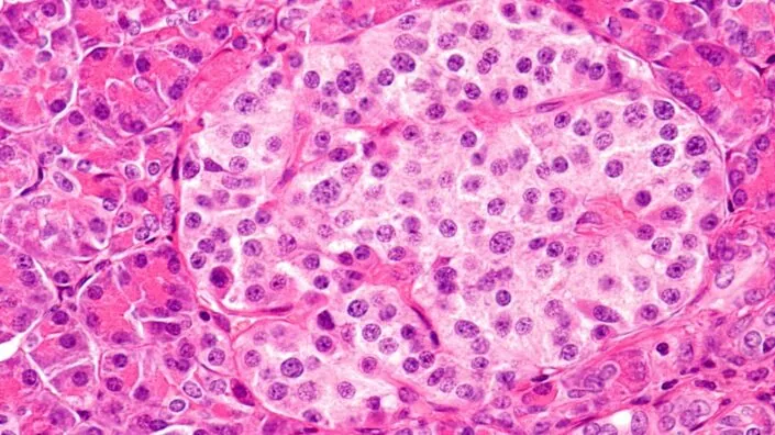
First, the scientists confirmed that human clusters of pancreatic endocrine cells (islets), including β-cells, from type 2 diabetes patients bear signs of mitochondrial dysfunction. Beta cells, but no other types of cells, from these patients had less mitochondrial DNA (mtDNA) and lower expression of 11 of 13 mitochondrial resident genes than in healthy controls. Mitophagy, the process of discarding malfunctioning mitochondria, was impaired as well.

These findings pointed at serious problems with the mechanisms of mitochondrial quality control. Interestingly, cells from simply obese donors or donors with insulin resistance did not show the same level of mitochondrial dysfunction, suggesting that this mitochondrial quality control loss was specific to diabetes patients.

How immature of you, β-cells

To see if impairing mitochondrial quality control can induce β-cell failure, the researchers engineered three mouse models with different mitochondrial pathways rendered deficient. The first model featured deletion of CLEC16A, a regulator of mitophagy, the second had reduced mtDNA content due to loss of TFAM, a regulator of mitochondrial genome integrity, and in the third, Mitofusins 1 and 2, proteins that promote mitochondrial fusion, were knocked out.

In all three models, messing with mitochondria triggered the integrated stress response (ISR). ISR is a cellular signaling network that is activated across various cell types to manage stress and maintain homeostasis by tweaking protein production. However, when persistently engaged, it can negatively impact cellular function. As they dug deeper to discover exactly how, the researchers received a surprise.

Apparently, mitochondria-to-nucleus (retrograde) ISR signaling dampened the expression of transcription factors that are central for β-cell maturity, identity, and function. As a result, the affected β-cells became less differentiated than their healthy counterparts.

“We wanted to determine which pathways are important for maintaining proper mitochondrial function,” said Dr. Emily M. Walker, a research assistant professor of internal medicine and first author of the study. “In all three cases, the exact same stress response was turned on, which caused β-cells to become immature, stop making enough insulin, and essentially stop being β-cells.”

Jamming the signal brings the cells back

Diabetes affects other metabolic tissues, such as liver tissue, muscle, and fat. To investigate further, the researchers ran similar experiments in mouse models of impaired mitochondrial quality control in liver cells (hepatocytes) and brown fat cells (adipocytes), with similar results.

“Diabetes is a multi-system disease: you gain weight, your liver produces too much sugar, and your muscles are affected. That’s why we wanted to look at other tissues as well,” said Scott A. Soleimanpour, M.D., director of the Michigan Diabetes Research Center and senior author of the study. “Although we haven’t tested all possible cell types, we believe that our results could be applicable to all the different tissues that are affected by diabetes.”

Can this be fixed? Several years ago, a potent ISR blocker called ISRIB was discovered and is currently in several clinical trials, including by Alphabet’s company Calico and the pharma giant AbbVie. The researchers treated mouse islets with ISRIB and found that it robustly restores β-cell identity markers specifically by inhibiting retrograde ISR signaling.

“Losing your β-cells is the most direct path to getting type 2 diabetes. Through our study we now have an explanation for what might be happening and how we can intervene and fix the root cause,” Soleimanpour said.

A possible basis for new treatments

Some other mitochondria researchers founded the study intriguing. “Using diet and genetic manipulations, the authors show the importance of robust mitophagy and retrograde signaling from mitochondria to nucleus in the involvement of mitochondrial function in type 2 diabetes,” said Dr. Amutha Boominathan, head of mitochondria research at the Longevity Research Institute. Boominathan and her team recently published an exciting study on nuclear expression of mitochondrial genes.

“What is interesting,” she added, “is that the authors find converging pathways in the β-Clec16aKO, β-Mfn1/2DKO and the Tfam-deficient mice in triggering the mitochondrial ISR influencing tissue specific blockade in cell differentiation not only in pancreas but also in other metabolic tissues such as liver and adipose tissues. The authors systematically address the causality for the role mitochondria play in age-associated metabolic diseases such as type 2 diabetes.”

Dr. Spring Behrouz, CEO of Vincere Biosciences, a mitochondria-targeting longevity biotech company, was impressed by the new study as well. “Reduced mtDNA levels, disrupted mitochondrial structure, and impaired mitophagy in metabolic tissues are often seen as secondary effects of other factors in type 2 diabetes,” she said. “However, this data suggests that early mitochondrial dysfunction actively contributes to the disease and potentially impacts other tissues.”

According to Behrouz, the study’s findings might be important for developing new mitochondria-based therapies. “By demonstrating the impact of mitochondrial quality control in metabolic tissue identity, this research opens up entirely new possibilities for treatment of diabetes as well as a range of other metabolic disorders,” she said.

We would like to ask you a small favor. We are a non-profit foundation, and unlike some other organizations, we have no shareholders and no products to sell you. All our news and educational content is free for everyone to read, but it does mean that we rely on the help of people like you. Every contribution, no matter if it’s big or small, supports independent journalism and sustains our future.

Literature

[1] Walker, E. M., Pearson, G. L., Lawlor, N., Stendahl, A. M., Lietzke, A., Sidarala, V., … & Soleimanpour, S. A. (2025). Retrograde mitochondrial signaling governs the identity and maturity of metabolic tissues. Science, eadf2034.

[2] Srivastava, S. (2017). The mitochondrial basis of aging and age-related disorders. Genes, 8(12), 398.

[3] Kwak, S. H., Park, K. S., Lee, K. U., & Lee, H. K. (2010). Mitochondrial metabolism and diabetes. Journal of diabetes investigation, 1(5), 161-169.

Pancreas

Creating a Functional Pancreas From Human Cells

In Cell Reports Medicine, researchers have described how they created a fully functional pancreas made from human cells and found it to work in mice.

A new era of organ replacement

In their introduction, the researchers discuss the well-known problems with insulin injections to treat Type 1 diabetes: the sort of constant monitoring that is required is difficult for patients to consistently comply with [1], and daily manual injections can’t adequately simulate the responsiveness of pancreatic tissue [2]. Direct injection of beta islet cells, which produce insulin, are limited by donor organs and require the immune system to be suppressed [3].

More modern techniques recognize that the extracellular matrix (ECM) governs a large part of how stem cells differentiate [4], and the effects of the ECM on the pancreas have been investigated in detail [5]. This led to a rapid increase in the work being done in this area, with ECM structures being created for the purpose [6]. The researchers of this paper had created a functional pancreas with insulin-producing cells from pigs, and it was functional in mice [7].

Therefore, the next step was to use cells derived from human beings.

Constructed organs are more effective than previous approaches

The researchers used two separate kinds of cells derived from human induced pluripotent stem cells (iPSCs): insulin-producing islet cells (SC-islets) and endothelial cells (iECs), which line the walls of arteries and veins. First, the researchers brought together these cells in a 9-to-1 ratio in order to produce spheroids (ViβeSs). Then, they populated decellularized rat lung tissue with more iECs and let them grow for two days, and finally, they injected this ready tissue with ViβeSs and more iECs to promote blood vessel formation (vascularization), creating a vascularized endothelia pancreas built from iPSCs: an iVEP.

iVEPs  

This approach worked. The injected ViβeSs did not come loose from the structure; instead, the iECs formed vascular tissue within the decellularized lungs, providing them with stability and the blood flow that they need to perform their duties. The iVEP structure was found to grant significant improvements to the cells’ survival and responsiveness, with the cells producing more insulin under high-glucose conditions.

In immunocompromised diabetic mice, the iVEP structure also performed much better than ViβeSs put into a a pre-vascularized pouch under the skin. In only two out of the thirteen mice given the latter, normal glycemia was established within a month; this happened in all the mice given iVEPs. Half of the iVEP-receiving mice had normal glycemia within two weeks of transplantation.

Unsurprisingly, removing the iVEPs from these mice led to diabetes within a week. An exaination of these structures revealed extensive vascular connections: the mice had successfully integrated the iVEPs into their bodies. Further investigation found that iECs were necessary in the creation of iVEPs; without endothelial cells, the structures fail to properly integrate into the vascular structure.

Decellularized, vascularized structures as a way forward

With these results in hand, the researchers compared their iVEP approach to previous work. They hold that their approach to vascularization improves many fundamental aspects of cellular development: for example, they note that by themselves, islet cells generated from iPSCs require 20 days to reach a mature and effective phenotype [8]. Meanwhile, in iVEPs, the cells require only a week to reach this phenotype.

While the researchers note that their approach is more complicated than products that are already in clinical trials, they believe that it will make for a better product. However, their existing scaffolds, derived from rats, are far too small to use in people. They plan to use pig organs instead, and they hope to use hypo-allergenic cells that completely get rid of the risk of immune rejection and the need for potentially risky immunosuppressants.

The researchers developed this approach to treat type 1 diabetes, but this work has implications for many other diseases, many of which are age-related. While replacing the pancreas cannot heal the insulin resistance inherent in type 2 diabetes, it may be a viable strategy for long-term loss of function. Similarly, this technology can potentially be applied to many other organs, including the lungs and heart. While wholesale replacement of human organs with bioengineered equivalents is still not on the table, this technology continues to advance to the clinic.

We would like to ask you a small favor. We are a non-profit foundation, and unlike some other organizations, we have no shareholders and no products to sell you. All our news and educational content is free for everyone to read, but it does mean that we rely on the help of people like you. Every contribution, no matter if it’s big or small, supports independent journalism and sustains our future.

Literature

[1] Beck, R. W., Bergenstal, R. M., Laffel, L. M., & Pickup, J. C. (2019). Advances in technology for management of type 1 diabetes. The Lancet, 394(10205), 1265-1273.

[2] Piemonti, L. (2021). Felix dies natalis, insulin… ceterum autem censeo “beta is better”. Acta Diabetologica, 58(10), 1287-1306.

[3] Pepper, A. R., Bruni, A., & Shapiro, A. J. (2018). Clinical islet transplantation: is the future finally now?. Current opinion in organ transplantation, 23(4), 428-439.

[4] Hogrebe, N. J., Augsornworawat, P., Maxwell, K. G., Velazco-Cruz, L., & Millman, J. R. (2020). Targeting the cytoskeleton to direct pancreatic differentiation of human pluripotent stem cells. Nature biotechnology, 38(4), 460-470.

[5] Berger, C., Bjørlykke, Y., Hahn, L., Mühlemann, M., Kress, S., Walles, H., … & Zdzieblo, D. (2020). Matrix decoded–A pancreatic extracellular matrix with organ specific cues guiding human iPSC differentiation. Biomaterials, 244, 119766.

[6] Peloso, A., Urbani, L., Cravedi, P., Katari, R., Maghsoudlou, P., Fallas, M. E. A., … & Orlando, G. (2016). The human pancreas as a source of protolerogenic extracellular matrix scaffold for a new-generation bioartificial endocrine pancreas. Annals of surgery, 264(1), 169-179.

[7] Citro, A., Neroni, A., Pignatelli, C., Campo, F., Policardi, M., Monieri, M., … & Piemonti, L. (2023). Directed self-assembly of a xenogeneic vascularized endocrine pancreas for type 1 diabetes. Nature Communications, 14(1), 878.

[8] Velazco-Cruz, L., Song, J., Maxwell, K. G., Goedegebuure, M. M., Augsornworawat, P., Hogrebe, N. J., & Millman, J. R. (2019). Acquisition of dynamic function in human stem cell-derived β cells. Stem cell reports, 12(2), 351-365.

Blood cells

Some Stem Cells Remain Youthful With Age

A team of scientists has discovered that some hematopoietic stem cells (HSCs) lose their ability to differentiate into useful somatic cells and that removing those bad HSCs is beneficial.

Blood creation diminishes with age

Hematopoiesis refers to the production of blood cells, both white and red. HSCs, which create these blood cells, are known to change with aging, developing mutations and losing the ability to perform their basic function [1]. Unsurprisingly, replacing the HSCs of older animals with those of younger animals imcreases lifespan [2] and putting older HSCs into younger animals decreases it [3].

HSCs age in several ways: genetic mutation is a crucial part [4], but epigenetic aging leading to altered gene expression [5] and mitochondrial changes [6] are also key factors. Some HSCs, however, remain quiescent, retaining their intrinsic abilities [7]. This work, therefore, focuses on determining which cells in aged animals retain useful abilities and which do not.

Younger stem cells perform better

In their first experiment, the researchers began with a population of young mice that had been lethally irradiated, killing all of their natural HSCs, then transplanting both young and old HSCs into the same mice. As expected, the older HSCs did not repopulate the bone marrow nearly as much as the younger HSCs did.

The researchers then transplanted either young or old HSCs into lethally irradiated, middle-aged (13-month-old) mice. These HSCs had a great many differences in the kinds of blood cells into which they differentiated: toung HSCs were more likely to differentiate into B cells, while old HSCs were more likely to differentiate into T cells and myeloid immune cells. However, the mice given younger HSCs had far more white blood cells and more robust immune systems in total.

As expected, the older HSCs led to epigenetically older blood, and mice given younger HSCs significantly outperformed mice given older HSCs on every metric that the researchers tested, including strength, balance, endurance, and fear conditioning.

Looking for the good ones

The researchers then performed RNA sequencing of both young and old HSCs. The gene expression of younger HSCs was largely similar between them, but old HSCs had significant distinguishing features, to the point that the quiescent cells were able to be clustered into three distinct groups. Surprisingly, many of the genes that are upregulated with aging were not upregulated in the third group (q3). Instead, the gene expression of this group was a lot more, although not entirely, like the gene expression of the young HSCs.

However, the researchers needed a good way to quickly determine which cells were in q3, looking for a marker that is readily identifiable with antibodies. They found that the surface marker CD150 increases with age-related gene expression markers but does not increase in the q3 cells.

This information was used to create distinct populations of aged cells, some with low CD150 and others with high CD150. Using their lethally irradiated young mice, the researchers determined that the cells had far different capabilities. The cells that were high in CD150 could proliferate but could not differentiate into functional cells. Genes related to stem cells activation were functional; the CD150-high cells simply could not create the basic blood cells that the mice needed.

On the other hand, the cells that were low in CD150 were able to do this, creating far more multipotent cells that led to the downstream creation of red and white blood cells. The researchers gave irradiated, 13-month-old mice cells that were derived from older donors but were separated to haave less CD150. These mice trended towards having better blood cell measurements than similar mice given unseparated HSCs. Mice that were only given cells high in CD150 performed much worse than either group, and there, the differences were statistically significant.

Similarly, the mice given CD150-low cells performed much better than the mice given CD150-high cells, with the mice given unseparated cells being in the middle. Epigenetically, the blood cells of the mice given CD150-low cells were found to be significantly younger. Most importantly, the mice given the CD150-low cells lived noticeably longer.

CD150 Survival

The researchers did not directly test the removal of CD150-high cells from naturally aged, unirradiated mice. However, their work shows that this may be a viable prospect. This, therefore, would be the next logical step to conduct, and if that is found to be viable and safe, the step after could be to test such an approach in people.

We would like to ask you a small favor. We are a non-profit foundation, and unlike some other organizations, we have no shareholders and no products to sell you. All our news and educational content is free for everyone to read, but it does mean that we rely on the help of people like you. Every contribution, no matter if it’s big or small, supports independent journalism and sustains our future.

Literature

[1] Jaiswal, S., & Ebert, B. L. (2019). Clonal hematopoiesis in human aging and disease. Science, 366(6465), eaan4673.

[2] Guderyon, M. J., Chen, C., Bhattacharjee, A., Ge, G., Fernandez, R. A., Gelfond, J. A., … & Li, S. (2020). Mobilization‐based transplantation of young‐donor hematopoietic stem cells extends lifespan in mice. Aging Cell, 19(3), e13110.

[3] Leins, H., Mulaw, M., Eiwen, K., Sakk, V., Liang, Y., Denkinger, M., … & Schirmbeck, R. (2018). Aged murine hematopoietic stem cells drive aging-associated immune remodeling. Blood, The Journal of the American Society of Hematology, 132(6), 565-576.

[4] Moehrle, B. M., & Geiger, H. (2016). Aging of hematopoietic stem cells: DNA damage and mutations?. Experimental Hematology, 44(10), 895-901.

[5] Sun, D., Luo, M., Jeong, M., Rodriguez, B., Xia, Z., Hannah, R., … & Goodell, M. A. (2014). Epigenomic profiling of young and aged HSCs reveals concerted changes during aging that reinforce self-renewal. Cell stem cell, 14(5), 673-688.

[6] Mansell, E., Sigurdsson, V., Deltcheva, E., Brown, J., James, C., Miharada, K., … & Enver, T. (2021). Mitochondrial potentiation ameliorates age-related heterogeneity in hematopoietic stem cell function. Cell Stem Cell, 28(2), 241-256.

[7] Foudi, A., Hochedlinger, K., Van Buren, D., Schindler, J. W., Jaenisch, R., Carey, V., & Hock, H. (2009). Analysis of histone 2B-GFP retention reveals slowly cycling hematopoietic stem cells. Nature biotechnology, 27(1), 84-90.

Phoenix Aerie logo

Phoenix Aerie: The Launchpad for Longevity Pioneers

Phoenix Aerie (P//A), the first-ever co-living house specifically dedicated to enriching, uplifting, and empowering young longevity pioneers, will be launching. Designed to catalyze breakthroughs in longevity-related biotech, implementation, and communication from a variety of angles, P//A offers a unique environment where emerging leaders live, learn, and grow together in the heart of the Bay Area’s thriving biotech ecosystem.

Why Phoenix Aerie?

As the longevity industry experiences unprecedented growth, there’s a critical need for spaces that nurture the next generation of people working for lasting innovation and implementation in the field—from myriad backgrounds and disciplines. Phoenix Aerie fills this gap by creating an intellectually vibrant, collaborative environment that accelerates innovation beyond traditional academic settings. Residents gain access to mentorship, networking opportunities, and an ecosystem that fuels both personal and professional growth.

Longevity work isn’t just about extending life—it’s about expanding the potential of human health. Phoenix Aerie exists to empower the minds that will lead this charge. We are creating a 24-7 launchpad where bold ideas and transformative collaborations will thrive.

Early Momentum and Growing Impact

Phoenix Aerie (P//A) has captured attention within the longevity community as it builds towards its inaugural cohort. P//A was recently featured in The SF Standard as part of the Bay Area’s burgeoning longevity movement and has received support from LongX, a leader in fostering youth-driven biotech initiatives.

A Call for Strategic Partnerships and Support

Phoenix Aerie is actively seeking sponsors, funders, and strategic partnerships to support its mission. Investors, nonprofits, and corporate leaders have the unique opportunity to be part of a groundbreaking initiative that cultivates future leaders in longevity science and biotech. Further, P//A’s plan for scalable growth and long-term impact makes it a pivotal force in shaping the future of health and aging.

About Phoenix Aerie

Phoenix Aerie is a co-living community for brilliant young thinkers and doers passionate about longevity, uniquely designed to foster interdisciplinary collaboration, rapid idea exchange, and direct access to the Bay Area’s vibrant biotech ecosystem. We foster and embolden the exploration of innovative ideas through curiosity and discourse. Phoenix Aerie’s core belief is that well-rounded groups make well-rounded solutions – which make lasting impacts. Phoenix Aerie isn’t just a place to live; it’s where bold ideas take root, driving innovations that will shape the future of human health and longevity.

Media Contact:

Hudson Eaton

Founder, Phoenix Aerie

Email: hudson@phoenixaeries.com

Website: www.phoenixaeries.com

We would like to ask you a small favor. We are a non-profit foundation, and unlike some other organizations, we have no shareholders and no products to sell you. All our news and educational content is free for everyone to read, but it does mean that we rely on the help of people like you. Every contribution, no matter if it’s big or small, supports independent journalism and sustains our future.

Sprague-Dawley Rat

How the Yamanaka Factors Affect Female Reproduction in Rats

The authors of a recent study reported that OSKM/Yamanaka factor gene therapy in rats results in higher fertility at an older age compared to controls and allows older rats to have regular cycles [1].

Rejuvenating Yamanaka factors

Multiple studies have suggested that gene therapy that uses the Yamanaka factors has profound regenerative and lifespan-extending potential. For example, David Sinclair’s lab has reversed glaucoma in mice using three of the four factors.

Based on this evidence, the authors of this paper designed a gene therapy that used an adenovirus to carry these four factors. The researchers chose the hypothalamus, a brain structure that is a part of the hypothalamic-pituitary-gonadal axis and is essential for many functions, including reproduction [2]. The goal of this long-term gene therapy was to slow down fertility decline.

Decreasing reproductive capacity

A human female’s reproductive system ages and fails to function correctly much earlier than other systems, with females spending a significant portion of their lives in the post-menopausal stage. A female rat’s reproductive capacity decreases in middle age, and hormonal changes lead to changes in estrous cyclicity, which is the rat’s equivalent of the menstrual cycle [3, 4].

This group of researchers had previously demonstrated that insulin-like growth factor-I (IGF-I) gene therapy, targeted to the hypothalamus and started at 8 months of age, could extend rats’ regular cycles beyond the age of 10 months, which is when untreated rats’ cycles become irregular. At 11 months, treatment with that gene therapy also “preserves the integrity of ovarian structure.” In contrast, age-matched controls mostly didn’t have cycles, and a high percentage had polycystic ovaries [5].

Extended regular cycles

In this study, the researchers used female Sprague-Dawley rats and divided them into groups of 12 animals each. At four months of age, they injected an specifically modified virus (adenoviral vector) carrying either OSKM and green fluorescent protein (GFP) genes or only GFP as a control into the rodents’ hypothalami, spurring the production of proteins. Then, they observed how the rat’s cycles changed as the animals aged.

The control group of rats had the typical age-related changes to their estrous cycles. The young rats had regular estrous cycles, which last for 4-5 days and have four stages: proestrus, estrus, metestrus, and diestrus.

This regularity changed around 9 months of age, when cycles became more irregular. Starting at 10 months of age, the researchers observed the prevalence of constant estrus status and the presence of numerous fluid-filled sacs (follicular cysts) in the ovaries. Past 20 months of age, female rats transitioned into the constant diestrus phase.

OSKM Rat Fertility 1

OSKM treatment impacted these cycles, and the OSKM-treated animals continued to have regular cycles at 10 months old.

Impact on fertility

The animals were mated for one week with a young male rat. The mating occurred almost half a year after the gene therapy. The authors point out that even though gene expression should still occur, the expression levels are most likely lower at the time of mating compared to the time of injection.

The researchers also admit that they expected viral vectors carrying OSKM to reach only a small proportion of hypothalamic cells. However, this was enough to impact the rate of reproductive aging.

The young group had the highest pregnancy rate at 83%, and the old control group had almost exactly a tenth of that, at 8.3%. The OSKM-treated group had a pregnancy rate of 25%, which, while lower than that of the young group, was still improved compared to the age-matched control group. However, it was not statistically significantly different (p=0.06552). An increase in the size of the experimental cohort would be beneficial in obtaining statistically significant and more robust experimental proof, but these results show a very positive trend.

The younger group also had a larger litter size (mean litter size 9.1 pups), while the mean litter size of the older animals, both control and OSKM-treated, was three pups. Pups from all groups survived and showed normal behavior.

The researchers observed differences in body weight at birth. It was lower for pups born to young mothers but similar in both groups of older mothers. However, there were differences between the pups from the older mothers at the time of weaning. The pups from the OSKM-treated animals gained more weight than the old control animals’ pups, suggesting that age affects milk supply. However, OSKM treatment can remedy the lack of milk supply and/or increase milk quality.

The weight of the pups from younger mothers remained lower, which was possibly caused by the larger litter size and more animals that the mother needed to feed.

OSKM Rat Fertility 2

A long road to optimizing fertility

The researchers believe that achieving regular cycles, and a 25% fertility rate in rats that are close to the cessation of their reproductive span, shows that this OSKM gene therapy significantly benefited the animals’ reproductive system; however, the fertility rate was lower than in young animals, suggesting that “regular ovulation is a necessary but not sufficient condition for keeping the rats at optimal fertility levels.” There are also other components of the reproductive system that impact fertility.

The researchers of this study were unable to assess any changes in hypothalamic DNA methylation following OSKM gene therapy. However, they expect that, as in previous work, OSKM treatment led to the reversal of age-related epigenetic changes.

The researchers suggest that this kind of therapy might be used to help extend women’s reproductive span. However, given that this research was done in rats, the impact and side effects of such a therapy on human beings need to be extensively studied in the future.

We would like to ask you a small favor. We are a non-profit foundation, and unlike some other organizations, we have no shareholders and no products to sell you. All our news and educational content is free for everyone to read, but it does mean that we rely on the help of people like you. Every contribution, no matter if it’s big or small, supports independent journalism and sustains our future.

Literature

[1] Gallardo, M. D., Girard, M., Portiansky, E. L., & Goya, R. G. (2025). Oct4, Sox2, Klf4, c-My (OSKM) gene therapy in the hypothalamus prolongs fertility and ovulation in female rats. Aging, null, 10.18632/aging.206191. Advance online publication.

[2] Neal-Perry, G., Nejat, E., & Dicken, C. (2010). The neuroendocrine physiology of female reproductive aging: An update. Maturitas, 67(1), 34–38.

[3] Neal-Perry, G. S., Zeevalk, G. D., Santoro, N. F., & Etgen, A. M. (2005). Attenuation of preoptic area glutamate release correlates with reduced luteinizing hormone secretion in middle-aged female rats. Endocrinology, 146(10), 4331–4339.

[4] Huang, H. H., Marshall, S., & Meites, J. (1976). Capacity of old versus young female rats to secrete LH, FSH and prolactin. Biology of reproduction, 14(5), 538–543.

[5] Rodríguez, S. S., Schwerdt, J. I., Barbeito, C. G., Flamini, M. A., Han, Y., Bohn, M. C., & Goya, R. G. (2013). Hypothalamic IGF-I gene therapy prolongs estrous cyclicity and protects ovarian structure in middle-aged female rats. Endocrinology, 154(6), 2166–2173.

Down arrows

Early Adult Mortality Remains High in the US

Mortality among Americans aged 25-44 has risen substantially between 2011 and 2023, a new study has found, and it remains high even after having passed the COVID-related peak [1].

Riches to rags

Despite the US being one of the world’s richest countries, Americans’ average life expectancy has been lagging behind that of comparable countries by more than four years (even more when compared to top performers such as Spain and Japan). The gap has widened substantially starting at around 2010, when US life expectancy growth ground to a halt and later took an unusually hard hit during the COVID-19 pandemic (interestingly, it was followed by a robust rebound in 2022 and 2023).

Part of this multifaceted phenomenon is increased mortality in young adults. While much of it has been driven by an epidemic of illegal drugs, deaths of natural causes such as cancer have also skyrocketed. This trend worries researchers and policymakers and is being actively investigated.

Not just COVID

In a new study that originated from the University of Minnesota and Boston University and was published in JAMA Open Network, the researchers took an unusually long perspective to analyze the dynamics of excess deaths in American adults aged 25-44. The team used 2010 as their baseline and defined excess mortality as that exceeding extrapolation of 1999-2010 trends.

Having analyzed 3,392,364 deaths, they found that excess mortality per capita in this category began to rise well before the pandemic. In 2019, it was 34.6% higher than expected (i.e., had pre-2011 trends continued).

Excess mortality

The pandemic brought another sharp increase in mortality, two major drivers being COVID-19 and drug overdoses. However, deaths from multiple other causes, including cancer, circulatory problems, and metabolic diseases, increased as well. Some of these may be connected to COVID, as studies have linked COVID infection to various complications such as cardiovascular conditions [2], or substance abuse. As a result, in 2021, all-cause excess mortality was almost three times what it had been in 2019 (116.2 vs 41.7 deaths per 100,000 population).

A complex phenomenon

Hinting at this interconnectedness is the fact that after 2021, excess mortality began to fall in several categories, such as cardiometabolic conditions. However, even for these causes, excess mortality remains much higher than pre-pandemic levels. All-cause early adult mortality was 70.0% higher in 2023 than it would have been had pre-2011 trends continued. In absolute numbers, this excess mortality translates into 71,124 deaths annually.

This might be partially due to the long-lasting effects of COVID, which are only beginning to attract researchers’ attention (“long COVID” and other complications). Another possible explanation is that drug- and alcohol-related deaths are still very high compared to a decade ago. Substance abuse is associated with multiple health hazards and might drive other causes of death.

The top five causes of death that collectively accounted for nearly three-quarters of the excess mortality in 2023 were drug poisoning (31.8% of excess mortality), the residual natural-cause category (16.0%), transport-related deaths (14.1%), alcohol-related deaths (8.5%), and homicide (8.2%). “Additionally, the combined contribution of cardiometabolic conditions, including circulatory and endocrine, metabolic, and nutritional, was substantial (9.2%),” the paper notes.

“The rise in opiate deaths has been devastating for Americans in early and middle adulthood,” said Elizabeth Wrigley-Field, lead author and an associate professor in the University of Minnesota College of Liberal Arts and Institute for Social Research and Data Innovation. “What we didn’t expect is how many different causes of death have really grown for these early adults. It’s drug and alcohol deaths, but it’s also car collisions, it’s circulatory and metabolic diseases – causes that are very different from each other. That tells us this isn’t one simple problem to fix, but something broader.”

“Our findings underscore the urgent need for comprehensive policies to address the structural factors driving worsening health among recent generations of young adults,” said author Andrew Stokes of Boston University. “Solutions may include expanding access to nutritious foods, strengthening social services and increasing regulation of industries that affect public health.”

Cancer is on the rise

While most natural causes are declining, cancer is an outlier, with excess mortality at an all-time high. A large-scale study in The Lancet last year found that cancer prevalence has increased in young compared to older cohorts, particularly in Generation Xers and millennials, for 17 out of 34 cancer types [3]. Alarmingly, cancer is often more aggressive in younger people.

This hike in cancer incidence has happened despite a declining prevalence of smoking and some other unhealthy behaviors. On the other hand, factors such as alcohol consumption, obesity, ultra-processed food consumption, and environmental pollution remain a challenge. A recent advisory from the US Surgeon General warns that alcohol is a major risk factor for cancer.

However, early detection might also have contributed to the rates of cancer in earlier cohorts. Some slow-growing cancers might be detectable now at earlier ages, affecting the statistic.

The lost Americans

Interestingly, the US was not always behind its peers in life expectancy. In 2023, a study by the University of Boston scientists aptly named “Missing Americans: Early death in the United States” stated that “The United States had lower mortality rates than peer countries in the 1930s-1950s and similar mortality in the 1960s and 1970s. Beginning in the 1980s, however, the United States began experiencing a steady increase in the number of missing Americans, reaching 622,534 in 2019 alone.” [4] The situation got much worse during the COVID-19 pandemic.

The authors of that study attributed the growing discrepancy to the lack of large-scale public health initiatives in the US. “While COVID-19 brought new attention to public health, the backlash unleashed during the pandemic has undermined trust in government and support for expansive policies to improve population health,” said the study’s lead and corresponding author Jacob Bor, associate professor of global health and epidemiology, at the time. “This could be the most harmful long-term impact of the pandemic, because expansion of public policy to support health is exactly how our peer countries have attained higher life expectancy and better health outcomes.”

We would like to ask you a small favor. We are a non-profit foundation, and unlike some other organizations, we have no shareholders and no products to sell you. All our news and educational content is free for everyone to read, but it does mean that we rely on the help of people like you. Every contribution, no matter if it’s big or small, supports independent journalism and sustains our future.

Literature

[1] Wrigley-Field, E., Raquib, R. V., Berry, K. M., Morris, K. J., & Stokes, A. C. (2025). Mortality Trends Among Early Adults in the United States, 1999-2023. JAMA Network Open, 8(1), e2457538-e2457538.

[2] Lee, C. C., Ali, K., Connell, D., Mordi, I. R., George, J., Lang, E. M., & Lang, C. C. (2021). COVID-19-associated cardiovascular complications. Diseases, 9(3), 47.

[3] Sung, H., Jiang, C., Bandi, P., Minihan, A., Fidler-Benaoudia, M., Islami, F., … & Jemal, A. (2024). Differences in cancer rates among adults born between 1920 and 1990 in the USA: an analysis of population-based cancer registry data. The Lancet Public Health, 9(8), e583-e593.Chicago

[4] Bor, J., Stokes, A. C., Raifman, J., Venkataramani, A., Bassett, M. T., Himmelstein, D., & Woolhandler, S. (2023). Missing Americans: early death in the United States—1933–2021. PNAS nexus, 2(6), pgad173.

New Nanoparticles for Treating Arthritis

In the Journal of Nanobiotechnology, researchers have described a new method of delivering a long-lasting treatment into cartilage.

A protein that promotes autophagy

Previous work has linked expression of the FGF18 protein with healthy cartilage and joints [1]. Problems with the gene responsible for FGF18 lead to osteoarthritis [2], and it has been found to be important alongside arthritis therapies, such as hydrogels that form a lattice for cartilage growth [3]. This is because FGF18 is known to have positive effects on the FOXO3 pathway, which stimulates autophagy, the cellular self-consumption process that removes unwanted and harmful components [4].

However, using a protein as a cartilage treatment has its own problems. Recombinant proteins directly delivered into tissue don’t last very long [5], and even mRNA-based therapies are vulnerable to rapid degradation in the human body [6]. To combat this, the researchers have chosen lipid nanoparticles (LNPs), which encapsulate the mRNA in order to deliver it into cells [7].

The researchers first confirmed the existence of a link between FGF18 and osteoarthritis. A broad gene expression database has reported that elderly people have only a quarter the FGF18 of young people. Tissue samples have revealed that people who undergo total knee arthroplasty have their FGF18 reduced by half. Similarly, mice that have had arthritis artificially induced by meniscus destabilization, as well as naturally aged mice, have approximately half the FGF-18-positive cells of healthy young mice.

Exposing cartilage-generating cells (chondrocytes) to an inflammatory environment characterized by TNF-α resulted in FGF18 expression being reduced to a fourth of its normal value. Driving chondrocytes senescent by exposing them to hydrogen peroxide reduced FGF18 to two-fifths of its normal value.

Effective delivery and therapeutic effects

LNP FGF18 Action

The mRNA delivery appeared to be effective. A cellular examination showed no toxicity to chondrocytes even at high concentrations. Unlike recombinant FGF18, the LNP-encapsulated mRNA nanoparticles penetrated relatively deeply into the cartilage of both young and old mice. The nanoparticles were just small enough to fit within the pores of the mice’s collagen networks, even the dense, cross-linked collagen of older animals.

Using a bioluminescent reporter, the researchers found out that this LNP treatment stays confined to where it needs to be and does not migrate to other organs, such as the liver, in appreciable amounts. Instead, it stays within the knee joint for approximately six days, and its effects diminish far slower than mRNA without LNP encapsulation. The LNP-mRNA was found to successfully cause cells to express significant amounts of the FGF18 protein.

This approach had significant beneficial effects in a cellular culture. Cellular senescence induced by the inflammatory cytokine IL-1β was cut approximately in half, as measured by p16, p21, p53, and SA-β-gal staining. Proliferation was approximately doubled as well. LNP-mRNA for FGF18 had very similar effects to pure FGF18 in this cellular experiment.

Autophagy was similarly upregulated. FOXO3 is downregulated when chondrocytes are exposed to IL-1β, but the LNP-mRNA was found to restore it nearly to the level of the control group. Cells that were only exposed to LNP-mRNA without IL-1β had even higher levels of FOXO3. This led to an increase of cartilage-producing proteins, and further experiments confirmed that this was due to an increase in autophagy.

After confirming its effects in cells, the researchers turned to mice: a control group, a group with a destabilized meniscus and no treatment, a group treated with FGF18 protein every week, and a group treated with LNP-mRNA every week. The damaged, untreated group was hypersensitive to pain, which was partially ameliorated by FGF18 and slightly moreso by the LNP treatment, although all of the damaged mice gradually got more sensitive to pain over eight weeks.

The LNP treatment was also found to benefit the mice’s gait and physical biomarkers, in both the destabilized meniscus model and in naturally aged mice. In many of the tests, there were no significant differences between FGF18-treated and LNP-treated mice, but there were some benefits to the new approach.

Most notably, the cartilage of the LNP-treated mice was significantly thicker, restoring the cartilage of damaged mice nearly to that of undamaged mice and, most critically, restoring the cartilage of aged mice nearly to that of young mice. A closer investigation found that the LNP injection was having the same effects in the mice as in the cellular culture, restoring proliferative capacity to the mice’s chondrocytes.

This is not a human study, but it appears that human trials are the next logical step for this approach, as it appears to be both safe and effective in animal models. Time will tell whether this particular LNP approach will be tested for the clinic or if it will undergo further refinement first.

We would like to ask you a small favor. We are a non-profit foundation, and unlike some other organizations, we have no shareholders and no products to sell you. All our news and educational content is free for everyone to read, but it does mean that we rely on the help of people like you. Every contribution, no matter if it’s big or small, supports independent journalism and sustains our future.

Literature

[1] Davidson, D., Blanc, A., Filion, D., Wang, H., Plut, P., Pfeffer, G., … & Henderson, J. E. (2005). Fibroblast growth factor (FGF) 18 signals through FGF receptor 3 to promote chondrogenesis. Journal of Biological Chemistry, 280(21), 20509-20515.

[2] Boer, C. G., Hatzikotoulas, K., Southam, L., Stefánsdóttir, L., Zhang, Y., de Almeida, R. C., … & Wilkinson, J. M. (2021). Deciphering osteoarthritis genetics across 826,690 individuals from 9 populations. Cell, 184(18), 4784-4818.

[3] Gothard, D., Rotherham, M., Smith, E. L., Kanczler, J. M., Henstock, J., Wells, J. A., … & Oreffo, R. O. (2024). In vivo analysis of hybrid hydrogels containing dual growth factor combinations, and skeletal stem cells under mechanical stimulation for bone repair. Mechanobiology in Medicine, 2(4), 100096.

[4] Cinque, L., Forrester, A., Bartolomeo, R., Svelto, M., Venditti, R., Montefusco, S., … & Settembre, C. (2015). FGF signalling regulates bone growth through autophagy. Nature, 528(7581), 272-275.

[5] Evans, C. H., Kraus, V. B., & Setton, L. A. (2014). Progress in intra-articular therapy. Nature Reviews Rheumatology, 10(1), 11-22.

[6] Hajj, K. A., & Whitehead, K. A. (2017). Tools for translation: non-viral materials for therapeutic mRNA delivery. Nature Reviews Materials, 2(10), 1-17.

[7] Hou, X., Zaks, T., Langer, R., & Dong, Y. (2021). Lipid nanoparticles for mRNA delivery. Nature Reviews Materials, 6(12), 1078-1094.

Rejuvenation Roundup January 2025

Rejuvenation Roundup January 2025

As rejuvenation research advances from theory to practice, more therapies start making their way into the clinic. 2025 continues with both mouse experiments and human clinical trials.

Interviews

Matthew O'ConnorCyclarity Launches Human Trial to Cure Atherosclerosis: Recently, Cyclarity Therapeutics announced the launch of a Phase 1 human clinical trial for a drug that aims to remove the arterial plaques that lead to heart attacks and strokes. Its primary cyclodextrin drug candidate, UDP-003, focuses on 7-ketocholesterol, a type of oxidized cholesterol that increases in cells and tissues as people age.

Marco Quarta on Cellular Senescence in Aging: Dr. Marco Quarta runs one of the most interesting start-ups in the longevity field: Rubedo, which focuses on utilizing the senolytic approach to cellular senescence. This company has developed ingenious ways to cope with the notorious heterogeneity of senescent cells and is one of the first to bring its senolytic drug candidate into clinical trials.

Advocacy and Analysis

The Battle for Long Life Has Been Accomplished: What’s Next?: How long can people live? This is not just a foundational question in science. The answer has important public policy implications and is of interest to us all. Recent scientific evidence has revealed the answer, so what’s next in humanity’s never-ending battle agaiS. Jay Olshanskynst disease and the persistent ravages of aging?

Research Roundup

Keeping Stem Cells Healthy and Young: A team of researchers has outlined a new approach that uses mRNA to prevent senescence and strengthen mesenchymal stem cells (MSCs) against aging before they are transplanted into patients.

Human ear anatomyA Potential Gene Therapy for Hearing Loss: In JCI Insight, researchers have explored the possibility of using gene therapy to restore a crucial protein and repair hearing loss. The most critical finding is that the adult cochlea, which processes hearing, is in fact capable of being remodeled through changes in gene expression after birth.

Drinking and Dying: Alcohol as a Risk Factor for Cancer: A new advisory by the US Surgeon General highlights a topic that – as the document itself notes – has been flying mostly under the public’s radar: the relationship between alcohol consumption and cancer.

Caloric restrictionReceiving Caloric Restriction Benefits Without Practicing It: In a new study, researchers have found that lithocholic acid, a metabolite found in the serum of calorically restricted mice, can mimic the effects of caloric restriction. While their research was conducted on model systems, they point to a previous study that observed that this metabolte was observed to be increased in the serum of healthy humans following 36 hours of fasting.

Precision Targeting of Senescent Cells: In a journal called Small, researchers have described a new targeting mechanism for delivering senolytic compounds where they need to go. These molecules are encapsulated in tiny soap bubbles rather than silica-based nanoparticles.

Time-restricted eatingIntermittent Fasting Improves Coordination in Mice: Researchers have discovered that intermittent fasting increases myelin in aged mice, leading to better neural function and coordination. Normally, neuronal axons are coated in a protein sheath made of myelin, which is necessary for their proper function, but demyelination occurs in aging.

A Gut Metabolite Reduces Senescence and Inflammation: In a preprint study, scientists from Lifespan Research Institute and the Buck Institute for Research on Aging have published their findings that Urolithin A, a molecule that has garnered a lot of attention in the longevity field, potently reduces senescence-related markers in human fibroblasts.

Gut bacteriaThe Impact of a Human Breast Milk Probiotic on Sarcopenia: The authors of this study, citing evidence of a link between the gut microbiome, muscle health, and sarcopenia, investigated the effect of the consumption of a probiotic on the muscle health of sarcopenia patients.

Enhancing NAD+ Efficiency by Energizing Sirtuins: Researchers publishing in Physical Review X have discovered compounds that can double the efficiency of the sirtuin SIRT3 in processing NAD+. Unlike previous efforts, this drug does not rely on a substrate to function.

Genetic examinationNew Study Links Epigenetic Changes to Genetic Mutations: A new paper published in Nature Aging suggests that somatic mutations cause significant remodeling of the epigenetic landscape. The findings might be relevant to future anti-aging interventions.

Fighting Alzheimer’s by Helping Neurons Consume Proteins: Researchers have found that kinesin family member 9 (KIF9), a protein that diminishes with aging, is instrumental in allowing cells to consume harmful proteins and fights Alzheimer’s in a mouse model.

TelomeresMaintaining Telomeres Extends Lifespan in Mice: A recent study has found that the overexpression of telomerase reverse transcriptase (TERT), which is a subunit of telomerase, an enzyme essential for telomere maintenance, leads to lifespan extension in mice without significant side effects.

Maintaining Muscle by Restoring Gut Bacteria: In Aging Cell, researchers have described how different combinations of gut bacteria impact muscle strength in mice. The link between gut bacteria and health is well-documented, and multiple biomarkers have confirmed that a healthy gut leads to health elsewhere.

Targeting cancerNew Drug Eliminates Breast Cancer in Mouse Study: Researchers have discovered a small molecule that effectively kills cancer cells in the most prevalent type of breast cancer. The new drug could help against cancer recurrence and decrease the need for surgery.

Restoring Cellular Proliferation Through Exosomes: In Cell Metabolism, researchers have described how a microRNA (miRNA) derived from exosomes generated by human embryonic stem cells (hESCs) restores function and fights senescence in cell cultures and mice.

Ultrasound deviceUltrasound as a Tool to Eliminate Senescent Cells: A new study suggests that low-intensity pulsed ultrasound (LIPUS) can be beneficial in eliminating senescent cells through the recruitment and activation of immune cells. LIPUS is a technology that can be easily applied in the clinic.

Inhibiting a Fundamental Factor in Brain Inflammation: Researchers have devised a method of reducing brain inflammation by creating a long-lasting inhibitor of the inflammatory factor NF-κB. These researchers believe that it “may serve as a potent therapeutic agent against pathological age-related inflammatory processes, especially those that target macrophages and microglia.”

Printed heartArtificially Grown Tissue Repairs Heart Failure in Monkeys: German scientists have created lab-grown “patches” of heart muscle tissue derived from pluripotent stem cells. Following a success with rhesus monkeys, they have obtained approval for a human trial.

Reducing functionally defective old HSCs alleviates aging-related phenotypes in old recipient mice: This study demonstrates the presence of “younger” HSCs in old mice and that aging-associated functional decline can be mitigated by reducing dysfunctional HSCs.

Tenascin-C promotes bone regeneration via inflammatory macrophages: Taken together, this study reveals the regulation of macrophage recruitment and its function in the activation of skeletal stem cells after bone injury, providing a strategy to accelerate bone regeneration by TNC delivery.

Innovative treatment of age-related hearing loss using MSCs and EVs with Apelin: These findings highlight the regenerative capabilities of MSCs and EV-mediated therapeutic approaches for this condition.

Delivery of FGF18 using mRNA-LNP protects the cartilage against degeneration via alleviating chondrocyte senescence: In summary, this study presents a novel approach superior to recombinant protein alone and holds promise as a new therapeutic strategy for OA.

Data-driven discovery of associations between prescribed drugs and dementia risk: A systematic review: Drug repurposing for use in dementia is an urgent priority. These findings offer a basis for prioritizing candidates and exploring underlying mechanisms.

Intestine-specific disruption of mitochondrial superoxide dismutase extends longevity: Combined, these results indicate that disruption of sod-2 in neurons, intestine, germline, or muscle is not required for lifespan extension, but that decreasing sod-2 expression in just the intestine extends lifespan.

Protection of Alzheimer’s disease progression by a human-origin probiotics cocktail: These results suggest that this unique probiotics cocktail could serve as a prophylactic agent to reduce the progression of cognitive decline and AD pathology.

The role of the Mediterranean diet in reducing the risk of cognitive impairement, dementia, and Alzheimer’s disease: a meta-analysis: These findings underscore the Mediterranean diet’s potential as a central element in neuroprotective public health strategies to mitigate the global impact of cognitive decline and dementia and to promote healthier cognitive aging.

Dietary carotenoid intakes and biological aging among US adults, NHANES 1999–2018: Increased dietary intakes of various carotenoids were associated with lower biological aging indices, which was possibly and mainly driven by lutein/zeaxanthin and β-carotene.

Multi-omics characterization of improved cognitive functions in Parkinson’s disease patients after the combined metabolic activator treatment: These results show that combined metabolic activator administration leads to enhanced cognitive function and improved metabolic health in Parkinson’s disease patients as recently shown in Alzheimer’s disease patients.

Combination of rapamycin and adipose-derived mesenchymal stromal cells enhances therapeutic potential for osteoarthritis: These findings suggest that the rapamycin and AD-MSC combination enhances the therapeutic efficacy of these cells in senescence-driven degenerative diseases such as OA, notably by improving their anti-fibrotic and anti-inflammatory properties.

Long-term intake of Tamogi-take mushroom (Pleurotus cornucopiae) mitigates age-related cardiovascular dysfunction and extends healthy life expectancy: Ingestion of Tamogi-take mushrooms could serve as a dietary intervention to promote cardiovascular health, support healthy aging and slow the progression of age-related diseases.

Ergothioneine improves healthspan of aged animals by enhancing cGPDH activity through CSE-dependent persulfidation: These findings elucidate this compound’s multifaceted actions and provide insights into its therapeutic potential for combating age-related muscle decline and metabolic perturbations.

Comprehensive evaluation of lifespan-extending molecules in C. elegans: These findings confirmed robust lifespan extension by many, but not all, of the 16 tested compounds from the literature and revealed that some of them could be combined to obtain additive effects.

Oct4, Sox2, Klf4, c-Myc (OSKM) gene therapy in the hypothalamus prolongs fertility and ovulation in female rats: Long-term OSKM gene therapy in the hypothalamus is able to extend the functionality of such a complex system as the hypothalamo-pituitary-ovarian axis.

Transcriptomic signatures and network-based methods uncover new senescent cell anti-apoptotic pathways and senolytics: Identifying new antiapoptotic resistance targets and drugs with potential senolytic activity paves the way for developing new pharmacological therapies to eliminate senescent cells selectively.

NAD World 3.0: the importance of the NMN transporter and eNAMPT in mammalian aging and longevity control: This approach features multi-layered feedback loops to provide a more comprehensive understanding of NAD.

Activation of Nuclear Receptor CAR: A Pathway to Delay Aging through Enhanced Capacity for Xenobiotic Resistance: These results suggest that the longevity effects of CAR agonists may be related to the enhancement of xenobiotic resistance of animals.

Long-Term Impact of Using Mobile Phones and Playing Computer Games on the Brain Structure and the Risk of Neurodegenerative Diseases: Lengthy mobile phone use is associated with a reduced risk of neurodegenerative diseases and improved brain structure compared to minimal usage.

News Nuggets

Futuristic CityVitalia Co-Founders Announce Split-up: Vitalia co-founders Niklas Anzinger and Laurence Ion today announced that they will be leading two new, separate organizations, Viva City and Infinita City. “Together, we built Vitalia from the ground up, establishing a foundation that has led us to this exciting new chapter,” said Anzinger and Ion in a joint statement.

Cyclarity Launches Human Trial for Atherosclerosis: Cyclarity Therapeutics, a biotechnology company based at the Buck Institute in California, has launched its first human clinical trial. Its primary candidate cyclodextrin drug, UDP-003, focuses on 7-ketocholesterol, an oxidized cholesterol variant that builds up in cells as we age.

Roswell ParkCutting-Edge Facility Expands to Support Cancer Therapy: New York Gov. Kathy Hochul and leaders from The Roswell Park Comprehensive Cancer Center came together on Monday to celebrate the opening of the newly expanded Roswell Park Good Manufacturing Engineering and Cell Manufacturing Facility (GMP).

New Database Lets You Know How Processed Your Food Is: Scientists have presented GroceryDB, an open-access online database that measures the degree of processing of tens of thousands of products sold in three major US grocery chains.

Coming Up

Founders Longevity ForumFounders Longevity Forum and NUS Announce Event: Founders Longevity Forum Singapore, hosted in collaboration with the National University of Singapore (NUS) Academy for Healthy Longevity, Yong Loo Lin School of Medicine, and Longevity.Technology is set to host a pivotal two-day event on 27-28 February 2025, in Singapore.

The Global Conference on Gerophysics: Chaired by Prof Brian Kennedy, Assoc Prof Jan Gruber and Dr Maximilian Unfried, this pioneering conference will bring together leading theoretical physicists and eminent researchers in ageing and rejuvenation biology to explore a transformative new field: ‘Gerophysics’.

Longevity Med Summit FullThe 3rd Longevity Med Summit Heads to Lisbon in May 2025: The Global 3rd Longevity Med Summit, the premier global event in longevity medicine, wellness, and healthcare innovation, is set to take place in Lisbon from May 6 to 8, 2025. This year’s summit promises an expanded agenda featuring groundbreaking topics, world-renowned speakers, and an exclusive Pre-Summit Day focused on the Future of Wellness.

Hevolution Foundation Hosts Second Global Healthspan Summit: On February 4-5, 2025, Hevolution Foundation will hold its second Global Healthspan Summit (GHS) in Riyadh, Saudi Arabia. The two-day event at the Four Seasons Hotel brings together international attendees, including world leaders, policymakers, researchers & scientists, and experts from the biotechnology, pharmaceutical, healthcare, and private sectors.

We would like to ask you a small favor. We are a non-profit foundation, and unlike some other organizations, we have no shareholders and no products to sell you. All our news and educational content is free for everyone to read, but it does mean that we rely on the help of people like you. Every contribution, no matter if it’s big or small, supports independent journalism and sustains our future.

Printed heart

Artificially Grown Tissue Repairs Heart Failure in Monkeys

German scientists have created lab-grown “patches” of heart muscle tissue derived from pluripotent stem cells. Following a success with rhesus monkeys, they have obtained approval for a human trial [1].

Wear and tear

As one of the most hard-working tissues in the body, the heart muscle is subject to incessant wear and tear due to aging and various health conditions. Unsurprisingly, heart failure is one of the most common age-related causes of death.

Scientists have tried to repair damaged heart tissue by injecting healthy heart muscle cells (cardiomyocytes), but retention and rejection issues are abundant. In a new study published in Nature, a group of German researchers has reported on an exciting new technique: growing entire patches of brand-new heart tissue from scratch.

Let it grow

The process starts with induced pluripotent stem cells (iPSCs), which are cells that were de-differentiated using cellular reprogramming methods into a stem-like pluripotent state. Such cells can then be re-differentiated into many cell types. Reprogramming also makes them epigenetically younger, so they are ready to do heavy lifting.

These newly differentiated cardiomyocytes are then mixed with stromal cells that provide structural support, and a patch of something closely resembling heart muscle tissue is grown in culture. The researchers call these structures engineered heart muscle (EHM).

After a series of previous experiments in rodent models, the group decided to take a major step up and move to non-human primates. While it is possible to produce iPSCs from the patient’s own cells, the researchers decided to use existing lines of iPSC-derived cardiomyocytes. The trade-off was the need for immunosuppression.

A group of rhesus macaques was subjected to a procedure imitating heart failure, and then their injured hearts were reinforced with EHMs in two different doses: either two or five patches. The higher dose uses about 200 million cardiomyocytes.

High retention, improved function

With both doses, but more so with the higher one, the researchers achieved a sustained and significant increase in heart wall thickness. Two of the three monkeys in the high dose group also showed increased heart wall contractility, indicating improved heart function.

The engrafted tissue, which initially lacked its own blood vessels, underwent vascularization upon implantation, even though blood perfusion was not as good as in the surrounding tissue. EHM cardiomyocytes were less developed, “younger,” than their resident counterparts, which is to be expected. It remains to be seen to what degree they can eventually develop.

Importantly, graft retention was confirmed for up to six months after the procedure, when the study ended. The researchers claim that this is the best result achieved by anyone so far.

Now, to humans!

In another experiment, the group previously implanted EHMs in a human patient who was awaiting a transplant for his severely damaged heart. After the new heart was transplanted, the researchers were able to study how their EHM patches performed on the old one.

Just like in monkeys, cardiomyocyte retention was good, and a high degree of vascularization was achieved. The patient demonstrated a stable disease course. “Collectively, the obtained clinical data confirmed the translatability of heart remuscularization by EHM allograft implantation from rhesus macaques to human patients with advanced heart failure,” the paper says.

“We have shown in rhesus macaques that cardiac patch implantation can be applied to re-muscularize the failing heart. The challenge was to generate and implant enough heart muscle cells from rhesus macaque induced pluripotent stem cells to achieve sustainable heart repair without dangerous side effects such as cardiac arrhythmia or tumor growth,” said Professor Wolfram-Hubertus Zimmermann, director of the Department of Pharmacology and Toxicology at the University Medical Center Göttingen, the study’s lead author.

Based on these results, the researchers have secured approval for a first-of-its-kind trial in human patients: “Safety and Efficacy of Induced Pluripotent Stem Cell-derived Engineered Human Myocardium as Biological Ventricular Assist Tissue in Terminal Heart Failure.”

We would like to ask you a small favor. We are a non-profit foundation, and unlike some other organizations, we have no shareholders and no products to sell you. All our news and educational content is free for everyone to read, but it does mean that we rely on the help of people like you. Every contribution, no matter if it’s big or small, supports independent journalism and sustains our future.

Literature

[1] Jebran, AF., Seidler, T., Tiburcy, M. et al. (2025). Engineered heart muscle allografts for heart repair in primates and humans. Nature.