×

The Blog

Building a Future Free of Age-Related Disease

Happy pig

World’s First Pig-to-Human Liver Transplant

Chinese scientists announced the world’s first successful transplantation of a genetically modified pig liver into a brain-dead patient. This represents an important step towards routinely using pig organs to save human lives [1].

Where do we get spare parts?

The shortage of organs for transplantation is a major cause of mortality, but transplantation also plays a role in the broader context of longevity, as it is one of the proposed strategies for radically addressing aging without fully understanding its complexities. Currently, a new organ is transplanted into a patient only when the original organ is failing, but in the future, we might want to proactively “service” our bodies to remain young and fit. However, we must first solve the supply problem.

While growing organs in a lab sounds exciting and might be possible someday, a more practical (albeit less humane) approach involves harvesting organs from one of the most humanlike animals: pigs. However, early attempts to do so had hit the wall of graft rejection. After all, even between humans, finding a good fit is complicated, and transplantations require immunosuppression.

Thankfully, recent advances in genetic engineering have allowed scientists to grow pigs that lack the handful of problematic genes responsible for acute rejection. These pigs also usually have certain human genes inserted to facilitate transplantation. Researchers have transplanted pig hearts and kidneys with relative success before and connected a pig liver to a brain-dead patient for several days. However, until now, no successful pig-to-human liver transplantation had been reported.

A “bridge transplantation”

A paper published in Nature several days ago by scientists from the Fourth Military Medical University in Xi’an, China, describes the first experiment of this kind. The researchers used a liver from a genetically modified Bama miniature pig for heterotopic auxiliary liver transplantation, meaning that the organ was implanted in addition to, and in a different location from, the recipient’s native liver. Specifically, the researchers knocked out the genes GGTA1, CMAH, and B4GALNT2, key mediators of hyperacute rejection, and inserted human transgenes coding for the proteins thrombomodulin, CD46, and CD55.

Although seemingly unusual, this approach mainly serves as a “bridge transplantation,” a temporary measure for patients whose livers have failed while waiting for a suitable human donor. However, this proof of concept lays the groundwork for full-scale transplantations in the future.

After the procedure, the pig’s liver began producing bile and porcine albumin, demonstrating a quick return to function. Liver enzymes were largely normal, aside from a transient spike in AST levels immediately after surgery, potentially originating from tissues other than the liver. The liver “functioned really well” and “smoothly secreted bile,” said study co-author Lin Wang, touting the transplantation as “a great achievement.”

The liver exhibited normal blood flow (hemodynamics) and, importantly, avoided the major coagulation problems encountered in earlier experiments, including the first human cardiac xenotransplantation. The chosen immunosuppression protocol mostly worked well. However, there were also unexpected signs of B-cell activation, which subsided after the immunosuppressant rituximab was used. This suggests that further optimization of immunosuppressive protocols might be needed.

The transplanted liver’s function remained stable for ten days. The experiment’s short duration was a limitation imposed by the patient’s family.

Kidney transplant by the same team

Peter Friend, Professor of Transplantation at the University of Oxford, who was not involved in the study, said: “This is an important study because it advances the field of xenotransplantation from non-human primates to humans, enabling assessment of transgenic xenografts in the context of human immunological and physiological systems.”

While stating that “a very elegant surgical technique” was used, Friend also urged some caution: “The presence of the brain-dead donor’s native liver means that we cannot extrapolate the extent to which this xenograft would have supported a patient in liver failure. However, this study does demonstrate that these genetic modifications allow the liver to avoid hyperacute rejection.”

Another researcher, Iván Fernández Vega, Professor of Pathological Anatomy at the University of Oviedo, Spain, who was also not involved in the study, said: “I found the work very relevant, but we have to be cautious. The quality of the work is very high, both in terms of scientific rigor and the exhaustive clinical, immunological, histological, and hemodynamic characterization of the procedure. It is the first study to demonstrate that a genetically modified porcine liver can survive and exert basic metabolic functions in the human body.”

The team at Xi’an has been working on xenotransplantations for over a decade. Around the same time as the liver transplantation, the researchers also reported one of the world’s first successful pig kidney transplantations. At the time of reporting, the 69-year-old female patient was nearly three weeks post-surgery and reportedly doing well. Pig kidney transplants have so far yielded mixed results: three out of five patients (including the latest one) remain alive, but two others died shortly after their procedures last year.

We would like to ask you a small favor. We are a non-profit foundation, and unlike some other organizations, we have no shareholders and no products to sell you. All our news and educational content is free for everyone to read, but it does mean that we rely on the help of people like you. Every contribution, no matter if it’s big or small, supports independent journalism and sustains our future.

Literature

[1] Tao, K. S., Yang, Z. X., Zhang, X., Zhang, H. T., Yue, S. Q., Yang, Y. L., … & Dou, K. F. (2025). Gene-modified pig-to-human liver xenotransplantation. Nature, 1-8.

Heartbeat

Extracellular Vesicles Restore Some Heart Function to Mice

Publishing in Stem Cell Research & Therapy, researchers have found that small extracellular vesicles (sEVs) derived from young mice alleviate heart dysfunction in older mice.

Reaffirming an approach

These researchers note that there are a few ways to classify extracellular vesicles. Many of the earlier classifications were based on their origin, with microvesicles coming from the membrane and exosomes being made from encapsulated endosomes [1]. While informative, these classifications are seldom useful in practice, as current methods of separating EVs are simply based on their size. The border between small and large EVs is the 200-nanometer mark; small ones can be as low as 30 nanometers, while large ones go up to a micrometer in length.

This is far from the first laboratory success for extracellular vesicles, particularly sEVs. We have previously reported on research demonstrating that taking sEVs from young mice extends life in older mice. These researchers, however, chose to focus on one particular organ that is very often directly related to lifespan: the heart.

Partially reversing an enlarged heart

These researchers derived sEVs from adipose-derived stem cells (ADSCs): mesenchymal stem cells (MSCs) taken from the fat of mice aged 3 to 6 months. The recipient mice were almost exactly 22 months old. The mice received two doses of sEVs, with a week between the doses.

Labeling found that, after injection, sEVs migrate throughout the bodies of mice, with a notable concentration in liver tissue. However, the heart received plenty of sEVs, and they were found to migrate into the muscle tissue that provides the basic functionality of the heart.

The researchers used an ultrasound device to conduct transthoracic echocardiography, an examination process that is commonly used to diagnose heart conditions in people. Heart rate didn’t change as these mice aged, and the treatment had no effect on heart rate either. In line with previous research [2], the ability of the left ventricle to contract (systolic function) was unaffected by aging or sEVs, and in mice, the valves of the aorta tend to become calcified, resulting in higher peak velocities of blood flow [3]; unfortunately, this treatment had no effect on this aging process.

However, the treatment had a substantial effect on diastolic function: the ability of the heart to decompress between beats, which is accompanied by enlargement of the left ventricle, in both mice and people [4]. Compared to a control group, old mice treated with sEVs had significantly reduced wall thickness. The echocardiogram showed a related restoration towards youthful function in the heart, with the left ventricle becoming better at properly expanding to receive more blood. While the mice were not fully restored in this respect, the effect was statistically significant.

sEVs wall thickness

Tissue and metabolic changes

Aging involves significant changes to the tissue of the heart. As mice age, they grow significantly larger overall, so the researchers found no statistically significant difference between total weight and heart weight between young and old mice. The old mice treated with sEVs, however, had hearts that were significantly smaller.

A closer look found less intense fibrosis in the treated mice compared to the control group, although young mice had practically no fibrosis at all. Angiogenesis, the ability to form new blood vessels, was almost completely absent in untreated old mice but somewhat restored in the treatment group.

Other markers were even more promising. Markers of oxidative damage to both fats and proteins increase with age, but with sEV treatment, these markers were significantly reduced. Inflammatory factors related to the senescence-associated secretory phenotype (SASP) were notably decreased, as were markers of cellular senescence itself. Tissue inflammation, as measured by T cell infiltration and the DNA damage marker γH2AX were also beneficially affected by sEV treatment.

There were also positive changes to metabolic markers. Older mice accumulate metabolites that younger mice do not, such as acetyl-CoA; this accumulatedhas been associated with a failure of cellular maintenance [5]. Treatment with sEVs partially reversed this trend as well, putting the metabolome of the treated mice closer to that of the young mice.

While this study did not fully reverse aging of the heart and this is only a mouse study, it builds upon a useful body of research suggesting that sEVs may be useful in lengthening lifespan and treating age-related conditions. Cardiovascular failure is the world’s leading cause of death; if such a treatment can be brought to market, it may save many, many lives.

We would like to ask you a small favor. We are a non-profit foundation, and unlike some other organizations, we have no shareholders and no products to sell you. All our news and educational content is free for everyone to read, but it does mean that we rely on the help of people like you. Every contribution, no matter if it’s big or small, supports independent journalism and sustains our future.

Literature

[1] Raposo, G., & Stoorvogel, W. (2013). Extracellular vesicles: exosomes, microvesicles, and friends. Journal of Cell Biology, 200(4), 373-383.

[2] Zhang, T. Y., Zhao, B. J., Wang, T., & Wang, J. (2021). Effect of aging and sex on cardiovascular structure and function in wildtype mice assessed with echocardiography. Scientific Reports, 11(1), 22800.

[3] De Moudt, S., Hendrickx, J. O., Neutel, C., De Munck, D., Leloup, A., De Meyer, G. R., … & Fransen, P. (2022). Progressive aortic stiffness in aging C57Bl/6 mice displays altered contractile behaviour and extracellular matrix changes. Communications biology, 5(1), 605.

[4] Paulus, W. J., & Tschöpe, C. (2013). A novel paradigm for heart failure with preserved ejection fraction: comorbidities drive myocardial dysfunction and remodeling through coronary microvascular endothelial inflammation. Journal of the American college of cardiology, 62(4), 263-271.

[5] Eisenberg, T., Schroeder, S., Andryushkova, A., Pendl, T., Küttner, V., Bhukel, A., … & Madeo, F. (2014). Nucleocytosolic depletion of the energy metabolite acetyl-coenzyme a stimulates autophagy and prolongs lifespan. Cell metabolism, 19(3), 431-444.

X chromosome

Activation of Silent X Chromosome Might Improve Cognition

Using a mouse model, researchers from UCSF have found that the genes that become activated on the silent X chromosome might explain some sex-dependent differences in cognitive abilities during aging [1].

XX and XY

It is widely known that women live longer than men [2]. Women also show differences in cognitive aging [3]. “In typical aging, women have a brain that looks younger, with fewer cognitive deficits compared to men,” said the senior author of this study, Dena Dubal, MD, PhD, a professor of neurology and the David A. Coulter Endowed Chair in Aging and Neurodegenerative Disease at UCSF.

This study’s authors link the genetic differences between sexes, specifically the X chromosome, of which women have two and men have one, to cognitive aging differences. “Cognition is one of our biggest biomedical problems, but things are changeable in the aging brain, and the X chromosome clearly can teach us what’s possible,” Dubal said.

Controlled inactivation

Even though women have two X chromosomes, they don’t simply express twice as many X-linked genes; instead, one X chromosome in each female cell is kept inactive or silent. However, some genes escape the inactivation of the second X chromosome. Such escapees from the silent X chromosome might be a part of sex-dependent differences, possibly those affecting cognition.

The inactivation of the X chromosome is a random process, and some cells in the same body might have an inactive maternal chromosome, while in others, the paternal X chromosome is inactive.

In experiments, it is challenging to distinguish whether a gene was expressed from the maternal or paternal chromosome. Therefore, the researchers crossed two mouse strains– Mus musculus and Mus castaneus. M. musculus is genetically modified to make the X chromosome from this strain always active, while the M. castaneus-derived X chromosome is inactive in every cell. Any gene expression that comes from M. castaneus, which can be assessed based on genetic differences, must be from the silent chromosome.

Changes on the X chromosome

The researchers used single-nucleus RNA sequencing to analyze the gene expression in 40,000 nuclei derived from different cell types in hippocampi taken from four young and four old female mice. The hippocampus is the the brain structure responsible for learning and memory.

An analysis of X chromosome-linked aging-impacted gene expression across hippocampal cell types uncovered that aging remodeled this expression from both X chromosomes in a cell-type-specific manner, suggesting differential responses to aging in different cells. Among the notable changes were several genes whose expression was activated from the silent X chromosome only in aged animals. “These results show that the silent X in females actually reawakens late in life, probably helping to slow cognitive decline,” said Dubal.

Many of these activated genes from the silent X chromosomes had neural-related functions. Additionally, nearly half of those genes are related to human X-linked conditions of intellectual disability, typically in males, who do not have the second X. In females, the second X can compensate for the mutation in a single X chromosome, suggesting the importance of those genes in cognitive functioning.

Female biology helping everyone

Among the identified genes, the researchers focused on the gene Plp1, which is activated on the silent X chromosome, and its expression increases with aging in a few cell types. Its protein, PLP1, is a component of myelin, the neuronal protective sheath essential for transmitting signals, and it is linked to Pelizaeus-Merzbacher disease, which results in intellectual disability.

A comparison of young and old mice’s hippocampi showed that Plp1 levels increased in aged female mice but not in the male parahippocampus, a region that surrounds the hippocampus and is involved in spatial memory, information, and context. These results were confirmed by measuring Plp1 expression in the aging human parahippocampus. Specifically, PLP1 levels were higher in older women than men in the parahippocampus but not in other tested brain regions.

To further test Plp1‘s effect on cognition in aging, the researchers created a genetically engineered virus that overexpressed Plp1 in oligodendrocytes. The focus on oligodendrocytes, cells that produce myelin, stems from the observation that “the highest overall expression and most robust aging-induced increase in Plp1” was in oligodendrocytes.

The researchers injected the dentate gyrus, one of the hippocampus regions, of mice of both sexes with the engineered virus expressing Plp1 or a control virus, which was identical except for the lack of Plp1 expression. The researchers chose the dentate gyrus because this brain region is essential for cognitive functions like spatial memory, and it exhibited the most differentially expressed genes in their analysis, suggesting sensitivity to aging.

Overexpression of Plp1 in oligodendrocytes of the hippocampi of aging mice didn’t change anxiety-like behaviors and total activity, but it improved learning and memory in both sexes.

These positive changes were observed in mice that received the treatment in relatively old age, showing that long-term treatment is not necessary. This is promising for developing future therapies for neurodegenerative diseases that occur later in life.

Understanding the biology

The authors discuss that the aging-induced activation of the silent X chromosome increases the dose of genes activated in the female hippocampus. They speculate that since the X chromosome is enriched in cognition-related genes, the increased dose of those genes might benefit cognitive abilities. “We immediately thought this might explain how women’s brains remain resilient in typical aging, because men wouldn’t have this extra X,” said Margaret Gadek, a graduate student in the MD PhD program at UCSF and the first author of the paper.

The authors suggest that aging-related epigenetic alterations, specifically methylation, might be responsible for making the chromatin more accessible, thus allowing some genes to be active on the silent X chromosome, but this still needs to be tested.

This research adds to a better understanding of sex-dependent differences in aging and what pathways and molecular processes are responsible for those differences. Understanding those differences might help find targets for interventions to increase both sexes’ healthspan and lifespan.

We would like to ask you a small favor. We are a non-profit foundation, and unlike some other organizations, we have no shareholders and no products to sell you. All our news and educational content is free for everyone to read, but it does mean that we rely on the help of people like you. Every contribution, no matter if it’s big or small, supports independent journalism and sustains our future.

Literature

[1] Gadek, M., Shaw, C. K., Abdulai-Saiku, S., Saloner, R., Marino, F., Wang, D., Bonham, L. W., Yokoyama, J. S., Panning, B., Benayoun, B. A., Casaletto, K. B., Ramani, V., & Dubal, D. B. (2025). Aging activates escape of the silent X chromosome in the female mouse hippocampus. Science advances, 11(10), eads8169.

[2] Zarulli, V., Barthold Jones, J. A., Oksuzyan, A., Lindahl-Jacobsen, R., Christensen, K., & Vaupel, J. W. (2018). Women live longer than men even during severe famines and epidemics. Proceedings of the National Academy of Sciences of the United States of America, 115(4), E832–E840.

[3] McCarrey, A. C., An, Y., Kitner-Triolo, M. H., Ferrucci, L., & Resnick, S. M. (2016). Sex differences in cognitive trajectories in clinically normal older adults. Psychology and aging, 31(2), 166–175.

Exosome inside

Scientists Discover a New Mitigator of Senescence

Working with flies, mice, and human cells, scientists have demonstrated that a highly conserved protein can modulate cellular senescence, potentially opening a new avenue for future therapies [1].

DNA damage and senescence

Imagine what would happen if the computer code in the software we use daily began accumulating errors with time. A somewhat similar process is going on in any living organism: despite robust repair mechanisms, DNA in our cells accumulates damage, rendering the genetic code incorrect. Sometimes, the damage is so extensive that it leads to the cell becoming senescent, which is when it stops dividing and performing many of its functions and starts emitting a mix of mostly pro-inflammatory factors: the senescence-associated secretory phenotype (SASP).

In a new study published in the journal Aging, a group of scientists from the Buck Institute for Research on Aging and the Lifespan Research Institute investigated this process in Drosophila flies, further validating their findings in mice and in human cells.

Less SASP and inflammation

Adult female flies were exposed to radiation to induce DNA damage and ultimately senescence in their guts. The researchers confirmed an increase in markers showing that DNA repair mechanisms were working overtime to cope with the damage. However, there was also a notable increase in the senescence marker β-galactosidase, showing that many cells became senescent nonetheless. A similar picture was observed in aged flies who were not subjected to radiation, as aging causes many of the same types of damage, just more slowly.

Previous research identified several Drosophila genes associated with increased gut permeability following irradiation, including meltrin. In this study, this gene’s expression was significantly lower following exposure to radiation.

To confirm the relationship, the researchers knocked down meltrin in adult flies using RNA interference and then ran a Smurf assay to assess gut permeability. This study gets its name from feeding flies blue dye, and if the intestine is damaged, the dye leaks outside, and the entire fly soon becomes blue. Knocking down meltrin in control flies caused little change, but in the irradiated group, half as many “Smurf flies” appeared. DNA repair markers and senescence markers were reduced as well.

Meltrin is a highly conserved gene, and it has a mammalian homolog: ADAM19. The latter has been tied to gut function by previous research: for instance, its expression is elevated in intestinal cells in inflammatory bowel disease (IBD) [2]. To investigate further, the researchers created a mouse model by treating mice with doxorubicin, a chemotherapy drug that causes gut permeability, and blocking ADAM19 with a different molecule, batimastat (BB-94).

As expected, doxorubicin treatment increased markers of intestinal permeability (fecal albumin), inflammation (IL-6), and cellular senescence (p21) – but not in treated mice. BB-94 essentially blocked this effect, preventing increases in all three markers.

BB-94 1

To look more closely into the effects of ADAM19 on cellular senescence, the researchers experimented with human fetal lung fibroblasts. Irradiation caused a large proportion of these cells to enter senescence and start churning out SASP-associated factors such as β-galactosidase and the pro-inflammatory cytokines IL-6, IL-8, and IL-1α. The researchers then tried either inhibiting ADAM19 with BB-94 or blocking it with an interfering RNA molecule. Both methods were effective in lowering those senescence markers.

BB-94 2

A Disintegrin and Metalloproteases (ADAMs) are a family of enzymes that are involved primarily in ectodomain shedding: the cleavage of transmembrane proteins, which releases their extracellular domain and leaves a “stub” inside the cellular membrane [3]. Ectodomain shedding is an important element of both extracellular and intracellular signaling. Although senescent cells are suspected of tightly regulating ectodomain shedding, the exact mechanism that ties ADAM19 to a reduction in SASP factors is a subject for further studies. Still, the authors suggest that “ADAM19 inhibitors might be a novel class of senomorphics” – drugs that modify senescent cells’ behavior, making it less destructive.

Similarity between flies and mice

“It is interesting to see how a common regulatory mechanism between flies, humans, and mice is involved in senescence programming,” said Dr. Amit Sharma, head of senescence-related programs at the Lifespan Research Institute and a co-author of this study. “To me, one of the most interesting and consequential effects of cellular senescence is in the SASP expression, and now, recognizing how ADAM19, along with other ADAMs, can manipulate the secretion of SASP offers another avenue to explore senescence heterogeneity and novel therapeutic possibilities. However, these data must be tested in other senescence and aging models in the future.”

Indeed, another notable element of this study is the use of fly models with further validation in mice and human cells. The paper notes that this is fast and cost-effective. “It’s surprising to see a senescence phenotype in adult Drosophila intestine, although it has been shown in the larval developing tissue,” said Dr. Kazutaka Akagi, Assistant Professor at the Institute of Natural Medicine, University of Toyama, who was not involved in this study. “The authors’ approach that uses multiple model systems to identify a conserved function of ADAM19 proved the power of Drosophila model as a screening system for aging research.”

“It’s remarkable to see conservation of senescence-related pathways from flies to humans, the significance of which I don’t think we understand well yet,” said Dr. Pankaj Kapahi of the Buck Institute, a co-author of the study. “This discovery of senescence in flies will open new areas of investigation.”

We would like to ask you a small favor. We are a non-profit foundation, and unlike some other organizations, we have no shareholders and no products to sell you. All our news and educational content is free for everyone to read, but it does mean that we rely on the help of people like you. Every contribution, no matter if it’s big or small, supports independent journalism and sustains our future.

Literature

[1] Bar, S., Hilsabeck, T. A., Pattavina, B., López-Domínguez, J. A., Basisty, N., Bons, J., Watson, M., Schilling, B., Campisi, J., Kapahi, P., & Sharma, A. (2025). Inhibition of the metalloprotease ADAM19 as a novel senomorphic strategy to ameliorate gut permeability and senescence markers by modulating senescence-associated secretory phenotype (SASP). Aging

[2] Franzè, E., Caruso, R., Stolfi, C., Sarra, M., Cupi, M. L., Ascolani, M., … & Monteleone, G. (2013). High expression of the “A Disintegrin And Metalloprotease” 19 (ADAM19), a sheddase for TNF-α in the mucosa of patients with inflammatory bowel diseases. Inflammatory bowel diseases, 19(3), 501-511.

[3] Lichtenthaler, S. F., Lemberg, M. K., & Fluhrer, R. (2018). Proteolytic ectodomain shedding of membrane proteins in mammals—hardware, concepts, and recent developments. The EMBO journal, 37(15), e99456.

Rejuvenation Roundup March 2025

Rejuvenation Roundup March 2025

It’s April Fool’s Day, but until we prevent our bodies from gradually deteriorating, the joke will always be on us. Here’s a no-fooling list of what’s been done to fight aging last month.

Interviews

Joshua McClure InterviewJoshua “Scotch” McClure: “Infectious Disease Drives Aging”: According to McClure, his company’s synthetic version of the ubiquitous but naturally unstable anti-microbial peptide LL-37 can fend off almost any infection, potentially leading to a considerable increase in human healthspan and lifespan.

Research Roundup

Genes Affect Aging and Mortality Less Than Extrinsic Factors: A massive study from the University of Oxford has evaluated the relative impact of genetic and non-genetic factors on aging, mortality, and disease prevalence.

Ginkgo bilobaGinkgolide B Improves Healthspan and Lifespan in Female Mice: The authors of a recent study describe Ginkgolide B, a compound with senotherapeutic potential that improved muscle health, metabolism, frailty, inflammation, and senescence metrics and increased lifespan in female mice.

A Klotho Gene Therapy Extends Life in Male Mice: In Molecular Therapy, a team of researchers has described how increasing the expression of a form of Klotho, a protein that has been frequently found to have rejuvenative effects, leads to longer lifespans in male mice.

Histone methylationAn Existing Diabetes Drug May Treat Aspects of Aging: In the Nature publication Signal Transduction and Targeted Therapy, researchers have described how glibenclamide, a drug used to treat type 2 diabetes, partially reverses epigenetic alterations and fights cellular senescence in mice.

A New Study Claims to Challenge Peto’s Paradox: Applying statistical methods to a large dataset spanning almost 300 species, scientists found a positive correlation between body size and cancer prevalence.

Europe from spaceHow Life Expectancy Has Changed in Europe: A recent country-level analysis of life expectancy among several European nations shows changes in life expectancy trends and how well-designed national policies can reduce or minimize exposure to risk factors, thus improving life expectancy.

Human Exposome Project Explores Environmental Disease Causes: Recent research confirms the relatively minor role that genetics plays in our health, with the ‘exposome’, defined as the totality of exposures individuals experience over their lives affecting their health, responsible for 10 times more variation in mortality risk than genetic predisposition.

Lab miceCellular Senescence Prevents Unlearning in Some Male Mice: In Aging Cell, researchers have established a link between cellular senescence and cognitive decline in unmodified male mice.

Using a Surface Biomarker to Target Senescent Cells: Scientists have identified a senescence-associated surface protein that can be targeted using antibodies and published their work as a pre-print.

Older woman lifting weightsElamipretide, a Potential New Drug, Reduces Frailty in Mice: In Aging Cell, Dr. Vadim Gladyshev and a team of researchers have described how elamipretide beneficially affects mitochondrial pathways and reverses frailty in mice.

Short-Term Overeating Alters Brain Insulin Sensitivity: A new study published in Nature Metabolism suggests that even a short period of eating loads of sweet and fatty snacks can cause brain changes reminiscent of those seen in obesity and type 2 diabetes.

HypothalamusA Hypothalamus Neuropeptide Reduces Aging in a Mouse Model: Researchers have improved multiple health metrics in prematurely aged mice by re-establishing the production of neuropeptide Y in the hypothalamus.

A Core Senescence Biomarker Fights Inflammation: Researchers publishing in Nature Communications have found that p53, a biomarker and inducer of senescence, suppresses both inflammation and DNA damage in senescent cells.

RNAScientists Create Cytoskeleton-Like Structures From RNA: In a new study, researchers report producing self-assembling nanotubes and rings made from RNA molecules inside artificial cell-like lipid vesicles. In the future, this technology could facilitate the creation of synthetic cells for various research, diagnostic, and therapeutic applications.

Anti-Amyloid Drug Reduces Alzheimer’s Risk in Small Subgroup: According to an open-label study from Washington University in St. Louis, the anti-amyloid drug gantenerumab reduced the risk of developing familial Alzheimer’s disease in a subgroup of participants.

Hot dayHeat May Speed Up Epigenetic Aging in Older Adults: A recent study reported significant associations between increased heat days and accelerated epigenetic aging.

Researchers Find New Target for Spinal Disc Degeneration: Researchers have found a new avenue for approaching spinal disc degeneration and published their work in Aging Cell.

Brain activityNeed for Cholesterol May Explain Alzheimer’s Brain Patterns: Some brain regions are more vulnerable to Alzheimer’s disease than others. A new study suggests that this might be due to how they uptake cholesterol.

Changes in Aging Adrenal Glands Disturb Hormonal Balance: In the Journal of Endocrinological Investigation, reviewers have described how aging affects the adrenal glands, which has downstream effects on the rest of the human body.

Optimal dietary patterns for healthy aging: These findings suggest that dietary patterns rich in plant-based foods, with moderate inclusion of healthy animal-based foods, may enhance overall healthy aging, guiding future dietary guidelines.

Butter and Plant-Based Oils Intake and Mortality: Substituting butter with plant-based oils, particularly olive, soybean, and canola oils, may confer substantial benefits for preventing premature deaths.

Effectiveness of exercise for improving cognition, memory and executive function: a systematic umbrella review and meta-meta-analysis: These findings provide strong evidence that exercise, even light intensity, benefits general cognition, memory and executive function across all populations.

Home-based heat therapy lowers blood pressure and improves endothelial function in older adults: This could be an alternative nonpharmacological intervention to reduce blood pressure and improve vascular function.

Effects of a natural ingredients-based intervention targeting the hallmarks of aging on epigenetic clocks, physical function, and body composition: These findings suggest that the Cel System supplement range may effectively reduce biological age and improve health metrics

Citrulline regulates macrophage metabolism and inflammation to counter aging in mice: These findings underscore the significance of citrulline deficiency as a driver of aging, highlighting citrulline supplementation as a promising therapeutic intervention.

Metformin and physical performance in older people with probable sarcopenia and physical prefrailty or frailty in England (MET-PREVENT): Metformin did not improve 4-m walk speed and was poorly tolerated in this population.

Neuroprotective effects of SGLT2 inhibitors empagliflozin and dapagliflozin on Aβ1–42-induced neurotoxicity and neuroinflammation in cellular models of Alzheimer’s disease: SGLT2i significantly reduced Aβ1–42-induced reactive oxygen species generation, downregulated NLRP3-inflammasome, and diminished tau pathology.

Sildenafil and risk of Alzheimer disease: a systematic review and meta-analysis: This meta-analysis showed that the use of Sildenafil is associated with a reduced risk of developing AD by two-fold.

Genetically supported targets and drug repurposing for brain aging: A systematic study in the UK Biobank: This study provides insights into the genetic basis of brain aging, potentially facilitating drug development for brain aging to extend healthspan.

Drug-Based Lifespan Extension in Mice Strongly Affects Lipids Across Six Organs: This finding implies that lifespan-extending treatments tend to reverse metabolic phenotypes to a biologically younger stage.

A torpor-like state in mice slows blood epigenetic aging and prolongs healthspan: These findings provide novel mechanistic insight into the decelerating effects of torpor and hibernation on aging

Small extracellular vesicles from young adipose-derived stem cells ameliorate age-related changes in the heart of old mice: These results denote the potential of ADSC-sEVs as a novel, noninvasive therapeutic strategy for mitigating cardiac aging-associated functional decline.

Senolytic treatment for low back pain: Together, these data suggest RG-7112 and o-vanillin as potential disease-modifying drugs for LBP and other painful disorders linked to cell senescence.

Low-intensity pulsed ultrasound inhibits chondrocyte senescence by inhibiting PI3K/AKT/mTOR signaling: findings expanded the clinical application of LIPUS and provide a new, non-invasive, and safe treatment approach to prevent and treat age-related degenerative joint disorders.

Rejuvenation of Senescent Cells, In Vitro and In Vivo, by Low-Frequency Ultrasound: These results indicate that mechanically induced pressure waves alone can reverse senescence and aging effects at the cellular and organismal level.

Reprogramming to restore youthful epigenetics of senescent nucleus pulposus cells for mitigating intervertebral disc degeneration and alleviating low back pain: Collectively, reprogramming by the OKS@M-Exo to restore youthful epigenetics of senescent NPCs may hold promise as a therapeutic platform to treat IVDD.

News Nuggets

Longevity Science SummitEvent Announcement: Longevity Science Summit: The Longevity Science Foundation (LSF) has organized the Longevity Science Summit in Miami – the future hub of longevity sciences in the United States.

LongX Launches 2nd Edition of the Xplore Program: LongX, an initiative dedicated to providing avenues into the longevity space, announced a call for applications for the 2025 Xplore Program.

Rejuve.ai logoRejuve.AI Launches App for Longevity Advice and Treatments: Longevity research network Rejuve.AI has launched Rejuve Longevity, an app that combines AI, cutting-edge research and blockchain technology to widen access to longevity treatments and advice.

We would like to ask you a small favor. We are a non-profit foundation, and unlike some other organizations, we have no shareholders and no products to sell you. All our news and educational content is free for everyone to read, but it does mean that we rely on the help of people like you. Every contribution, no matter if it’s big or small, supports independent journalism and sustains our future.

Adrenal glands

Changes in Aging Adrenal Glands Disturb Hormonal Balance

In the Journal of Endocrinological Investigation, reviewers have described how aging affects the adrenal glands, which has downstream effects on the rest of the human body.

Small but vital

This paper begins with an in-depth description of the anatomy of the adrenal glands, which sit atop the kidneys, and the various hormones that each of their four zones produces; only the inner portion of these glands is responsible for adrenaline itself. The levels of these hormones vary with aging; some are elevated and others are reduced.

This work focuses on one compound in particular. Many tissues, including the adrenal glands, produce the biologically active DHEA, an androgenic steroid that is a precursor to testosterone, but only the zona reticularis of the adrenal glands sulfates it into DHEAS [1]. Production of this hormone is surprisingly rapid before birth, but it falls dramatically after birth until the age of five [2]. Its levels are highest in teenagers and young adults, with men having more than women; after the age of sixty, a man might have a tenth to a fifth of his younger level [3]. Under normal circumstances, levels of DHEA fluctuate during the day, but with aging, that rhythm is disrupted [2].

DHEA fulfills multiple roles within the human body: it has a natural antidepressant function and improves brain performance [2] and it has effects on the immune system, although they are complex and are modulated by other hormones [4]. Low DHEAS levels are associated with increased severity of arthritis and breast cancer along with increased mortality [2] and brain pathologies, including Alzheimer’s [5].

However, these are associations rather than causative proofs, and a lot of work involving DHEA supplements has reached negative or inconclusive results. Four separate studies focusing on daily living found that DHEA supplements provide no tangible benefit. Older women, who are at risk of osteoporosis, may have benefits in bone health, but there is no benefit in this area for men [6]. It also may benefit arterial health [7], although the extent is unclear.

These reviewers suggest that short time periods, a focus on healthy participants, and small sizes prevented most DHEA research from providing useful information.

Other hormonal and physical changes

In non-human primates, previous work has found that all the zones of the adrenal glands are impacted by senescence, as evidenced by increasing amounts of the biomarker p21 along with an increase in amyloid-β peptides, which are seldom discussed outside the context of Alzheimer’s. Signs of inflammaging, such as increased numbers of T cells and macrophages, are present as well [8]. These reviewers note that few examinations of human adrenal glands have been carried out, and they urge more work in this area.

The adrenal glands also produce corticosteroids, most notably the stress hormone cortisol. Under normal circumstances, this hormone spikes in the morning and is low in the evening, and it serves a very wide variety of bodily functions, including dealing with fats and reducing inflammation [9].

Unlike DHEA, however, cortisol levels somewhat increase about 20 to 50 percent between the ages of 20 and 80. While there is still a diurnal rhythm, it is less pronounced and shifted forward with age [10]. However, this may be due to inflammaging and other stressors [11], and while the brain’s receptiveness to cortisol has been found to diminish with aging in animal models [12], the increased levels are correlated with a great many problems, including failures of memory and reduced brain volume [13].

The researchers also point to associations between cortisol and other issues, such as diabetes, although they acknowledge that this work is largely inconclusive; for example, there appears to be no link between cortisol and body mass index. However, confirming common wisdom, elevated cortisol appears to be connected with an increase in blood pressure [14].

It is unclear if another hormone, aldosterone, increases or decreases with age. Some work has found a decrease [15], while other work has found an increase that is explainable by a migration of function from one part of the adrenal glands to another; a system that fluctuates in response to stimuli may be replaced by a constant flow with aging [16].

This is a review article, and it did not provide any novel research on its own. However, the collected information paints a picture of a fairly well-known but poorly explored target. The reviewers believe that adrenal aging should be considered an age-related pathology with its own diagnosis and disease stages, which would provide a framework for future interventions. As the adrenal glands are responsible for hormones throughout the body, their proper function may be key in preventing imbalances and damage in seemingly unrelated organs.

We would like to ask you a small favor. We are a non-profit foundation, and unlike some other organizations, we have no shareholders and no products to sell you. All our news and educational content is free for everyone to read, but it does mean that we rely on the help of people like you. Every contribution, no matter if it’s big or small, supports independent journalism and sustains our future.

Literature

[1] Samaras, N., Samaras, D., Frangos, E., Forster, A., & Philippe, J. (2013). A review of age-related dehydroepiandrosterone decline and its association with well-known geriatric syndromes: is treatment beneficial?. Rejuvenation research, 16(4), 285-294.

[2] Arlt, W. (2004). Dehydroepiandrosterone and ageing. Best Practice & Research Clinical Endocrinology & Metabolism, 18(3), 363-380.

[3] Orentreich, N. O. R. M. A. N., Brind, J. L., Vogelman, J. H., Andres, R., & Baldwin, H. O. W. A. R. D. (1992). Long-term longitudinal measurements of plasma dehydroepiandrosterone sulfate in normal men. The Journal of Clinical Endocrinology & Metabolism, 75(4), 1002-1004.

[4] Prall, S. P., & Muehlenbein, M. P. (2018). DHEA modulates immune function: a review of evidence. Vitamins and hormones, 108, 125-144.

[5] Quinn, T., Greaves, R., Badoer, E., & Walker, D. (2018). DHEA in prenatal and postnatal life: implications for brain and behavior. Vitamins and hormones, 108, 145-174.

[6] Lin, H., Li, L., Wang, Q., Wang, Y., Wang, J., & Long, X. (2019). A systematic review and meta-analysis of randomized placebo-controlled trials of DHEA supplementation of bone mineral density in healthy adults. Gynecological Endocrinology.

[7] Weiss, E. P., Villareal, D. T., Ehsani, A. A., Fontana, L., & Holloszy, J. O. (2012). Dehydroepiandrosterone replacement therapy in older adults improves indices of arterial stiffness. Aging cell, 11(5), 876-884.

[8] Wang, Q., Wang, X., Liu, B., Ma, S., Zhang, F., Sun, S., … & Zhang, W. (2024). Aging induces region-specific dysregulation of hormone synthesis in the primate adrenal gland. Nature Aging, 4(3), 396-413.

[9] Yiallouris, A., Filippou, C., Themistocleous, S. C., Menelaou, K., Kalodimou, V., Michaeloudes, C., & Johnson, E. O. (2024). Aging of the adrenal gland and its impact on the stress response. In Vitamins and Hormones (Vol. 124, pp. 341-366). Academic Press.

[10] Van Cauter, E., Leproult, R., & Kupfer, D. J. (1996). Effects of gender and age on the levels and circadian rhythmicity of plasma cortisol. The Journal of Clinical Endocrinology & Metabolism, 81(7), 2468-2473.

[11] Moffat, S. D., An, Y., Resnick, S. M., Diamond, M. P., & Ferrucci, L. (2020). Longitudinal change in cortisol levels across the adult life span. The Journals of Gerontology: Series A, 75(2), 394-400.

[12] Lee, S. Y., Hwang, Y. K., Yun, H. S., & Han, J. S. (2012). Decreased levels of nuclear glucocorticoid receptor protein in the hippocampus of aged Long-Evans rats with cognitive impairment. Brain research, 1478, 48-54.

[13] Ferrari, E., Cravello, L., Falvo, F., Barili, L., Solerte, S. B., Fioravanti, M., & Magri, F. (2008). Neuroendocrine features in extreme longevity. Experimental Gerontology, 43(2), 88-94.

[14] Di Dalmazi, G., Fanelli, F., Zavatta, G., Ricci Bitti, S., Mezzullo, M., Repaci, A., … & Pagotto, U. (2019). The steroid profile of adrenal incidentalomas: subtyping subjects with high cardiovascular risk. The Journal of Clinical Endocrinology & Metabolism, 104(11), 5519-5528.

[15] Hegstad, R., Brown, R. D., Jiang, N. S., Kao, P., Weinshilboum, R. M., Strong, C., & Wisgerhof, M. (1983). Aging and aldosterone. The American journal of medicine, 74(3), 442-448.

[16] Nanba, K., Vaidya, A., & Rainey, W. E. (2018). Aging and adrenal aldosterone production. Hypertension, 71(2), 218-223.

Rejuve.ai logo

Rejuve.AI Launches App for Longevity Advice and Treatments

Longevity research network, Rejuve.AI, today launches Rejuve Longevity, an app that combines AI, cutting-edge research and blockchain technology to widen access to longevity treatments and advice. Rejuve Longevity analyzes users’ demographic, medical, and lifestyle data to give them a biological age estimate, alongside other personalized health insights. At its core, Rejuve Longevity is designed to help its users understand how to extend their healthy lifespans while putting power back into the hands of its users by ensuring they receive a fair share of proceeds gained from the use of their data.

Health Data

The data on which insight is built is gathered using demographic, medical, and lifestyle data from wearables or self-inputted by the user. Users then have the option to opt-in to studies and databases, being paid for participation using Rejuve.AI’s RJV crypto token, which can be used in-app to purchase products such as supplements and DNA tests. The app is designed to not only help users maximise their health, but also provide a deeper dive into data concerning lifespan. The app enables users to plunge into cutting-edge rejuvenation therapies as they come onboard, alongside a range of AI insights.

Rejuve Longevity calculates longevity recommendations using over 370 biomarkers, with over 300 present in the AI, one of the largest sets available in today’s market. Alongside this, the app’s systems are built on Bayes Expert, Rejuve.AI’s revolutionary approach to integrating diverse scientific studies into a holistic understanding of health risks and intervention. Rejuve.AI’s cutting-edge attitude to the intersection between technology and health makes today’s app launch an exciting development for the health technology community.

Jasmine Smith, Chief Executive Officer of Rejuve.AI, said: “The Longevity app is one-of-a-kind. While other wellbeing apps monitor one or a few aspects of a person’s health, Rejuve Longevity allows users to earn, discover trusted products and providers, and eventually combine various data types such as genetics, epigenetics and telomeres to get a truly holistic view of their health and longevity progress. It also, crucially, allows individuals to take back control of, and benefit from their personal data.

“The level and speed of progress being made in the longevity space is astounding. But that progress shouldn’t be gatekept. Anyone who wants to live a healthier, longer life should have the tools and insight to do so. This is the mission on which we founded Rejuve, and it’s so exciting to see this come to life two years after we penned the initial whitepaper.”

Dr Ben Goertzel, Chief AI Scientist at Rejuve.AI and CEO of SingularityNET, is just one of the AI leaders who has contributed to the app’s development, as well as Chief Technology Officer Dr Deborah Duong, an industry leader in AGI research. This expertise, paired with the app’s presence within the SingularityNET ecosystem (part of the ASI Alliance), means that the app is built on first-class technology.

Goertzel commented: “AI is advancing with remarkable speed, as is the accumulation of valuable biomedical data. There seems little doubt that the application of advanced AI to all this data has tremendous potential to move forward the science of longevity toward deeper understanding and toward powerful therapies for extending human healthspan. The pharmaceutical establishment is being distressingly slow at actualizing this potential. Their business models and ways of thinking are stuck in a previous era, focused on siloing data and insights in proprietary vaults and attacking one disease at a time in isolation and in a generic way, rather than approaching health in a holistic and personal fashion. Rejuve.AI has the modest mission of solving all this, by rolling out the world’s most advanced AI and systems biology modeling technology on decentralized networks leveraging crowdsourced biomedical data, and applying these tools to the reduction and eventual elimination of involuntary human death. It’s a complex and intensive job but someone’s got to do it, our lives are very literally at stake!”

Alongside developing Rejuve Longevity, Rejuve.AI has been building a network of best-in-class partners, spanning supplements, wearables, testing, DNA sequencing, and epigenetics. This includes companies like Garmin, Travala, TruDiagnostics, Glycanage, Lifelength and AVEA. This all happened while carrying out a 4,000-person beta test of the app to ensure the app was ready for launch.

The app is launching both on Android and iOS. The app is free to download, with premium services available by subscription, which is set to arrive later this year. Users can sign up here for Android and here for IOS devices.

About Rejuve.AI

Rejuve AI People

Rejuve.AI, the world’s first decentralized AI longevity research network, brings together blockchain, artificial intelligence, and cutting-edge longevity research. With a firm belief that an enhanced, healthy lifespan shouldn’t be an elite privilege, Rejuve.AI promotes equitable access to longevity benefits.

Users contribute health data via the Longevity app on iOS and Android, earning RJV tokens in return. These tokens unlock a wealth of wellness products and personalized longevity insights.

Central to Rejuve.AI is its unique tokenomic model, encompassing both the RJV utility token and innovative non-fungible tokens (NFTs) – the Data NFT (dNFT) and the Product NFT (pNFT). This structure guarantees a fair reward system for all contributors.

Beyond its platform, Rejuve.AI is carving out strategic partnerships across the longevity ecosystem, from supplement providers to biopharma companies, amplifying its impact.

In essence, Rejuve.AI isn’t just a platform—it’s a movement. Merging the technological promise of Web3 with the age-old quest for longevity, Rejuve.AI envisions a world where healthy aging is democratically accessible to all.

We would like to ask you a small favor. We are a non-profit foundation, and unlike some other organizations, we have no shareholders and no products to sell you. All our news and educational content is free for everyone to read, but it does mean that we rely on the help of people like you. Every contribution, no matter if it’s big or small, supports independent journalism and sustains our future.
Brain activity

Need for Cholesterol May Explain Alzheimer’s Brain Patterns

Some brain regions are more vulnerable to Alzheimer’s disease than others. A new study suggests that this might be due to how they uptake cholesterol [1].

Why do some parts of the brain succumb earlier?

Despite decades of research and vast amounts of funding, scientists still have limited understanding of how Alzheimer’s disease develops and progresses. The field urgently needs new approaches, and a recent study from the University of California, San Francisco, published in Alzheimer’s and Dementia, is an example of one.

The researchers capitalized on the fact that some brain regions appear to be much more vulnerable to Alzheimer’s than others. To understand why, the authors analyzed 22 post-mortem brain samples from patients at various stages of the disease, focusing on two regions: the locus coeruleus (LC) and the substantia nigra (SN). The former is among the first regions to exhibit Alzheimer’s-related damage, while the latter remains relatively resilient.

“These two regions are remarkably similar despite their markedly different vulnerabilities to Alzheimer’s disease,” said study first author Alexander Ehrenberg, Ph.D., an investigator at the UCSF Memory and Aging Center and translational health fellow at the Innovative Genomics Institute.

Both are anatomically and neurochemically similar, and both are also similarly vulnerable to other neurodegenerative diseases like Parkinson’s. Given this, we reasoned that the differences between the brain regions at the beginning of the study would offer clues into the baseline selective vulnerability of the LC to Alzheimer’s disease.

Someone’s hungry for cholesterol!

The researchers analyzed gene expression in these two regions and found that some genes and pathways were expressed markedly differently in the LC versus the SN. These included inflammation-related pathways, which is expected given the known connection between neuroinflammation and Alzheimer’s; estrogen pathways; and, notably, significant differences in cholesterol-related genes.

Cholesterol has previously been linked to Alzheimer’s disease [2]. Importantly, the gene most strongly associated with sporadic Alzheimer’s, APOE, is involved in cholesterol transport. The brain contains about one-fifth of the body’s total cholesterol, mostly within neuronal membranes and the myelin sheaths that cover axons, the long, slender projections of neurons transmitting electrical impulses. Brain cholesterol is primarily produced by glial cells, especially astrocytes, and delivered to neurons through specialized transport mechanisms.

“One key difference between the brain regions had to do with cholesterol metabolism and homeostasis,” said Ehrenberg. “The LC neurons exhibit signatures suggesting that they are super cholesterol-hungry—these neurons are doing both their best to produce their own cholesterol and take in as much as possible. The SN, on the other hand, doesn’t have the same level of demands.”

The paper makes an interesting suggestion as to why the LC has a higher cholesterol demand: “The LC projects widely throughout the neocortex to regions with high metabolic demand, while the SN, comparatively, projects less diffusely.” Essentially, longer and more extensive projections might increase cholesterol requirements for maintaining neuronal membranes, supporting synaptic function, and, where present, sustaining myelin sheaths, although this is currently hypothetical.

Same receptors take up amyloid beta

The researchers identified significantly increased expression of the LDLR gene in the LC compared to the SN. This gene codes for part of the transmembrane receptor complex Sigma-2, which facilitates the uptake of extracellular lipoproteins, including the notorious LDL (low-density lipoprotein, or “bad cholesterol”) and apoE, produced by its namesake gene. However, Sigma-2 also takes in soluble oligomers (small clumps) of amyloid beta, the misfolded peptide central to Alzheimer’s pathology.

While amyloid beta is commonly associated with extracellular plaques, research suggests that its soluble oligomers that enter cells might also be harmful. It should be noted that in 2022, fraud was found in several influential studies dealing with amyloid beta oligomers, but these primarily involved a specific oligomer type (Aβ*56). Nonetheless, substantial unrelated evidence supports a role for amyloid beta oligomers in Alzheimer’s [3].

The study was limited by its small sample size and exploratory nature. Further experimental research is required to confirm this hypothesis. However, the authors’ comparative approach provides valuable insights into some of the mechanisms underlying Alzheimer’s. While focusing on cholesterol metabolism, the study also highlights other gene expression differences between these two brain regions, which are also potentially relevant to Alzheimer’s mechanisms.

“The study highlights how cholesterol regulation not only explains differences between people’s vulnerability to Alzheimer’s but also differences in vulnerability between brain regions at early disease stages,” said senior author Lea Grinberg, MD, Ph.D., the John Douglas French Alzheimer’s Foundation Endowed Professor at the UCSF Memory and Aging Center.

A deeper understanding of the causal factors underlying LC degeneration—and the development of strategies to mitigate its vulnerability—could have a profound impact on the treatment of Alzheimer’s. LC dysregulation impairs critical functions such as sleep regulation and neuroinflammatory control, both of which are recognized as key risk factors that can accelerate Alzheimer’s disease progression.

We would like to ask you a small favor. We are a non-profit foundation, and unlike some other organizations, we have no shareholders and no products to sell you. All our news and educational content is free for everyone to read, but it does mean that we rely on the help of people like you. Every contribution, no matter if it’s big or small, supports independent journalism and sustains our future.

Literature

[1] Ehrenberg, A. J., Sant, C., Pereira, F. L., Li, S., Buxton, J., Langlois, S., Trinidad, M., Oh, I., Paraizo Leite, R. E., Diehl Rodriguez, R., Ribeiro Paes, V., Pasqualucci, C. A., Seeley, W. W., Spina, S., Suemoto, C. K., Temple, S., Kaufer, D., & Grinberg, L. T. (2025). Pathways underlying selective neuronal vulnerability in Alzheimer’s disease: Contrasting the vulnerable locus coeruleus to the resilient substantia nigra. Alzheimer’s & Dementia, 21, Article e70087.

[2] Feringa, F. M., & Van der Kant, R. (2021). Cholesterol and Alzheimer’s disease; from risk genes to pathological effects. Frontiers in Aging Neuroscience, 13, 690372.

[3] Jongbloed, W., Bruggink, K. A., Kester, M. I., Visser, P. J., Scheltens, P., Blankenstein, M. A., … & Veerhuis, R. (2015). Amyloid-β oligomers relate to cognitive decline in Alzheimer’s disease. Journal of Alzheimer’s disease, 45(1), 35-43.

Older man with back pain

Researchers Find New Target for Spinal Disc Degeneration

Researchers have found a new avenue for approaching spinal disc degeneration and published their work in Aging Cell.

A very common problem

Back problems in the elderly are widely known to be caused by a deteriorating spine. Some of these problems are due to the vertebrae themselves losing cohesion, such as in osteoporosis, but the soft discs between the vertebrae also wear out. This condition, intervertebral disc degeneration (IDD), makes discs less elastic, decreasing their ability to bear loads and hold the spine together [1].

Previous work has found that cellular senescence is a major part of disc degeneration, as the SASP has been linked to the degradation of the nucleus pulposus (NP) cells responsible for maintaining spinal discs [2]. Some recent work has directly contradicted other recent work in the mechanisms behind IDD; one paper concluded that the senescence-related STING pathway is responsible for it [3], while another concluded that it is not [4].

This paper, however, does not focus on STING. Instead, it focuses on BRD4, a compound that regulates transcription and gene expression. Previous work has found that BRD4 is directly related to the degradation of NP cells and thus IDD in diabetic patients [5], and these researchers have previously found that inhibiting BRD4 suppresses IDD in rats [6].

However, that prior study did not go into precisely why this is the case. Therefore, the researchers took a very close look at the biochemistry involved, discovering a mechanistic pathway that had not been previously described.

Confirming the target

In their first experiment, the researchers examined cells derived from IDD patients, including pathway analysis. They found that BRD4 indeed spurs senescence in NP cells, including upregulation of the well-known senescence inducer NF-κB. Senescence and severity are strongly correlated in IDD; patients with merely Grade II IDD had far less than patients with Grade V IDD, whose discs had become yellowed and solidified.

The researchers then turned to their rat population. Wild-type Sprague-Dawley rats naturally develop IDD over time in much the same way as humans; 2-month-old rats had no signs of it, while 9-month-old rats began to develop it and 20-month-old rats had notable degeneration. As expected, the SASP and other senescence biomarkers increased over time in these animals’ NP cells. Here, too, BRD4 was directly linked to this increase.

The researchers confirmed this link by cultivating NP cells and driving them senescent by either TNF-α or repeated replication. In both cases, the cells’ BRD4 increased alongside their SASP production. Overexpressing BRD4 caused these maintenance-focused cells to secrete compounds that break down the extracellular matrix (ECM); inhibiting BRD4 caused them to build the ECM instead.

Fundamental mechanisms

An examination of biochemical pathways found that the gene MAP2K7 is expressed alongside BRD4. Previous work has found this gene to be related to the well-known MAPK signaling pathway [7]. Knocking down BRD4 also knocked down MAP2K7, confirming that MAP2K7 is downstream of BRD4 and suggesting that it plays a role in cellular senescence.

Directly upregulating and downregulating MAP2K7 had the same effects as in BRD4, with overexpression leading to cellular senescence and ECM deterioration; similarly, inhibition led to a decrease in senescence and an increase in ECM construction.

Similar work on MAP2K7 and BRD4 found yet another downstream target, PGF. In cells derived from IDD patients, the expression of all three of these genes increased alongside disease severity and cellular senescence. Rat tissues were found to have similar results. Once more, direct upregulation and downregulation of PGF had the same effects as in BRD4 and in MAP2K7. These effects were confirmed in rats; in an IDD model induced by needle puncture, rats with BRD4 knocked down through a silencing lentivirus had reduced levels of both MAP2K7 and PGF, along with reduced senescence and better disc and ECM healing, compared to a control group.

Regulating these three components differently had interesting and sometimes contradictory effects. Downregulating BRD4 while upregulating MAP2K7 decreased senescence but also decreased ECM construction. Doing the reverse seemed to be beneficial, decreasing senescence and increasing ECM construction. Overexpressing MAP2K7 while inhibiting PGF also led to benefits in fighting senescence and improving ECM construction, while doing the reverse of this also reduced senesence but harmed construction.

Overall, the researchers concluded that BRD4, MAP2K7, and PGF form a signaling axis that modulates senescence and ECM maintenance in NP cells. They suggest that the components of this axis are potentially druggable targets, and focusing on this area may lead to effective therapies for age-related back pain and disc deterioration.

We would like to ask you a small favor. We are a non-profit foundation, and unlike some other organizations, we have no shareholders and no products to sell you. All our news and educational content is free for everyone to read, but it does mean that we rely on the help of people like you. Every contribution, no matter if it’s big or small, supports independent journalism and sustains our future.

Literature

[1] Azril, Huang, K. Y., Hobley, J., Rouhani, M., Liu, W. L., & Jeng, Y. R. (2023). Correlation of the degenerative stage of a disc with magnetic resonance imaging, chemical content, and biomechanical properties of the nucleus pulposus. Journal of Biomedical Materials Research Part A, 111(7), 1054-1066.

[2] Gao, J. W., Shi, H., Gao, F. P., Zhou, Z. M., Peng, X., Sun, R., … & Wu, X. T. (2025). Inhibition of OLR1 reduces SASP of nucleus pulposus cells by targeting autophagy-GATA4 axis. The Journals of Gerontology, Series A: Biological Sciences and Medical Sciences, 80(2), glae204.

[3] Wang, P., Zhang, S., Liu, W., Lv, X., Wang, B., Hu, B., & Shao, Z. (2024). Bardoxolone methyl breaks the vicious cycle between M1 macrophages and senescent nucleus pulposus cells through the Nrf2/STING/NF-κB pathway. International Immunopharmacology, 127, 111262.

[4] Ottone, O. K., Kim, C. J., Collins, J. A., & Risbud, M. V. (2022). The cGAS-STING pathway affects vertebral bone but does not promote intervertebral disc cell senescence or degeneration. Frontiers in immunology, 13, 882407.

[5] Wang, J., Hu, J., Chen, X., Huang, C., Lin, J., Shao, Z., … & Zhang, X. (2019). BRD4 inhibition regulates MAPK, NF‐κB signals, and autophagy to suppress MMP‐13 expression in diabetic intervertebral disc degeneration. The FASEB Journal, 33(10), 11555-11566.

[6] Zhang, G. Z., Chen, H. W., Deng, Y. J., Liu, M. Q., Wu, Z. L., Ma, Z. J., … & Kang, X. W. (2022). BRD4 inhibition suppresses senescence and apoptosis of nucleus pulposus cells by inducing autophagy during intervertebral disc degeneration: an in vitro and in vivo study. Oxidative Medicine and Cellular Longevity, 2022(1), 9181412.

[7] Zhang, H., Shao, Y., Yao, Z., Liu, L., Zhang, H., Yin, J., … & Bai, X. (2022). Mechanical overloading promotes chondrocyte senescence and osteoarthritis development through downregulating FBXW7. Annals of the rheumatic diseases, 81(5), 676-686.

Hot day

Heat May Speed Up Epigenetic Aging in Older Adults

A recent study reported significant associations between increased heat days and accelerated epigenetic aging [1].

Heat alters DNA

While warm summer days on the beach are pleasant, extreme heat is not as enjoyable, especially in daily life. It is also not healthy, since extreme heat has been linked to cardiovascular diseases and death [2, 3].

DNA methylation is a biological process that is known to respond to stressors, such as heat. By altering how DNA is methylated, organisms can react to stress by modifying their gene expression. These short-term, stress-related changes might have wider, long-term consequences that can eventually impact lifespan.

While there is ample evidence in different organisms, from worms [4] to mice [5], that heat impacts DNA methylation patterns, studies in humans are scarce. Therefore, these researchers used epigenetic clocks, which measure DNA methylation patterns, to assess whether outdoor heat has an effect on the speed of aging. They analyzed data from a representative sample of over 3,500 adults, aged 56 and up, in the United States.

Hot and humid

Those researchers calculated a daily heat index based on a National Weather Service (NWS) formula for each day between 2010 and 2016 in the contiguous United States. The heat index considers the daily maximum ambient temperature and the minimum relative humidity to approximate how the human body feels temperature. It can differ with the same ambient temperature but different humidity; with lower humidity, the apparent temperature can be lower than the air temperature.

“It’s really about the combination of heat and humidity, particularly for older adults, because older adults don’t sweat the same way. We start to lose our ability to have the skin-cooling effect that comes from that evaporation of sweat,” said Jennifer Ailshire, senior author of the study and professor of gerontology and sociology at the USC Leonard Davis School. “If you’re in a high-humidity place, you don’t get as much of that cooling effect. You have to look at your area’s temperature and your humidity to really understand what your risk might be.”

The heat index is an estimate of the stress that heat exerts on the human body and the potential for adverse health effects, and it is divided into categories that reflect it. Values between 80° to 90°F (26.7° to 32.2°C) and labeled “caution” and suggest increasing concern. Values between 90° and 103°F (32.2° and 39.4°C) are labeled “extreme caution” and indicate a moderate health risk, and values between 103° and 124°F (39.4° and 51.1°C) are labeled “danger”, indicating a high risk of adverse health effects. Finally, values above 124°F (51.1°C) are labeled “extreme danger”, as such temperatures are known to cause rapid heatstroke.

Heat epigenetics 1

The researchers examined different time windows in their analysis. By analyzing shorter time windows, they estimated the effects of immediate heat waves. Mid-length windows allowed them to observe delayed responses to heat exposure, and long-term windows show the consequences of prolonged exposure to heat, including its cumulative health impact and potential dose-response effect.

Heat epigenetics 2

However, the researchers were not able to take multiple methylation measurements at various time points throughout the study. This would have allowed them to track methylation changes in response to heat days.

Hotter days, quicker aging

Based on the available data, the researchers reported “significant associations between heat and accelerated epigenetic aging that differ across epigenetic clocks.”

A co-author of the study, Eunyoung Choi, USC Leonard Davis PhD in Gerontology alumna and postdoctoral scholar, commented on the results: “Participants living in areas where heat days, as defined as Extreme Caution or higher levels (≥90°F), occur half the year, such as Phoenix, Arizona, experienced up to 14 months of additional biological aging compared to those living in areas with fewer than 10 heat days per year,” Choi said. “Even after controlling for several factors, we found this association. Just because you live in an area with more heat days, you’re aging faster biologically.”

The longer, the worse

While the general conclusions were clear, there were differences in the more granular levels depending on the epigenetic clock used.

The PCPhenoAge epigenetic clock showed an association between the number of heat days and accelerated aging, at all levels of heat intensity and in all time windows. PCGrimAge and DunedinPACE also indicated the effect of heat on epigenetic age, but these results were only significant for longer time windows, not for the short and mid-length periods.

These differences might stem from variations in the methylation sites selected to build each clock, the various aspects of aging on which each clock focuses, and the different sensitivities to environmental stresses that each clock represents. If researchers want to understand the differences more deeply, they need to investigate the changes in methylation of specific sites and their short- and long-term impact on biological processes.

Heat epigenetics 3

Additionally, the differences in results regarding different time frames might reflect the differences in biological processes activated as a response to short-term compared to long-term heat stress. The effects observed on a short-term scale might be less robust and possibly transient. On the other hand, the long-term effects of heat exposure might accumulate over time. This is supported by previous studies that have also seen more pronounced effects of heat on methylation over longer periods than shorter ones [6, 7].

The authors also point to the possibility that extended periods of heat can change behavior, such as reducing physical activity and leading to increased stress and anxiety, which results from heat-related sleep disruptions and discomfort. Accumulated over time, these factors can result in health decline and accelerated aging.

Everyone is affected

When the researchers analyzed different sociodemographic subgroups based on age, gender, race/ethnicity, education, and wealth, they concluded that there was consistency among the subgroups, and their results did not suggest a higher vulnerability of any specific group. However, the researchers suggest that a lack of a subgroup-specific epigenetic clock might limit this part of the analysis.

This analysis is based on the weather conditions outdoors. However, as researchers admit, they cannot assess how much time the participants spend outdoors and whether they use air-conditioning. Therefore, those results should be interpreted “as reflecting the potential for heat exposure rather than direct, personal heat exposure.”

Risk mitigation

The authors stress that their results show the importance of including hot weather when discussing morbidity and mortality risk factors. Mitigating the risks of excessive heat should be considered when designing public policy and developing public health interventions.

“If everywhere is getting warmer and the population is aging, and these people are vulnerable, then we need to get really a lot smarter about these mitigation strategies,” Ailshire concluded.

We would like to ask you a small favor. We are a non-profit foundation, and unlike some other organizations, we have no shareholders and no products to sell you. All our news and educational content is free for everyone to read, but it does mean that we rely on the help of people like you. Every contribution, no matter if it’s big or small, supports independent journalism and sustains our future.

Literature

[1] Choi, E. Y., & Ailshire, J. A. (2025). Ambient outdoor heat and accelerated epigenetic aging among older adults in the US. Science advances, 11(9), eadr0616.

[2] Cleland, S. E., Steinhardt, W., Neas, L. M., Jason West, J., & Rappold, A. G. (2023). Urban heat island impacts on heat-related cardiovascular morbidity: A time series analysis of older adults in US metropolitan areas. Environment international, 178, 108005.

[3] Khatana, S. A. M., Werner, R. M., & Groeneveld, P. W. (2022). Association of Extreme Heat With All-Cause Mortality in the Contiguous US, 2008-2017. JAMA network open, 5(5), e2212957.

[4] Wan, Q. L., Meng, X., Dai, W., Luo, Z., Wang, C., Fu, X., Yang, J., Ye, Q., & Zhou, Q. (2021). N6-methyldeoxyadenine and histone methylation mediate transgenerational survival advantages induced by hormetic heat stress. Science advances, 7(1), eabc3026.

[5] Murray, K. O., Brant, J. O., Iwaniec, J. D., Sheikh, L. H., de Carvalho, L., Garcia, C. K., Robinson, G. P., Alzahrani, J. M., Riva, A., Laitano, O., Kladde, M. P., & Clanton, T. L. (2021). Exertional heat stroke leads to concurrent long-term epigenetic memory, immunosuppression and altered heat shock response in female mice. The Journal of physiology, 599(1), 119–141.

[6] Ni, W., Nikolaou, N., Ward-Caviness, C. K., Breitner, S., Wolf, K., Zhang, S., Wilson, R., Waldenberger, M., Peters, A., & Schneider, A. (2023). Associations between medium- and long-term exposure to air temperature and epigenetic age acceleration. Environment international, 178, 108109.

[7] Chiu, K. C., Hsieh, M. S., Huang, Y. T., & Liu, C. Y. (2024). Exposure to ambient temperature and heat index in relation to DNA methylation age: A population-based study in Taiwan. Environment international, 186, 108581.

Alzheimer's disease

Anti-Amyloid Drug Reduces Alzheimer’s Risk in Small Subgroup

According to an open-label study from Washington University in St. Louis, the anti-amyloid drug gantenerumab reduced the risk of developing familial Alzheimer’s disease in a subgroup of participants [1].

Is it about amyloid beta?

Despite billions of dollars invested in developing therapies against Alzheimer’s disease, the progress has been slow. The decades-old amyloid cascade hypothesis postulates that plaques of the insoluble peptide amyloid beta in the brain, first noticed by Alois Alzheimer himself more than a century ago, are the main culprit. The researchers learned to effectively clear those plaques, but this didn’t lead to a revolution in Alzheimer’s treatment. For instance, the recently approved state-of-the-art drug leqanemab (Leqembi) is only able to moderately slow the disease’s progression, despite being effective in removing the plaques.

As these successes are so modest, the amyloid hypothesis comes into question: after all, if amyloid plaques are the main cause, their removal should lead to the reversal of symptoms. The hypothesis’ advocates counter that interventions might occur too late, when the cascade of deterioration, which also includes the accumulation of tau-protein tangles inside brain cells, is already well underway.

Unfortunately, showing that anti-amyloid drugs can prevent Alzheimer’s rather than slow its advance is a massive multi-year undertaking. Treatment must start early in a big sample of healthy people and show that fewer of them eventually develop Alzheimer’s than those without the treatment. Such long-term trials have been largely unsuccessful – at least until now.

A tale of two studies

A paper published in Lancet Neurology presents data from a trial of the anti-amyloid drug gantenerumab. This trial focused on the dominantly inherited Alzheimer’s disease (DIAD), which is associated with certain genetic variants. People with DIAD have a very high risk of getting Alzheimer’s in their 30s to 50s. While the familial version of the disease is not similar to sporadic Alzheimer’s, working with the former simplifies things.

The study was an open-label extension (OLE) of a larger study, DIAN-TU-001, the world’s first Alzheimer’s prevention trial. That study ran between the years 2012 and 2019, and gantenerumab failed to impress. OLE is when participants in the original study, including the placebo arm, are invited to continue (or start) taking the drug, sometimes at different regimens.

OLEs are not blinded, as the participants know they’re taking the drug; they are not randomized, as participant selection is not random and is skewed towards those who didn’t drop out of the original study; and they are not controlled, as there is no control group. OLEs are conducted to gather additional safety or efficacy data about the drug, but their drawbacks make them less valuable than randomized controlled trials (RCTs).

In 2023, the drug’s sponsor, pharma giant Roche, gave up on gantenerumab. Since then, the researchers have been crunching OLE data and now released the results. The study goes on, but due to the discontinuation of gantenerumab, most participants are now receiving lecanemab. However, finishing this new part of the study requires more funds, and the grant is now under review by NIH.

Risk reduction in a small subgroup

Among the 73 participants in the open-label study, the researchers claim to have detected a statistically significant effect on the risk of developing Alzheimer’s in a subgroup of 22. Those were people who had little to no symptoms prior to their enrollment in the original study and took the drug for the longest period of time (eight years on average). According to the authors’ calculations, the risk of developing the disease in this subgroup was slashed by half by the treatment.

Since the study lacked a control group, scientists pitted the results against those from a comparable group of participants in the placebo arm of the original study and in a sister study, DIAN Observational. The researchers controlled for the expected age of onset derived from the participants’ familial history of Alzheimer’s.

“Everyone in this study was destined to develop Alzheimer’s disease, and some of them haven’t yet,” said senior author Randall J. Bateman, MD, the Charles F. and Joanne Knight Distinguished Professor of Neurology at WashU Medicine. “We don’t yet know how long they will remain symptom-free – maybe a few years or maybe decades. In order to give them the best opportunity to stay cognitively normal, we have continued treatment with another anti-amyloid antibody in hopes they will never develop symptoms at all. What we do know is that it’s possible at least to delay the onset of the symptoms of Alzheimer’s disease and give people more years of healthy life.”

Glass half empty

Not all researchers sounded as excited. Dr. Sebastian Walsh, NIHR Doctoral Fellow in Public Health Medicine, University of Cambridge, who was not involved in this study, said: “The results of this very small trial are actually ’null’ – meaning there is no strong evidence of a positive finding. A more accurate interpretation of the findings would be that this drug, like several other drugs before, demonstrated its effectiveness at removing the amyloid protein from the brain. But there was no convincing evidence in this trial that this led to any actual benefit for the participants in terms of the development or worsening of dementia symptoms. This is either because the effects were too small, or the study was too small, or a combination of both.”

Walsh cited several additional limitations of the study, including the fact that it was not blind and that familial Alzheimer’s differs from the sporadic form of the disease: “Evidence from population studies tells us that we cannot assume that findings from groups like this will translate to the majority of people who develop clinical Alzheimer’s disease – who often have other things going wrong in their brain, beyond the amyloid protein being targeted in this study, and typically also have other medical conditions affecting the rest of the body. These people are mostly older, frailer, and more complex.”

Bateman, however, is keeping his spirits up. “If late-onset Alzheimer’s prevention trials have similar results to the DIAN-TU trials, there soon could be Alzheimer’s preventions available for the general population,” he said. “I am highly optimistic now, as this could be the first clinical evidence of what will become preventions for people at risk for Alzheimer’s disease. One day soon, we may be delaying the onset of Alzheimer’s disease for millions.”

We would like to ask you a small favor. We are a non-profit foundation, and unlike some other organizations, we have no shareholders and no products to sell you. All our news and educational content is free for everyone to read, but it does mean that we rely on the help of people like you. Every contribution, no matter if it’s big or small, supports independent journalism and sustains our future.

Literature

[1] Bateman, R. J., Li, Y., McDade, E. M., Llibre-Guerra, J. J., Clifford, D. B., Atri, A., … & Schofield, P. R. (2025). Safety and efficacy of long-term gantenerumab treatment in dominantly inherited Alzheimer’s disease: an open-label extension of the phase 2/3 multicentre, randomised, double-blind, placebo-controlled platform DIAN-TU trial. The Lancet Neurology, 24(4), 316-330.

Joshua McClure Interview

Joshua “Scotch” McClure: “Infectious Disease Drives Aging”

Like many people, I’m both wary of and intrigued by people who make bombastic claims. Years in the longevity field have taught me caution but also that big claims are not necessarily outlandish, and few people make bigger claims than Joshua “Scotch” McClure, founder and CEO of Maxwell Biosciences. The company developed Claromers™, synthetic small molecules that mimic the natural immune system. According to McClure, his company’s synthetic version of the ubiquitous but naturally unstable anti-microbial peptide LL-37 can fend off almost any infection, potentially leading to a considerable increase in human healthspan and lifespan.

McClure is a fixture at geroscience conferences, where he pushes his company’s simple yet revolutionary idea with typical fervor, and he has some serious proof to back it up. He raises eyebrows, but he also raises money. Maxwell has entered numerous collaborations with the US military and countries like India. It also recently conducted successful non-human primate trials, a perfect prelude to the upcoming clinical trials. All of this meant one thing: it was time for us to have a chat.

People in our field who are not trained biologists usually have the most interesting stories of how they got here. What’s yours?

Ever since I was a kid, I’ve been very interested in genetics. I’ve been reading genetics books and scientific publications for a long time. I became very interested in longevity science when the Conboys started publishing about the benefits of heterochronic parabiosis. This convinced me that there was something in the plasma that would allow us to live longer.

I was working in commercial real estate AI at the time, making a lot of money. I thought this might be something I could fund as a lab or a side project, but then my dad and my daughter got really sick with antibiotic-resistant infections. One was a viral infection; the other was a bacterial infection.

At that point, I couldn’t look at healthspan as a side project anymore. This was a major emergency in my family. I had to take a leave of absence from my job to figure out what was going on with my daughter and my dad. Eventually, they both pulled through.

I understand that you’re a scientist, just not a biologist.

Yes, I’m a data scientist. Not a biologist, though I was always into physics and genetics. You could call me a biophysicist – understanding electromagnetic fields and electrostatic interactions of biology. That’s the kind of stuff I was studying on the side.

I looked at various business models, and the one I ultimately selected was mass-scale, hyper-affordable medicine. If you’re going to raise the average lifespan of humanity, the way to do it is by making just about everybody immune to whatever the biggest killer is. And the biggest killer is infectious disease, by far.

Pretty much everybody has chronic subacute infections, such as Epstein-Barr virus. Subacute means it hasn’t popped up above the radar. You’re not feeling sick, but you don’t know what your highest level of performance is. You wake up in the morning, you’re a bit groggy, so you take some coffee, and then you feel better. That could very well be Epstein-Barr virus, or herpes, or whatever.

You need a certain amount of energy to function. Underneath all the active operations are passive operations – your heart beating, your breathing, your thought processes – but also maintaining homeostasis, which draws core energy. This is like a spaceship’s life support system. If life support fails, the body dies, because it’s constantly being attacked by bacteria.

E. coli, Staphylococcus live inside us. They are our enemies; they just have their swords sheathed. They’re ready to eat us just the same way they’re ready to eat a hamburger coming through our gastrointestinal system, because we are essentially a hamburger.

The reason they’re not hurting us is because of our innate immune system peptides that reside inside the mucosal membranes of the gastrointestinal system, the nose, eyes, everywhere. They’re like pokey swords that are holding the bacteria and all the microbiome at sword point. If they go pathogenic, there’s an immediate attack within less than a second, so they don’t.

But as soon as you get weaker because of stress or old age, and you’re not able to have quite so many swords out there poking at them, they think, “He’s not really paying attention.” And then suddenly you get ulcerative colitis, gut permeability, and that’s the slow chronic disease decline that we’re trying to get rid of.

That may be one of the sources of inflammaging.

Correct. That’s the fundamental, multi-million-year competition between the tiny and the big, the ongoing battle that goes far beyond our species. If you look at sheer biomass on the planet, humans and all mammals don’t even show up on a graph. About 98 percent of the planet’s biomass is fungi and bacteria. It’s not a safe place for us to live.

The major thing killing us is infectious disease. It’s life eating life. And we are creating a synthetic immune system that is much better than your innate immune peptides and makes it impossible for you to be eaten when you get stressed. You still have all those blades out there keeping your E. coli in check, and you also completely get rid of the toxic microbiome. That’s where I think we’re headed – getting rid of infectious disease.

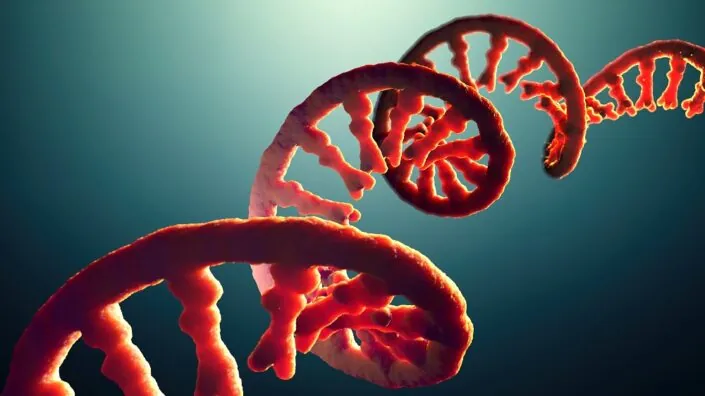
So, you have a technology that can mimic peptides and you’re specifically interested in the antimicrobial peptide LL-37. I understand it took you a long time to sift through a huge peptide database and zero in on LL-37 as a major regulator of the immune system.

Yes, and when we figured that out, we thought we had discovered something really amazing, but then we found that LL-37 is one of the most studied peptides in the human body. There are thousands of published articles about it.

So, I only discovered something for myself. Many bioengineers, chemists, and biologists knew that LL-37 is important, but people in our field also tend to think infectious disease doesn’t matter. There are sexier things like mapping the genome. People don’t care about some peptide that everybody has.

But the abundance of previous knowledge corroborated your science.

Yes, it totally corroborated it. Then I got involved with some of the top minds in what are called defensins, the antimicrobial peptides. I started talking with them, and they told me about this invention where the Department of Energy and DARPA got together to mimic peptides with a small molecule form factor.

How does this peptide actually work against pathogens?

It’s a positively charged linear peptide that looks like a corkscrew. It’s attracted to negatively charged pathogens, which includes almost all of them. Then, this positively charged corkscrew just drills into the pathogen and rips it apart. The only way a pathogen has a chance is to keep the peptide away or avoid detection.

LL-37 works against both Gram-positive and Gram-negative bacteria and enveloped viruses, and humans only have this one core peptide for their innate immune system. If it didn’t work against everything, you’d be dead. A lot of people don’t produce enough of it – babies, senior citizens, people not getting enough exercise, sunlight, good nutrition – so they are vulnerable.

I understand that LL-37 is as unstable as it is important, which was the main problem you had to overcome.

Yes, it’s like wet toilet paper – it falls apart very quickly.

So, you realized that to use it, you must stabilize it somehow, and you went for the peptoid architecture?

That’s right. We took the functional side chains that do all the work on the peptide and did a lot of extra math. We went through many very frustrating years of difficult work to do something that you’d think would be pretty easy: attaching the side chain to the nitrogen on the peptide’s backbone instead of the alpha-carbon.

The math of replicating the biological function of a peptide – that’s the thing. You wonder how that peptide works, and then you find out that even with thousands of published articles on it, people don’t really know that.

LL-37

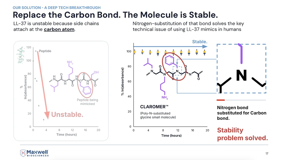
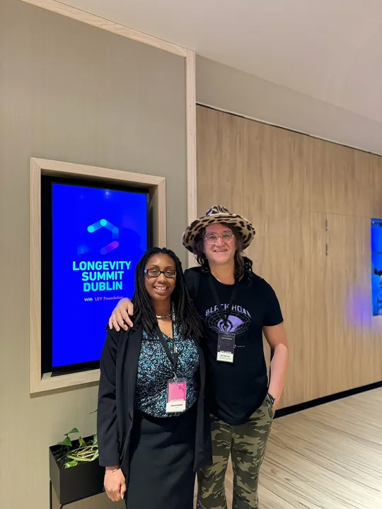
A slide from McClure’s presentation at last year’s ARDD conference in Copenhagen

Are Claromers™ your proprietary version of peptoids?

Yes, the antimicrobial version of peptoids. We’ve got lots of new ones coming out that are way better than the ones we’re taking through the FDA right now, but even those are so incredible that the FDA loves them.

First, there is no toxic concentration that we have found yet. Second, they’re anti-inflammatory and extremely broad spectrum. We have one that works against bacteria and fungi, and another against viruses: all influenza, all coronaviruses, SARS-1, SARS-2, regular cold. It’s literally the cure for the common cold. It’s crazy but amazing.

What about long COVID?

It’s going to knock it out completely. I have no doubt about it. Of course, these are my opinions. I’m the CEO of the company, and I am paid to be the optimist. But these are not promissory statements. And I’m not asking for investment. We don’t need it.

The fact is that, in partnership with the government, we’ve passed non-human primate studies with a single molecule killing the worst type of bacteria and the worst fungus we could select – the worst multi-drug-resistant versions of both that would kill you because there’s no drug to treat it.

You barely need diagnostics anymore. Just use this all the time, and if it doesn’t work, then you can do some expensive diagnostics to figure out what wild thing is in there that isn’t being killed. It kills all the bad things and leaves the good stuff in the microbiome.

Because of how it mimics the natural thing?

Yes, it’s exactly the same natural mechanism of action, so it leaves your microbiome alone.

You’ve already touched on this, but let’s talk some more about how this relates to aging.

Infectious disease is the primary driver of aging. It is the primary disruptor of homeostasis. As soon as we start fighting infectious disease with our drug – the first time we’re approved by the FDA for any kind of infectious disease – it will start lengthening healthspan immediately.

I think it’s inevitable at this point because we passed the non-human primate study. The only thing that could stop us now would be lack of cash flow, but we don’t have a problem with that. We’re working on multiple big contracts right now with the US military, the governments of India, UAE – direct government contracts. We’re working on two big contracts with commercial companies.

This is going to be world-shaking. Just like AI – even though it’s not currently replacing your job, you can pretty much bet it’s going to replace most jobs. Same with this technology: it’s going to eradicate infectious disease and more.

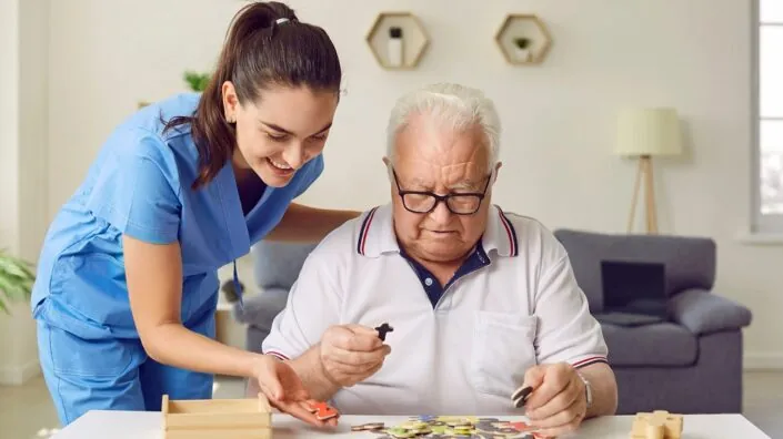
The first thing that came to my mind when I heard about your technology is that infections are an insanely prevalent cause of death in the oldest old. Many think this is a major factor that limits our maximal lifespan.

It truly is. Many times, people say, “Great-grandma fell and broke her hip,” and everyone thinks that’s natural, that’s how very old people die, but why does breaking a leg or a hip make someone fall apart and die?

It’s because their body has been fending off infectious disease pretty well up to this point. Now it must redirect just a little bit of energy to healing the hip, and not enough is left to defend against infectious disease. You’re breathing millions of fungal spores every day that will eat you just like a rotten tree if you don’t produce enough of this peptide to keep them destroyed.

This big change – pretty much eliminating the ever-present attack of pathogens against us – is going to greatly expand our healthspan. I’m predicting 20 to 40 additional years for the whole population just from our drug, comparable to what advances against infectious diseases have already done for lifespan.

Once we solve this, once we clean everything up and you’re like, “Okay, bird flu is a big issue right now, but I don’t need to worry about it because I’ve got this drug; everything is clean – my food is clean, my water is clean, my air is clean,” – then you can start sort of moving up Maslow’s hierarchy of needs and getting into the sexy stuff – can I enhance my vision? Can I enhance other things?

How does a collaboration with a country like India work?

Countries have their own priorities and budgets established for those priorities. With India, it’s for rare, neglected tropical diseases. This budget has been around since 2017, but no one has gotten it because no one wanted to tackle the indications they want us to address.

With India, it’s more about antibiotic-resistant septic infections that happen out there in the villages. India has plenty of antibiotics – they’re the home of generic antibiotics. What they need is help with difficult situations where somebody is going to die before they can get diagnostics. There’s no diagnostics out there to tell you what type of bacteria it is, so you must have something very broad-spectrum at hand to cure whatever it is.

The whole pharma industry says, “No, that’s not how medicine works.” We come in and say, “This is exactly how medicine should work.” Our product is shelf-stable and thermal-stable. It can be hot enough to cook an egg – and our drug will still be effective.

It’s a $300 million deal over three years, and it’s helping us while we pursue bigger things like tuberculosis and long COVID. It’s non-dilutive funding, essentially.

You saw no toxicity in your preclinical studies?

Zero toxicity. Not only it’s not toxic to human cells, but it’s not toxic to the commensal microbiome. We specifically measured the commensal microbiome in rhesus macaque monkeys in the India project, where we went up against methicillin-resistant Staphylococcus aureus (MRSA).

In the microbiome test, we found that they have a huge amount of Staphylococcus aureus in their sinus cavity – it’s one of the predominant bacteria there. Our compound works great against Staph aureus, but all our data said it doesn’t affect the commensal microbiome.

So, we asked: does that mean it’s selective for commensal Staph aureus and only kills the pathogenic Staph aureus? That seemed crazy, but that’s what we found. The commensal Staph aureus was untouched and in fact helped to protect the primate against the infection.

We had to remove some of the commensal microbiome to even give our MRSA strain a seat on the bus, so to speak. It took us a long time to actually establish an infection. This tells me that many human infections have to do with microbiome disruption. We eat preservatives and other things that disrupt the protective microbiome, which is a major way to get infected. The good bacteria form a shield that prevents pathogenic bacteria from getting through.

There must be some mechanism that prevents the peptide or peptoid from attacking beneficial bacteria. Do you know what it is?

We do, and we’ve published on it. Like I said, LL-37 is extremely well studied. The mechanism of action of LL-37 and our compounds is that they target negatively charged phospholipids on the membrane of bacteria, fungi, or viruses. That negatively charged phospholipid in the membrane is required to merge with a human cell by connecting to TIM receptors (TIM-2, TIM-3, TIM-4). So, it’s a hallmark of aggressive pathogens.

The negatively charged phospholipids are present in the membrane of the commensal microbiome, too, but they are covered with a V-shaped or shield-shaped molecule called annexin-5 (and the whole annexin family). The annexin goes over the top of the negatively charged phospholipids, saying “I’m not going to attack you.”

If they take their annexin-5 off, they become pathogenic. That’s how Staphylococcus stays commensal as long as it’s getting fed inside your gut. But if you stop eating and stop producing antimicrobial peptides, it’s like “You’re what’s for dinner” – it comes after you.

Anything you can tell me about your collaboration with the military?

They want things like prophylactic wound care for combatants on the run; it’s pre-first aid, where you can squirt something into a puncture wound to stop bleeding and potential infection.

We have about seven Collaborative Research and Development Agreements (CRADAs) with the Pentagon. Some are for quite exotic things like anthrax and Ebola, things that could be weaponized. The military says, “We have to have something against that, and we’re willing to pay a billion dollars to stockpile it, just in case somebody uses a weaponized version against us.”

We’re also working on more common things, such as anti-diarrheal applications, which we could use for commercial purposes as well. How many battles have been won by dysentery rather than by the enemy army?

How far out are you in terms of human trials and approvals?

This year, we got the data back on the non-human primate studies, which is pretty much equivalent to a human Phase I. It was totally safe and very effective, so we have a very high probability for the human trials outcome. The value of the company has gone up significantly, and the certainty of the outcome has increased, which is why we’ve gotten all these contracts with governments and companies.

We would like to ask you a small favor. We are a non-profit foundation, and unlike some other organizations, we have no shareholders and no products to sell you. All our news and educational content is free for everyone to read, but it does mean that we rely on the help of people like you. Every contribution, no matter if it’s big or small, supports independent journalism and sustains our future.
RNA

Scientists Create Cytoskeleton-Like Structures From RNA

In a new study, researchers report producing self-assembling nanotubes and rings made from RNA molecules inside artificial cell-like lipid vesicles. In the future, this technology could facilitate the creation of synthetic cells for various research, diagnostic, and therapeutic applications [1].

Paperless origami

DNA and RNA molecules are central to life, carrying essential genetic information for protein production. However, their unique properties also make them excellent building materials. Nature discovered this eons ago; ribosomes, for instance, are partly built from RNA.

Scientists have experimented extensively with designing DNA and RNA sequences that cause molecules to self-assemble into predetermined shapes. The technique has become known under the poetic name “DNA (or RNA) origami” [2].

These engineered structures can be impressively intricate: for example, DNA “boxes” that carry drug molecules directly to target sites, then each open a molecular “door” to release their contents [3]. A new study from Heidelberg University, published in Nature Nanotechnology, takes RNA origami to the next level.

Tubes, rings, and networks

The researchers designed RNA molecules to self-assemble into structures resembling cellular cytoskeletons. The cytoskeleton, naturally composed of protein filaments and microtubules, is critical for maintaining cell shape and stability. “We designed RNA origami tiles that fold upon transcription and self-assemble into micrometer-long, three-dimensional RNA origami nanotubes,” the authors explain. Creating an artificial cytoskeleton is an important milestone on the path toward synthetic cells.

The team encapsulated DNA templates and RNA polymerase, the enzyme needed to transcribe DNA into RNA, into giant unilamellar lipid vesicles (GUVs), effectively creating proto-cells. To supply these synthetic cells with RNA building blocks (nucleotides), they used the bacterial transmembrane pore protein α-haemolysin. Magnesium ions (Mg2+) served as a trigger to prevent premature transcription, and the pore protein facilitated the removal of transcription byproducts.

When transcription was initiated, RNA strands immediately folded and assembled into nanotubes inside these synthetic cells. Remarkably, some nanotubes reached several micrometers in length, comparable to actual cellular cytoskeletal structures.

The researchers observed that subtle variations in the DNA template sequence significantly altered the RNA origami structures, demonstrating the method’s flexibility and ease of control. For instance, slight modifications switched the resulting structure from nanotubes to rings.

RNA cytoskeleton 1

To scale up their creations, the researchers included aptamers: RNA sequences capable of binding to specific molecular targets, including other RNA molecules. With aptamers incorporated, the nanotubes formed “cytoskeleton-like networks tens of micrometers across.”

RNA cytoskeleton 2

Aptamers also proved useful in creating cortex-like structures adhering to the lipid membrane, mirroring the cortices seen in real cells. Whether aptamers were used or not, a prolonged nucleotide supply led to structural networks becoming so extensive that they physically deformed the GUVs. Being able to maintain and alter the cell’s shape is another hallmark of a true cytoskeleton.

Possible applications

A major advantage of RNA origami is that these structures can be produced directly inside cells. Once DNA templates are introduced into cells, a single enzyme, T7 polymerase, can create numerous RNA products from these templates. In comparison, the full biological transcription-translation machinery requires over 150 genes.

“In contrast to DNA origami, RNA origami enables synthetic cells to manufacture their building blocks by themselves,” explained Dr. Kerstin Göpfrich, the study’s lead author, whose team, “Biophysical Engineering of Life,” conducts research at the Center for Molecular Biology of Heidelberg University (ZMBH). “This could open new perspectives on the directed evolution of such cells.”

This early-stage discovery has broad implications, including in aging research. It could help scientists better understand early cellular evolution, develop biomimetic systems, and engineer cells designed for specific tasks.

While creating fully functional synthetic eukaryotic cells remains distant due to their enormous complexity, the pathway toward viable, simplified prokaryotic “proto-cells” with limited but useful functions has just become shorter. Currently, bacteria are used extensively to produce biological molecules. However, minimal synthetic cells might simplify this process and even enable protein production directly within living organisms, circumventing bacterial immunogenicity issues.

Such proto-cells could, for example, produce essential proteins like collagen and elastin to maintain youthful extracellular matrix (ECM) function. Additionally, RNA origami structures could be introduced into existing cells to provide structural support and other functionalities.

The authors anticipate that future RNA origami structures will become more than passive scaffolds; they will actively perform complex cellular tasks by integrating ribozymes, RNA molecules capable of enzymatic activity. According to Göpfrich, the long-term research goal is the creation of fully functional molecular machinery for RNA-based synthetic cells.

We would like to ask you a small favor. We are a non-profit foundation, and unlike some other organizations, we have no shareholders and no products to sell you. All our news and educational content is free for everyone to read, but it does mean that we rely on the help of people like you. Every contribution, no matter if it’s big or small, supports independent journalism and sustains our future.

Literature

[1] Tran, M. P., Chakraborty, T., Poppleton, E., Monari, L., Illig, M., Giessler, F., & Göpfrich, K. (2025). Genetic encoding and expression of RNA origami cytoskeletons in synthetic cells. Nature Nanotechnology, 1 – 8.

[2] Dey, S., Fan, C., Gothelf, K. V., Li, J., Lin, C., Liu, L., … & Zhan, P. (2021). DNA origami. Nature Reviews Methods Primers, 1(1), 13.

[3] Udomprasert, A., & Kangsamaksin, T. (2017). DNA origami applications in cancer therapy. Cancer science, 108(8), 1535-1543.

DNA structure

A Core Senescence Biomarker Fights Inflammation

Researchers publishing in Nature Communications have found that p53, a biomarker and inducer of senescence, suppresses both inflammation and DNA damage in senescent cells.

Senescence against cancer

One of the main reasons why cells become senescent is to prevent cancer. The senescence-inducing compound p53, in particular, is known in the literature as a tumor suppressor, and its presence has been found to reduce, rather than exacerbate, the SASP [1]. The biochemical reasons behind this seemingly contradictory connection have not been previously explained.

However, these researchers have previously found that a biochemical pathway from the mitochondria to the nucleus is partially responsible for driving the SASP [2]. This pathway drives the expulsion of chromatin from the nucleus into the rest of the cell, which activates the cGAS/STING pathway and thus turns on NF-κB, the SASP’s master regulator [3]. Therefore, they sought to determine a link between p53 and this well-established pathway.

Preventing the SASP at its root

For their first experiment, the researchers created fibroblasts in which 53BP1, a suppressor of DNA damage that works with p53, was somewhat increased. These fibroblasts, after they were driven senescent through irradiation, had reduced amounts of chromatin in the nucleus and, thus, less SASP. Driving cells senescent by introducing p53 had similar effects, as did knocking down its target, MDM2.

Mutating 53BP1, on the other hand, had significant negative effects, spurring the release of chromatin into the nucleus after the cells were driven senescent with radiation. Silencing p53 had similar effects; four days after irradiation, cells with silenced p53 had significantly more chromatin in the nucleus. Further work found that mitochondria are required for this SASP upregulation; ablating away mitochondria prevented the chromatin from being expelled into the nucleus and thus prevented the upregulation of SASP elements.

Protecting cells from DNA damage

p53 was also found to be instrumental in DNA repair. γH2AX is a marker of DNA damage, and its levels in the nucleus were reduced in cells where MDM2 was downregulated and p53 was upregulated. The researchers found that this could be accomplished by introducing RG7388, a compound that promotes p53.

Similarly, silencing p53 increased the prevalence of this DNA damage marker. Interestingly, p21, another well-known biomarker of senescence, was found to be necessary for the effects of p53; without p21 being present, neither MDM2 nor p53 made any difference.

This was not just a change in a biomarker; p53 had real effects on the genome. In cells treated with RG7388 shortly after irradiation, the number of DNA amplifications and deletions was relatively small. In untreated cells, however, these signs of genetic damage were rampant. Most of this damage was located near the ends of the chromosomes, where the telomeres are. Unlike with chromatin ejection, however, mitochondria had nothing to do with these DNA damage effects.

Effects in female mice

The researchers then turned to in vivo experiments, introducing HDM201, a compound that suppresses MDM2, to a population of naturally aged mice for two weeks. In both males and females, HDM201 had no effects on body weight, blood cell counts, or liver pathological biomarkers. However, as the liver is where senescent cells normally accumulate in mice [4], the researchers focused their attentions there. Both p53 and p21 were significantly increased in these mice.

Interestingly, in gene expression, this treatment had substantially greater effects on female mice than on male mice. While it had no senolytic effects, it reversed many of the gene expression changes caused by aging. As expected, many of these genes were related to the SASP, and, likewise, the number of immune cells that infiltrated into these mice’s livers was decreased as well.

Both p53 and p21 have been investigated in the context of senescence, with previous researchers considering them as potential targets to be suppressed. This work, however, demonstrates that these compounds are both necessary and beneficial for preventing senescent cells from getting out of control. The researchers describe p53 as a senomorphic compound; while it clearly does not remove senescent cells, it significantly blunts their negative effects on the cells around them. They hold that “it may be possible to one day design a treatment that targets p53 to promote healthier aging.”

We would like to ask you a small favor. We are a non-profit foundation, and unlike some other organizations, we have no shareholders and no products to sell you. All our news and educational content is free for everyone to read, but it does mean that we rely on the help of people like you. Every contribution, no matter if it’s big or small, supports independent journalism and sustains our future.

Literature

[1] Coppé, J. P., Patil, C. K., Rodier, F., Sun, Y. U., Muñoz, D. P., Goldstein, J., … & Campisi, J. (2008). Senescence-associated secretory phenotypes reveal cell-nonautonomous functions of oncogenic RAS and the p53 tumor suppressor. PLoS biology, 6(12), e301.

[2] Vizioli, M. G., Liu, T., Miller, K. N., Robertson, N. A., Gilroy, K., Lagnado, A. B., … & Adams, P. D. (2020). Mitochondria-to-nucleus retrograde signaling drives formation of cytoplasmic chromatin and inflammation in senescence. Genes & development, 34(5-6), 428-445.

[3] Dou, Z., Ghosh, K., Vizioli, M. G., Zhu, J., Sen, P., Wangensteen, K. J., … & Berger, S. L. (2017). Cytoplasmic chromatin triggers inflammation in senescence and cancer. Nature, 550(7676), 402-406.

[4] Ogrodnik, M., Miwa, S., Tchkonia, T., Tiniakos, D., Wilson, C. L., Lahat, A., … & Jurk, D. (2017). Cellular senescence drives age-dependent hepatic steatosis. Nature communications, 8(1), 15691.

Hypothalamus

A Hypothalamus Neuropeptide Reduces Aging in a Mouse Model

Researchers have improved multiple health metrics in prematurely aged mice by re-establishing the production of neuropeptide Y in the hypothalamus [1].

An essential protein

Age-related decline in the function of the hypothalamus, a core region of the brain, has been suggested to be a “key factor in the development of whole-body aging” [2]. Due to its essential role and implication in several aging-related processes, the hypothalamus may be a target for lifespan- and healthspan-extending therapeutic strategies.

The authors of this study focused specifically on hypothalamic neuropeptide Y (NPY). Several studies suggest that neuropeptide Y plays a role in lifespan through its involvement in lifespan-extending processes such as autophagy induction [3] and stress resistance [4], and it appears to play an essential role in caloric restriction-induced lifespan extension [5, 6]. Decreased levels have been connected to neurodegenerative diseases [7]. Therefore, the authors hypothesized whether re-establishing hypothalamic neuropeptide Y levels might slow down aging.

Aging too quickly

For this study, the researchers used animals that lack a gene encoding the protein Zmpste24 (Z24-KO), and this lack results in accelerated aging and premature death. Z24-KO mice have multiple defects in skin, bone, cardiovascular tissues, and skeletal muscles, similar to those observed in human accelerated aging processes. Therefore, the authors decided to test if their hypothalami exhibit the same changes as the hypothalami of naturally aged mice.

They tested an area of the hypothalamus called the arcuate nucleus (ARC), which contains neurons that release neuropeptide Y. They found lower levels of neuropeptide Y compared to age-matched wild-type mice and lower levels of a neuronal marker called NeuN, suggesting neuronal aging and fewer hypothalamic neurons.

Markers connected to neuroinflammation were also altered. The authors observed increased levels of a gliosis marker in Z24-KO mice. Gliosis is a process that occurs in glial cells (non-neuronal cells located in the central nervous system) in response to damage. Therefore, an increase in gliosis suggests neuroinflammation. On the other hand, a marker showing the activity of microglia, the immune cells of the brain, decreased in Z24-KO mice. This, according to the authors, suggests that immune response capacity is impaired.

Restoring youth

To re-establish decreased neuropeptide Y levels, the researchers used a genetically modified adeno-associated virus (AAV) that causes an increase in the expression of neuropeptide Y (AAV-NPY) and injected it into the mice. The virus increased neuropeptide Y in the ARC for at least four months, which is when the mice were sacrificed to analyze their organs.

The researchers tested the AAV-treated Z24-KO mice for the same biomarkers as the untreated Z24-KO mice. They observed an increase in NeuN and a reduction of the gliosis biomarker that is normally elevated in these mice. However, the biomarker of microglial immune capacity didn’t significantly change. Overall, AAV treatment made the brains of Z24-KO mice similar to those of age-matched wild-type mice, suggesting that neuropeptide Y plays a role in reducing neuroinflammation.

Further testing suggested positive changes in aging-related processes. The authors observed a reduction in NF-κB, a protein that increases with aging and is associated with neuroinflammation in the hypothalamus [8]. Other positive effects were a decrease in a marker of tau pathology, which is associated with neurodegenerative diseases [9], and an increase in autophagy, a process that plays an essential role in longevity.

Better looks and healthier minds

The authors also report positive changes in body weight, body composition, mobility, vitality, and fur among the virus-treated animals. The treatment also showed neuroprotective effects, as the spatial memory improvements accompanied physical health improvements, but there were no significant differences in locomotor activity.

One of the characteristics of the prematurely aged Z24-KO mice is lipodystrophy. This condition results in the organism losing fat from some parts of the body, including under the skin surface of different body parts, while gaining it in others, such as the liver.

The AAV-treated mice had fewer lipodystrophy symptoms. The treatment resulted in a thicker outermost skin layer (epidermis) and thicker layers of under-skin (subcutaneous) fat, probably resulting from increased proliferation of fat cells as suggested by increased levels of a cell proliferation marker. It also increases skin collagen, which decreases with age. Autophagy markers in the skin indicated increased autophagic activity, suggesting better cellular health.

Liver structure was also improved in the Z24-KO mice after treatment. The researchers observed increased cell proliferation, which might indicate improved liver protection and regeneration, a decrease in cellular death by apoptosis, and increased autophagic activity in the liver, suggesting better cellular health.

There were no differences between treated and untreated mice in different parameters of kidney health and heart structure, but heart cells showed improved cellular health.

Promising, but there are still many unknowns

Overall, this study’s results suggest that re-establishing neuropeptide Y has a positive effect on aging-associated hypothalamus-related symptoms. However, in most of the experiments performed, the researchers compared untreated, prematurely aged mice to prematurely aged mice treated with a virus expressing neuropeptide Y. It would be beneficial if a wild-type control was included, as this would allow for assessing whether the observed changes make the prematurely aged mice more similar to wild-type mice or if the changes are significant but modest.

Additionally, long-term studies are essential to determine the long-lasting effects and side effects of this approach, whether it induces the desired amount of neuropeptide Y expression, and how improvements in different biomarkers of cellular health translate to increases of healthspan and lifespan.

We would like to ask you a small favor. We are a non-profit foundation, and unlike some other organizations, we have no shareholders and no products to sell you. All our news and educational content is free for everyone to read, but it does mean that we rely on the help of people like you. Every contribution, no matter if it’s big or small, supports independent journalism and sustains our future.

Literature

[1] Ferreira-Marques, M., Carmo-Silva, S., Pereira, J., Botelho, M., Nóbrega, C., López-Otín, C., de Almeida, L. P., Aveleira, C. A., & Cavadas, C. (2025). Restoring neuropetide Y levels in the hypothalamus ameliorates premature aging phenotype in mice. GeroScience, 10.1007/s11357-025-01574-0. Advance online publication.

[2] Kim, K., & Choe, H. K. (2019). Role of hypothalamus in aging and its underlying cellular mechanisms. Mechanisms of ageing and development, 177, 74–79.

[3] Aveleira, C. A., Botelho, M., Carmo-Silva, S., Pascoal, J. F., Ferreira-Marques, M., Nóbrega, C., Cortes, L., Valero, J., Sousa-Ferreira, L., Álvaro, A. R., Santana, M., Kügler, S., Pereira de Almeida, L., & Cavadas, C. (2015). Neuropeptide Y stimulates autophagy in hypothalamic neurons. Proceedings of the National Academy of Sciences of the United States of America, 112(13), E1642–E1651.

[4] Michalkiewicz, M., Knestaut, K. M., Bytchkova, E. Y., & Michalkiewicz, T. (2003). Hypotension and reduced catecholamines in neuropeptide Y transgenic rats. Hypertension (Dallas, Tex. : 1979), 41(5), 1056–1062.

[5] Chiba, T., Tamashiro, Y., Park, D., Kusudo, T., Fujie, R., Komatsu, T., Kim, S. E., Park, S., Hayashi, H., Mori, R., Yamashita, H., Chung, H. Y., & Shimokawa, I. (2014). A key role for neuropeptide Y in lifespan extension and cancer suppression via dietary restriction. Scientific reports, 4, 4517.

[6] de Rijke, C. E., Hillebrand, J. J., Verhagen, L. A., Roeling, T. A., & Adan, R. A. (2005). Hypothalamic neuropeptide expression following chronic food restriction in sedentary and wheel-running rats. Journal of molecular endocrinology, 35(2), 381–390.

[7] Duarte-Neves, J., Pereira de Almeida, L., & Cavadas, C. (2016). Neuropeptide Y (NPY) as a therapeutic target for neurodegenerative diseases. Neurobiology of disease, 95, 210–224.

[8] Zhang, G., Li, J., Purkayastha, S., Tang, Y., Zhang, H., Yin, Y., Li, B., Liu, G., & Cai, D. (2013). Hypothalamic programming of systemic ageing involving IKK-β, NF-κB and GnRH. Nature, 497(7448), 211–216.

[9] Samudra, N., Lane-Donovan, C., VandeVrede, L., & Boxer, A. L. (2023). Tau pathology in neurodegenerative disease: disease mechanisms and therapeutic avenues. The Journal of clinical investigation, 133(12), e168553.