×

The Blog

Building a Future Free of Age-Related Disease

Europe from space

How Life Expectancy Has Changed in Europe

A recent country-level analysis of life expectancy among several European nations shows changes in life expectancy trends and how well-designed national policies can reduce or minimize exposure to risk factors, thus improving life expectancy [1].

Slowdown in life expectancy increase

Life expectancy has grown in high-income countries since at least 1900, except during the two World Wars and the 1918 influenza pandemic [2]. However, the speed of the growth differed; for example, since 2011, Europe’s trend towards life expectancy increases was reduced, and this was followed by a decline in life expectancy in most European countries due to the COVID-19 pandemic [3, 4].

The authors of this recent study used the data from the Global Burden of Diseases, Injuries, and Risk Factors Study (GBD) 2021 to compare changes in life expectancy and exposure to risk factors in the 16 founding European Economic Area countries and four UK nations.

The life expectancy at birth was defined as “the mean number of years that a newborn infant could expect to live, if he or she were to pass through life exposed to the sex-specific and age-specific death rates prevailing at the time of his or her birth, in a given country.”

They compared periods of 1990 to 2011 (pre-slowdown in life expectancy), 2011 to 2019 (slowdown in life expectancy to pre-COVID-19 pandemic), and 2019 to 2021 (COVID-19 pandemic).

Country-level analysis

When analyzed separately, all countries showed improved life expectancy from 1990 to 2011 and 2011 to 2019; however, the rate varied. Confirming the observations of bulk data analysis reported previously, the rate of life expectancy improvement was higher in the 1990-to-2011 period than the 2011-to-2019 period.

Norway was the only exception from that observation. In Norway, the trend of life expectancy increased more during the 2011-to-2019 period compared to the 1990-to-2011 period.

During the COVID-19 pandemic and post-pandemic period, all countries but Ireland, Iceland, Sweden, Norway, Denmark, and Belgium experienced an absolute fall in life expectancy, with Greece and England observing the most significant decrease.

Life expectancy Europe 1 Life expectancy Europe 2

Cardiovascular diseases, cancers, and COVID-19

The life expectancy improvements seen in the 1990-to-2011 period stem from improvements related to causes of death attributed to cardiovascular diseases and neoplasms, which are tissue masses that result from abnormal growth, whether benign or cancerous.

Unsurprisingly, the decrease in life expectancy in years 2019–21 can be attributed to the deaths from respiratory infections and other COVID-19-related health problems. However, before the COVID-19 pandemic period, reductions in improvements in life expectancy were primarily driven by cardiovascular diseases.

The researchers also made an interesting observation: “among the studied countries, those with the greatest slowdown in life expectancy improvements before the COVID-19 pandemic were generally most severely affected by COVID-19 and had some of the largest decreases in life expectancy in 2019-21.”

Avoiding risk

The researchers analyzed risk factors, attributed to different causes of death, for both sexes in all countries combined in 2019. The top three risk factors for cardiovascular disease were high systolic blood pressure, dietary risks, and high LDL cholesterol. For neoplasms, the top risk factors included tobacco smoke, dietary risks, and occupational risks.

Life expectancy Europe 3

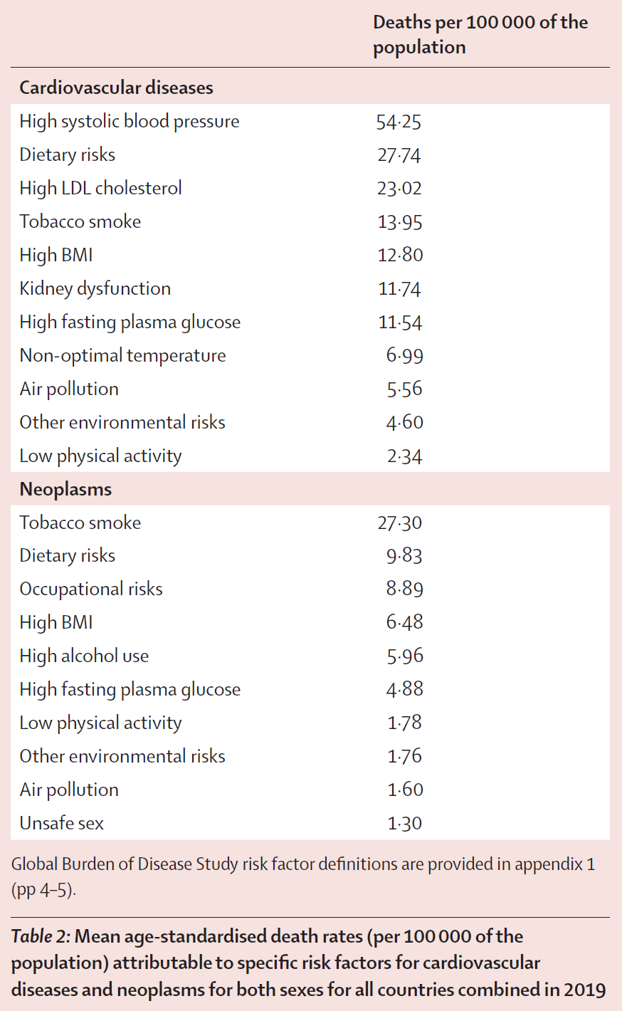
The levels of different risk factors changed with time, such as exposure to tobacco smoke; even though it is still a high population risk, it has decreased over time. On the other hand, BMI has increased in all countries, and dietary risks, high alcohol use, and low physical activity remain high in most.

The authors also point to high LDL cholesterol and systolic blood pressure, which declined until before 2011; however, this trend reversed after 2011 in many countries.

The danger of a risk factor varies by the time between exposure to it and the start of the disease that it causes, the length of exposure, and its interactions with other risk factors. Unfortunately, this dataset doesn’t provide some of that information.

Funding healthcare

Following their analysis, the authors discuss governmental policies and their impact on life expectancy. For example, they mention national fiscal and healthcare policies that impact the population’s life expectancy, especially for people in the worst socioeconomic situations.

An example of policies aimed at increase access to healthcare are Belgian, French, and Norwegian national policies, which, in recent years, have focused on increasing cancer diagnosis and treatment. The authors hold that these policies improved life expectancy related to neoplasms between 1990 and 2019. Additionally, some research has suggested that funding cuts to health, social care, and welfare since 2010 contributed to the slowdown in life expectancy improvement [5, 6].

Diet and physical activity are the foundation of health and longevity

Diagnosis and treatment happen after a person suffers from a disease. Preventing diseases from occurring through proper diet and physical activity might be more effective at increasing life expectancy.

The authors give examples of how healthy food consumption can be influenced by effective policy. An example is Norway, which had implemented a sugar tax as early as 1922. Similarly, starting in the 1980s, the Norwegian government talked with the industry about reducing the amount of salt in food products. This was complemented by Norway’s ‘Action Plan on Nutrition 2007–2011’, which, apart from education, also focused on other nutritional aspects, such as increasing focus on nutrition in a health care setting.

This broader approach has proven more effective than focusing only on education and voluntary dietary changes. Apart from diet, physical activity is the cornerstone of health and reducing premature mortality. Unfortunately, accordingly to this analysis, at the population level, there were no improvements in the levels of even low physical activity across the studied countries. The authors believe that systematic strategies and incentives are necessary to change that.

Ultimately, the authors intend for policymakers to utilize this analysis as a guide to reverse their countries’ slowdown in life expectancy improvement. They also hold that countries that implement successful policies can be used as examples for others to follow.

We would like to ask you a small favor. We are a non-profit foundation, and unlike some other organizations, we have no shareholders and no products to sell you. All our news and educational content is free for everyone to read, but it does mean that we rely on the help of people like you. Every contribution, no matter if it’s big or small, supports independent journalism and sustains our future.

Literature

[1] GBD 2021 Europe Life Expectancy Collaborators (2025). Changing life expectancy in European countries 1990-2021: a subanalysis of causes and risk factors from the Global Burden of Disease Study 2021. The Lancet. Public health, 10(3), e172–e188.

[2] Roser M. (2020) The Spanish flu: the global impact of the largest influenza pandemic in history. https://ourworldindata.org/spanish-flu-largest-influenza-pandemic-in-history

[3] Raleigh VS. (2019) Trends in life expectancy in EU and other OECD countries. OECD Health Working Papers 108.

[4] Organisation for Economic Co-operation and Development, EU. (2018) Health at a glance: Europe 2018: state of health in the EU Cycle.

[5] Alexiou, A., Fahy, K., Mason, K., Bennett, D., Brown, H., Bambra, C., Taylor-Robinson, D., & Barr, B. (2021). Local government funding and life expectancy in England: a longitudinal ecological study. The Lancet. Public health, 6(9), e641–e647.

[6] McCartney, G., McMaster, R., Popham, F., Dundas, R., & Walsh, D. (2022). Is austerity a cause of slower improvements in mortality in high-income countries? A panel analysis. Social science & medicine (1982), 313, 115397.

Elephant and mouse

A New Study Claims to Challenge Peto’s Paradox

Applying statistical methods to a large dataset spanning almost 300 species, scientists found a positive correlation between body size and cancer prevalence [1]. Other researchers dispute that these findings invalidate the famous paradox.

A paradox of size

Cancer has forever been a mortal enemy of multicellular life. Sometimes, the cell’s “program” malfunctions, causing it to divide uncontrollably, until its progeny takes over the organism and kills it. This can happen to almost any cell, and one cell is enough.

A logical conclusion is that the more cells an animal has, the greater its risk of developing cancer. Hence, larger animals with many times more cells should be getting cancer much more frequently, but this does not appear to occur in nature. Instead, lifespan is positively correlated with body size (barring a few outliers), and small animals often get more cancer, not less. For instance, cancer is a highly prevalent cause of death in lab mice.

This became known as Peto’s paradox, after Sir Richard Peto, a British epidemiologist and statistician, who first articulated the idea in 1977 while studying cancer risks in different species. Of course, there’s nothing mystical about it: many large, long-lived species have developed superior anti-cancer defenses, such as DNA repair mechanisms, robust immune systems, and enhanced methods of removing cancerous cells through apoptosis. A lower cell division rate in some large species might also explain a part of the paradox.

For example, elephants have at least 20 copies of the TP53 gene, which plays crucial roles in detecting and repairing DNA damage and in triggering cell death in potentially cancerous cells, while humans have just one. Studying those mechanisms is a rapidly growing field in geroscience.

The correlation between body size and cancer

Since Peto’s original observation, several studies have investigated this question. Some of them found no correlation between cancer prevalence and body sizes, seemingly confirming the paradox [2]. However, those earlier studies were plagued by low data availability; after all, it’s hard to amass enough necropsies for multiple species.

Enter this new paper by scientists from the University of Reading, University College London and the Johns Hopkins University School of Medicine. This paper was published in Proceedings of the National Academy of Sciences (PNAS) and ambitiously titled “No evidence for Peto’s paradox in terrestrial vertebrates.”

The paper is based on a dataset created for a slightly earlier study by Compton et al. [3] The unprecedentedly large dataset consists of 16,049 necropsy records for 292 species, which made better statistical analysis possible. Interestingly, that paper did not reach the same definitive conclusion but instead highlighted “limitations to Peto’s paradox, by showing that large animals do tend to get somewhat more neoplasms and malignancies when compared with smaller animals.”

The authors of the PNAS paper claim to have applied more robust statistical analysis to the same dataset, which allowed them to extract a clearer signal. Professor Chris Venditti, senior author of the research at the University of Reading, said, “Everyone knows the myth that elephants are afraid of mice, but when it comes to cancer risk, mice are the ones who have less to fear. We’ve shown that larger species like elephants do face higher cancer rates—exactly what you’d expect given they have so many more cells that could go wrong.”

The researchers separately analyzed birds and mammals, which stop growing at certain points in their life, along with amphibians and reptiles, many of which never do. In the first subset, the authors controlled for body mass, while in the second, for body length (which itself might have affected the results). They also controlled for species’ longevity, although the longevity data was limited.

They found a significant positive association between neoplasia (this included both benign and malignant tumors, which were strongly correlated) and body size. For mammals, the relationship was β = 0.129, indicating a relatively flat slope (a linear relationship would have β = 1). In amphibians and reptiles, the correlation was stronger: β = 0.433. “Across all four vertebrate classes, larger species have an increased prevalence of malignancy compared to smaller species, thus demonstrating no evidence of Peto’s paradox,” the paper concludes.

The paradox is dead, long live the paradox

However, this suggests a narrow reading of Peto’s paradox as nothing short of zero positive correlation between cancer and body size. “The conclusion of this paper is not supported by the results,” said Dr. Vera Gorbunova of the university of Rochester, a researcher of long-lived species who was not involved in this study. “Even if there is a small statistical trend towards increased cancer with increased body size, it is not proportional to the number of cells or cell divisions experienced by larger species. An elephant still has much lower cancer incidence than a mouse. The authors themselves conclude that larger species have evolved better control of the cell cycle. This means they did evolve additional anticancer defenses, which is what Peto’s paradox posits.”

Indeed, the authors point to some instances of animals clearly “outsmarting” cancer, with elephants having 56% lower cancer rates than the researchers’ model expected for their body size, and naked mole rats, rodents famous for their longevity, performing even stronger. On the opposite side of the spectrum lie the notoriously cancer-prone ferrets and opossums. Interestingly, bats and turtles, highlighted in Compton et al. as supporting Peto’s paradox, are not mentioned in this new study.

Dr. Joanna Baker, co-author from the University of Reading, said, “When species needed to grow larger, they also evolved remarkable defenses against cancer. Elephants shouldn’t fear their size—they developed sophisticated biological tools to keep cancer in check. It’s a beautiful example of how evolution finds solutions to complex challenges.”

An important aspect of this study is that the researchers were able to count in some evolutionary differences. In particular, they found that species that evolved larger body sizes more rapidly, such as through a series of evolutionary ‘bursts’, were more likely to have stronger anti-cancer defenses.

“These studies represent a more comprehensive quantitative evaluation of some of the theories of evolution of aging and life history strategies,” said Dr. Emma Teeling of the University College Dublin, who also was not involved in this study. “Collecting these malignancy and life history studies requires decades if not centuries for long-lived species. This is why these studies are confined to captive species, where perhaps what was measured is actually the potential stress of captivity rather than true rate of malignancy. We are limited to species that we are able to maintain in captivity, which are not necessarily those that have evolved the most robust and therefore the most interesting anti-cancer mechanisms.”

“The authors detected a signal of evolution in action, where indeed with increased body size, there is a trend towards increased cancer incidence that then gets compensated by evolution of additional tumor suppressor mechanisms,” Gorbunova said. “Overall, I think the title of the paper is somewhat ‘sensationalized’. If this study found that upon certain phylogenetic comparisons larger species have slightly increased cancer risk, it does not eliminate Peto’s paradox.”

“The outliers in both of these studies, the species that were observed to have more or less than predicted cancer regardless of the methods used, are the most interesting candidates,” Teeling added. “Some of these species have been the focus of previous anti-aging research, such as the naked mole rat. Both studies will stimulate new ways to consider the evolution of cancer and anti-cancer mechanisms across the tree of life, new methods, datasets, and conclusions.”

We would like to ask you a small favor. We are a non-profit foundation, and unlike some other organizations, we have no shareholders and no products to sell you. All our news and educational content is free for everyone to read, but it does mean that we rely on the help of people like you. Every contribution, no matter if it’s big or small, supports independent journalism and sustains our future.

Literature

[1] Butler, G., Baker, J., Amend, S. R., Pienta, K. J., & Venditti, C. (2025). No evidence for Peto’s paradox in terrestrial vertebrates. Proceedings of the National Academy of Sciences, 122(9), e2422861122.

[2] Boddy, A. M., Abegglen, L. M., Pessier, A. P., Aktipis, A., Schiffman, J. D., Maley, C. C., & Witte, C. (2020). Lifetime cancer prevalence and life history traits in mammals. Evolution, medicine, and public health, 2020(1), 187-195.

[3] Compton, Z. T., Mellon, W., Harris, V. K., Rupp, S., Mallo, D., Kapsetaki, S. E., … & Boddy, A. M. (2025). Cancer prevalence across vertebrates. Cancer discovery, 15(1), 227-244.

Histone methylation

An Existing Diabetes Drug May Treat Aspects of Aging

In the Nature publication Signal Transduction and Targeted Therapy, researchers have described how glibenclamide, a drug used to treat type 2 diabetes, partially reverses epigenetic alterations and fights cellular senescence in mice.

A system tightly tied together

This paper begins with a discussion of the relationship between epigenetic alterations and cellular senescence. The histone H3K4me3 upregulates the senescence-related genes Cdkn1a, which is responsible for the biomarker p21 [1], and Cdkn2a, which is responsible for the biomarker p16 [2]. H3K27me3, on the other hand, downregulates these genes. Another histone, H3K9me3, suppresses repetitive genetic elements that cause an inflammatory response related to senescence [3].

While research has been done on directly targeting these histones [4], doing this with small molecules is difficult because they are structurally similar [3]. These researchers point to evidence suggesting that it may be more effective to target metabolism instead, as fundamental aspects of metabolism are linked to histone methylation [5].

These researchers had previously found that chlorpropamide provides a rejuvenation effect in C. elegans worms through a mitochondrial pathway [6]. They began this work to more definitively determine how and why this was happening, looking for a therapeutic target.

A metabolic target

This work began with a study on lung fibroblasts. Using a chemical probe based on chlorpropamide, the researchers looked at protein functions in order to determine what was being affected by this compound. They found MDH2 to be a potential target, as it had similar mitochondrial effects as chlorpropamide.

Further work in lung fibroblasts found that MDH2 was directly related to cellular senescence, whether it was induced by doxycycline or by excessive replication. The researchers created two cell lines, one with suppressed MDH2 and the other with overexpressed MDH2, to determine whether this relationship was causative in nature. They found that suppressing MDH2 reduced key senescence biomarkers, including SA-β-gal and p16, while overexpressing it increased them. The difference was not large, but it was statistically significant.

The researchers then tested how MDH2 interacts with five different sulfonylureas, a class of drugs that includes chlorpropamide. Of these drugs, the researchers found that glibenclamide has the strongest interaction with MDH2, far stronger than that of chlorpropamide.

In doxycycline-induced senescent lung fibroblasts, glibenclamide was found to reduce senescence biomarkers, including SA-β-gal, p16, and interleukins. Its overall effects in this area seemed to be roughly on par with those of metformin, another diabetes drug; it was not as good at reducing the inflammatory cytokine IL-6, but it reduced IL-1β in these cells, which metformin did not do. These beneficial effects were confirmed to be dependent on MDH2, as glibenclamide had no effects in cells with silenced MDH2.

Increases in histones and ROS

While the effects on lifespan and frailty were positive, glibenclamide increased, rather than decreased, mitochondrial reactive oxygen species (ROS) in lung fibroblasts. The researchers found that this was due to the inhibition of the TCA metabolic cycle, which forced the mitochondria to use oxygen-burning glycolysis instead [7]. The researchers hold that these effects on the TCA cycle are core to its beneficial effects, as they relate to the methionine cycle, which affects methylation.

Glibenclamide effects

On the first day of treatment in lung fibroblasts, glibenclamide immediately upregulated the senescence suppressor H3K27me3. Interestingly, when given for five days, glibenclamide upregulated both H3K4me3 and H3K27me3 in these cells while having no effect on H3K9me3.

Benefits for mice

The researchers also experimented on Black 6 mice. One group of 12-month-old mice was given glibenclamide, another was given NMN, and a third served as a control group. At 26 and 27 months of age, the glibenclamide group had significantly less frailty than either of the other groups. Mice given NMN appeared to live slightly longer on average, but the effect was not statistically significant; mice given glibenclamide, on the other hand, lived significantly longer.

Glibenclamide lifespan

Another experiment found that, while its physical benefits were not apparent at this age, glibenclamide reduces liver fibrosis and senescence in 20.5-month-old mice. In these animals, the drug was found to have significant effects on H3K27me3 but not H3K4me3.

While its anti-aging effects have not been tested in human beings, glibenclamide is a drug that is already being prescribed in the clinic. If these beneficial effects can be confirmed in human beings, this drug might be more widely prescribed to slow cellular senescence, particularly in the liver. The researchers also suggest that derivatives of this drug could be developed to more precisely target MDH2 to further slow cellular senescence.

We would like to ask you a small favor. We are a non-profit foundation, and unlike some other organizations, we have no shareholders and no products to sell you. All our news and educational content is free for everyone to read, but it does mean that we rely on the help of people like you. Every contribution, no matter if it’s big or small, supports independent journalism and sustains our future.

Literature

[1] Yan, K., Ji, Q., Zhao, D., Li, M., Sun, X., Wang, Z., … & Liu, G. H. (2023). SGF29 nuclear condensates reinforce cellular aging. Cell Discovery, 9(1), 110.

[2] Kotake, Y., Zeng, Y., & Xiong, Y. (2009). DDB1-CUL4 and MLL1 mediate oncogene-induced p16 INK4a activation. Cancer research, 69(5), 1809-1814.

[3] Zhang, B., Long, Q., Wu, S., Xu, Q., Song, S., Han, L., … & Sun, Y. (2021). KDM4 orchestrates epigenomic remodeling of senescent cells and potentiates the senescence-associated secretory phenotype. Nature aging, 1(5), 454-472.

[4] Hsu, C. L., Lo, Y. C., & Kao, C. F. (2021). H3K4 methylation in aging and metabolism. Epigenomes, 5(2), 14.

[5] Salminen, A., Kauppinen, A., Hiltunen, M., & Kaarniranta, K. (2014). Krebs cycle intermediates regulate DNA and histone methylation: epigenetic impact on the aging process. Ageing research reviews, 16, 45-65.

[6] Mao, Z., Liu, W., Huang, Y., Sun, T., Bao, K., Feng, J., … & Li, J. (2022). Anti-aging effects of chlorpropamide depend on mitochondrial complex-II and the production of mitochondrial reactive oxygen species. Acta Pharmaceutica Sinica B, 12(2), 665-677.

[7] Li, X., Yang, Y., Zhang, B., Lin, X., Fu, X., An, Y., … & Yu, T. (2022). Lactate metabolism in human health and disease. Signal transduction and targeted therapy, 7(1), 305.

Lab mouse

A Klotho Gene Therapy Extends Life in Male Mice

In Molecular Therapy, a team of researchers has described how increasing the expression of a form of Klotho, a protein that has been frequently found to have rejuvenative effects, leads to longer lifespans in male mice.

Klotho has various forms

In their introduction, the researchers distinguish between the various forms of Klotho. The full mRNA sequence that generates Klotho creates two homologous effective sections (KL1 and KL2) and a membrane that is meant for transportation between cells: this full version is m-KL [1]. Once enzymes take away this membrane, however, it becomes p-KL, with each section being p-KL1 and p-KL2.

However, this full version interferes with the function of FGF23, a protein that manages the metabolism of minerals [2]. Artificially upregulating this protein, therefore, is not safe [3].

Another form of Klotho, secreted Klotho (s-KL), does not have this problem. s-KL has been found to have multiple anti-aging effects according to a review of 65 studies [4]. Of course, without the transmembrane protein, it is not fit for intercellular transportation. Therefore, the researchers used an adeno-associated virus (AAV) to deliver a gene therapy that upregulates Klotho in the mice in this experiment.

Improvements in lifespan

The researchers used a total of 96 mice of the Black 6 strain: a group that received the AAV at 6 months, a group that received the AAV at 12 months, and a control group that received an ineffective AAV at 6 months. Both male and female mice were included in this experiment.

The treatment had far different effects in males and females. In female mice, the treatment had similar effects at 6 months and 12 months, but the increase in s-KL was accompanied by serious health problems during the course of the experiment, including skin ulcers and bleeding from the anus. In male mice, the AAV upregulated s-KL much more than in female mice, and it was much more effective at 12 months than at 6 months. Despite having far more s-KL, the males did not experience any of the health problems that the females did; instead, they received significant improvements to their lifespan.

s-KL Effectiveness

Improvements to tissues and performance

Interestingly, at the age of 24 months, females showed improvements on the rotarod balance test that males did not. Both sexes given the s-KL AAV at 12 months were able to hold onto a horizontal bar longer than their control groups. In a three-trial grip strength test, the AAV-treated males performed far better, while the females performed better during only the first trial. The males also had significant reductions in fibrosis.

Regeneration capability was tested by transplanting muscles from old mice into younger mice. The muscle fibers in the AAV-treated animals became much larger than those from the control group. Muscles derived from the animals treated at 6 months old grew a wider variety of fiber sizes than those derived from animals treated at 12 months. Proliferation markers, and markers relating to a muscle-related fate of stem cells, were increased in the muscles derived from the mice that had received the s-KL AAV.

The researchers also tested bone tissue, seeing significant improvements to bone structure in females treated at 6 months and non-significant improvements in males treated at 12 months. Curiously, while FGF23 was upregulated along with many other bone-related factors in male mice, it was downregulated in female mice. This may be beneficial for females, as age-related increases in FGF23 have been linked to osteoporosis [5].

While no behavior testing was done in this study, the researchers did examine the mice’s brains. They found that, in the treated animals, there were more functional neurons and a thicker cellular layer, and markers of cellular proliferation were increased in the hippocampus. An examination of differently expressed genes revealed that the treated animals had fewer age-related changes than the control group.

The researchers note that this is the first time an AAV for s-KL has demonstrated lifespan increases in wild-type mice; previous experiments used transgenic mice. They believe that further experiments should test mice with different genetic backgrounds, because the side effects they saw in this experiment may or may not be limited to the AAV’s effects on Black 6 mice. Further work may elucidate exactly why klotho treatment has such different effects on males and females.

We would like to ask you a small favor. We are a non-profit foundation, and unlike some other organizations, we have no shareholders and no products to sell you. All our news and educational content is free for everyone to read, but it does mean that we rely on the help of people like you. Every contribution, no matter if it’s big or small, supports independent journalism and sustains our future.

Literature

[1] Chen, C. D., Tung, T. Y., Liang, J., Zeldich, E., Tucker Zhou, T. B., Turk, B. E., & Abraham, C. R. (2014). Identification of cleavage sites leading to the shed form of the anti-aging protein klotho. Biochemistry, 53(34), 5579-5587.

[2] Kurosu, H., Ogawa, Y., Miyoshi, M., Yamamoto, M., Nandi, A., Rosenblatt, K. P., … & Kuro-o, M. (2006). Regulation of fibroblast growth factor-23 signaling by klotho. Journal of Biological Chemistry, 281(10), 6120-6123.

[3] Roig-Soriano, J., Sánchez-de-Diego, C., Esandi-Jauregui, J., Verdés, S., Abraham, C. R., Bosch, A., … & Chillón, M. (2023). Differential toxicity profile of secreted and processed α-Klotho expression over mineral metabolism and bone microstructure. Scientific reports, 13(1), 4211.

[4] Abraham, C. R., & Li, A. (2022). Aging-suppressor Klotho: Prospects in diagnostics and therapeutics. Ageing Research Reviews, 82, 101766.

[5] Sirikul, W., Siri-Angkul, N., Chattipakorn, N., & Chattipakorn, S. C. (2022). Fibroblast growth factor 23 and osteoporosis: evidence from bench to bedside. International Journal of Molecular Sciences, 23(5), 2500.

Ginkgo biloba

Ginkgolide B Improves Healthspan and Lifespan in Female Mice

The authors of a recent study describe Ginkgolide B, a compound with senotherapeutic potential that improved muscle health, metabolism, frailty, inflammation, and senescence metrics and increased lifespan in female mice [1].

From East Asia to the clinic

Ginkgolide B is a compound that can be extracted from Ginkgo biloba, an East Asian tree known as the maidenhair tree. Previous research indicates that Ginkgolide B may offer many health benefits, such as improvements in osteoporosis and muscle regeneration in aged mice [2-7].

Therefore, these researchers hold that Ginkgolide B’s good safety, tolerability, and pharmacokinetic profile in humans [8, 9] and promising beneficial effects seen in model organisms make it a good candidate for healthspan and lifespan studies.

Extending lifespan

The researchers tested Ginkgolide B’s impact on female mouse lifespan. They started Ginkgolide B administration at 20 months (equivalent to 70- to 80-year-old humans). Ginkgolide B significantly extended the median lifespan by 8.5% and “extended the mean maximal lifespans of the 10% and 20% longest-lived mice by approximately 55” days.

Additionally, the researchers observed a reduced incidence of tumors; however, even the Ginkgolide B-treated mice with tumors still had longer lives, suggesting that Ginkgolide B extends lifespan not only by reducing tumors but through its beneficial impact on multiple organs and molecular processes.

Gingkolide B effects

Strengthening muscle

Apart from increasing lifespan, an increase in healthspan was also observed.

First, the researchers tested the impact of Ginkgolide B on muscle mass and strength. Ginkgolide B treatment improved female mice’s muscle strength, exercise capacity, and balance. It also reversed aging-related muscle wasting symptoms, such as a decreased skeletal muscle-to-body ratio, alterations to protein content in muscle, and muscle atrophy markers in aged skeletal muscle. Ginkgolide B also led to enlargement in the thigh’s main (femoral) artery and capillary density, which allowed for increased accessibility of oxygen and nutrients in muscles. However, it didn’t improve fatigue resistance or muscle recovery rate.

On the molecular level, Ginkgolide B treatment reversed several age-related changes associated with declining physical performance and muscle contraction; for example, it reduced aging-related increases in intramuscular lipid infiltration and collagen deposition.

Since the researchers focused on female mice, they also investigated sex hormones’ role in age-dependent muscle functioning. They surgically removed the mice’s ovaries, resulting in estrogen-deficient mice. Loss of estrogen led to muscle deterioration and decreased physical performance. Ginkgolide B treatment restored those functions in a dose-dependent manner, with high doses of Ginkgolide B almost completely restoring measured muscle functions.

Improved aging markers

Apart from declining muscular health, aging results in changes to metabolism, increased frailty, the chronic, low-grade inflammation known as inflammaging, and declining organ health. Ginkgolide B treatment helped alleviate those symptoms, such as by reducing the frailty index by 64.8% and benefiting the heart, kidney, spleen, and liver.

After two months of Ginkgolide B treatment, the body composition of aged mice resembled that of young mice. Similarly, it reversed disruption in biochemical measurements, such as serum triglyceride and total cholesterol levels, in aged mice and improved glucose tolerance and disruptions in glucose metabolism-related genes in skeletal muscle and liver.

Treatment with Ginkgolide B also positively impacted the inflammatory profile of aged mice, making it similar to that of young mice. The researchers also observed changes in the profiles of immune cells in aged mice, such as decreases in pro-inflammatory M1 macrophages and increases in anti-inflammatory M2 macrophages.

Inflammaging, among other aging-related processes, is linked to senescent cells. The researchers observed that Ginkgolide B treatment positively impacted the expression of several senescence-associated markers, such as the senescence-associated secretory phenotype (SASP) along with DNA damage, cell cycle, cell size, and cell proliferation in different organs and in cell culture models of induced senescence.

Molecular pathways

The authors of this study also examined aging-induced molecular changes by conducting multiple analyses of gene expression in the mouse leg muscle using either bulk expression data or expression data from single nuclei.

The results indicated that both aging and Ginkgolide B impacted gene expression. Ginkgolide B treatment had slight but measurable impacts in this area, partially reversing some of the changes that are brought about by normal aging in mice.

An analysis that focused on the hallmarks of aging showed that multiple genes related to these hallmarks are disrupted during normal aging. Ginkgolide B “partially restored intercellular communication, cellular senescence, nutrient sensing deregulation and mitochondrial dysfunction.”

Further gene expression analysis was performed separately for different subtypes of nuclei. The authors observed that one of the subtypes, called Runx1+ type 2B myonuclei, which appear in muscle cells, had the most significant alterations to gene expression. They refer to this subtype of cells as having a “host of age-related and GB-rescued signatures at the single-nucleus level.”

These myonuclei were enriched with apoptosis and ROS markers during aging, which were reversed by Ginkgolide B treatment. The authors hypothesize that the enrichment of apoptosis markers suggests that age-related apoptosis in Runx1+ type 2B myonuclei contributes to muscle degeneration.

The authors conducted further database searches and experiments to find a molecular pathway linking Ginkgolide B treatment and Runx1, a transcription factor that controls the expression of multiple genes. They identified miR-27b-3p, a microRNA whose levels are decreased in aged muscles and restored by Ginkgolide B treatment. Restoration of miR-27b-3p levels leads to reduced expression of Runx1.

Senotherapeutic potential

The researchers concluded that Ginkgolide B has a strong senotherapeutic potential, even when started late in life, and can help address aging-related conditions that current senotherapeutics fail to address, such as sarcopenia. However, the obtained results should be investigated in different mouse strains and eventually in humans to confirm their therapeutic value.

We would like to ask you a small favor. We are a non-profit foundation, and unlike some other organizations, we have no shareholders and no products to sell you. All our news and educational content is free for everyone to read, but it does mean that we rely on the help of people like you. Every contribution, no matter if it’s big or small, supports independent journalism and sustains our future.

Literature

[1] Lee, C. W., Wang, B. Y., Wong, S. H., Chen, Y. F., Cao, Q., Hsiao, A. W., Fung, S. H., Chen, Y. F., Wu, H. H., Cheng, P. Y., Chou, Z. H., Lee, W. Y., Tsui, S. K. W., & Lee, O. K. (2025). Ginkgolide B increases healthspan and lifespan of female mice. Nature aging, 5(2), 237–258.

[2] Wu, T., Fang, X., Xu, J., Jiang, Y., Cao, F., & Zhao, L. (2020). Synergistic Effects of Ginkgolide B and Protocatechuic Acid on the Treatment of Parkinson’s Disease. Molecules (Basel, Switzerland), 25(17), 3976.

[3] Zhao, Y., Xiong, S., Liu, P., Liu, W., Wang, Q., Liu, Y., Tan, H., Chen, X., Shi, X., Wang, Q., & Chen, T. (2020). Polymeric Nanoparticles-Based Brain Delivery with Improved Therapeutic Efficacy of Ginkgolide B in Parkinson’s Disease. International journal of nanomedicine, 15, 10453–10467.

[4] Yao Y. (2020). Ginsenosides reduce body weight and ameliorate hepatic steatosis in high fat diet‑induced obese mice via endoplasmic reticulum stress and p‑STAT3/STAT3 signaling. Molecular medicine reports, 21(3), 1059–1070.

[5] Zhu, B., Xue, F., Zhang, C., & Li, G. (2019). Ginkgolide B promotes osteoblast differentiation via activation of canonical Wnt signalling and alleviates osteoporosis through a bone anabolic way. Journal of cellular and molecular medicine, 23(8), 5782–5793.

[6] Lee, C. W., Lin, H. C., Wang, B. Y., Wang, A. Y., Shin, R. L., Cheung, S. Y. L., & Lee, O. K. (2021). Ginkgolide B monotherapy reverses osteoporosis by regulating oxidative stress-mediated bone homeostasis. Free radical biology & medicine, 168, 234–246.

[7] Wang, B. Y., Chen, Y. F., Hsiao, A. W., Chen, W. J., Lee, C. W., & Lee, O. K. (2023). Ginkgolide B facilitates muscle regeneration via rejuvenating osteocalcin-mediated bone-to-muscle modulation in aged mice. Journal of cachexia, sarcopenia and muscle, 14(3), 1349–1364.

[8] Shen, C., Jin, X., Wu, M., Huang, X., Li, J., Huang, H., Li, F., Liu, J., Rong, G., & Song, S. (2020). A sensitive LC-MS/MS method to determine ginkgolide B in human plasma and urine: application in a pharmacokinetics and excretion study of healthy Chinese subjects. Xenobiotica; the fate of foreign compounds in biological systems, 50(3), 323–331.

[9] Shao, F., Zhang, H., Xie, L., Chen, J., Zhou, S., Zhang, J., Lv, J., Hao, W., Ma, Y., Liu, Y., Ou, N., & Xiao, W. (2017). Pharmacokinetics of ginkgolides A, B and K after single and multiple intravenous infusions and their interactions with midazolam in healthy Chinese male subjects. European journal of clinical pharmacology, 73(5), 537–546.

Smoking and drinking

Genes Affect Aging and Mortality Less Than Extrinsic Factors

A massive study from the University of Oxford has evaluated the relative impact of genetic and non-genetic factors on aging, mortality, and disease prevalence.

A deeper dive

The question of how strongly genes affect our longevity has been asked before, and the emerging answer is not very much [1]. Most of the variation in human lifespan seems to come from extrinsic factors, such as smoking and physical activity. However, quantifying the correlation between these myriad factors and longevity is tricky. In this new study coming from the University of Oxford and published in Nature Medicine, the researchers use the trove of data accumulated in the UK Biobank (UKB), a unique repository of various health data on hundreds of thousands of British citizens, to bring us closer to the answer.

The authors sought to quantify the relative contributions of environmental factors (the exposome) versus genetics in determining aging, disease risk, and premature mortality. “A strong argument that nongenetic environmental factors play a key role in aging and premature mortality,” the paper says, “comes from the observation that global human lifespan has increased nearly twofold during the past 200 years, while the human genome is expected to have been stable in such a short period.”

The exposome and mortality

The researchers started with a list of 164 environmental exposures recorded in the UKB. They conducted an exposome-wide analysis to identify environmental exposures (in this term’s broadest sense) associated with all-cause mortality and employed various methods of addressing causality and confounding. For instance, some factors, such as household income and the number of cars owned, are interconnected and had to be disentangled using statistical models. These associations were validated in independent replication and validation cohorts.

Most factors associated with mortality were modifiable (such as physical activity) rather than non-modifiable (such as ethnicity). Smoking predictably ended up on top of the list of detrimental factors, along with the frequency of feeling tired and various measures of deprivation, such as renting a home from the local council versus owning one.

On the opposite side, some of the most beneficial factors included high household income, being employed and educated, physical activity, and living with a partner as opposed to living alone. Each of those factors was associated with a hazard ratio of less than 0.8, meaning a 20% or more reduction in mortality risk.

Cornelia van Duijn, St Cross Professor of Epidemiology at Oxford Population Health and senior author of the paper, said, “Our research demonstrates the profound health impact of exposures that can be changed either by individuals or through policies to improve socioeconomic conditions, reduce smoking, or promote physical activity.”

Interestingly, being of any ethnic background other than White (Asian, Black, mixed, or other ethnicity) was also strongly associated with less mortality risk. This correlation has been confirmed for the UK by previous research [2] and stands in stark contrast with the US, where being Black is associated with a shorter lifespan. The possible reasons for this discrepancy include differences in the two healthcare systems, cultural factors such as diet, and self-selection (“the healthy immigrant effect”), as most non-White people in the UK are immigrants.

Connection to aging and diseases

The researchers were also able to tie these exposures to biological aging via the proteomic aging clock that they developed in an earlier study [3]. The clock has been shown to be associated with mortality, major chronic age-related diseases, multimorbidity, and aging-related phenotypes, including frailty and cognitive function.

Each exposure that was associated with both mortality and proteomic aging (in the same direction) was also linked to concurrent incidence of multiple age-related diseases, “indicating that the exposome is a potential catalyst of disease multimorbidity,” the paper says. Smoking (both current status and pack-years for former smokers) was associated with 21 out of 25 diseases included in the analysis, while household income, Townsend deprivation index, home ownership, and frequency of feeling tired were associated with 19 diseases. In other words, age-related diseases rarely come alone, and many environmental factors drive numerous diseases simultaneously.

“Studies on environmental health have tended to focus on individual exposures based on a specific hypothesis,” van Duijn said. “While this approach has seen many successes, the method has not always yielded reproducible and reliable findings. Instead, we have followed a ‘hypothesis free’ exposome approach and studied all available exposures to find the major drivers of disease and death.”

Genes are less important (exceptions apply)

While some diseases are caused by single-gene variants, most do not have such a clear genetic connection and are instead associated with polygenic risk scores. The researchers analyzed these scores for 22 major diseases to find that, on average, they were much less associated with mortality and aging than the exposome.

Most of the variation in mortality was explained by age and sex (with women having lower mortality risk). Genetics only explained less than 2% of additional variation, whereas the exposome explained an additional 17%.

While this was the case for most outcomes, there were some notable outliers. Polygenic risk explained more variation than the exposome in the incidence of dementias along with breast, prostate, and colorectal cancer. The exposome, on the other hand, was much more predictive of lung, heart, and liver diseases.

“While genes play a key role in brain conditions and some cancers, our findings highlight opportunities to mitigate the risks of chronic diseases of the lung, heart and liver which are leading causes of disability and death globally,” van Duijn said. “The early life exposures are particularly important as they show that environmental factors accelerate ageing early in life but leave ample opportunity to prevent long-lasting diseases and early death.”

Dr. Austin Argentieri, lead author of the study at Oxford Population Health and Research Fellow at Massachusetts General Hospital, said, “Our exposome approach allowed us to quantify the relative contributions of the environment and genetics to ageing, providing the most comprehensive overview to date of the environmental and lifestyle factors driving ageing and premature death. These findings underscore the potential benefits of focusing interventions on our environments, socioeconomic contexts, and behaviors for the prevention of many age-related diseases and premature death.”

Professor Bryan Williams, Chief Scientific and Medical Officer at the British Heart Foundation, added, “Your income, postcode and background shouldn’t determine your chances of living a long and healthy life. But this pioneering study reinforces that this is the reality for far too many people.”

We would like to ask you a small favor. We are a non-profit foundation, and unlike some other organizations, we have no shareholders and no products to sell you. All our news and educational content is free for everyone to read, but it does mean that we rely on the help of people like you. Every contribution, no matter if it’s big or small, supports independent journalism and sustains our future.

Literature

[1] Ruby, J. G., Wright, K. M., Rand, K. A., Kermany, A., Noto, K., Curtis, D., … & Ball, C. (2018). Estimates of the heritability of human longevity are substantially inflated due to assortative mating. Genetics, 210(3), 1109-1124.

[2] White, C. (2021). Ethnic differences in life expectancy and mortality from selected causes in England and Wales: 2011 to 2014. Office for National Statistics.

[3] Argentieri, M. A., Xiao, S., Bennett, D., Winchester, L., Nevado-Holgado, A. J., Ghose, U., … & van Duijn, C. M. (2024). Proteomic aging clock predicts mortality and risk of common age-related diseases in diverse populations. Nature medicine, 30(9), 2450-2460.

Longevity Science Summit

Event Announcement: Longevity Science Summit

The Longevity Science Foundation (LSF) is thrilled to announce that it is organizing the Longevity Science Summit in Miami – the future hub of longevity sciences in the United States. The event will spotlight the latest advancements in healthy human longevity research and unite the local community for an evening of networking, collaboration, and celebration. With a fundraising goal of $250,000, the summit aims to support cutting-edge research projects on biological aging and chronic disease.

The event will welcome a mix of participants, including longevity enthusiasts, venture capitalists, private equity professionals, philanthropists, PhDs, MDs, students, community members, local government representatives, business owners, and ultra-high-net-worth (UHNW) individuals. Attendees can look forward to a presentation by LongeVC, panel discussions, and an exclusive interview with leading experts in the longevity sector and beyond.

With attendance limited to just 120 people, the LSF is fostering an intimate environment for high-profile engagement and innovative action. The summit presents a great opportunity to delve into this rapidly expanding field and contribute to the development of innovative, life-saving therapies.

“In addition to raising funds for high-quality fundamental research, our ambition is to position Miami as a hub for accessible longevity sciences, treatments, and care, building strong collaborations that yield tangible results for the advancement of groundbreaking research,” says Joshua C. Herring, President & CEO of the Longevity Science Foundation. “This summit serves as a catalyst, setting the stage for future growth and innovation in the field.”

The Longevity Science Summit will take place in Downtown Miami. The final venue details and agenda are currently being finalized and will be communicated to those who express interest in attending. The summit will span 6 to 7 hours, featuring opening remarks by Joshua C. Herring (the Longevity Science Foundation) and Michaeljohn Green (Miami Downtown Development Authority). Additional programming may be announced, with updates on the schedule and location shared as plans solidify.

Confirmed Speakers:

  • Kayla Barnes-Lentz (LYW Wellness)
  • Dr. Jose Antonio (International Society of Sports Nutrition)
  • Ella Davar, RD (Longevity Dietician, Founder of Gut-Brain Method)
  • Dr. James Galvin (University of Miami, Miller School of Medicine)
  • Dr. Ravindra Ganesh (Baptist Health Concierge Medicine)
  • Sergey Jakimov (LongeVC)
  • Dr. Richard Siow (Ageing Research at King’s College London)
  • Prof. Pawel Swietach (University of Oxford, Department of Physiology, Anatomy & Genetics)
  • Dr. Walter Neto (Serucell)

Supporting Companies and Sponsors Include:

  • Miami Downtown Development Authority
  • LongeVC
  • Baptist Health Concierge Medicine
  • Muhdo Health
  • Bean Intellisphere Group
  • Ageing Research at King’s College London (ARK)

Ambassadors and Partners:

  • European Society of Preventive Medicine (ESPM)
  • Longevity Journal
  • Dr. Deborah A. Finley
  • Demetri Kachevas

Proceeds from the event will continue to drive forward vital scientific research, exemplified by the LSF’s grant to the University of Oxford to investigate metabolic mechanisms of aging in the heart, as well as initiatives focused on female fertility, women’s health, and longevity. Attendees are encouraged to make donations either before or during the summit.

Even for those unable to attend, contributions are invaluable in supporting transformative longevity initiatives. For more information, to express interest, or to donate, please visit https://longevity.foundation/events

Join us in shaping the future of longevity research.

​​Press & Attendance

Lev Dvornik

ld@longevity.foundation

We would like to ask you a small favor. We are a non-profit foundation, and unlike some other organizations, we have no shareholders and no products to sell you. All our news and educational content is free for everyone to read, but it does mean that we rely on the help of people like you. Every contribution, no matter if it’s big or small, supports independent journalism and sustains our future.
Rejuvenation Roundup February 2025

Rejuvenation Roundup February 2025

February might be the shortest month of the year, but it still has room for plenty of research and advancements. Here’s what’s happened over the last four weeks.

Interviews

Janine Sengstack InterviewJunevity Is Silencing RNA to Treat Obesity and Diabetes: Some longevity biotech companies fit neatly into one of the big buckets we have in this field, like senolytics or cellular reprogramming. Others, such as Junevity, a small spin-out from the University of California, San Francisco, dare to walk an unbeaten path.

Advocacy and Analysis

The Underexplored Applications of Longevity Biotechnology: No other human endeavor today holds more promise than understanding and targeting aging. The molecular mechanisms that drive aging impact overall vigor, environmental stress resistance, reproductive health, and broad disease risk, and they fundamentally change what life means by radically changing our relationship to death.

Hevolution GHS 2025Impressions from Hevolution’s Global Healthspan Summit 2025: These are some of the best talks from the largest healthspan conference in the world, which was held for the third time in Riyadh by the Hevolution Foundation.

Research Roundup

New Nanoparticles for Treating Arthritis: In the Journal of Nanobiotechnology, researchers have described a new method of delivering a long-lasting treatment, which involves the crucial FGF18 protein, into cartilage.

Down arrowsEarly Adult Mortality Remains High in the US: Mortality among Americans aged 25-44 has risen substantially between 2011 and 2023, a new study has found, and it remains high even after having passed the COVID-related peak.

How the Yamanaka Factors Affect Female Reproduction in Rats: The authors of a recent study reported that OSKM/Yamanaka factor gene therapy in rats results in higher fertility at an older age compared to controls and allows older rats to have regular cycle.

Blood cellsSome Stem Cells Remain Youthful With Age: A team of scientists has discovered that some hematopoietic stem cells (HSCs) lose their ability to differentiate into useful somatic cells and that removing those bad HSCs is beneficial.

Creating a Functional Pancreas From Human Cells: In Cell Reports Medicine, researchers have described how they created a fully functional pancreas made from human cells and found it to work in mice, paving the way for a new era of organ replacement.

Pancreatic isletMitochondrial Damage May Drive Type 2 Diabetes: A new study suggests that damaged mitochondria activate the integrated stress response, which causes pancreatic β-cells, as well as liver and fat cells, to lose their identity and malfunction. Blocking this response had benefits in mouse models.

Probiotics Slow Down Alzheimer’s Disease in Mice: A new study dives into a human-derived probiotic cocktail meant to protect against Alzheimer’s disease. The treatment improves gut health and reduces inflammation in mice.

Brain agingA Potential New Target for Normal Brain Aging: In Aging Cell, researchers published their findings that using gene therapy to overexpress a synaptic promoter increases cognitive ability in ordinary, middle-aged mice.

A Key Protein for Tendon Health: Researchers publishing in Matrix Biology Plus have discovered that cochlin, a protein that decreases with age and has significant effects on the extracellular matrix, is vital for the health of tendons.

Pharmaceutical shelfRepurposing Drugs to Lower Dementia Risk: The authors of a recent review analyzed the results of 14 studies that included 139 million people to identify patterns that connect dementia risk to commonly used medications.

Young Plasma Decreases Inflammation After Surgery in Trial: In the Journal of Translational Medicine, researchers have published the results of a randomized, controlled clinical trial demonstrating that plasma proteins from young donors have beneficial effects against inflammation in a surgical context.

Computing DNAA Generative, Foundational AI Model for Genetics: The Arc Institute, a nonprofit research organization, has published a manuscript on its creation of Evo 2, an AI foundation model that is capable of both understanding and building full genomes of organisms.

Receiving Care in Your Language Linked to Lower Health Risks: A new study suggests that people with hypertension who receive care in their preferred language are less likely to have a major cardiovascular event or die from any cause.

Woman with knee osteoarthritisEffects of Estradiol and Progesterone on Knee Osteoarthritis: Recent research has addressed menopause-related molecular processes that impact the high prevalence of knee osteoarthritis in post-menopausal women. Restoration of female sex hormones in a post-menopausal mouse model improved joint health.

A Protein That Controls Senescent Cell Structure: Researchers publishing in Cellular Signaling have explained how the protein AP2A1 affects stress fibers that change with cellular senescence, altering how cells adhere to the extracellular matrix.

Individual and additive effects of vitamin D, omega-3 and exercise on DNA methylation clocks of biological aging in older adults from the DO-HEALTH trial: In summary, this trial indicates a small protective effect of omega-3 treatment on slowing biological aging over 3 years across several clocks, with an additive protective effect of omega-3, vitamin D and exercise based on PhenoAge.

Senolytic compounds reduce epigenetic age of blood samples in vitro: Of eight tested compounds, JQ1, RG7112, nutlin-3a, and AMG232 reduced epigenetic age, indicating that this approach may be useful in drug screening for senolytic compounds.

Golgi-restored vesicular replenishment retards bone aging and empowers aging bone regeneration: Collectively, these findings provide insights into Golgi regulation in stem cell senescence and bone aging, which further highlight CA-EVs as a potential rejuvenative approach for aging bone regeneration.

Reduction of DNA Topoisomerase Top2 Reprograms the Epigenetic Landscape and Extends Health and Life Span Across Species: These observations suggest that Top2 reduction confers a pro-longevity effect across species, possibly through a conserved mechanism, and may be a promising strategy for longevity intervention.

Changing life expectancy in European countries 1990–2021: a subanalysis of causes and risk factors from the Global Burden of Disease Study 2021: The countries that best maintained improvements in life expectancy after 2011 (Norway, Iceland, Belgium, Denmark, and Sweden) did so through better maintenance of reductions in mortality from cardiovascular diseases and neoplasms, underpinned by decreased exposures to major risks, possibly mitigated by government policies.

Enhanced paracrine action of FGF21 in stromal cells delays thymic aging: These findings establish that paracrine FGF21 improves thymic function and delays immune aging.

AI-Driven Robotics Laboratory Identifies Pharmacological TNIK Inhibition as a Potent Senomorphic Agent: Thus, TNIK inhibition as a novel senomorphic strategy may inform future therapeutic approaches for diverse aging-related diseases.

Glibenclamide targets MDH2 to relieve aging phenotypes through metabolism-regulated epigenetic modification: This research not only identified MDH2 as a potential therapeutic target and Gli as a lead compound for anti-aging drug development, but also shed light on the intricate interplay of metabolism and epigenetic modifications in aging.

A pilot study of senolytics to improve cognition and mobility in older adults at risk for Alzheimer’s disease: This study suggests that intermittent dasatinib and quercetin treatment is feasible and safe; data hint at potential functional benefits in older adults at risk of Alzheimer’s disease.

Augmenting Cognitive Function in the Elderly with Mild Cognitive Impairment Using Probiotic Lacticaseibacillus rhamnosus CBT-LR5: These findings provide foundational evidence suggesting that MH-Pro supplementation may serve as a potential intervention for enhancing cognitive function through gut–brain axis pathways in the elderly population.

Restoring neuropeptide Y levels in the hypothalamus ameliorates premature aging phenotype in mice: Moreover, these results suggest that strategies that promote maintenance of hypothalamic NPY levels might be relevant to counteract aging progression and age-related deteriorations.

Circular RNA Telomerase Reverses Endothelial Senescence in Progeria: These data suggest that TERT circRNA is superior to linear TERT mRNA in reversing processes involved in senescence.

Long-term effects of s-KL treatment in wild-type mice: enhancing longevity, physical well-being, and neurological resilience: These results show the potential of elevating s-KL expression to simultaneously reduce the age-associated degeneration in multiple organs, increasing both life and health span.

Playful brains: a possible neurobiological pathway to cognitive health in aging: Engaging in exploratory social activities and stimulating the LC through social playfulness may offer a promising pathway to promote cognitive health and support healthy aging.

News Nuggets

Phoenix Aerie logoPhoenix Aerie: The Launchpad for Longevity Pioneers: Phoenix Aerie (P//A), the first-ever co-living house specifically dedicated to enriching, uplifting, and empowering young longevity pioneers, will be launching. P//A offers a unique environment where emerging leaders live, learn, and grow together in the heart of the Bay Area.

Junevity Launches to Develop Cell Reset Therapeutics: Junevity, a biotechnology company on a mission to extend lifespan and healthspan by resetting cell damage from age-related diseases, announced $10 million in seed funding led by Goldcrest Capital and Godfrey Capital.

We would like to ask you a small favor. We are a non-profit foundation, and unlike some other organizations, we have no shareholders and no products to sell you. All our news and educational content is free for everyone to read, but it does mean that we rely on the help of people like you. Every contribution, no matter if it’s big or small, supports independent journalism and sustains our future.

Fibroblasts

A Protein That Controls Senescent Cell Structure

Researchers publishing in Cellular Signaling have explained how the protein AP2A1 affects stress fibers that change with cellular senescence.

Stress fibers

Stress fibers naturally hold cells into their proper shape. They are made out of the common protein actin and linked together by α-actinin and a form of myosin that is not directly related to muscles. Mesenchymal cells, which are found in the extracellular matrix (ECM), use these stress fibers to pull the ECM into the necessary shapes [2]. These miniature mechanical processes affect how cells develop [3] and have even been implicated in cancer [4].

These fibers change with senescence [5], and these researchers have recently discovered how considerable the changes are: out of 135 proteins, the researchers found that 63 of them were upregulated as cells become senescent [6]. Many of these proteins have been thoroughly researched, but despite being generally known in its biological functions and implicated in multiple diseases [7], AP2A1 had not previously been investigated in the context of senescence.

This work began by allowing human fibroblasts to divide 30 times, which is when they were considered aged and approached replicative senescence, with the typical changes in morphology and senescence-related biomarkers. Fibroblasts passaged for 10 and 20 times were referred to as young and adult, respectively.

Part of why senescent cells look different

Consistent with the protein upregulation that these researchers previously found, the stress fibers in the aged fibroblasts were thicker than their young and adult counterparts. The natural turnover rate of these fibers was also significantly lower. These cells also kept, rather than recycled, the structural protein integrin β1, which is used to bolster fiber thickness. Additionally, the researchers confirmed that these senescent cells lacked the motility that the younger cells had.

Senescent fibroblasts were found to adhere differently, and more firmly, to the ECM than their younger counterparts. In younger cells, two proteins related to focal adhesion, vinculin and paxillin, were located at the edges, as were stress fibers; meanwhile, in older cells, these cellular features were located more centrally.

AP2A1 was found to increase with age in both proteomic and gene expression analyses. While in younger cells, this protein is diffusely spread throughout the cellular structure, it is aligned along the fibers of senescent cells. AP2A1 is known to affect endocytosis, the process that transports materials into the cell, and this process was found to also be increased with senescence. The movement of AP2A1 within the cell was found to be slowed down with age as well.

These age-related changes were confirmed to be associated with multiple forms of senescence. In addition to repeated replication, cells can be driven senescent by radiation or chemicals. Using either of these approaches led to the same increases in AP2A1, and associated changes in morphology, as replicative senescence did.

A two-way street

While senescence clearly affects AP2A1, the researchers also wanted to know whether this relationship also works in reverse. Using silencing RNA (siRNA), the researchers stopped the aged cells in their culture from expressing AP2A1. Unsurprisingly, the modified cells were smaller and had fewer stress fibers, but most critically, they also had reduced levels of senescence biomarkers, including p53, p21, and the well-known SA-β-gal. Cellular proliferation, which declines with senescence, was enhanced by this removal.

Overexpression, on the other hand, appeared to lead to senescence. Young cells that were induced to express more AP2A1 had the characteristic increases in stress fibers and overall size, and their senescence biomarkers were increased as well.

The researchers believe that these facts make AP2A1 a good target for further study. Still, this is a cellular study in fibroblasts, and its findings have not been confirmed in animal models. As this protein appears to be a fundamental building block of cellular function, broad reductions may have significant side effects in living animals. However, if it can be more precisely targeted, this protein may be of key interest to research groups looking how to mitigate the increase in senescence that comes with aging.

We would like to ask you a small favor. We are a non-profit foundation, and unlike some other organizations, we have no shareholders and no products to sell you. All our news and educational content is free for everyone to read, but it does mean that we rely on the help of people like you. Every contribution, no matter if it’s big or small, supports independent journalism and sustains our future.

Literature

[1] Burridge, K., & Wittchen, E. S. (2013). The tension mounts: stress fibers as force-generating mechanotransducers. Journal of Cell Biology, 200(1), 9-19.

[2] Burridge, K., & Guilluy, C. (2016). Focal adhesions, stress fibers and mechanical tension. Experimental cell research, 343(1), 14-20.

[3] Ridley, A. J., Schwartz, M. A., Burridge, K., Firtel, R. A., Ginsberg, M. H., Borisy, G., … & Horwitz, A. R. (2003). Cell migration: integrating signals from front to back. Science, 302(5651), 1704-1709.

[4] Tojkander, S., Gateva, G., & Lappalainen, P. (2012). Actin stress fibers–assembly, dynamics and biological roles. Journal of cell science, 125(8), 1855-1864.

[5] Chen, Q. M., Tu, V. C., Catania, J., Burton, M., Toussaint, O., & Dilley, T. (2000). Involvement of Rb family proteins, focal adhesion proteins and protein synthesis in senescent morphogenesis induced by hydrogen peroxide. Journal of cell science, 113(22), 4087-4097.

[6] Liu, S., Matsui, T. S., Kang, N., & Deguchi, S. (2022). Analysis of senescence-responsive stress fiber proteome reveals reorganization of stress fibers mediated by elongation factor eEF2 in HFF-1 cells. Molecular biology of the cell, 33(1), ar10.

[7] Wang, C., Zhao, D., Shah, S. Z. A., Yang, W., Li, C., & Yang, L. (2017). Proteome analysis of potential synaptic vesicle cycle biomarkers in the cerebrospinal fluid of patients with sporadic Creutzfeldt–Jakob disease. Molecular Neurobiology, 54, 5177-5191.

Janine Sengstack Interview

Junevity Is Silencing RNA to Treat Obesity and Diabetes

Some longevity biotech companies fit neatly into one of the big buckets we have in this field, like senolytics or cellular reprogramming. Others, such as Junevity, a small spin-out from the University of California, San Francisco, dare to walk an unbeaten path. Junevity pursues the ambitious goal of fixing age-related transcriptional dysregulation using short interfering RNAs, also known as silencing RNAs (siRNAs), a powerful mechanism of regulating gene expression.

The company recently closed a $10 million funding round and is charging ahead with drug candidates against obesity and type 2 diabetes. We decided it was a good time to have a chat with Junevity’s co-founder and CSO, Dr. Janine Sengstack.

Everyone has a personal story of how they got into our field, that moment when you decide to study or even realize you’re specifically studying the biology of aging. What’s your story?

Ever since I was a little kid – since middle school, really – my goal has been to help people live longer, healthier lives. Originally, I wanted to do medicine because in middle school, a whole bunch of people important to me died of various diseases. I thought, “This is terrible, I want to go into healthcare and make a difference in people’s lives.”

As I progressed through education and started my undergrad at Cal Poly in San Luis Obispo, I realized what I’m super passionate about is doing the science behind the medicine and coming up with novel therapeutics to help people live healthier lives. That’s really where the passion for discovering new science and coming up with new ideas got fostered.

For aging biology specifically, I have a lot of great older role models in my life. I want to be like them when I grow up. Some of them include my mom’s hiking group friends who are in their 80s and still climbing mountains.

I can relate. I live in Seattle, where hiking is the favorite pastime, and those 70–80-year-olds breezing past me on trails are both inspiring and mildly annoying.

Right! They’re inspirational. My German grandma is 91 and still living by herself, super sharp, has so much energy. That inspired me to want to be like them when I grow up but also to study aging and help people live these longer, healthier lives.

I did a summer rotation with Dr. Hao Li at UCSF as part of a summer internship program. I had a phenomenal time working with him in his aging biology lab and quickly became enthralled with that science. I also really loved working with him as a mentor and the lab in general. Just a few months later, I applied for grad school thinking I would love to work in and do my PhD in Hao Li’s lab at UCSF, and that’s what I ended up doing.

We did what Hao and I called a high-risk, high-reward PhD project where we wanted to find brand new transcription factors to target to take cells from a diseased, old state and bring them back to a healthy state. We were inspired by the partial reprogramming work with the Yamanaka factors because it shows the power of targeting transcription factors to undo time in a way. That’s very powerful, but we wanted to focus on not de-differentiating cells – we want them to stay the same cell type, just bring them back to a healthier version of themselves.

I guess it all starts with the understanding that aging is a massive, very heterogeneous dysregulation of things, including transcriptional signatures. Due to the complexity of this, we probably need AI’s help, and that’s what you are using to discover those most upstream factors, correct?

Yes, that’s exactly right. We have this underlying hypothesis that in aging and in many complex diseases related to aging, transcriptional dysregulation plays a major role. There are so many changes across different pathways like inflammation, mitochondrial function.

But we don’t want to target one mutated gene, which is what a lot of traditional drug discovery does. We want to focus on the underlying gene expression changes and then, like you said, use AI and machine learning tools to predict that upstream regulator of those genes to then repress and bring cells back to a healthy state.

Are you also trying to develop a mechanistic understanding of the relationships you find?

Yes, absolutely. We think a lot about first predicting the factor, but then a very important part of our platform is the validation and follow-up analysis of each factor and what it does in the cells at a mechanistic level. Let’s say we knock down a transcription factor; what changes in the gene expression patterns downstream? We could do RNA sequencing in cells that have been treated to knock down that factor and look at specific pathways.

In our collaboration with Novo Nordisk last year, we were looking at specific readouts in metabolism. We did some exploration of metabolic rate in cells and could look at the mechanism there and see if perturbing this factor changes the energy expenditure of these cells, for example.

You have said in your presentations that one of the differences between your way of doing things and that of Yamanaka is that overexpressing transcription factors might be unsafe, in part because they bind off-target, while silencing them is safer, and that’s what you do using siRNAs, right?

Yes, that’s right: overexpressing transcription factors has more inherent risk because if you’re adding more to a system, they can bind to the wrong things. It’s also harder to dose therapeutically. In our case, we’re focused on siRNA for downregulation of transcription factors – that is safer from a cell biology perspective but also a well-established modality now. I would not have said that ten years ago. We are fortunate that the timing is perfect for us to go after using siRNA.

How does RNA silencing work?

SiRNAs are about 20 base pairs long, double-stranded RNA molecules. Other scientists in the last 20 years or so have figured out a lot of the really challenging biology and therapeutics development of siRNA, so we are thankful for how much work has gone into developing this modality. That involves backbone chemistry modifications to the siRNA so that it avoids nucleases and doesn’t get degraded.

Right, you basically use various tricks to reinforce those RNAs to make them longer-lived.

Yes, exactly, and you can do specific tissue targeting, which is important for a small molecule that can go anywhere. The most well-established way to do that is with what’s called a GalNAc conjugation: a sugar-amino type of attachment. That makes it only go to hepatocytes, so it’s super specific to liver targeting. We’re starting with that because it’s very established and very safe.

The beauty of siRNA is: repression is safer, you can do very specific targeting, and the effects are very durable. Sometimes, you can have duration up to six months. This ability to have long-lasting effects is impressive because that helps patient compliance. You don’t have to take a pill every single day, you might just get an injection every six months.

Yes, we usually think about RNA as something easily degradable, but siRNAs bind to a protein complex called RISC, and in this form, they can persist for weeks and months.

Yes, they just sit there, attacking and degrading those target mRNAs, correct.

I understand that methylation clocks didn’t show rejuvenation in the skin cells that you worked on during your PhD.

We’re not working with methylation clocks in our current research, but it’s correct: in those fibroblasts, we saw rejuvenation, but the methylation clocks didn’t show it. My speculation would be that we pushed them back towards something like a middle-passage state and not a stem cell state, but I didn’t look into it super deeply. It was more like a supplemental figure.

This caught my attention because even partial reprogramming with Yamanaka factors causes epigenetic rejuvenation.

Yes, that’s interesting. Again, my guess would be that it’s because our technology doesn’t move cells towards a stem-like cell state but rather makes them a younger, healthier version of themselves. It’s just a different mechanism of rejuvenation.

So, you started with fibroblasts, and you were able to essentially rejuvenate them. After something like that, how do you move towards more concrete indications where you want to spin out and become a company?

That is something we thought very deeply about for a long time. My PhD work developing the RESET platform proved that we can use computational and experimental methods to take cells from an older state and bring them back to a younger state by targeting different transcription factors. We used that underlying proof point to then further develop the RESET platform at Junevity to focus on targeting specific diseases related to healthspan and lifespan.

We want to help people live longer, healthier lives, but we don’t want to do a 30-year clinical trial and see if they live longer. We need to be more focused on very practical, measurable, established clinical endpoints as a company. We think that’s the most likely way to get approvals, move things forward, and make a big impact.

That’s why we focused our platform on specific indications that are deeply related to aging. If you have type 2 diabetes, your lifespan tends to be shorter, and your healthspan is certainly shorter. Obesity, too. We’re considering other indications like osteoarthritis – clearly aging-related, healthspan-related, but having a distinct clinical endpoint is still a key part of that.

Currently, your two main indications are obesity and type 2 diabetes, right? Can you tell me a little bit about how your candidates actually work on them?

I can tell you some things, but not too many. We identified our target transcription factors by looking at very large-scale human datasets of people with diabetes and obesity – over 500 patients – and looking at their transcriptomics data in the liver, at the underlying gene expression changes, and then what transcription factor is likely regulating those things.

Having identified our factor, we could create an siRNA for it. We’re very excited about it because we do see significant improvements in insulin sensitivity, which is a really big deal in the diabetes field. A lot of patients are taking several drugs – there’s a lot of amazing improvements in the space, but they still need many different drugs and have complicated dosing regimens.

Some drugs, like pioglitazone, do work for insulin sensitivity but have a negative side effect of weight gain. If you have type 2 diabetes, you really don’t want to gain more weight. We’re very excited that we have similar insulin sensitivity improvement as pioglitazone but without the weight gain.

You have very promising results from your preclinical studies for obesity, right?

Yes. The obesity space has grown dramatically with the advent of GLP-1 receptor agonists. Those drugs are very successful, and it shows that the market is huge. Many public company CEOs have been saying they don’t think this is the top of the obesity market – it’s just getting started, and there’s going to be a lot of next iterations and learnings on new approaches.

A downside of the current drugs is muscle loss because they function as a caloric restriction model. You basically just don’t eat, and you lose fat, which is great, but you also lose muscle, which is less great, especially in older patients where muscle retention really matters.

We’re excited because our candidate leads to fat loss but no muscle loss, and only a little bit reduced caloric intake. Those things together are very promising from a monotherapy direction but also as a combinatorial therapy possibility in the obesity space.

Today, longevity companies have no choice but to work with particular indications instead of targeting aging itself. Do you envision a better paradigm for aging research in the future?

I would love to do more preventative medicine as a general goal for the world. That would be phenomenal. To some degree, the GLP-1 class of drugs is doing that by helping pre-diabetics stop from becoming diabetics. So, there’s some of that happening.

I imagine that the future will involve multiple different drugs for different aspects of what’s going wrong in a person as they age. You might have one that helps with your knee cartilage, one that helps with your liver if you have pre-diabetes. Something that helps slow heart aging and something that helps slow brain aging. It’s unlikely we’ll have one drug to solve all the different problems, but I imagine there will be layers of different tissue- and cell-specific therapies that can help us across many different parts of aging biology.

A lot of this age-related dysregulation, including in transcriptional pathways, is adaptive. Basically, there’s damage and there is a compensatory reaction to it, and we do not want to hit those adaptive pathways. Is this a concern for you?

We definitely think a lot about if something is protective or maladaptive. We were literally just talking about this a few days ago at our company. Something like cell division, when you need to repair a tissue: in the short term, it’s a good idea to divide and fill up the space, but then prolonged cell division is a bad idea because then you can get cancer. There’s a temporal balance where short-term activation of something is beneficial, but long-term activation is actually bad.

You’re right – in some cases, for instance, a DNA damage repair enzyme gets upregulated as you age because you have more DNA damage to fix. We try our best to be as thorough as we can in the computational selection process, but then, of course, we do many experiments in cells and animal models to answer precisely this question: “Does repressing this factor reset the cells back to a healthy state, or was it actually something protective?”

I understand you jumped into the world of biotech straight from your graduation. What was it like?

Some four years into my PhD, Professor Hao and I realized that the science was going great, and we wanted to move it towards patients as fast as we could. I thought about that a lot and realized that if it stays in academia, it might take too long to move towards a therapeutic. It’s something that’s very near and dear to my heart.

Hao and I talked about it and decided we wanted to spin it out and create a company. I have great friends and mentors in the biotech and tech space – my husband was a founder, two of my best friends are CEOs of tech companies and biotech companies. So, I have a great network of other founders that gave me advice and helped me.

During grad school, I did a six-month program called Nucleate, which was incredibly helpful. It taught grad students what starting a company would look like: offering workshops, connecting us with founders, and giving us practice pitching and fundraising. Through that process, I went from “Do I want to start a company?” to “I definitely want to start a company.”

I really wanted co-founders because flying solo would be very difficult. I’m thrilled to have my co-founders Rob Cahill and John Hoekman. Rob has great business experience, has started his own tech company, got it acquired, ran a hundred-person team at a public company. During that time, he also shifted to studying aging biology and became super passionate about it, got a bioinformatics degree and was really wanting to start a longevity company. We met at the perfect time.

Then John, he took his PhD work from an idea all the way through FDA approval, went public, and raised several hundred million dollars. He’s one of the only people I’ve ever met that actually took PhD work all the way to FDA approval.

Still, you’re a pretty small company at this point. The funding you have received gives you some runway, but I can’t help but wonder how it feels to be a small longevity biotech startup. A bit scary, maybe?

I’m having so much fun. It is just the best. Not scary at all. I get great joy from the people that I work with and the science that we’re tackling. It’s challenging, of course, but that’s part of the fun – we really care about solving these difficult questions. I just get happy when I go to work every day.

What is your impression of the longevity biotech space in the last couple of years, how would you describe the climate?

I would say there is more enthusiasm around longevity science over the past couple of years, which is great. It’s been building through a variety of successes along the way – partial reprogramming, parabiosis, some of the small molecules that have shown lifespan extension in animal models. Those things happening give more credibility and believability to the notion that we can affect healthspan and lifespan, that you don’t have to start suffering as you age; there are ways to make things better.

Our approach, and what some other companies are also doing, is less like “we’re going to make everyone live forever” and more “here are some specific things that big pharma is used to seeing”: clear endpoints of specific indications that will make people live healthier lives. That makes it more digestible and approachable from a traditional biotech background.

How do we even define a longevity company? Sometimes, it feels like people simply either want to designate themselves as such or they don’t. For instance, Altos Labs is fighting this label tooth and nail, although many would say it’s clearly a longevity company.

I guess it would be the underlying mission: that our long-term goals are really to help people live healthier, longer lives. We’re approaching it from a specific indication angle, because that’s the most practical way to move forward in the near-ish term, but our mission in terms of selecting indications, in terms of how we approach our science, always comes back to “Will treating this help people have a healthier lifespan and healthspan?”

It actually feeds well into the notion that it’s more about ideology than technology. If you think of yourself as a longevity company, you probably are one.

Yes, something like that.

Do you have any other indications in mind?

We think our technology has great potential across many aging-related indications. Something in the neuro space would be very exciting to tackle, or in muscle wasting or bone health. They’re all critical things that deteriorate with age and have specific indications associated with them. They also have transcriptional dysregulation as an underlying core reason that we think our approach could be well applied to.

We would like to ask you a small favor. We are a non-profit foundation, and unlike some other organizations, we have no shareholders and no products to sell you. All our news and educational content is free for everyone to read, but it does mean that we rely on the help of people like you. Every contribution, no matter if it’s big or small, supports independent journalism and sustains our future.
Woman with knee osteoarthritis

Effects of Estradiol and Progesterone on Knee Osteoarthritis

Recent research has addressed menopause-related molecular processes that impact the high prevalence of knee osteoarthritis in post-menopausal women. Restoration of female sex hormones in a post-menopausal mouse model improved joint health [1].

A centuries-old observation

The authors start the article with a quote from the English physician John Haygarth from 1805:

the nodosities [irregularities] of the joint are almost peculiar to women and begin when the menses naturally cease.

While this 200-year-old observation has been confirmed by contemporary medicine, an inadequate amount of attention is still given to knee osteoarthritis in post-menopausal females, even though age and sex are risk factors for this condition and knee osteoarthritis is more prevalent and severe in post-menopausal women than men [2, 3, 4]. The researchers of this study aimed to fill this gap and focused explicitly on post-menopausal models.

Mimicking human menopause

The researchers mention a few ways to mimic menopause in mice, which do not have the same menopausal transition as humans. One of them is the surgical removal of the ovaries (ovariectomy), although this model has many drawbacks. Human females normally transition into menopause through a period of perimenopause, during which regular cycles become irregular and finally cease. However, ovariectomy results in a sharp cessation of cycles and the abrupt disruption of ovarian sex hormones, including those that do not change during normal menopause.

Due to those shortcomings, ovariectomy is not the best model for investigating the effects of the menopause transition on the trajectory of knee osteoarthritis. Therefore, the researchers used a different model that chemically induced menopause. They injected mice with the ovarian toxin 4-vinylcyclohexene diepoxide (VCD). This approach is not an exact representation of menopause and might cause some unwanted changes in biology. However, it seems to have more benefits than ovariectomy; for example, the animals undergo perimenopause and have intact ovaries.

The authors modified previous protocols and treated 14- to 16-month-old female C57BL/6N mice with VCD. These middle-aged mice roughly correspond to 47- to 52-year-old humans, an average perimenopause age.

As expected, animals treated with VCD experienced perimenopause and the menopausal transition, including body temperature, weight, and hormonal changes that mirrored that of humans. The researchers called the VCD-treated group the ‘menopause group’ and control mice the ‘non-menopause group.’

Declining joint health

The researchers assessed the impact of menopause on cartilage, the layer of bone below the cartilage in a joint (subchondral bone), and the membranous structure located on the inner surface of joint capsules (synovium). Cartilage, subchondral bone, and synovium health didn’t differ between these groups early in the experiment, 11 days after VCD injections.

However, when cartilage integrity was scored in the menopausal group during perimenopause and the menopausal transition, the researchers noted “progressively increased degeneration,” which was not observed in the non-menopause group. Synovium health worsened in both the menopause and non-menopause groups, but the menopause group had worse scores compared to the non-menopause group. The impact of menopause on subchondral bone depended on the bone region, with some areas showing no differences between groups and others showing a decrease in bone volume and density in the menopause group.

Modeling the molecular changes

The researchers aimed to understand the molecular mechanisms behind their observation. They used mass spectrometry to identify proteins present in cartilage samples of mice at mid-perimenopause, the start of menopause, and late menopause. After identifying what proteins changed, the researchers identified pathways impacted by the menopausal transition and integrated them into a network to identify changes in pathways over time.

Their analysis revealed cellular signaling changes followed by extracellular matrix (ECM) changes, such as changes in collagen expression, in the menopause group. Conducted experiments also suggested an increased susceptibility to collagen degradation caused by menopause.

Further analysis of menopause-associated protein changes showed that, besides changes to the ECM, cellular senescence and actin cytoskeleton stress were also impacted by the menopausal transition.

Then, the researchers used a simulation system to estimate how altering sex hormone levels and administering senolytics would impact health. In this simulation, administering 17β-estradiol plus progesterone eliminated ‘cellular senescence’ and ‘ECM disassembly’ processes and improved other measurements. A senolytic, dasatinib, had a similar effect.

Testing the predictions

The researchers tested their simulation’s results in vivo. They induced menopause in mice and treated them daily from mid-perimenopause to the start of menopause with either 17β-estradiol, progesterone, 17β-estradiol plus progesterone, or dasatinib.

Cartilage integrity was improved in mice treated with 17β-estradiol and 17β-estradiol plus progesterone compared to controls. However, synovium and subchondral bone tissue were not affected by the treatments.

While analyzing a few mice from the groups for side effects, the researchers noticed abnormalities and excessive growth of tissues in the intestine of some of the animals treated with either 17β-estradiol or progesterone, but further research is needed to confirm that it was indeed caused by the treatment since the sample size was too small to be conclusive.

To learn about the functionally relevant impact of the treatments, the researchers tested behavioral outcomes. The differences were seen only in step length (increased in menopausal mice) and stride length (decreased in menopausal mice). Both progesterone and 17β-estradiol plus progesterone treatments restored those measurements to non-menopausal levels.

Improved chondrocytes health

Encouraging results in mouse models led the researchers to further experimentation on available human material. They isolated cells responsible for cartilage formation (chondrocytes) from post-menopausal patients undergoing knee surgery.

Culturing human-derived chondrocytes with 17β-estradiol, progesterone,17β-estradiol plus progesterone, and dasatinib resulted in a decreased proportion of cells expressing senescence markers, an increase in the cells expressing proliferation markers, and a reduction in the senescence-associated secretory phenotype (SASP).

Additionally, progesterone, 17β-estradiol plus progesterone, and dasatinib led to a modest increase in the expression of transcription factors essential in regulating key genes related to cartilage formation and development (chondrogenicity). 17β-estradiol plus progesterone improved the health of chondrocytes and positively regulated the expression of different types of collagen.

The researchers concluded that “these findings support our network medicine analyses suggesting that restoration of progesterone signaling alters the senescent phenotype of aged, post-menopausal chondrocytes.”

Restoring health with hormones

This study suggests a link between changes in sex hormone signaling during the menopausal transition and knee osteoarthritis development in post-menopausal females. Restoration of those hormones improves cartilage and chondrocyte health. Future studies are required to address whether such a therapy could help women with knee osteoarthritis.

We would like to ask you a small favor. We are a non-profit foundation, and unlike some other organizations, we have no shareholders and no products to sell you. All our news and educational content is free for everyone to read, but it does mean that we rely on the help of people like you. Every contribution, no matter if it’s big or small, supports independent journalism and sustains our future.

Literature

[1] Gilmer, G., Iijima, H., Hettinger, Z. R., Jackson, N., Bergmann, J., Bean, A. C., Shahshahan, N., Creed, E., Kopchak, R., Wang, K., Houston, H., Franks, J. M., Calderon, M. J., St Croix, C., Thurston, R. C., Evans, C. H., & Ambrosio, F. (2025). Menopause-induced 17β-estradiol and progesterone loss increases senescence markers, matrix disassembly and degeneration in mouse cartilage. Nature aging, 5(1), 65–86.

[2] Prieto-Alhambra, D., Judge, A., Javaid, M. K., Cooper, C., Diez-Perez, A., & Arden, N. K. (2014). Incidence and risk factors for clinically diagnosed knee, hip and hand osteoarthritis: influences of age, gender and osteoarthritis affecting other joints. Annals of the rheumatic diseases, 73(9), 1659–1664.

[3] Hame, S. L., & Alexander, R. A. (2013). Knee osteoarthritis in women. Current reviews in musculoskeletal medicine, 6(2), 182–187.

[4] Srikanth, V. K., Fryer, J. L., Zhai, G., Winzenberg, T. M., Hosmer, D., & Jones, G. (2005). A meta-analysis of sex differences prevalence, incidence and severity of osteoarthritis. Osteoarthritis and cartilage, 13(9), 769–781.

Patient and caregiver

Receiving Care in Your Language Linked to Lower Health Risks

A new study suggests that people with hypertension who receive care in their preferred language are less likely to have a major cardiovascular event or die from any cause [1].

Let’s find a common language

Communication between the doctor and the patient is important, and it is becoming clear how important. A new study by Canadian scientists, published in JAMA Network Open, suggests that the impact of communication quality on health outcomes can be drastic.

Few things hinder communication as much as a language barrier. In immigrant-rich countries like the US and Canada, where people speak dozens of languages and often struggle to express themselves in the dominant one, this presents a major challenge.

In a large cohort of more than 100,000 patients with hypertension, the researchers identified 5,229 who primarily spoke an allophone language, which in Canada is a language other than English, French, or an Indigenous language. The researchers asked how communicating with a regular primary care provider in the patient’s preferred language affects the incidence of major adverse cardiovascular events (MACE), which, in this study, included hospitalization with acute coronary syndrome, heart failure, or stroke along with death by a cardiovascular cause.

Populational studies cannot establish causation and have to deal with multiple variables, but the researchers did a thorough job of accounting for possible confounding factors. Those included age, sex, marital status, educational level, household income, geographic region, urban or rural residence, Indigenous identity, immigrant status, knowledge of English, smoking, diabetes, obesity, history of heart disease, and history of stroke. All in all, the respondents reported speaking nearly 100 different languages.

More understanding means less risk

The study showed that participants whose preferred language was not English or French were 36% less likely to have a major adverse cardiovascular event if they received care from their regular primary care physician in their preferred language (including via translation services). A secondary analysis showed a similar correlation for all-cause hospitalization and mortality. They were 27% and 28% lower, respectively, for people who received primary care in their preferred language.

This is not the first study to explore the connection between language-concordant care (in which the physician speaks the patient’s native or preferred language) and health outcomes. The paper notes that studies conducted in the US have shown better glycemic control, blood pressure, and low-density lipoprotein cholesterol (LDL) levels in non–English-speaking patients who received primary care from physicians in their preferred language compared to those who communicated with their doctor strictly in English [2].

Talking about solutions

Michael Reaume, a resident in the Faculty of Medicine’s Department of Nephrology at the University of Ottawa and the study’s lead author, said, “If there was a new medication that decreased the risk of major adverse cardiovascular event by 36% or all-cause mortality by 28%, this medication would immediately be offered to our patients. We need to start thinking about language barriers in our health care systems in a similar way.”

“This starts by collecting preferred language for all patients systematically,” he noted. “This information is critical as it allows us to match patients to health care providers who have proficiency in their preferred language, while also identifying patients who would benefit from professional interpretation services.”

However, there might be a simple solution: AI. Several studies have recently shown that large language models (LLMs) are superior to human primary care providers in communicating with patients. The chatbots outperform humans on nearly all parameters, including thoroughness and empathy [3]. What’s more, these models can naturally converse in multiple languages.

We would like to ask you a small favor. We are a non-profit foundation, and unlike some other organizations, we have no shareholders and no products to sell you. All our news and educational content is free for everyone to read, but it does mean that we rely on the help of people like you. Every contribution, no matter if it’s big or small, supports independent journalism and sustains our future.

Literature

[1] Reaume, M., Labossière, M. N., Batista, R., Van Haute, S., Tangri, N., Rigatto, C., … & Lix, L. M. (2025). Patient-Physician Language Concordance and Cardiovascular Outcomes Among Patients With Hypertension. JAMA Network Open, 8(2), e2460551-e2460551.

[2] Fernandez, A., Schillinger, D., Warton, E. M., Adler, N., Moffet, H. H., Schenker, Y., … & Karter, A. J. (2011). Language barriers, physician-patient language concordance, and glycemic control among insured Latinos with diabetes: the Diabetes Study of Northern California (DISTANCE). Journal of general internal medicine, 26, 170-176.

[3] Goh, E., Gallo, R., Hom, J., Strong, E., Weng, Y., Kerman, H., … & Chen, J. H. (2024). Large language model influence on diagnostic reasoning: a randomized clinical trial. JAMA Network Open, 7(10), e2440969-e2440969.

Computing DNA

A Generative, Foundational AI Model for Genetics

The Arc Institute, a nonprofit research organization, has published a manuscript on its creation of Evo 2, an AI foundation model that is capable of both understanding and building full genomes of organisms.

A new step in understanding biology

The authors of this paper, a group of professionals largely from the Arc Institute and well-known universities in California, begin by discussing Evo 2’s unprecedented size. Unlike the original Evo, which was only trained on organisms that lack nuclei (prokaryotes), this model was trained on organisms with nuclei (eukaryotes) as well, a classification that includes everything from amoebae to human beings, and a total of 9.3 trillion base pairs were included in its training set.

The researchers created two variants, one with 7 billion parameters (7B) and another with 40 billion parameters (40B), and both models use a context window of a million single base pairs. This model is open source, including both the training and inference code along with its parameters and the training data originating from OpenGenome2.

This paper goes into detail describing how the model was trained. Like the commonly known large language models (LLMs), this model was fundamentally trained to predict the next “token”; instead of predicting the next word in the English language, however, Evo 2 was built to predict the next DNA base pair. This model was built on StripedHyena2, a convolutional, multi-hybrid system that directs it to think in different, layered ways (stripes) about the training information it’s receiving.

Predicting the effects of mutations

The researchers found that Evo 2 was able to predict whether or not a genetic mutation would impact essential function, which had never been accomplished before in eukaryotes. Evo 2 had learned to predict the likelihood of mutations as they related to start and stop codons; this, the researchers claimed, meant that it had an understanding of such “fundamental genetic features” despite solely being trained on base pairs and not taught what they meant.

Furthermore, by testing its predictions against known effects in RNA sequences, the researchers determined that the model was able to accurately ascertain whether any given mutation would affect the essential function of the sequence, and it was even able to grasp that effects in noncoding regions would have significant consequences. The 40B model was found to be substantially better than the 7B model at this.

This held true even for sequences derived from human beings. Mutations in the BRCA1 gene often lead to breast cancer, and 40B Evo2 was able to predict whether or not any given mutation in this gene would be dangerous or not, especially when it was specifically supervised to do so, even beating out specialized models made for the purpose. This, the researchers note, is in spite of the model being trained on only one reference human genome within its expansive dataset; its predictions are fundamentally derived from how organisms work, not humans in particular.

Grasping genetics from the ground up

The researchers took a close look at Evo2’s thought process. They realized that it was accurately able to identify features associated with CRISPR-related phage sequences within E.coli bacteria. Rather than memorizing the bacterial phages themselves, the model identified the CRISPR spacers instead. Similarly, the model was able to identify frameshift mutations and premature stop codons. It was able to identify exons and introns that it learned from the human genome and notice them in the woolly mammoth genome, which it had never been trained on.

As this is a generative AI, the researchers set it to the task of generating genomes. The genomes it created were found to have many natural features, including reasonable chromatin accessibility, although the authors judged its performance based on other algorithms and did not actually create any physical structures based on Evo2’s outputs. They posit that their model can, with further training related to sequences and their associated functions, be used to generate effective genetic structures.

To prevent this open-source model from being used for bioterrorism, the researchers intentionally excluded infectious diseases from its training set, and they red-teamed their model to ensure that it was no better than random chance in generating or understanding the effects of infectious diseases. However, they did note that they cannot prevent malefactors from training the model with such diseases.

This model may have significant benefits for diagnosing and treating both mitochondrial dysfunction and genomic instability, such as by identifying and better understanding the age-related mutations that give some cells or mitochondria a reproductive advantage over others at the expense of the overall organism. It may even be possible for future research to use this model to test individual people for mutated cells or even to create individually targeted gene therapies. It is still a foundational model, however, and nothing based on Evo2 has been put to such tasks.

This manuscript was published on the Arc Institute’s website and not in a peer-reviewed publication. However, the depth and detail of this paper’s explanations, along with its authorship of researchers from reputable institutions, lend weight to its claims being correct. As this is an open-source tool for the research community, it will swiftly become clear whether or not it can be used to advance oncology, develop treatments for genetic diseases, or directly impact aging at the genetic level.

We would like to ask you a small favor. We are a non-profit foundation, and unlike some other organizations, we have no shareholders and no products to sell you. All our news and educational content is free for everyone to read, but it does mean that we rely on the help of people like you. Every contribution, no matter if it’s big or small, supports independent journalism and sustains our future.
Mitchell Lee Ora Op-Ed

The Underexplored Applications of Longevity Biotechnology

No other human endeavor today holds more promise than understanding and targeting aging. The molecular mechanisms that drive aging impact overall vigor, environmental stress resistance, reproductive health, and broad disease risk, and they fundamentally change what life means by radically changing our relationship to death. Intriguingly, aging mechanisms are highly evolutionarily conserved, so much so that the same subcellular changes that drive aging in single-celled organisms and small animals also drive aging in larger mammals, including companion pets and humans. Truly, from applications to beneficiaries, the potential of targeting aging to transform life is vast.

Seeking to realize the promise of longevity technologies (defined here as any intervention that extends healthy organismal lifespan), longevity biotechnology (LongBio) has continued to mature over the last 10 years.

LongBio’s focus has largely centered on its biopharmaceutical applications. Indeed, many people have even ventured to assert that biopharmaceutical applications define longevity biotechnology itself [1]. Biopharma LongBio has understandably led the charge, as the need for new disease treatments is great, and geroscience theory makes a compelling case that aging targets overlap with myriad disease states.

However, longevity biotech applications are not limited to treating disease; they hold the key to a radical transformation in industries far beyond healthcare.

The foundational science behind longevity is simple: maintaining and extending the healthy function of biological systems. Yet, outside of applications into current medical practice, the potential of longevity biotech remains vastly underexplored. From conservation biology to space travel, the ability to preserve, optimize, and extend the vitality of living organisms is an untapped goldmine.

Expanding the scope of longevity biotechnology

Dietary supplements and consumer products

Products marketed as “anti-aging” existed well before LongBio. The major challenge with consumer-ready LongBio products is the same with any other product: does it work as claimed? With no regulatory body that assesses non-clinical product claims and the morass around treating longevity/aging as an FDA-approved indication, quality assurance across consumer LongBio products is left to the manufacturer. Interestingly, the dominance of biopharma LongBio and the lack of clarity around aging as an indication have set the stage for consumer LongBio groups becoming best positioned to truly move our understanding of longevity technologies forward.

To find something, the most obvious strategy is to directly look for it. This is true for longevity technologies. However, the target-based reductionism prevalent in biopharma drug development has created a situation where, instead of directly measuring extended lifespan (the gold standard analysis for a longevity technology), longevity technologies are called such based on whether they modify a known “Hallmark of Aging” [2].

At its most extreme, biopharma LongBio is fundamentally misaligned to assessing whether an intervention extends healthy lifespan. For biopharma LongBio, disease indications are the focus, not longevity. Consumer LongBio products, on the other hand, are focused on directly extending healthy lifespan. This creates a major opportunity for these groups to validate longevity technologies by directly measuring healthy lifespan.

A major unmet need in this space is an unbiased, third-party system to evaluate longevity claims made by consumer LongBio products. Elevating standards of quality and supporting companies’ scientific efforts by choosing their products will create alignment and lead to better longevity technologies being developed faster.

Biomarkers and precision longevity

Along with effective longevity technologies, biomarkers that predict successful extended healthy lifespan are the biggest unmet need in LongBio. Today’s biomarkers, most prominently the numerous “clocks” in the consumer marketplace, have questionable use beyond serving as entertainment products.

Advancements in longevity biomarkers can refine personalized medicine by predicting disease risk, optimizing interventions, and even guiding lifestyle choices based on real-time biological data. In a commercial sense, longevity-focused biomarker technology could create a new wave of diagnostics, health optimization services, and AI-driven longevity coaching.

Reproductive health

The connection between reproductive health and longevity is becoming clearer, opening doors for new fertility-enhancing treatments that also promote long-term vitality. Longevity biotech could support extended reproductive windows, healthier pregnancies, and delayed reproductive aging, creating opportunities in both clinical and consumer health markets.

Veterinary and pet longevity

Veterinary medicine is already seeing interest in longevity applications for pets, but why stop there? A broader application of these technologies could enhance the health and lifespan of pets outside of veterinary care. The pet care industry alone, valued at over $200 billion globally, is primed for disruption through longevity-driven innovations.

Longevity in unexplored commercial frontiers

Conservation biology & remediation

Longevity technologies could revolutionize conservation efforts by extending the lifespans and/or reproductive viability of endangered and threatened species. A longer-lived, healthier wildlife population could improve biodiversity conservation, making longevity biotech a potential tool in ecosystem restoration.

Completely overlooked as of now is how improved healthy lifespan and stress resistance could improve the function of organisms that perform bioremediation and carbon fixing. Longevity technologies may also provide benefit in the context of environmental mitigation, which seeks to offset environmental and ecological damage produced during real estate and other development. Combined, remediation, environmental mitigation, and carbon markets have a combined market size of over $1 trillion.

Agriculture & food security

Healthier, longer-living crops and livestock could transform agriculture, reducing losses due to disease and environmental stress while increasing yield stability. Longevity technologies could enhance plant resilience, while longevity-focused veterinary applications could improve livestock productivity, addressing both economic and ethical concerns in food production.

Military performance & readiness

Servicemember readiness, retention, and performance are all priorities for leaders across the Department of Defense. Longevity biotech offers unprecedented potential for improving soldier performance, recovery, and retention. Enhancing cellular resilience, injury recovery, and cognitive longevity could create a new era of soldier optimization: one where extended healthspans mean fewer medical discharges and greater operational readiness. The military has always been a driver of cutting-edge biotech, and LongBio should be no exception.

Space medicine & interstellar longevity

Human space travel is limited by biological aging, radiation exposure, and long-term health deterioration. Longevity biotechnologies could mitigate these risks by promoting cellular repair, bolstering immune resilience, and extending astronaut viability for deep-space missions. As commercial space travel expands, so too will the demand for longevity-focused space medicine solutions.

Industrial & environmental applications

Any system reliant on biological organisms—whether it be wastewater treatment using microbial life or bio-based manufacturing—stands to benefit from longevity advancements. Healthier, longer-lived microbes, plants, and engineered biological systems could drive new efficiencies in bioindustrial processes.

The next commercial revolution

The potential of longevity biotechnology extends far beyond personal health and wellness; it is a foundational tool for any industry that relies on the health of living systems. The next wave of biotech-driven commercialization will not just be about treating disease but about optimizing the very biology of life itself. As we expand our understanding of aging and cellular health, we will uncover new markets, drive unprecedented innovation, and reshape industries that have yet to recognize the longevity revolution knocking at their door.

The question isn’t whether longevity biotechnology will expand into new industries—it’s how quickly we’ll seize the opportunity.

Literature

[1] Boekstein, N. et al. Defining a longevity biotechnology company. Nature Biotechnology 41, 1053-1055 (2023).

[2] López-Otín, C., Blasco, M. A., Partridge, L., Serrano, M. & Kroemer, G. Hallmarks of aging: An expanding universe. Cell 186, 243-278 (2023).

Blood plasma

Young Plasma Decreases Inflammation After Surgery in Trial

In the Journal of Translational Medicine, researchers have published the results of a randomized, controlled clinical trial demonstrating that plasma proteins from young donors have beneficial effects against inflammation in a surgical context.

From parabiosis experiments to the clinic

It has been documented for two decades that giving young blood to older animals, a process known as heterochronic parabiosis, has been found to rejuvenate them in multiple respects [1]. This work has been confirmed multiple times, with researchers finding that it has benefits for the brain [2], the kidneys [3], and bone tissue [4]. While some of these effects have been attributed to the simple dilution of proteins that originate from older tissue, some proteins from young tissue have been found to have benefits: for example, tissue inhibitor of metalloproteinase 2 (TIMP2), which is derived from umbilical cord blood, restores cognitive function in older mice [5].

However, as these researchers note, plasma from young donors has not been confirmed as a clinical treatment. In fact, back in 2019, the FDA warned consumers against receiving plasma for rejuvenation purposes, as neither safety nor efficacy could be guaranteed and some of the people marketing it were untrustworthy.

A controlled trial for human plasma proteins

These researchers, however, did not test raw plasma itself. Instead, they tested GRF6021, a proprietary 5% plasma fraction that is derived from young donors and has been approved by the FDA; the batch of GRF6021 used in this trial was derived from people with an average age of 35. Using simple saline as a control group, this trial tested GRF6021’s effects on inflammation among older people who had received hip and knee replacements (joint arthroplasty), as a poor inflammatory response slows down healing [6].

A total of 697 patients were assessed for participation in this trial; however, a great many of them were unable to participate due to having serious medical conditions or substance abuse. Furthermore, as this study was conducted during the COVID pandemic, some of the planned surgeries were cancelled. Of the 164 eligible patients, only 55 consented to the trial, and only 36 made it through to the end of this study.

The surgeries were conducted as normal, except that lidocaine, corticosteroids, and ketamine were all prohibited from being used as infusions, as all three have been reported to affect the immune response. GRF6021 was administered four times: one day before surgery, immediately before and after surgery, and one day after surgery.

Biomarkers were significantly affected

The treatment did not appear to have any significant effects on the proteome on the two administrations before surgery. However, immediately and one day after surgery, the effects were statistically significant and highly noticeable. Pathways relating to inflammation were strongly affected, including PI3K-AkT, cytokine receptors, and the cytokine-related JAK-STAT.

GRF6021’s effects on the immune system, as expected, matched these proteomic effects. Before surgery, there were no significant effects; after surgery, JAK-STAT and MAPK signaling pathways were significantly affected. There were significant effects on the adaptive immune system, including a decrease in inflammatory factors released by monocytes; innate immune cells, on the other hand, seemed to be unaffected. NF-κB signaling, which is often affected by aging, was also unaffected by this treatment.

Unfortunately, and possibly due to the limited number of participants, there were few significant differences in the patients’ quality of life, and the researchers could find no correlations between immune and patient outcomes. There were trends towards a more rapid reduction of pain and fatigue. Opioid use for pain relief was significantly less in the treatment group, and the effects here seemed to be strongest in the patients experiencing the most pain. The researchers hold that “while speculative, this observation is compatible with the view that patients at risk for a prolonged and impaired recovery may benefit most from” this administration of GRF6021.

The authors of this paper present this study as a proof of principle, demonstrating that proteins from relatively young donors have beneficial immune effects. They note that the donors, with an average age of 35, were not particularly young; other sources, such as umbilical cord blood, may have had stronger effects. They also note that it is not clear which proteins in the proprietary GRF6021 cocktail were responsible for the effects seen in this study; if these proteins can be identified, it may be possible to synthesize them, better controlling the intervention and removing the need for donor plasma.

We would like to ask you a small favor. We are a non-profit foundation, and unlike some other organizations, we have no shareholders and no products to sell you. All our news and educational content is free for everyone to read, but it does mean that we rely on the help of people like you. Every contribution, no matter if it’s big or small, supports independent journalism and sustains our future.

Literature

[1] Conboy, I. M., Conboy, M. J., Wagers, A. J., Girma, E. R., Weissman, I. L., & Rando, T. A. (2005). Rejuvenation of aged progenitor cells by exposure to a young systemic environment. Nature, 433(7027), 760-764.

[2] Villeda, S. A., Luo, J., Mosher, K. I., Zou, B., Britschgi, M., Bieri, G., … & Wyss-Coray, T. (2011). The ageing systemic milieu negatively regulates neurogenesis and cognitive function. Nature, 477(7362), 90-94.

[3] Huang, Q., Ning, Y., Liu, D., Zhang, Y., Li, D., Zhang, Y., … & Chen, X. (2018). A young blood environment decreases aging of senile mice kidneys. The Journals of Gerontology: Series A, 73(4), 421-428.

[4] Baht, G. S., Silkstone, D., Vi, L., Nadesan, P., Amani, Y., Whetstone, H., … & Alman, B. A. (2015). Exposure to a youthful circulation rejuvenates bone repair through modulation of β-catenin. Nature communications, 6(1), 7131.

[5] Castellano, J. M., Mosher, K. I., Abbey, R. J., McBride, A. A., James, M. L., Berdnik, D., … & Wyss-Coray, T. (2017). Human umbilical cord plasma proteins revitalize hippocampal function in aged mice. Nature, 544(7651), 488-492.

[6] Gaudillière, B., Fragiadakis, G. K., Bruggner, R. V., Nicolau, M., Finck, R., Tingle, M., … & Nolan, G. P. (2014). Clinical recovery from surgery correlates with single-cell immune signatures. Science translational medicine, 6(255), 255ra131-255ra131.