×

The Blog

Building a Future Free of Age-Related Disease

Michael Levin Interview

Michael Levin on Bioelectricity in Development and Aging

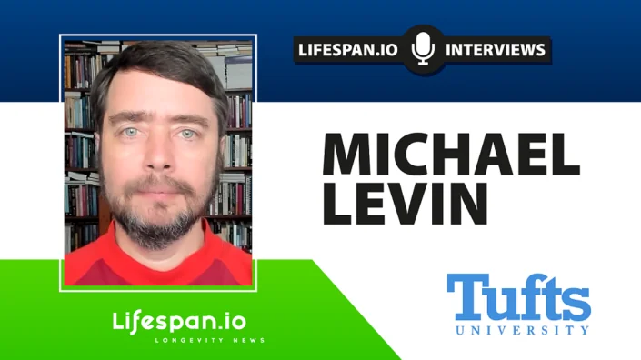
Michael Levin, professor at Tufts University and director of Allen Discovery Center, has been working for years on how bioelectrical patterns affect development and aging. His research proves that this often-overlooked part of biology is immensely important and that mastering its mechanisms might one day do wonders for human health and longevity. By manipulating ion channels in cells other than neurons, Levin’s team has been able to engineer growth of new limbs and organs, suppress cancer, and produce something that looks like entirely new forms of life. However, this research also touches on some deeply philosophical questions.

You started your career as a software engineer. How did you end up doing biology?

I was interested in biology from an early age, along with electrical engineering. As a child, I had asthma, and we didn’t have any medications [Michael’s family emigrated to the US from Russia when he was a child]. So, my dad used to take the back off of the TV set—we had this giant wooden thing with a tiny screen in the front and vacuum tubes in the back—and we would sit there and look at it as a way to distract me during attacks. I was absolutely fascinated that somebody knew how to put all that stuff together in just the right order to make the show come out on the other side. I remember asking my dad, “How did somebody know to do this?” and hhe said, “Well, you can learn to do this. It’s called engineering,” and I thought, “There you go. That’s the best magic.”

Then, I had an older friend who was into insects, bugs, and beetles. We would go outside and look at all this stuff, and he would point out that this is the egg that turns into a caterpillar that turns into a butterfly. I was always interested in those things, watching them develop and interact with each other and their environment.

When I came to the US, I discovered computers and realized that software was fascinating. Before college, I worked in scientific computing, writing code for companies and scientists, and wanted to work in AI, but having observed the state of computer science once I got to college, it became clear that we didn’t have any AI, and it wasn’t going to happen without learning about the one obvious case we do know: biology, where you watch right in front of your eyes a little blob of chemistry and physics become a creature with a mind, preferences, goals, and behavioral competencies.

So, I finished two BS degrees, one in computer science and one in biology, focusing on developmental biology. As I tell my students, there are two magical classes in the university. The first is developmental biology, where you see physics and chemistry becoming mind. The second is electrical engineering 101, where you start with Ohm’s Law and these very basic pieces of physics, and before you know it, you build something that does logic: the basic elements of thought.

How does your background affect your approach to biology?

Computer science and engineering really inform my thinking because they teach you to be comfortable with abstractions and coarse graining. Computer scientists are good at cutting biology’s endless complexity into chunks that matter, and we can say something useful without having to say absolutely everything. It’s like math, because it’s rigorous and allows you to explore the consequences of whatever ideas you can imagine, but it forces you to make things that actually run and work (the best parts of abstract thought and practical engineering).

You chose an area that’s not really popular with biologists, and not a lot of people are working in it. It’s actually amazing how few people are working in bioelectricity.

The reason I chose bioelectricity is that what I’m interested in fundamentally is how minds enter the physical world, how they scale, how they transform. If you think about what makes us greater than just a pile of neurons or cells in general, there must be some kind of cognitive glue. There must be policies that allow larger-scale systems to operate in spaces that their parts don’t operate in.

Which is what emergence is.

In neuroscience, we know what that is: electrophysiology enables this amazing network that underlies all these things that we do. I wanted to understand what the cognitive glue is for intelligence in other spaces. For example, the anatomical morphospace: development, regeneration, cancer suppression. All these things are not just a set of sequential chemical steps; they’re actually navigating a problem space.

One of the most amazing things about developmental and regenerative biology is the incredible plasticity and problem-solving capacity of groups of cells as they navigate that space. How is it that all the cells take that journey together? When you look at an embryo with hundreds of thousands or millions of cells, there’s alignment, the commitment of all the cells to the same story of where they’re going to in anatomical space.

I was very interested in understanding how that alignment happens, how an entire collective buys into the same story. In an important sense, models of the world, AKA stories, hold collectives together. All the cells are solving problems together because they’ve all bought into the same goal state they want to achieve.

To understand the physical mechanisms that enable a collective of cells to implement the same story, it turns out it’s exactly the same as in the brain. I was interested in this for a very long time, but for many years, I couldn’t say that. When you’re just starting out in such an unconventional area, you can’t tell people you’re studying cognition. The first grant proposal I wrote was, “I’m studying ion channel genes in development.” Reviewers said, ion channels are kind of weird, but genes in development makes sense, people study that, so okay.

After a while, having done some work on it, I was able to say, “Well, actually it’s voltage gradients in development.” Again, reviewers said that was pretty weird, but it followed from the prior work, so maybe it’s okay. After a while I could say, “No, it’s really computations in morphogenesis.” By now, I can say, “Actually, it’s cognition: decision making, memories, goal-directed problem solving.”

So, you’re studying cognition based on cells other than neurons, and you use an interesting definition of intelligence introduced by William James, which is basically the ability to get to a certain goal via different routes or means.

Yes, it’s a very nice definition because it’s very cybernetic. It doesn’t talk about brains, it doesn’t constrain what space a goal is in. It doesn’t say that you’re a brain or that you’re evolved or engineered. It’s a very generic definition that I think gets to the heart of what we mean by intelligence, which is the ability to adaptively navigate a problem space with some degree of competency to get your needs met.

Is this something that requires a certain level of algorithmic complexity?

That’s an interesting point. In some of the latest work on memories, I talk about this issue of a bow tie, like an hourglass turned on its side.

At any given moment, you don’t have access to the past. What you can access is the memory traces, the engrams that were left in your brain and body by things you did in the past. Your job is to continuously interpret those memory traces to build and update a model of what’s going on so that you can act in the future. It’s a model of continuous reinterpretation of your own memory traces.

I think the left side of that bow tie, the compression of experiences into the thin middle (the bow tie has this bottleneck, like an auto-encoder), that part is algorithmic. We can say how we do the compression, but the right side of that funnel, in my opinion, can’t be algorithmic. This is what is special about life, which is not the case with modern computer architectures.

The right side of that funnel has to be creative because you lost information going through that bottleneck. You cannot reinflate it algorithmically, meaning with steps that will get you exactly what you had at the beginning. What you can do, though, is have creative problem-solving: you get a prompt from your memories, and now you have this whole system for figuring out what that means for you.

I think the same thing is happening in anatomical space. The DNA you receive from past generations, we know it’s not a blueprint. I think of it as a prompt for a generative model. You receive these ‘memories,’ but now it’s up to you to interpret them. Under most circumstances, all things being equal, you will end up interpreting them the same way, which is why dogs have puppies and cats have kittens.

But we know that if you make some changes in their circumstances, the material is extremely clever at making other things. With a normal human genome, you could make an anthrobot as well as a human embryo. With a normal frog genome, you could make a xenobot as well as a frog embryo. With a planarian genome, you could make planarians with heads of other species. We’ve done this in our lab.

There’s a lot of interpretation going on; morphogenesis is a creative process. That’s important because the debate about whether some sort of computational analogy is a good tool for these kinds of things rages on. I think parts of it are algorithmic and parts of it are not algorithmic.

You mentioned a few important concepts that are part of your work: morphogenesis, planarian experiments, xenobots. The readers, at this point, might not know what all this means. So, can you briefly describe your research and where it currently stands?

One of the things we’re very interested in is that creative aspect of plasticity. This notion comes straight out of computer science, the idea of reprogrammability. Your genome tells every cell what computational materials it can have. Specifically, we’re most interested in ion channels and electrical synapses; the genome sets these materials for you.

This is already conceptually different from what many people are used to, which is that the genome is the software, right? But you’re introducing a different candidate, which is bioelectricity patterns. How does it work as software on top of the genetic hardware?

Hardware and software are, of course, a metaphor. The question is, “How do we apply this metaphor to biology?” The most popular way of mapping them is to say the genome is what matches the concept of software, and maybe the transcriptional machinery is the hardware. That’s the standard view.

I’m not saying it’s a bad metaphor; I’m saying it’s limited. There are scenarios where it prevents you from making novel discoveries. I’ll give you examples in bioelectricity, but there are other examples. There is also software that’s biochemical and runs on gene regulatory networks and chemical gradients. There’s also software that runs biomechanically on various physics gradients and forces. There are probably other things like biophotons.

I think bioelectricity makes the strongest case for this. There are numerous examples—we’ve developed a whole bunch of them—where knowing the genetic information leads you astray. It doesn’t tell you what’s actually going on.

For example, frog tadpoles don’t have legs, while axolotl embryos do have legs. We know their genomes. Now, in my lab, I make a “frogolotl.” A frogolotl is partly frog, partly axolotl. Looking at their genomes, can you tell me if a frogolotl is going to have legs or not? There’s no way to know. If it does have legs, will those legs contain frog cells or only axolotl cells? No models make predictions on this yet. Knowing the genetics is not enough to tell you what the pattern will be.

So, when it comes to developmental patterns, the genetics cannot really explain how an organism develops into its final form, how this type of intercellular communication works.

The genetics will certainly help you with the hardware of the communication. It will tell you that these cells have certain ion channels, certain ways to secrete chemical signals, gap junctions or synapses. What it doesn’t tell you is how the decision-making is going to work.

When a frog cell finds itself next to an axolotl cell, and all the axolotl cells are saying “build the leg” and the frog cells are not expecting to build a leg, what happens then? That’s a software question. You don’t get there from knowing the hardware.

Similarly, if I take two species of planaria (flatworms) with different shaped heads, and I take stem cells from one shape, stick them into the body of the other, and then cut the head off, what head shape is it going to make? Is one going to be dominant? Is it going to be an intermediate shape? Is it never going to stop regenerating because neither set of cells is ever happy about what’s happening?

These are questions that require understanding the decision-making of a collective intelligence: the group of cells that are navigating anatomical space. That’s very much a software question.

In our lab, we’ve studied examples where the software is a very important target for reprogramming the outcome. That’s critical because you can have philosophical debates about what’s software and what’s hardware, but the only reason any of that matters is: given however you’re using your metaphor, what does it enable you to do that couldn’t be done before?

Let me give you a few examples. We’ve shown that when you cut a normal planarian flatworm into pieces, every piece always develops one head, one tail. You can ask, how does it know how many heads it’s supposed to have? The standard answer is, “Well, the genome, of course.” Even though we know the genome doesn’t actually say anything about heads or head size or shape or number.

We developed the first molecular tools to read and write electrical information in living tissue, outside the brain. We’ve decoded bioelectrical patterns to understand how they say, “One head, one tail.” Then we said, “Let’s change the pattern so that it says, ‘two heads.'”

We don’t apply fields, there are no electrodes, frequencies, magnets, electromagnetic waves, nothing like that. We manipulate ion channels and gap junctions because that’s the electrical interface that the cells use to manipulate each other. So, we use ion channel drugs to manipulate the voltage pattern so that it says, “Two heads.”

If you do that, that fragment will regenerate with two heads. Not only that, but if you continue to cut that fragment in the future as many times as you want, with no more drugs of any kind, just regular water, they will continue generating two-headed worms forever. So now you have a strain of worms that has one head and a strain of worms that has two heads. The difference is not genetic; we didn’t touch the genome. There’s nothing genetically wrong with them. If you sequence them, you are none the wiser about why they have two heads.

So where does it say how many heads the worm should have? What the genetics actually gives you is a hardware specification that by default acquires a pattern that says, “One head”. Just like when you buy a programmable calculator from the store, when you turn it on, it says zero by default, but it’s reprogrammable, and living tissue is reprogrammable too. If you change the pattern it holds, it has a memory, and once you’ve set it to “two heads,” it holds.

Another example we have is in cancer, where we can introduce nasty human oncogenes into tadpoles, and they develop tumors. If we manage the voltage appropriately, you don’t get tumors even though the oncoprotein is still blazingly expressed.

We don’t kill the cells, we don’t fix the DNA, we don’t do anything with the DNA itself. Some hardware problems are fixable in software. In other words, you still have the oncoprotein, but if we keep that cell connected to the electrical network, then the collective works on nice things like building healthy skin or muscle, instead of being a unicellular organism and doing metastasis.

Your insights into cancer are very interesting: you say that it’s a cell reverting to its local unicellular goals, sort of losing communications with other cells.

I describe cancer as a dissociative identity disorder, quite literally. The right question is not “Why is there cancer?” It’s “Why is there anything but cancer all the time?”

Individual cells are very competent; they used to be unicellular organisms. But, how do you convince a bunch of cells to work together toward building this larger organism? Everything they do is focused on establishing metabolic, physiological, and other kinds of goals in a tiny 10-micron radius. That’s their size. I call this the “cognitive light cone”: it’s the size of the biggest goal you can work toward.

Individual cells have a quite small cognitive light cone; they pursue tiny local pH and metabolic states. But, you have this amazing thing in evolution and development where you suddenly pull cells together into a large network where the network has grandiose goals.

For example, in a salamander, the cells of the leg have the goal of building a leg. Why do I call it a goal? Because if I deviate them from that goal by cutting off the leg, they will spring into action, rebuild the leg, and then stop when it’s done. That’s the definition of a goal: it’s a cybernetic thing.

How did this happen? How is it that a bunch of cells together now have this giant cognitive light cone that’s centimeters in size and projects into a new space? Whereas before, it was projecting in physiological and metabolic space, now their goal exists in this anatomical space that individual cells don’t have access to.

What happened is that the cells are connected into a network that enables them to remember very large goal states.

Basically, a society of cells.

It’s a society, but it’s more than that. Any homeostatic process has to store the set point. There has to be somewhere where the set point is stored. In your thermostat, what you need to know is how to change the set point. Once you change it, the whole thing will act toward the new set point, no matter how the rest of it works.

Remembering what a salamander limb looks like is too much for one cell. Individual cells don’t know what a finger is or how many fingers you’re supposed to have, but groups of cells can, because they’re a larger computational network.

They’re a society, yes, but specifically they form a larger-scale processing network that can store these enormous goal states. That process builds larger intelligences that project with bigger cognitive light cones into new problem spaces.

That process obviously has a failure mode. What happens when cells disconnect from that electrical network? They can no longer remember that large goal. They revert back to their unicellular tiny little goals.

Before, the whole organism was “me,” and now the rest of the body is just the external environment. “I’m a single cell, but this external world—I don’t care what happens to it. I’m going to do whatever’s good for me.” That’s metastasis: “I’m going to replicate as much as I can, go where I want, eat what I want.”

If you have this view, you can approach cancer differently. Instead of killing the cells or trying to fix the genomics, you can forcibly reconnect them together. That’s what we’ve done, and it’s a therapeutic modality. We’re now moving from frogs to human tissues.

How is this information stored in that single cell where it all begins?

We can see how it’s stored: in the electrical circuits that are formed when the cell starts dividing. The really weird question is, where does the information come from? We have to dive into the philosophical question of what it means for something to come from somewhere.

Typically, biologists like information that comes either from history—meaning evolution or some kind of past events guided and made sure that the pattern was this and not something else. So, there’s a historical set of selection forces that made sure this is the information that’s here.

The other source is physics. In this case, both of those things are present. The history of evolution made sure that your cell has certain ion channels. By having certain ion channels, the excitable medium that you make when you have a collection of cells has certain properties. The electrical circuit has certain properties of memory, plasticity, symmetry breaking, amplification, and those processes are in part derived from the properties of the ion channels that are there. So, evolution is plugged in here.

Physics is absolutely also plugged in because some aspects of how electric circuits function are not part of biology; they’re part of physics and the laws of computation.

There’s a third component here. A similar problem has been discussed with respect to chemical patterns. Alan Turing, one of my heroes, was very interested in intelligence and unfamiliar embodiments. He wrote this paper on chemical self-organization and embryogenesis, weird for a guy interested in math, computers, and intelligence.

I think he saw a profound truth, and if he had lived longer, he would’ve written about it: the formation of the body and the formation of a mind are parallel, symmetrical processes. I think he was trying to get at this question of where the goal patterns of novel beings come from.

Typically, when you look at a goal-directed system such as any biological system, and you want to know why it makes the shape it makes or has the behavior it has, the answer is “Oh, evolution” because eons of selection made sure that’s what it has. But when you make new things, new bodies and new minds, as in AI or swarm robotics or any kind of collective intelligence, where do their goals come from?

When we make synthetic beings, such as xenobots and anthrobots (which are synthetic life forms made of cells), they’ve never existed before. There is no history of selection that makes for a good xenobot or tells an anthrobot what it has to be.

A xenobot, just to explain this to the readers, is sort of a clump of frog cells that begins its own life.

It’s what happens when you liberate some frog prospective skin cells from the embryo and take them away from the other cells that normally bully them into having a boring two-dimensional life as the outside of the embryo. Now, they get their own life, and they do something completely different. They organize into a little self-motile construct that runs around and does all sorts of interesting things, including reacting to sound stimuli.

They also reproduce, which I find pretty amazing.

It’s crazy. They do this kinematic self-replication. If you give them a bunch of loose epithelial cells, they will make more xenobots out of those. Then we do anthrobots, which are the same thing made out of adult human tracheal epithelial cells. We have a protocol that lets them assemble into this self-motile thing that does interesting things like heal neural wounds by joining them across the gap.

So there’s the history of selection, there’s stuff from physics, and the third thing is what Turing studied in his model of chemical symmetry breaking and amplification, reaction-diffusion systems and things like that. You have two chemicals that interact with each other in some particular way, and they make spirals or spots such as on a leopard, or stripes on a zebra.

Where do those patterns come from? There are two ways to address that question. One thing you can say is they’re emergent from the physical properties of the two chemicals that you have. You can say that, but it doesn’t really say anything. What emergence really says is “we got surprised.” We had things that we knew, we put them together, and then we got some new thing that we didn’t see coming.

I don’t like that because it feels like stamp collecting we’re just going to be surprised when we are, and we’ll write it down in our big book of emergent amazing things. What I like more is how many mathematicians look at it: there is an ordered, structured space of mathematical truths, like the Platonic space. Pythagoras also saw this very clearly—there is an ordered space of truths that matter functionally in the physical world.

Evolution makes use of them. They provide all kinds of “free lunches” for evolution. They make a difference in the physical world, but the key is that they themselves are not set by any facts of physics.

For example, if we look at a fractal, like these Halley plots, these amazing, beautiful, very organic-looking fractals that come from a very small formula, if you ask, “Where does that shape come from?” there are no facts of physics that determine it.

It emerges from a set of simple rules.

Basically, it’s a very simple rule, a very simple procedure that reveals this thing. When you want to ask where it comes from, you have two choices. You can say there’s a space of these patterns, and what you found is a pointer into that space. For a computer scientist, this looks exactly like pointers into a preexisting space of patterns.

When we make an embryo or a biobot or any kind of living or engineered thing, we’re providing a bunch of pointers or interfaces into that space of patterns. I like to assume, and this is a metaphysical stance, that it’s a structured, ordered space so that we can study it.

To me, Xenobots and Anthrobots are vehicles for studying the contents of that latent space. I don’t think that emergence as just “the surprising thing that sometimes happens” helps us enough. I think we have to assume it’s an ordered space. We have to make more tools to study it.

There are mathematical structures that will ingress into the physical world and guide what happens when you provide an appropriate interface. What’s an appropriate interface? If you want to see some basic facts around simple machines, you can make a lever and a fulcrum, and then you’ll see those patterns. If you want to see some facts of computation, you can make a bioelectric medium, and then you will see some of these patterns come through.

When we ask where these patterns come from, I think there are three factors: the two that are standard biology fare, evolution and physics, and then there’s this third thing, which I take very seriously, this space of patterns that we are now able to investigate systematically.

Rarely do I get to discuss the Platonic heaven with a geroscientist, but how is your research related to aging and age-related diseases?

The bare fact of the existence of planarians, I’m talking about the asexual forms that we have, is telling us that it is not the case that inevitably you accumulate mutations and get old and die.

Right, they’re one of the non-aging species, basically immortal.

Yes, the asexual planarians are immortal. So, we know that these ‘inevitable’ thermodynamic theories of aging cannot be the full explanation. Planarians give me hope that aging can be defeated.

Also, because planarians reproduce by asexual fission, they accumulate somatic mutations, and their genomes are a total mess. So, the fact that the animal with the messiest genome is also highly regenerative, cancer-resistant, and immortal also tells us that all this focus on genetics is somewhat misplaced.

After 20 years of driving myself crazy about it, I think we finally understand why there are no abnormal genetic strains of planaria. You can get flies with red eyes and crooked wings, and you can get mice with weird toes, but there are no weird kinds of planaria except for the two-headed strains that we made, and ours are not genetic. There’s a deep reason for that: planaria basically ignore most of their genetic information.

In planaria, individual cells die all the time and need to be replaced. When new cells come in, if you don’t know where to place them, you’re going to degrade over time. So, the ship of Theseus that is our body, the ship of Theseus isn’t the boat, it’s the plan in the mind of the workers who fix up the ship.

The collective must have a memory of what our body looks like so that it can continuously maintain anatomical homeostasis: that’s how regeneration works and how aging is resisted.

We study in our lab what happens to the bioelectrical pattern during aging. Two things can happen. First, the pattern can get degraded over time. If the bioelectrical pattern gets degraded, everything else is going to go wrong afterward.

We know this from our work in birth defects. We’ve shown that whether caused by chemical teratogens or by mutations in important genes, we can rescue terrible birth defects by forcing the appropriate bioelectrical pattern. If the bioelectrical pattern is good, it covers up a lot of defects in the hardware.

So, one possibility is that the bioelectrical pattern is getting fuzzy, and we’re now studying this. We have evidence for this in humans with senescent cells. The other possibility, of course, is that over time, the cells might become less able to execute the pattern; we have evidence for that too.

The first aspect is the bioelectric part of it, and how that relates to the goal of keeping the body plan implemented. Then there are two other things about aging that I think are quite different.

One is a cognitive theory of aging. The standard theories focus on whether it’s a thermodynamic noise issue or programmatic theories of aging where there’s an evolutionary reason we age and there are genes for this. But, there’s another option: what happens, given that the body is a goal-seeking agent that works to implement a certain anatomical goal in morpho-space, when that goal-seeking cognitive system has finished making its goal?

Think about the Judeo-Christian concept of heaven. You and your pet snake go to heaven, everything’s great. There is no noise, no degradation, no radiation, nothing. Everything’s perfect physically, for an infinity of years.

It’s like “happily ever after.” I’ve always asked myself this question too; it seems lazy as a concept.

Well, the question is: the snake maybe will be fine doing snake things every day, but for the human, do you really think we could stay sane? You’ll keep yourself busy for the first 10,000 years. What about after a billion years?

We did simulations of a goal-seeking morphogenetic system that was given a goal to build a body. It does, but then there is no new goal. What we observe, which is quite amazing, is that in the absence of noise and external stimulation, you have a fundamentally cognitive problem, which is dissociation. The pattern starts to degrade.

During embryogenesis, the goal gets met. Then, for some amount of time, there’s stasis, and then this thing just starts to degrade. We think that’s part of aging; what you need to do is either reinforce the goal or provide new goals. By new goals, I mean it’s possible that you’re not going to do a thousand years in the standard human body, but you could if every so often you switched it up a little bit, made some changes.

I think this is what planarians do when they split. The reason these asexual planarians are immortal is that every so often they provide a new challenge to themselves: they tear themselves in half, and that reminds all the cells what the morphogenetic goal is, and they rebuild. That gives them another few weeks to hang around, and then they do it again.

This kind of “boredom theory of aging,” that it’s fundamentally a cognitive dissociation at the bottom of it, is another thing we’re studying. The question is: can we use our bioelectric interface to provide a reminder to the cells of what they should be doing?

The third thing Leo Lopez in my group did was use phylostratigraphy, which is a bioinformatics tool that looks at gene expression and tries to estimate the age of various genes: “On the phylogenetic tree, how far back did these things appear?”

When he looks at transcriptomics from young humans and old humans, he finds something very interesting. As you get older, two things happen. First, some tissues start to differentially express genes that are further back on the phylogenetic tree. Not all tissues do this, but some do. They start to up- and downregulate more and more ancient genes. There’s this old observation that embryonic stages of development look like stages of evolution.

Where embryos of all kinds of species look like each other, and then they start to diverge.

Exactly. Very much like development traverses embryonic stages of past lineages, it looks like aging goes in the opposite direction. The cells start focusing on genes that are further and further back in evolution. Not only that, but you get this crazy dissociation where different tissues in the body do it quite differently.

When you’re young, all cells have a tight concordance of where you are on that phylogenetic tree, but when you get older, you start to get this divergence where the cells no longer agree as much.

One of the really wild possibilities is that whereas cancer is a kind of spatial dissociation, aging may be more of a temporal dissociation, floating off backwards on the scale of evolution. This gives rise to some of our efforts to remind cells what their priors are.

You have co-founded some interesting companies, including one working on regeneration in mammals.

There are three companies. Morphoceuticals works on using bioelectric cues to regenerate limbs. We did it in frogs some years ago, and we are now working on this in rodents. I don’t have anything to report yet; we’re still working on it.

Fauna Systems is formed around designing bespoke synthetic living machines for applications in the environment: sensing, cleanup. Right now, it’s mostly focused on xenobots, but there many other technologies are coming. The idea is using AI to help communicate novel goals to the materials so that you get biological robots of desired form and function.

The third company, which may be of the most relevance here, is called Astonishing Labs. It’s working on aging; all the stuff I told you today about aging are things that Astonishing Labs is doing. It’s also working on anthrobots, which are personalized in-the-body agential interventions. Anthrobots can be made of your own cells, so you don’t need immunosuppression.

If you had an infinite timeline, what do you think could be done with your work on bioelectricity for aging? What’s the best thing you can imagine?

Where I think this is going, assuming we all survive and live into the future to do science, I am looking at a world that has what I call “freedom of embodiment.” The children of the future will look back on this time (and it’s not that far off, I think we actually have a roadmap to it), and it’ll be like when we first learned about ancient humans, and we said, “My God, imagine living in a world where if you step on a sharp stick, you get sepsis and die.” The children of the future will look back at us and say, “You’re telling me that these people had to live in whatever body they were randomly given? And they had no choice about their IQ, capabilities, hopes and dreams?”

And, of course, lifespan.

Lifespan, diseases, and they were susceptible to all sorts of stupid viruses and bacteria, and they got lower back pain and astigmatism and brain degeneration, and everything was up to this random walk of evolution. Incredible. How could anybody live like this?

In the future, we’re going to have an “anatomical compiler” to specify what living form you want and compile it into a list of stimuli that have to be given to cells to get them to build what you want. At that point, forget about regeneration, aging, and injury. You will be able to have the body you want. You want an extra brain hemisphere? You want to directly sense the solar weather and the stock market? Why not? We’ve made them in the lab in model systems. Biology is incredibly plastic, and I absolutely think it’s possible.

We would like to ask you a small favor. We are a non-profit foundation, and unlike some other organizations, we have no shareholders and no products to sell you. All our news and educational content is free for everyone to read, but it does mean that we rely on the help of people like you. Every contribution, no matter if it’s big or small, supports independent journalism and sustains our future.
Intestines

Young Microbiota Transfer Reduces Aging Aspects in Mice

In a recent study, lifelong, repeated microbiota transfer from young mice to old mice improves intestinal permeability, coordinative ability, and metabolic profiles while reducing pro-inflammatory responses [1].

Small in size, but mighty in impact

Previous research has found that the composition and function of gut microbes (microbiota) changes as we age. These changes are linked to health and lifespan [2], suggesting that the microbiota can be targeted for lifespan and healthspan extension.

Initial experiments with microbiota transfer in model organisms gave positive results, suggesting the possibility of lifespan extension [3] or improvements in brain function [4]. However, such studies usually involve only one transfer and use mostly germ-free mice, which exhibit many changes in physiology, from an attenuated immune system to problems with nutrient absorption [3]. Being raised germ-free, without any microbiota in the gut, is also not an approach that can be translated into human therapeutic strategies in the future.

A different approach

The authors of this study took a different approach. They performed recurring (every 8 weeks) fecal microbial transfers and used conventionally raised mice. Before each colonization procedure, they treated them with an antibiotic to wipe the intestinal microbiota and improve the efficiency of microbiota transfer. Using antibiotics comes with some downfalls, such as the possibility of developing antibiotic resistance and the potential impact of antibiotics on aging processes.

To achieve a rejuvenating effect, the microbiota was derived from 8-week-old mice (young microbiota); in the control animals, the researchers transferred the microbiota from animals of the same age.

Optimizing for a longer lifespan

The researchers monitored the animals until the end of the experiment (week 120), when a large proportion of mice in the control group died quickly. This suggested lifespan extension in the group receiving the young microbiota transfer; however, this result is not statistically significant due to a low number of remaining animals.

The researchers suggest that the lack of statistical significance, despite the relatively large number of animals at the beginning of the study (20 per group), could be due to variability in biological response to the treatments. They also note that the death of several animals in both groups of early time points could contribute to the lack of statistical significance. Those deaths were induced by lesions caused by forcibly feeding the animals when transferring the microbiota.

In future studies, they recommend optimizing the treatment regimen for optimal lifespan extension. They believe that such parameters as antibiotic dosing, frequency of transfers, and less invasive fecal transplant methods are essential.

Improvements, but not everywhere

The researchers analyzed a few aging-related phenotypes. They noted no difference between groups regarding glucose homeostasis, as measured by the glucose tolerance test.

Muscle function, assessed by measuring grip strength, also didn’t show differences between groups, but the researchers observed coordination improvements in the mice receiving young microbiota transplants.

Rejuvenated microbiota rejuvenates the host

Since the microbiota has direct contact with the intestinal walls, the researchers analyzed the impact of the microbiota transplants on the intestinal barrier. Young microbiota transfers reduced the amount of bacterial antigen leakage, suggesting improved intestinal barrier function.

Changes were also observed in the composition of microbiota. The group that received the young microbiota transplant had a microbiota composition more similar to that of the 8-week-old animals. The metabolic functions of the microbiome were also rejuvenated. This rejuvenated microbiota “provided beneficial metabolic functions to the host and thereby may contribute to delaying physiological processes associated with aging.”

Among the improved composition of microbiota content in the animals receiving young microbiota transfer, the researchers pointed to the increased abundance of Akkermansia, a bacterium linked to improved health and lifespan in mice [5]. However, some results need further investigation and clarification, including the reduced abundance of Lactobacillus bacteria, which are normally considered beneficial, in animals receiving the young microbiota transfer. The researchers speculate that differences in specific strains might drive the differences, but they require further investigation.

Rejuvenated biological processes

Transplanting young microbiota led to gene expression changes in immune, colon, and small intestine cells. Many of those changes were cell-type specific, and they pointed to effects on some aging-related processes.

First, the researchers assessed mesenchymal scores by measuring the expression of signature mesenchymal genes in epithelial cells. More epithelial cell transition into a mesenchymal-like state is associated with older age. They observed lower mesenchymal scores in several types of intestinal epithelial cells in animals who received young microbiota transfers compared to the control group, suggesting rejuvenation processes.

Next, the researchers used gene expression associated with aging-related inflammation to create an inflammatory score. This score was lower in young mice compared to old mice.

In mice who underwent lifelong microbiota transplants, the inflammatory score was lower in multiple immune cells from mice who received young microbiota transplanta. This observation aligns with the previously observed restored intestinal barrier in those animals, as reduced leakage through the barrier helps reduce inflammation.

Apart from gene expression, the researchers also investigated ligand-receptor interactions, interactions between two proteins that, after recognizing each other, can initiate and regulate many biological processes.

An analysis suggested fewer ligand-receptor interactions in epithelial and immune cells derived from mice receiving young microbiota. However, there were differences between those two cell types, with epithelial cells showing stronger interactions. According to the authors, these results suggest “more focused transcriptional response after microbiome rejuvenation.”

However, in the immune cells, the interaction strength decreased, with macrophages and T cells contributing the most to the decrease in the number and strength of interactions. Further investigation is needed to understand those differences and their connection with rejuvenation.

Optimizing for better health in humans

While this study didn’t report significant differences regarding lifespan extension following continuous young microbiota transfers in mice, it reported substantial healthspan-related improvements such as better coordinative ability, a tightened intestinal barrier, reduced inflammation, and cell type-specific changes in gene expression and rejuvenated metabolic profiles. This study’s results suggest that microbiota transfer can be an interesting treatment for healthspan or lifespan extension, but it needs further optimization and testing in humans.

We would like to ask you a small favor. We are a non-profit foundation, and unlike some other organizations, we have no shareholders and no products to sell you. All our news and educational content is free for everyone to read, but it does mean that we rely on the help of people like you. Every contribution, no matter if it’s big or small, supports independent journalism and sustains our future.

Literature

[1] Sommer, F., Bernardes, J. P., Best, L., Sommer, N., Hamm, J., Messner, B., López-Agudelo, V. A., Fazio, A., Marinos, G., Kadibalban, A. S., Ito, G., Falk-Paulsen, M., Kaleta, C., & Rosenstiel, P. (2025). Life-long microbiome rejuvenation improves intestinal barrier function and inflammaging in mice. Microbiome, 13(1), 91.

[2] Sommer, F., & Bäckhed, F. (2013). The gut microbiota–masters of host development and physiology. Nature reviews. Microbiology, 11(4), 227–238.

[3] Smith, K., McCoy, K. D., & Macpherson, A. J. (2007). Use of axenic animals in studying the adaptation of mammals to their commensal intestinal microbiota. Seminars in immunology, 19(2), 59–69.

[4] Parker, A., Romano, S., Ansorge, R., Aboelnour, A., Le Gall, G., Savva, G. M., Pontifex, M. G., Telatin, A., Baker, D., Jones, E., Vauzour, D., Rudder, S., Blackshaw, L. A., Jeffery, G., & Carding, S. R. (2022). Fecal microbiota transfer between young and aged mice reverses hallmarks of the aging gut, eye, and brain. Microbiome, 10(1), 68.

[5] Bárcena, C., Valdés-Mas, R., Mayoral, P., Garabaya, C., Durand, S., Rodríguez, F., Fernández-García, M. T., Salazar, N., Nogacka, A. M., Garatachea, N., Bossut, N., Aprahamian, F., Lucia, A., Kroemer, G., Freije, J. M. P., Quirós, P. M., & López-Otín, C. (2019). Healthspan and lifespan extension by fecal microbiota transplantation into progeroid mice. Nature medicine, 25(8), 1234–1242.

Organelles

TFEB Lets Cells Live Long Enough to Become Senescent

In Aging Cell, researchers have explained how transcription factor EB (TFEB) is related to cellular senescence and keeps stressed cells alive.

Inactivated by mTOR

Yesterday, we reported on TFEB’s effects on proteostasis and how it has downstream functions on protein chaperones. This research, however, focuses on an somewhat different aspect of this transcription factor: its effects on the lysosomes that digest unwanted proteins and its role in autophagy, the process by which cells consume their own organelles as a maintenance process [1].

Previous work has found that mTOR has effects on TFEB, phosphorlyating it and rendering it inactive within the cytosol of the cells. Reversing this process leads to the activation of a gene network that alters lysosomal function. When nutrients are abundant, mTOR kicks in; when there are fewer nutrients or when the lysosome is stressed, TFEB becomes active [1].

The researchers note a documented oddity in mTOR’s function and reactions. During senescence, it activates components of the SASP [2]. However, the stresses associated with senescence also render mTOR inactive, thus activating TFEB; this has been suggested to be a reason why senescent cells stay alive [3]. This work, therefore, was done to codify the relationship between mTOR, TFEB, cellular senescence, and oxidative stress sensors.

Surviving the storm

For their first experiment, the researchers chemically induced senescence in a population of human dermal fibroblasts, which are commonly used in senescence studies. Four days of twice-daily administration of this toxin put the cells under significant stress (the “stress phase”), driving them senescent five days after that.

The researchers found that the lysosomes were highly activated during the stress phase, but by the time the cells became fully senescent, this overactivation had ceased. Similar results were found when ultraviolet radiation was used to drive cells senescent instead of a chemical.

The stressed cells were clearly having problems performing autophagy. The researchers found that autophagic flux, a measurement of this maintenance process, decreases when the lysosomes are stressed. Removing the stressors allows the return of proper autophagy, even when the cells have been driven to senescence.

During the stress phase, TFEB was found to be in the nucleus, becoming activated while mTOR was found to be deactivated. However, during senescence, TFEB was found to reside inactivated in the cytosol, presumably due to the effects of mTOR. The transcription factor that had allowed these cells to survive to senescence had no need to be active when they were actually senescent.

Both of the oxidative stress indicators AMPK and Akt affect mTOR. Just like TFEB, AMPK activation was increased during the stress phase but dwindled during senescence. Akt, on the other hand, decreased during stress and increased during senescence instead. These findings dovetail with previous work showing a signaling relationship between AMPK and Akt [4].

More TFEB means more survival and more senescence

The researchers created cells that overexpress TFEB. Compared to a control group, more of these cells survived the chemical that drove them senescent, preventing far more deaths by apoptosis. However, despite their increased survival rate, these cells progressed into senescence. As expected, depleting TFEB decreased the cells’ ability to survive.

Crucially, there appears to be no truly direct link between TFEB and senescence, as senescence-related pathways were unaffected by its depletion. TFEB is a cellular survival mechanism; the fact that the cells survive to become senescent is not within its biochemical purview.

The researchers float the idea that TFEB inhibitors could be used as a pre-senolytic, causing stressed cells to die rather than linger around excreting the inflammatory SASP. However, given TFEB’s effects on other cells, such an inhibitor would need to be precisely targeted.

We would like to ask you a small favor. We are a non-profit foundation, and unlike some other organizations, we have no shareholders and no products to sell you. All our news and educational content is free for everyone to read, but it does mean that we rely on the help of people like you. Every contribution, no matter if it’s big or small, supports independent journalism and sustains our future.

Literature

[1] Napolitano, G., & Ballabio, A. (2016). TFEB at a glance. Journal of cell science, 129(13), 2475-2481.

[2] Carroll, B., Nelson, G., Rabanal-Ruiz, Y., Kucheryavenko, O., Dunhill-Turner, N. A., Chesterman, C. C., … & Korolchuk, V. I. (2017). Persistent mTORC1 signaling in cell senescence results from defects in amino acid and growth factor sensing. Journal of Cell Biology, 216(7), 1949-1957.

[3] Curnock, R., Yalci, K., Palmfeldt, J., Jäättelä, M., Liu, B., & Carroll, B. (2023). TFEB‐dependent lysosome biogenesis is required for senescence. The EMBO journal, 42(9), e111241.

[4] Zhao, Y., Hu, X., Liu, Y., Dong, S., Wen, Z., He, W., … & Shi, M. (2017). ROS signaling under metabolic stress: cross-talk between AMPK and AKT pathway. Molecular cancer, 16, 1-12.

Protein folding

Limiting One Protein Maintenance Pathway Enhances Another

In Aging Cell, researchers have explored how transcription factor EB (TFEB) promotes proteostasis in a common aging model.

Maintaining protein function

Proteostasis is naturally maintained by a quality control system that utilizes a network of chaperones and co-chaperones in order to handle all aspects of protein maintenance, including both folding and unfolding along with destruction of misfolded or otherwise damaged proteins [1]. This process is partially controlled by stress responses that act on transcription factors, which upregulate and downregulate proteostasis-related genes [2].

One of these key factors is coenzyme A (CoA), which is widely used throughout the body in multiple fundamental biochemical reactions, including mitochondrial energy-gathering processes, natural steroid synthesis, and protein management. To synthesize CoA, the body uses pantothenate kinase (PanK), which is the key bottleneck in its production [3]. As expected, genetic mutations that cause PanK deficiencies lead to severe diseases, such as brain degeneration [4].

CoA also produces clusters of iron and sulfur (ISCs) that are used as electron banks for biochemical reactions. While they are assembled in the mitochondria [5], they are transported throughout the cell through a complicated network, and disruptions to this network lead to serious diseases [6].

This work explores how CoA and ISCs are related to the common transcription factor TFEB. TFEB has an ortholog, HLH-30, in C. elegans worms, and so the researchers refer to it as HLH-30/TFEB throughout their paper.

A possible benefit of limitation

In C. elegans, full PanK depletion leads to similar problems as it does in humans [7]. However, halving its production does not lead to decreased lifespans in these worms.

Instead, worms that had a genetic mutation that led to PolyQ expansion, a proteostasis disorder that leads to formation of aggregates that cluster as foci within their bodies. Worms that had both this disorder and halved PanK production had fewer of these foci in their muscles, along with more activity and muscle movement, than worms that only had the disorder, although they had the same number of aggregates. Interestingly, this only occurred in worms treated with RNA interference (RNAi) to reduce PanK expression rather than worms that were created with halved PanK at the genetic level.

Intrigued, the researchers then turned to a model with a mutation that mis-encodes a crucial muscle protein, making it temperature sensitive. At the wrong temperature, worms with this mutation find it much more difficult to move as these proteins misfold. However, reducing PanK in these worms, through either a different genetic modification or RNAi, improves their movements at the wrong temperature, suggesting that proteostasis mechanisms are working harder.

Work with a labeled insulin protein recapitulated these findings. Worms that can properly process it place it, properly folded, into the body cavity, while worms that cannot process it leave the misfolded protein in the cells. Causing worms to undergo chronic stress through a mutation harms their proteostasis, meaning that they leave it in their cells; however, decreasing PanK through mutation or RNAi leads to more properly folded protein.

The resarchers then took a closer look at stress. They found that worms with less PanK were better at handling stress, whether that was due to a chemical insult or to heat shock; the difference was slight compared to unstressed controls, but it was still clearly visible in the data and statistically significant.

Finally, and critically, the same was found to be true of human cells. Bone cancer cells that were given a PanK inhibitor survived against heat shock more than cells not given the inhibitor. Similar results were found in another cell type. PanK inhibition did not affect these cells’ viability when there was no heat stress applied.

These results were found to be contingent on the resulting decreased levels of CoA. Worms that were given CoA in addition to decreased PanK had none of the previously observed benefits.

Protein chaperones to the rescue

Their findings led the researchers to investigate possible causes, looking closely at the mitochondria in an effort to find out why decreasing something as fundamental as CoA would lead to increased, not decreased, proteostasis. Most of the CoA-related pathways that they investigated were unfruitful; interfering with them only made things worse. However, silencing two genes relating to ISCs was found to be beneficial.

Further work found that interfering in these ISCs at any point was of benefit, whether it was their origination in the mitochondria or their transport throughout the cell. These effects were not additive with PanK silencing, demonstrating a chain of causality: the decrease in ISCs caused by PanK suppression leads to improvements in proteostasis.

These proteostasis improvements were not related to the proteasome nor the lysosome, which degrades misfolded proteins, nor did they have anything to do with the cellular clearing function known as autophagy. Instead, they were found to be related to an increase in chaperone-mediated protein folding; targeting these chaperones destroyed the ability of PanK reduction to increase performance under stress.

The role of TFEB

There were 13 specific chaperones found to improve heat shock survival, and 10 of them were related to HLH-30/TFEB in an existing data set. The mutants with less PanK had significant increases in a multitude of related genes. As with the CoA supplementation, interfering with HLH-30/TFEB stopped these chaperones from working and so prevented PanK reduction from increasing survival under heat shock conditions.

Therefore, the researchers concluded that reducing CoA and ISCs through decreased PanK is responsible for upregulating HLH-30/TFEB, which has substantial and beneficial downstream effects on protein folding through chaperones.

While very detailed in its biochemistry, this is a preliminary study. None of these treatments led to significant lifespan increases in worms, whether they were placed under stress or not. While they mentioned that it is a clear target, the researchers did not increase HLH-30/TFEB through a genetic intervention or other means. Further work, on worms and mice, will need to be done in order to determine if directly boosting chaperones could possibly be an effective treatment for proteostasis disorders such as Alzheimer’s and Parkinson’s.

We would like to ask you a small favor. We are a non-profit foundation, and unlike some other organizations, we have no shareholders and no products to sell you. All our news and educational content is free for everyone to read, but it does mean that we rely on the help of people like you. Every contribution, no matter if it’s big or small, supports independent journalism and sustains our future.

Literature

[1] Jayaraj, G. G., Hipp, M. S., & Hartl, F. U. (2020). Functional modules of the proteostasis network. Cold Spring Harbor Perspectives in Biology, 12(1), a033951.

[2] Pessa, J. C., Joutsen, J., & Sistonen, L. (2024). Transcriptional reprogramming at the intersection of the heat shock response and proteostasis. Molecular cell, 84(1), 80-93.

[3] Robishaw, J. D., & Neely, J. R. (1985). Coenzyme A metabolism. American Journal of Physiology-Endocrinology And Metabolism, 248(1), E1-E9.

[4] Gregory, A., & Hayflick, S. J. (2017). Pantothenate kinase-associated neurodegeneration.

[5] Paul, V. D., & Lill, R. (2015). Biogenesis of cytosolic and nuclear iron–sulfur proteins and their role in genome stability. Biochimica Et Biophysica Acta (BBA)-Molecular Cell Research, 1853(6), 1528-1539.

[6] Maio, N., & Rouault, T. A. (2020). Outlining the complex pathway of mammalian Fe-S cluster biogenesis. Trends in Biochemical Sciences, 45(5), 411-426.

[7] Samuelson, A. V., Carr, C. E., & Ruvkun, G. (2007). Gene activities that mediate increased life span of C. elegans insulin-like signaling mutants. Genes & development, 21(22), 2976-2994.

Peter Fedichev Op-Ed

Playing the Long Game Towards Radical Life Extension

For most of the history of human civilization, humanity expanded at an astonishing pace: faster than exponential, nearly hyperbolic. This trend was famously described in a 1960 paper by Heinz von Foerster and colleagues, who extrapolated global population data to predict a so-called “Doomsday”, a demographic singularity in which human numbers would become infinite by 2026, assuming growth continued unchecked [1].

However, something unexpected happened: birth rates began to fall even as lifespans continued to rise. This shift wasn’t directed by any authority; it was a spontaneous outcome of billions of individual choices. It was a striking example of emergent behavior in a complex system. In doing so, humanity collectively dodged what had once been seen as its greatest existential threat: overpopulation.

Yet, the success of this transition has brought a new crisis: as we live longer, we don’t necessarily live better. The burden of chronic disease rises sharply with age, driving healthcare costs higher and threatening to destabilize pension systems worldwide. The same demographic shift that saved us from overpopulation now demands a new kind of solution; we must either decouple aging from disease or stop aging altogether.

This imperative has fueled the rise of longevity biotechnology, a modern crusade to unlock the secrets of extended life. Billions of dollars have poured into startups, research labs, and bold promises of reversing aging. We have made progress: we know that human life, and especially the lives of lab animals, can be stretched impressively.

Caloric restriction (CR), cutting food intake while maintaining nutrition, remains the gold standard, consistently extending lifespan in mice, worms, and even monkeys by slowing metabolism and reducing cellular wear. It’s a trick that nature has been hinting at for eons. Here’s the rub: despite all our high-tech tools, no cutting-edge intervention, whether cellular reprogramming with Yamanaka factors or advanced drug cocktails, has outperformed rapamycin or caloric restriction in animal models, whether tested alone or in combination. As Matt Kaeberlein has emphasized, nothing yet beats the effect of simply eating less.

This stagnation echoes a tale from 800 years ago, when Genghis Khan, the conqueror of empires, turned his gaze to conquering death itself. Around 1222, as he ravaged all of the taxable-at-the-time world, the aging Khan summoned Qiu Chuji, a Taoist monk famed for his wisdom on longevity. Genghis, nearing 60 and feeling the weight of his relentless campaigns, demanded the secret to eternal life: an elixir to defy time.

After a grueling two-year trek to meet the Khan, Qiu offered no potion, no magic. Instead, he counseled moderation: curb your excesses, avoid overindulgence in wine and war, live simply. It was caloric restriction and a balanced life in all but name, pragmatic advice rooted in observation, not mysticism. Genghis, perhaps disappointed, still honored the monk but died just five years later in 1227. Even then, with all his power and the wisdom of the age, nothing better than moderation emerged.

Ironically, it is traditional pharma that has edged closer to practical anti-aging interventions. Drugs like Ozempic, which were originally developed for diabetes and obesity, have shown real, though modest, mortality benefits across a growing list of conditions. These effects are meaningful, but they still fall short of fundamentally altering the aging process.

Meanwhile, the longevity field projects a contradictory message. On one hand, it claims we are close to developing a drug against aging; on the other, it acknowledges that we still lack a shared understanding of what aging actually is. We are like early aviators tinkering with wings and engines, achieving powered flight through trial and error. Drugs that mimic the effects of caloric restriction, like rapamycin and metformin, are our first creaky airplanes: promising, but still crude.

The ambition to truly defeat aging is not just about building better airplanes; it’s about realizing that no airplane, no matter how refined, can reach the Moon. To get there, humanity needed rockets, which are based on entirely different principles. Similarly, halting aging will demand not just incremental improvements, but a deep, principled mastery of the fundamental mechanics that drive the aging process.

As with the case of flying machines, nature provides numerous examples of evolutionarily advanced creatures, including some mammals, that exhibit little to no signs of aging and live up to 10 times longer than expected for animals of their size: an effect size far greater than that achieved by CR. This phenomenon, known as negligible senescence, is increasingly recognized as a distinct regime of aging. Notably, recent studies by the Calico team, using increasingly large animal cohorts of naked mole rates, have shown the absence of the acceleration of mortality, a defining feature of aging in humans.

We must begin thinking more deeply to make sense of these observations. In a 2007 paper in PLOS Genetics, Leonard Hayflick proposed entropy, the universal force driving systems toward disorder, as the fundamental cause of aging. Building on this idea and using modern molecular-level data, our team developed a comprehensive, quantitative theory of aging that integrates both dynamic (reversible) and entropic (irreversible) components of the aging process.

In a series of peer-reviewed studies, published in Nature Communications [2] and Aging Biology [3], we demonstrated that human aging is governed by a dual mechanism: a slow, linear accumulation of entropy that expands the footprint in physiological configuration space and leads to irreversible information loss, and, alongside it, dynamic fluctuations—short-term, reversible stress responses—that gradually destabilize the system and drive the onset of chronic diseases with age.

This combined framework not only confirms Hayflick’s hypothesis but extends it, providing a coherent and, crucially, quantitative explanation for how different biological systems age over time and eventually fail. Critically, it allowed us to classify organisms into two distinct aging regimes: relatively short-lived (“unstable”) species, like mice, whose aging is dominated by dynamic instabilities, and longer-lived (“stable”) species, like humans, where aging is driven primarily by the slow, irreversible accumulation of entropic damage. A key insight from this model is that interventions targeting only the dynamic components of aging, such as senescence or inflammation, have little to no effect on the underlying entropic damage, which is consistent with the expectations of macroscopic irreversibility as dictated by the second law of thermodynamics.

These findings are not merely of academic interest; they form the foundation for a new, quantitative theory of aging: one that explains mortality trends, biomarker divergence, and why long-lived species age differently from short-lived ones. At the heart of this theory lies a simple but powerful principle: aging in humans is driven by the accumulation of microscopic molecular insults—each individually benign and reversible, but collectively irreversible—that gradually erode physiological resilience. As this process unfolds, the organism becomes increasingly fragile, until even small fluctuations (biological noise) can push it past critical thresholds, triggering disease or death.

These findings and the underlying theory not only explain much of what is known about aging biology, but also generate important new predictions. In our 2021 Nature Communications publication, we provided one of the first direct measurements of the maximum human lifespan using longitudinal physiological data. By analyzing changes in biological markers, such as blood composition and physical activity patterns, we demonstrated that the variance and recovery time of physiological signals diverge near a critical age, between 120 and 150 years. This divergence signals a fundamental loss of resilience, indicating a hard upper limit on human lifespan. Our results suggest that maximum lifespan is not merely a statistical artifact of demographic data, but an objective, measurable, and potentially modifiable feature of human physiology.

Understanding the physics and biology behind this upper bound on lifespan is critical for evaluating the potential of longevity interventions. Our theory identifies three primary levers for intervention, which we classify into three distinct levels based on their potential effect size [4].

  1. Level-1 therapies target key molecular hallmarks of aging. These include CR mimetics, cellular rejuvenation therapies, senolytic therapies, telomere activators, and most other areas of research currently in the drug development pipeline. These work well in short-lived organisms where aging is unstable and markers of aging are tightly coupled. Level-1 therapies hold significant promise for addressing individual age-related diseases, including those with the largest market potential. Of those, diabetes alone reduces human lifespan by up to 8 years (depending on the age at diagnosis). This is why we expect that drugs aimed at improving metabolic health are expected to deliver the greatest benefits in this category.
  2. We predict the emergence of a new class of drugs, Level-2 therapies, designed to reduce physiological noise: the random fluctuations that destabilize health as organisms approach the limits of resilience. By damping this noise, these therapies could decouple aging from the onset of diseases, effectively “squaring” the survival curve. In practical terms, Level-2 interventions could add 30-40 years of healthy life by bridging the gap between today’s average lifespan (70-80 years, depending on the country) and the maximum natural lifespan of 120-150 years. However, they would not significantly extend the maximum lifespan itself.
  3. Level-3 therapies would aim to halt—or, as some hope, reverse—the accumulation of entropic damage itself. These therapies would not merely extend life; they would arrest functional decline and push maximum lifespan beyond the current 120-150 year limit. Because entropic damage accumulates slowly, future experiments and clinical trials will require the development of actionable biomarkers to track it. Targeting or controlling this damage will be challenging and will likely demand novel technologies, such as advanced organ replacement and new animal models of aging. Nevertheless, our theoretical framework provides a solid analytical foundation and brings these ambitious goals within conceptual reach.

Without a clear theoretical understanding of the aging process, drug development efforts often fall into the trap of focusing narrowly on specific disease indications. This is why most researchers, investors, and entrepreneurs in longevity biotechnology are currently centered on Level-1 therapies. These interventions may delay the onset or progression of individual diseases and modestly improve healthspan. However, they will not alter the fundamental dynamics of aging or extend the maximum lifespan.

The theoretical picture sends a dire warning. Level-1 biology addresses diseases in humans but has only a modest effect on lifespan. Level-2 interventions may further reduce the incidence of diseases and mortality. However, neither Level-1 nor even Level-2 therapies alone can alter the rate of functional decline. Aging is not merely the sum of diseases. Even in the absence of illness, humans grow increasingly fragile over time. A 90-year-old free of disease remains a diminished, less resilient version of their younger self.

The reason is that irreversible damage accumulates over time, leading to the progressive and likely irreversible decline of key functional indicators such as IQ, VO₂max, kidney function, and others that collectively define physiological resilience and quality of life. While squaring the curve, extending healthspan toward maximum lifespan, could significantly prolong life compared to current averages, it would not intercept the aging process itself. Without directly addressing underlying decline, it risks becoming a path to prolonged disability rather than true rejuvenation.

Only a combination of Level-2 therapies that decouple aging from disease—and, even more critically, Level-3 therapies that target or reverse damage accumulation—will extend lifespan and preserve function, opening the path to negligible senescence in our species.

The era of low-hanging fruit is coming to an end. Rather than chasing isolated hallmarks of aging or targeting individual diseases, we must now approach aging as a system-level, entropy-driven process. My scientific aspirations are firmly at Level-3, but my instincts tell me that Level-2 therapies, those that reduce biological noise, can be discovered and developed into real medicines much sooner with today’s technology.

This is the current focus of our research and development at Gero. We are investigating the biology of physiological noise using longer-lived mammals, such as dogs, as model organisms. Whether it is us or another research team that ultimately cracks the code of Level-2 or Level-3 interventions, true success will come only through a comprehensive understanding of the aging process and by raising the bar for what we expect from interventions. Without that foundation, no amount of billions spent will carry us much farther than the same old wisdom Genghis Khan got almost exactly 800 years ago.

We would like to ask you a small favor. We are a non-profit foundation, and unlike some other organizations, we have no shareholders and no products to sell you. All our news and educational content is free for everyone to read, but it does mean that we rely on the help of people like you. Every contribution, no matter if it’s big or small, supports independent journalism and sustains our future.

Literature

[1] Von Foerster, H., Mora, P. M., & Amiot, L. W. (1960). Doomsday: Friday, 13 November, AD 2026: At this date human population will approach infinity if it grows as it has grown in the last two millenia. Science, 132(3436), 1291-1295.

[2] Pyrkov, T. V., Avchaciov, K., Tarkhov, A. E., Menshikov, L. I., Gudkov, A. V., & Fedichev, P. O. (2021). Longitudinal analysis of blood markers reveals progressive loss of resilience and predicts human lifespan limit. Nature communications, 12(1), 2765.

[3] Tarkhov, A. E., Denisov, K. A., & Fedichev, P. O. (2024). Aging clocks, entropy, and the challenge of age reversal. AgingBio, 2, e20240031.

[4] Denisov, K. A., Gruber, J., & Fedichev, P. O. (2024). Discovery of Thermodynamic Control Variables that Independently Regulate Healthspan and Maximum Lifespan. bioRxiv, 2024-12.

Rejuvenation Roundup April 2025

Rejuvenation Roundup April 2025

While computer technology continues to accelerate, human beings still have to contend with age-related disease and death. Here’s some of what’s been done to support longer, healthier lives in April.

A Lifespan.io Project Bears Fruit

Results of a Crowdfunded One-Year Human Rapamycin Trial: In Aging, Dr. Sajid Zalzala and his team have published the results of Participatory Evaluation of Aging with Rapamycin for Longevity (PEARL), a randomized, controlled human clinical trial that was crowdfunded by Lifespan.io.

Research Roundup

Scientists Discover a New Mitigator of Senescence: Working with flies, mice, and human cells, scientists have demonstrated that a highly conserved protein can modulate cellular senescence, potentially opening a new avenue for future therapies.

Activation of Silent X Chromosome Might Improve Cognition: Using a mouse model, researchers from UCSF have found that the genes that become activated on the silent X chromosome might explain some sex-dependent differences in cognitive abilities during aging.

Extracellular Vesicles Restore Some Heart Function to Mice: Publishing in Stem Cell Research & Therapy, researchers have found that small extracellular vesicles (sEVs) derived from young mice alleviate heart dysfunction in older mice.

Reprogramming Epigenetics to Fight Back Pain: A new paper published in the Nature journal Bone Research has found that reprogramming the epigenetics of spinal disc cells reduces senescence and alleviates pain in a rat model.

Fasting Affects the Immune System via the Brain: A new study has found that the immune remodeling associated with fasting can be recapitulated by activating a subset of neurons in the hypothalamus. The findings could be important in the context of fasting mimicking, metabolic disorders, and cancer.

Dietary Patterns Associated With Healthy Aging: A recent study investigated the impact of eight different mid-life dietary patterns on the odds of healthy aging, including cognitive, mental, and physical health.

How Exercise May Fight Parkinson’s Disease: Experimenting on rodents, researchers have found a pathway through which exercise could fight Parkinson’s disease and a molecule that might recapitulate this effect.

Neurons Hidden to Immune Cells Improve Parkinson’s in Rats: By tweaking genes used by placental and cancerous cells to evade immune detection, scientists have created non-immunogenic neuronal grafts that may help Parkinson’s patients.

Senolytics Decrease Low Back Pain in Mice: Researchers have tested a synthetic and natural senolytic combination of RG-7112 and o-vanillin in mice with early-onset low back pain and disc degeneration. They observed reduced signs of back pain; decreased senescence and disc degeneration; and improvements in vertebral bone quality.

Researchers Use Big Data to Find a Longevity Target: Researchers publishing in Aging Cell have used large databases to discover a causal relationship between multiple genes and overall mortality risk, finding a new potential target in the process.

A Senescence-Related Target for Blood Vessel Formation: In Aging Cell, researchers have linked macrophage senescence to the failure of new blood vessel formation, finding a key target that might make it easier to treat arterial clogs.

Researchers Fight Some Mutations by Targeting Mitochondria: Clonal hematopoiesis, a condition linked to numerous age-related disorders, can be caused by overachieving mitochondria, and it may be susceptible to drugs such as MitoQ and metformin.

Technology Use Associated With Reduced Cognitive Impairment: New research suggests that digital technologies provide beneficial effects on cognition in middle-aged and older adults who did not grow up with such technologies.

How Fisetin May Fight Blood Vessel Calcification: In Aging, researchers have described how the well-known supplement fisetin may fight calcification of the blood vessels, seeing significant successes in both cellular and mouse models.

OS-01 Peptide Tested in Skin Aging Pilot Study: A recent study featured in the Journal of Cosmetic Dermatology has analyzed the impact of a topical product containing OS-01. This is a senotherapeutic peptide that targets senescence, affecting the skin’s barrier function and multiple aging biomarkers.

How Extracellular Vesicles From Stem Cells Fight Senescence: Researchers have discovered a cocktail of micro-RNA strands that make some extracellular vesicles (EVs) effective in reducing cellular senescence and published their findings in Aging Cell.

Immune Resilience Is a Strong Determinant of Mortality: A new study defines the concept of immune resilience and positions it as a central determinant of aging trajectories, linking it to survival, inflammation control, and the body’s ability to withstand stress.

Impact of Butter and Plant-Based Oils on Mortality: A study published in JAMA Internal Medicine suggests that a higher intake of butter is associated with increased mortality while a higher intake of plant-based oils is associated with reduced mortality.

Dietary methionine restriction started late in life promotes healthy aging in a sex-specific manner: The observed benefits provide a translational rationale to develop methionine restriction mimetics as an anti-aging intervention.

Targeting Senescence with Apigenin Improves Chemotherapeutic Efficacy and Ameliorates Age-Related Conditions in Mice: This study demonstrates the feasibility of exploiting a natural compound with senomorphic capacity to achieve geroprotective effects by modulating the SASP.

Niclosamide extends health span and reduces frailty by ameliorating mTORC1 hyperactivation in aging models: It extended lifespan and improved frailty-related phenotypes in C. elegans and effectively ameliorated frailty in aging mice, particularly in muscle aging.

Associations between five indicators of epigenetic age acceleration and all-cause and cause-specific mortality among US adults aged 50 years and older: AAGrimAge and AAGrimAge2 outperformed AAHorvathAge, AAHannumAge and AAPhenoAge in predicting mortality risk, and the association pattern was positive.

Maximum lifespan and brain size in mammals are associated with gene family size expansion related to immune system functions: The researchers conducted a genome-wide analysis not restricted to specific functional gene categories and analyzed the overlap between MLSP-associated genes identified in this study with gene sets previously associated with human longevity and molecular processes relevant to aging and longevity evolution.

AI-Driven Identification of Exceptionally Efficacious Polypharmacological Compounds That Extend the Lifespan of Caenorhabditis elegans: Rationally designing polypharmacological compounds enables the design of geroprotectors with exceptional efficacy.

Life-long microbiome rejuvenation improves intestinal barrier function and inflammaging in mice: Life-long and repeated transfer of microbiota material from young mice improved age-related processes, including coordinative ability (rotarod), intestinal permeability, and both metabolic and inflammatory profiles mainly of macrophages but also of other immune cells.

Social relationships and immune aging in early midlife: Evidence from the National Longitudinal Study of Adolescent to Adult Health: Higher quantity and quality of social relationships may help protect against immune aging, particularly in the CD4+ T cell compartment, prior to midlife.

Compression of morbidity by interventions that steepen the survival curve: A subset of interventions that extend lifespan and steepen the shape of the survival curve are predicted to compress the relative sickspan.

From geroscience to precision geromedicine: Understanding and managing aging: It is anticipated that, pending results from randomized clinical trials and regulatory approval, gerotherapeutics will be tailored to each person based on their genetic profile, high-dimensional omics-based biomarkers of aging, clinical and digital biomarkers of aging, psychosocial profile, and past or present exposures.

Age reprogramming: Innovations and ethical considerations for prolonged longevity: With interdisciplinary collaboration, robust ethical frameworks, and scalable technological innovations, cellular rejuvenation therapies have the potential to transform healthcare.

Evaluation of exploratory fluid biomarkers from a phase 1 senolytic trial in mild Alzheimer’s disease: The levels and treatment responses of the analytes identified here may help inform trial design and outcomes for senolytic studies.

News Nuggets

The mTOR Inhibitors Lifespan Project Enters Next Phase: Ora Biomedical, in partnership with Rapamycin Longevity Lab, announces the successful funding of the first subproject under its ambitious initiative to conduct a rapid lifespan analysis of 601 mTOR inhibitors in roundworms. With $50,000 secured, Ora Biomedical will now commence the next phase of the first subproject.

World’s First Pig-to-Human Liver Transplant: Chinese scientists announced the world’s first successful transplantation of a genetically modified pig liver into a brain-dead patient. This represents an important step towards routinely using pig organs to save human lives.

Coming Up

The Aging Code Summit and Pitch Fest: Longevity Global, Mindvyne, and 3cubed.ai announced that they will be partnering to co-host the Aging Code Summit and Pitch Fest, a two-day event on June 11th and 12th that brings the latest advancements in aging research to Boston and strengthens the East Coast longevity ecosystem.

We would like to ask you a small favor. We are a non-profit foundation, and unlike some other organizations, we have no shareholders and no products to sell you. All our news and educational content is free for everyone to read, but it does mean that we rely on the help of people like you. Every contribution, no matter if it’s big or small, supports independent journalism and sustains our future.

Aging Code Summit

The Aging Code Summit and Pitch Fest

Longevity Global, Mindvyne, and 3cubed.ai announced that they will be partnering to co-host the Aging Code Summit and Pitch Fest, a two-day event on June 11th and 12th that brings the latest advancements in aging research to Boston and strengthens the East Coast longevity ecosystem.

The summit welcomes leading scientists, biotech entrepreneurs, investors and industry pioneers to explore the latest breakthroughs in longevity science with a focus on the use of AI in drug discovery and healthspan extension. It is the first gathering of its kind in Boston and is positioned as a pre-BIO event, the most comprehensive biotechnology gathering in the world. Longevity Global has held its annual December summit at the Buck Institute for Research on Aging in Novato, California, for the past four years; this June meeting extends that footprint.

Keynote speaker George Church, renowned synthetic biologist and co-founder of Colossal Biosciences, will headline the program. Colossal has recently drawn global attention for its work in reviving the dire wolf via a genetic “de-extinction” protocol.

The program will showcase the use of AI in advances in lifespan, healthspan, and wellness research, focusing on decoding aging patterns to tailor interventions to each individual’s anatomy, lifestyle, and risk profile.

“AI has been an essential part of aging science for decades. Progress is rapidly advancing in biomarkers, drug discovery, and personalized medicine. As we learn to recognize the biological patterns of aging we will be able to target them to prevent and even reverse aging.” said Justin Taylor, VP of Communications, Longevity Global.

The second day of the event will be a pitch competition, with longevity and aging-related startups from around the country pitching to an audience of top investors.

Event Schedule

Day 1 — Summit

Venue: The Foundry, Cambridge, MA

Time: 8:00 a.m.–5:30 p.m., followed by a networking reception

Day 2 — Pitch Fest

Venue: Portal Innovations, Southline Boston

Time: noon – 8:00 p.m.

Format: Ten seed-stage and ten Series A startups will pitch to longevity experts, receive complimentary feedback, and display poster findings on Day 1.

Judging begins at noon; doors open to the public at 4:00 p.m.

Confirmed Speakers

Professor George Church (Harvard Medical School & MIT) • Michael Ringel (Life Biosciences) • Jamie Heywood (Alden Scientific) • Ali Siam (Rubedo Life Sciences) • Dr Ulrike Trendelenburg (Novartis) • Christin Glorioso MD PhD (Longevity Global, NeuroAge) • Fiona Miller (quadraScope) • Heer Joisher (Nucleate) • Jaynee Sasso (LinkCo) • José Navarro Betancourt MD PhD (quadraScope) • Junaid Mian RPh (1435 Capital Management) • Leon Peshkin (Harvard Medical School) • Mahdi Moqri (Harvard Medical School) • Petrina Kamya PhD (Insilico Medicine) • Raiany Romanni PhD (Amaranth Foundation) • Renée Deehan (InsideTracker) • Robert Kirk (InterGen Data) • Ronjon Nag (R42 Group) • Shane Hegarty (Axonis Therapeutics) • Spring Behrouz (Vincere Biosciences) • Tayo Sanders II (Arrowpoint Labs) • Yuxi Lin (Eli Lilly)

Sponsors

Leader Bank • Portal Innovations • InsideTracker • CDD Vault • Pliancy and more!

Media Partners

Lifespan.io • Foresight Institute and more!

For full agenda details and registration, visit agingcodesummit.com. Sponsorship and media partnership opportunities are still available.

About Longevity Global

Longevity Global, a branch of nonprofit Academics for the Future of Science (AFS), connects longevity researchers, investors, and entrepreneurs through the annual Longevity Summit at the Buck Institute, curated networking events, hikes, beach gatherings, and virtual sessions. Its programs span San Francisco, New York, Boston, San Diego, Miami, and Abu Dhabi, with additional cities planned. For more on Longevity Global, visit https://longevitygl.org.

About Mindvyne

Mindvyne transforms scientific networking by combining a social platform—offering event calendars, promotion tools, and rich user profiles—with Mindvyne Live, a full-service event host that secures premier venues and delivers memorable experiences. Its mission is to connect, collaborate, and cultivate innovation, making professional interaction engaging for scientists worldwide. For more on MIndvyne, visit https://www.mindvyne.com/.

About 3cubed.ai

3cubed.ai partners with organizations to expose inefficiencies and outdated workflows, then designs tailored AI-powered solutions that accelerate processes, cut costs, and future-proof operations. By helping teams become willing, able, and ready for change, 3cubed.ai delivers tomorrow’s automation, speed, and strategic insight today. For more on 3cubed.ai, visit https://3cubed.ai/.

Media Contact

Justin Taylor, VP of Communications, Longevity Global

justin@longevitygl.org

Cell: 860-377-9851

Butter and oils

Impact of Butter and Plant-Based Oils on Mortality

A study published in JAMA Internal Medicine suggests that a higher intake of butter is associated with increased mortality while a higher intake of plant-based oils is associated with reduced mortality [1].

Fats: the good, the bad, and the ugly

Previous research has found that not all dietary fats are created equal. Results of different studies encourage the consumption of unsaturated fats, the avoidance of saturated fats, and the elimination of trans fats from the diet [2].

However, in real life, people do not consume fats in isolation. Instead, each food item contains a mix of fats. Therefore, the researchers in this study focused not on fat categories but on two dietary items: butter and plant-based oils. While both are a mix of fats, the former is rich in saturated fats, while the latter contains mainly unsaturated fats.

Given its predominant composition, it’s easy to assume that butter would be the ‘bad guy’ in this duo. Some older research supports this, linking butter to increased cardiovascular risk [3]; however, recent studies have shown mixed results regarding the relationship between butter consumption and mortality [4-8].

On the other hand, studies and clinical trials have linked unsaturated fat-rich plant-based oils to improved lipid profiles, reduced inflammation [9], and lower risks of cancer, cardiovascular disease, and type 2 diabetes [10-13]. However, those studies mainly focus on olive oil, and other oil types are not given enough attention.

Broad databases for comparison

Given the gaps and conflicting results in previous research, the researchers of this study investigated the associations between butter and plant-based oils intake and the risk of total and cause-specific mortality among adults in the United States.

The researchers used the data from three large cohorts: the Nurses’ Health Study, the Nurses’ Health Study II, and the Health Professionals Follow-up Study. In total, those studies included 221,054 adults followed up for up to 33 years.

The researchers assessed the intake of butter and plant-based oils (safflower, soybean, corn, canola, and olive oil) based on the food frequency questionnaires that study participants completed every 4 years.

Bread and butter, or maybe not?

The researchers analyzed all three cohorts, adjusting for potential confounders, and concluded that participants who consumed the highest volumes of butter, compared to those who consumed the least, had a 15% higher risk of total mortality.

This study differentiated between the uses of butter. They reported no association between mortality and the use of butter for baking and frying, which is likely because the amounts are relatively small and unlikely to have any significant effect. However, among participants who added butter to their food or bread, a 4% increase in total mortality was observed for every 5g of butter consumed daily.

Not all oils are created equal

On the other hand, people who who consumed the most plant-based oils, compared to those who consumed the least, showed 16% lower mortality rates. When olive oil was removed from the plant-based oil group, an 8% reduction in mortality remained.

This category encompasses a wide range of oils, and the researchers discovered differences between them. They observed a significant association between higher consumption of canola oil, soybean oil, and olive oil and lower total mortality; however, no associations were found for corn and safflower oil consumption and mortality.

The authors propose several explanations for the lack of association between corn oil consumption and mortality. Corn oil contains small amounts of omega-3 fatty acids, which can potentially be oxidized during frying, producing harmful byproducts. Some studies also suggest the presence of trans fats in corn oil. The authors caution against drawing firm conclusions about this piece of data due to the low consumption of corn oil and safflower oil, which makes the analysis less reliable.

The results regarding plant-based oils and butter associations with mortality, in general, were consistent across different subgroups of participants divided by age, BMI, and lifestyle factors.

Cause-specific mortality

Mortality is a very general term, and the causes of death for each individual can be different. Therefore, the researchers also examined the association between butter and plant-based oil consumption and cause-specific mortality, focusing on cancer and cardiovascular diseases.

These results were similar to those for all-cause mortality, with plant-based oils being associated with reduced mortality for cancer-related mortality and cardiovascular diseases: 11% and 6%, respectively, per 10-gram increase in daily consumption. In contrast, butter was associated with an increased risk of cancer mortality but not cardiovascular disease mortality.

The researchers discuss a possible mechanism linking butter intake and cancer mortality. They link the high saturated fat levels in butter to adipose tissue inflammation, which is one of the pathways in cancer development. Additionally, dietary saturated fats impact hormonal activity, which, in turn, can affect the development of hormone-sensitive cancers such as breast and prostate cancer.

Looking into specific oils, the researchers showed an inverse association between the increased consumption of olive oil, canola oil, and soybean oil and cancer mortality. Higher consumption of olive oil was also inversely associated with mortality from cardiovascular disease.

The positive impact of olive oil is not surprising, as it has been repeatedly shown to have beneficial effects on cardiovascular health. Previous studies have also shown that including polyunsaturated fat-rich soybean oils in the diet offers several benefits, including lower circulating cholesterol levels and a reduced risk of coronary heart disease and total mortality [14, 15]. However, in this study, the researchers did not find a significant association between soybean oil and reduced cardiovascular mortality. Therefore, further investigation is needed to untangle those two observations.

Making better dietary choices

A substitution analysis sheds some light on how substituting butter in the diet with plant-based oils might be of benefit. This analysis found that substituting 10 grams of butter consumption per day with the equivalent amount of plant-based oils is associated with a 17% reduction of total mortality and cancer mortality. For cardiovascular mortality, the researchers observed a 6% reduction, but this result was not statistically significant.

Small change, big difference

This research provides additional evidence supporting the reduction of saturated fatty acids in the diet and substituting them with unsaturated fatty acids, as recommended by the American Heart Association’s dietary guidelines and the Dietary Guidelines for Americans.

This study suggests that even such a small change as “replacing 3 small pats of butter (approximately 15 g) with 1 tablespoon of plant-based oil (approximately 15 g) in the daily diet could contribute to lowering the risk of premature mortality.”

We would like to ask you a small favor. We are a non-profit foundation, and unlike some other organizations, we have no shareholders and no products to sell you. All our news and educational content is free for everyone to read, but it does mean that we rely on the help of people like you. Every contribution, no matter if it’s big or small, supports independent journalism and sustains our future.

Literature

[1] Zhang, Y., Chadaideh, K. S., Li, Y., Li, Y., Gu, X., Liu, Y., Guasch-Ferré, M., Rimm, E. B., Hu, F. B., Willett, W. C., Stampfer, M. J., & Wang, D. D. (2025). Butter and Plant-Based Oils Intake and Mortality. JAMA internal medicine, e250205. Advance online publication.

[2] Jakobsen, M. U., O’Reilly, E. J., Heitmann, B. L., Pereira, M. A., Bälter, K., Fraser, G. E., Goldbourt, U., Hallmans, G., Knekt, P., Liu, S., Pietinen, P., Spiegelman, D., Stevens, J., Virtamo, J., Willett, W. C., & Ascherio, A. (2009). Major types of dietary fat and risk of coronary heart disease: a pooled analysis of 11 cohort studies. The American journal of clinical nutrition, 89(5), 1425–1432.

[3] Trevisan, M., Krogh, V., Freudenheim, J., Blake, A., Muti, P., Panico, S., Farinaro, E., Mancini, M., Menotti, A., & Ricci, G. (1990). Consumption of olive oil, butter, and vegetable oils and coronary heart disease risk factors. The Research Group ATS-RF2 of the Italian National Research Council. JAMA, 263(5), 688–692.

[4] Goldbohm, R. A., Chorus, A. M., Galindo Garre, F., Schouten, L. J., & van den Brandt, P. A. (2011). Dairy consumption and 10-y total and cardiovascular mortality: a prospective cohort study in the Netherlands. The American journal of clinical nutrition, 93(3), 615–627.

[5] Montonen, J., Järvinen, R., Heliövaara, M., Reunanen, A., Aromaa, A., & Knekt, P. (2005). Food consumption and the incidence of type II diabetes mellitus. European journal of clinical nutrition, 59(3), 441–448.

[6] O’Sullivan, T. A., Hafekost, K., Mitrou, F., & Lawrence, D. (2013). Food sources of saturated fat and the association with mortality: a meta-analysis. American journal of public health, 103(9), e31–e42.

[7] Larsson, S. C., Männistö, S., Virtanen, M. J., Kontto, J., Albanes, D., & Virtamo, J. (2009). Dairy foods and risk of stroke. Epidemiology (Cambridge, Mass.), 20(3), 355–360.

[8] Hu, D., Huang, J., Wang, Y., Zhang, D., & Qu, Y. (2014). Dairy foods and risk of stroke: a meta-analysis of prospective cohort studies. Nutrition, metabolism, and cardiovascular diseases : NMCD, 24(5), 460–469.

[9] Mensink, R. P., Zock, P. L., Kester, A. D., & Katan, M. B. (2003). Effects of dietary fatty acids and carbohydrates on the ratio of serum total to HDL cholesterol and on serum lipids and apolipoproteins: a meta-analysis of 60 controlled trials. The American journal of clinical nutrition, 77(5), 1146–1155.

[10] Markellos, C., Ourailidou, M. E., Gavriatopoulou, M., Halvatsiotis, P., Sergentanis, T. N., & Psaltopoulou, T. (2022). Olive oil intake and cancer risk: A systematic review and meta-analysis. PloS one, 17(1), e0261649.

[11] Neuenschwander, M., Barbaresko, J., Pischke, C. R., Iser, N., Beckhaus, J., Schwingshackl, L., & Schlesinger, S. (2020). Intake of dietary fats and fatty acids and the incidence of type 2 diabetes: A systematic review and dose-response meta-analysis of prospective observational studies. PLoS medicine, 17(12), e1003347.

[12] Guasch-Ferré, M., Liu, G., Li, Y., Sampson, L., Manson, J. E., Salas-Salvadó, J., Martínez-González, M. A., Stampfer, M. J., Willett, W. C., Sun, Q., & Hu, F. B. (2020). Olive Oil Consumption and Cardiovascular Risk in U.S. Adults. Journal of the American College of Cardiology, 75(15), 1729–1739.

[13] Hu F. B. (2003). Plant-based foods and prevention of cardiovascular disease: an overview. The American journal of clinical nutrition, 78(3 Suppl), 544S–551S.

[14] Sacks, F. M., Lichtenstein, A. H., Wu, J. H. Y., Appel, L. J., Creager, M. A., Kris-Etherton, P. M., Miller, M., Rimm, E. B., Rudel, L. L., Robinson, J. G., Stone, N. J., Van Horn, L. V., & American Heart Association (2017). Dietary Fats and Cardiovascular Disease: A Presidential Advisory From the American Heart Association. Circulation, 136(3), e1–e23.

[15] Messina, M., Shearer, G., & Petersen, K. (2021). Soybean oil lowers circulating cholesterol levels and coronary heart disease risk, and has no effect on markers of inflammation and oxidation. Nutrition (Burbank, Los Angeles County, Calif.), 89, 111343.

White blood cells

Immune Resilience Is a Strong Determinant of Mortality

A new study defines the concept of immune resilience and positions it as a central determinant of aging trajectories, linking it to survival, inflammation control, and the body’s ability to withstand stress [1].

Keeping the balance

Geroscientists have long suspected that the immune system plays an outsized role in aging, one that has deep evolutionary roots and stems from the fundamentally double-edged nature of inflammation. On one hand, inflammation is a crucial part of the immune response that helps stave off the incessant, lifelong onslaught of pathogens. On the other, it destroys cells and tissues. Health and lifespan may depend, in large part, on how well the body performs this balancing act [2]. A well-honed immune system combines effectiveness with relatively low inflammation levels.

A new study by researchers from the University of Texas published in Aging Cell proposes immune resilience (IR) as a major determinant of aging trajectories, linking it to survival, inflammation control, and the body’s ability to withstand stress. By analyzing about 17,500 participants across multiple cohorts, the researchers identified IR as a dynamic trait that can predict health outcomes more strongly than age alone.

They began by stratifying participants using several standard immune markers, including CD4/CD8 T cell ratios, and longitudinally analyzed how people with different immune profiles react to inflammatory stress events, such as infections or hospitalization. This allowed the team to classify individuals into IR-preservers, reconstituters, or degraders.

IR-preservers maintained robust immune defenses and relatively low inflammation throughout the stress event. IR-reconstituters experienced temporary loss of IR but eventually regained it. In IR-degraders, stress events caused an irreversible exacerbation of the “pathogenic triad”, a cluster of processes accelerating biological aging: chronic inflammation (inflammaging), immunosenescence, and the accumulation of senescent cells.

The team then delved deeper into the molecular traits associated with the three subsets. Using transcriptomics and proteomics, the researchers derived two molecular signatures linked to IR status: survival-associated (SAS-1) and mortality-associated (MAS-1). The former was characterized by upregulation of proteins supporting immune competence, and the latter by proteins associated with inflammation and programmed cell death. Interestingly, components of the insulin-like growth factor 1 (IGF-1) pathway were positively associated with SAS-1 and negatively with MAS-1, aligning IR with established aging regulators.

The master regulator

One gene stood out: TCF7, a transcription factor essential for maintaining stem-like, multipotent T cells. TCF7 was strongly associated with the SAS-1 signature and predicted long-term survival across multiple contexts, including chronic conditions like HIV, tuberculosis, and lupus. People with high TCF7 expression were far more likely to preserve immune function under stress.

Interestingly, in the Framingham Heart Study [3], TCF7 expression was linked to increased lifespan and reduced cardiovascular risk. TCF7 is also highly evolutionarily conserved: it is one of only four genes consistently conserved in T cells across species [4].

Immune resilience

“Our work shows that immune resilience is associated with TCF7, a central master regulator that maintains T cell health,” said Muthu Manoharan, MS, co-first author and senior research scientist at UT Health San Antonio.

The researchers view IR as a health-promoting (salutogenic) trait that protects against diseases and aging and can be targeted to increase healthy longevity. “When salutogenesis declines and pathogenesis emerges, this may create a state of inflammation and immune aging that promotes disease,” explained Sunil K. Ahuja, MD, professor in the department of medicine at the Joe R. and Teresa Lozano Long School of Medicine. “Individuals with TCF7-linked immune resilience appear better equipped to resist inflammatory stressors and maintain a low-inflammatory immune profile promoting survival and better health.”

The warranty period

Importantly, the researchers identified the period between ages 40 and 70 as the one where the differences between the three IR subtypes are most pronounced. People with low IR at 40 faced nearly tenfold higher mortality, equivalent to someone 15.5 years older with preserved immune resilience.

The resilience gap narrowed after age 70, as general systemic aging overtook the benefits of strong immunity. Per the researchers, this suggests a critical intervention window in midlife, when preserving or restoring IR could have the greatest impact. However, even beyond age 70, people with strong IR signatures continued to show molecular signs of better biological aging and some survival advantage.

Framing their findings within human evolution, the researchers proposed that immune resilience evolved to balance the benefits of inflammation with its long-term damage. This “biological warranty period,” as the authors call it, extending to around age 70, reflects the span during which IR offers a strong survival benefit. After that, the advantage diminishes as age-related pathologies accumulate. However, the more we know about how those processes shaped by evolution work, the better we can become at affecting them to promote health and longevity.

“We envision a future in which immune resilience is routinely assessed, much like cholesterol testing,” said Justin Meunier, BS, a bioinformatician at the Center for Personalized Medicine. “Optimal immune resilience is associated with a unique blood biomarker profile that reflects higher levels of growth and immune factors, along with lower levels of inflammation.”

“The study provides compelling evidence that immune resilience — not just the absence of disease — is a key determinant of longevity,” said Dr. David Furman, associate professor at the Buck Institute for Research on Aging, who was not involved in this study, to Lifespan.io. “This work highlights a critical reality: chronic inflammation, cumulative cell stress, and lifelong environmental exposures such as pollutants, diet, and lifestyle factors — what we call the exposome — are central drivers of aging. The exposome relentlessly interacts with our immune system, either preserving resilience or degrading it.”

“We’ve known for years that ‘inflammaging’ — the chronic, sterile inflammation that builds with age — is a core mechanism behind nearly every age-related disease,” Furman explained. “This paper elegantly frames healthy aging as an active, dynamic process. The implications are clear: strengthening immune resilience could be one of the most powerful and actionable strategies we have to extend healthspan, especially as we increasingly recognize that aging is shaped not only by our genes, but by the totality of our lived experiences and exposures.”

While the study controlled for major confounders like age, sex, and comorbidities, it remains observational and cannot fully account for unmeasured factors such as lifestyle or medication use. Experimental validation of TCF7’s role, which was not performed in this study, will certainly be needed.

We would like to ask you a small favor. We are a non-profit foundation, and unlike some other organizations, we have no shareholders and no products to sell you. All our news and educational content is free for everyone to read, but it does mean that we rely on the help of people like you. Every contribution, no matter if it’s big or small, supports independent journalism and sustains our future.

Literature

[1] Manoharan, M. S., Lee, G. C., Harper, N., Meunier, J. A., Restrepo, M. I., Jimenez, F., Karekatt, S., Branum, A. P., Gaitan, A. A., Andampour, K., Smith, A. M., Mader, M., Noronha, M., Tripathy, D., Zhang, N., Moreira, A. G., Pandranki, L., Sanchez-Reilly, S., Trinh, H. D., . . . Ahuja, S. K. The 15-Year Survival Advantage: Immune Resilience as a Salutogenic Force in Healthy Aging. Aging Cell, e70063.

[2] Furman, D., Campisi, J., Verdin, E., Carrera-Bastos, P., Targ, S., Franceschi, C., … & Slavich, G. M. (2019). Chronic inflammation in the etiology of disease across the life span. Nature medicine, 25(12), 1822-1832.

[3] Ho, K. K., Pinsky, J. L., Kannel, W. B., & Levy, D. (1993). The epidemiology of heart failure: the Framingham Study. Journal of the American College of Cardiology, 22(4), A6-A13.

[4] Jiao, A., Zhang, C., Wang, X., Sun, L., Liu, H., Su, Y., … & Zhang, B. (2024). Single-cell sequencing reveals the evolution of immune molecules across multiple vertebrate species. Journal of Advanced Research, 55, 73-87.

Cells and DNA

How Extracellular Vesicles From Stem Cells Fight Senescence

Researchers have discovered a cocktail of micro-RNA strands that make some extracellular vesicles (EVs) effective in reducing cellular senescence and published their findings in Aging Cell.

A new approach to senescence

The researchers began their paper discussing the well-trodden ground of senolytics, which kill senescent cells, and senomorphics, which alter them instead. They noted that cellular senescence is often targeted in aging because it is presumably easier to drug than other aspects are, and they listed various senolytics and senomorphics, such as fisetin and the well-known combination of dasatinib and quercetin. They even noted rapamycin’s senomorphic qualities [1].

However, they have chosen a different, and possibly more effective, approach. EVs are signaling molecules sent from one cell to another, and they contain various molecules that control how cells behave. We have reported on researchers finding EVs to be effective against a variety of conditions, such as enlarged hearts, and even extending lifespan in mice. With an eye towards how they affect senescent cells, these researchers took a look at EVs in depth, attempting to find which of their components, specifically micro-RNA strands (miRNAs), lead to such benefits.

A variety of EVs demonstrate benefits

This study began by driving fibroblasts senescent through etoposide for 48 hours, then removing it and waiting for six days. The researchers then confirmed that EVs derived from stem cells, including embryonic stem cell-derived vesicles (AC83) known to be effective in mice, reduce the senescence of these cells. EVs from endothelial stem cells, different embryonic progenitor stem cells, and human liver stem cells were all compared to AC83 and a control group.

All of these cells had some degree of statistically significant effect, although AC83 appeared to be slightly more effective than the other types in rapidly reducing the proportion of senescent cells, from 100% to approximately 70%, as measured by SA-β-gal. Other senescence biomarkers, including p16, p21, and the inflammatory interleukins IL-1β and IL-6, were also reduced, although AC83 was the only one to reduce p21 and the human liver stem cells were the least effective overall.

Critically, they were all found to be senomorphic rather than senolytic; the number of total cells did not significantly decrease due to EVs derived from any source.

Looking for the right combination

The next step was to take a look at the specific miRNAs involved. Using the miRNA Enrichment Analysis and Annotation Tool (miEAA) and an age-related genetic database called GenAge, the researchers sought to determine which of the miRNAs they extracted from their EVs might be having these beneficial effects, intentionally filtering out miRNAs that also appear in EVs derived from non-stem cell fibroblasts. They found eight different ones that scored highly based on this metric.

Interestingly, none of these miRNAs had complete benefits on their own. One compound decreased SA-β-gal but significantly increased inflammatory interleukins. Another compound did nothing to SA-β-gal but decreased these interleukins. One miRNA that decreased senescence also decreased p16 but provided no benefits elsewhere.

Intrigued by these results, the researchers began testing various combinations and screening them for effectiveness. After multiple attempts, they found that a combination of four of these miRNAs (E5) was sufficient to have broad, positive effects on senescent fibroblasts, reducing the senescent proportion even more than AC83 and having broad benefits against inflammation and other senescence markers.

A genetic investigation found a potential reason why this is the case. Specifically, the genes PCAF and HIPK2 work together to activate p21 in response to genetic damage, beginning a senescence response. These genes were downregulated by E5 at different times, apparently blocking this source of senescence. E5 was also found to affect multiple other pathways, including those related to inflammation, mTOR, and the cell cycle.

Effectiveness in mice

Unsuprisingly, the expression of all four of the E5 miRNAs was downregulated in old mice compared to young mice. To determine its possible therapeutic effectiveness, the researchers injected two-year-old mice with E5 three times over two weeks. They found that, in liver tissue, there was a reduction of both senescence and a DNA damage marker along with reductions in other senescence markers. Inflammation markers trended toward reduction, and the difference was barely outside of statistical significance.

There were several aspects of these miRNAs that went unexplored; for example, it is not known what precise effects they had on other types of cells, and this paper did not include a lifespan study. It may be that miRNA cocktails that specifically instruct cells not to become senescent, delivered through nanoparticles or genetated EVs, may be more potent than senolytic or senomorphic small molecules. Significant work needs to be done to determine the extent of miRNAs’ potential effectiveness.

We would like to ask you a small favor. We are a non-profit foundation, and unlike some other organizations, we have no shareholders and no products to sell you. All our news and educational content is free for everyone to read, but it does mean that we rely on the help of people like you. Every contribution, no matter if it’s big or small, supports independent journalism and sustains our future.

Literature

[1] Selvarani, R., Mohammed, S., & Richardson, A. (2021). Effect of rapamycin on aging and age-related diseases—past and future. Geroscience, 43, 1135-1158.

Skin aging

OS-01 Peptide Tested in Skin Aging Pilot Study

A recent study featured in the Journal of Cosmetic Dermatology has analyzed the impact of a topical product containing OS-01. This is a senotherapeutic peptide that targets senescence, affecting the skin’s barrier function and multiple aging biomarkers [1].

Conducted by a team from OneSkin alongside academic partners, this initial trial aimed to determine if skin treatment could yield significant results both at the epidermal layer and throughout the body, particularly regarding inflammation and signs of biological aging.

Destroy or modify?

As people age, senescent cells build up in various tissues, such as the skin. These old, damaged cells release persistent inflammatory signals, which harm tissue integrity and function. These inflammatory signals are collectively called the senescence-associated secretory phenotype (SASP).

Senotherapeutics have shown promise in targeting aging cells and improving overall health. These are therapies that target senescent cells, which have stopped dividing and contribute to aging and age-related diseases.

By eliminating or altering these cells to be less harmful, senotherapeutics aim to improve overall health, reduce inflammation, and potentially extend lifespan.

The elimination of these cells using senolytics has emerged as the primary approach in research. A number of companies are actively developing senolytic drugs to remove these potentially harmful cells from the body.

While some researchers initially believed that all senescent cells were a problem and that it would be better to remove them all, the situation appears to be nuanced. Some scientists warn that targeting all senescent cells may result in reduced functions, including impaired wound healing [2].

The late Dr. Judith Campisi, a pioneer in senescent cell research, cautioned that there are many kinds of senescent cells in tissues and that their roles are not fully understood. This is part of why there are ongoing efforts to better understand the different kinds of senescent cells in tissues and what they do.

While interest in senolytics continues apace, some researchers hold that senomorphics, which prevent, modify, or reverse senescence, might be a better approach [3]. OS-01, or Pep 14, can be considered a senomorphic because rather than actively seeking and destroying senescent cells, it instead reduces SASP markers. In other words, it does not kill old, damaged cells; it modifies the harmful signals they secrete.

OS-01 and other senomorphics have previously shown promise in various studies, particularly in relation to skin aging and systemic inflammation. We reported back in 2023 how the OS-01 peptide puts the brakes on cellular senescence.

Anti-inflammatory results

As the skin is the body’s biggest organ, it can potentially generate a large number of senescent cells as it ages. Therefore, these cells’ contribution to whole-body inflammation may be highly significant. Any therapy that can remove them, slow down their accumulation, or modify their signals to be less harmful could have a significant impact on inflammation.

This study appears to suggest that the use of OS-01 correlates with a reduction of systemic inflammatory biomarkers. The data also appears to show an improvement in skin function.

The study looked at cytokines and how they changed following OS-01 treatment. The activity of IL-8, a pro-inflammatory cytokine, was reduced. Interestingly, IL-10, another cytokine that normally has an anti-inflammatory function, was also reduced. The study’s authors suggest that this drop in IL-10, which still remained within its typical range, may be due to a balancing of the wider cytokine landscape.

The researchers reported that TNF-α and IFN-γ both significantly increased in the control group. TNF-α is a master regulator of inflammatory responses and is involved in some age-related and autoimmune diseases. IFN-γ is a cytokine that has many functions, mainly in the immune system. It helps activate macrophages and boosts their ability to eat and kill germs. This cytokine coordinates both innate and adaptive immune responses against viruses, bacteria, and tumors.

The team used mass spectrometry to analyze blood components. They confirmed that the OS-01 peptide was not present in the circulatory system before initial treatment and 12 weeks following application. This confirms that OS-01 remains localized within the skin area where it is administered.

However, even though it was not present in the bloodstream, test group participants showed a reduction in inflammatory markers. This suggests that simply improving the skin’s integrity and function may reduce systemic inflammation. This does make sense given that the skin is the first line of defense for keeping invading pathogens out.

Finally, instrumental evaluations indicated that the OS-01 cohort demonstrated more significant enhancements in skin moisture levels and transepidermal water loss than the control group. Participants also reported that their skin quality had improved, including skin elasticity, hydration, and visual appearance.

An interesting study, but there are issues

Unfortunately, the paper isn’t too specific on the size of the skin area that was treated, so it is hard to understand the scale of the treatment and the quantity of the peptide that was used.

Another issue was that the control group was given another commercial skin product. It would have been useful to have opted for a control group given DMSO or even the OneSkin product with the OS-01 peptide removed. Because they opted to use another skin product, it makes a comparison challenging.

The study group was also quite small. The smaller the study group, the more outliers can skew the results, which may have happened here. The authors have acknowledged some of these limitations, and there is certainly enough evidence to justify further, larger-scale studies.

Finally, and perhaps most obviously, many of the study’s authors have a commercial interest in selling products containing OS-01. OneSkin at the end of the day is a company that needs to sell products and while being directly involved in the research is not a deal breaker, it is something to keep in mind when evaluating the published data.

The skin is an ideal target for testing senotherapeutics due to ease of access and ability to take measurements. Hopefully, we will see more studies using OS-01 or other senolytics or senomorphics in the near future.

We would like to ask you a small favor. We are a non-profit foundation, and unlike some other organizations, we have no shareholders and no products to sell you. All our news and educational content is free for everyone to read, but it does mean that we rely on the help of people like you. Every contribution, no matter if it’s big or small, supports independent journalism and sustains our future.

Literature

[1] Zonari, A., Brace, L. E., Buhrer, L. B., Harder, N. H., Harker, C., Aronson, A. B., … & Carvalho, J. L. (2025). OS‐01 Peptide Topical Formulation Improves Skin Barrier Function and Reduces Systemic Inflammation Markers: A Pilot 12‐Week Clinical Trial. Journal of Cosmetic Dermatology, 24(4), e70169.

[2] Demaria, M., Ohtani, N., Youssef, S. A., Rodier, F., Toussaint, W., Mitchell, J. R., … & Campisi, J. (2014). An essential role for senescent cells in optimal wound healing through secretion of PDGF-AA. Developmental cell, 31(6), 722-733.

[3] Kim, E. C., & Kim, J. R. (2019). Senotherapeutics: emerging strategy for healthy aging and age-related disease. BMB reports, 52(1), 47.

Strawberry basket

How Fisetin May Fight Blood Vessel Calcification

In Aging, researchers have described how the well-known supplement fisetin may fight calcification of the blood vessels, seeing significant successes in both cellular and mouse models.

When calcium goes where it doesn’t belong

Calcification is not the same as ‘hardening’ of blood vessel walls (atherosclerosis), which occurs due to plaque deposits. Calcification occurs when phosphates in the blood cause calcium to precipitate, forming crystals; normally, regulatory processes prevent this from happening, but conditions such as chronic kidney disease [1] and systemic inflammation [2] can disrupt them, leading to stiff, dangerously narrow arteries.

Senescence of the smooth muscle cells of the vasculature (VSMCs) has been found to play a part. Exposing these cells to excessive phosphates, or excessive glucose, drives them senescent [3], and suppressing phosphate has been found to be beneficial in a rat model of kidney disease [4]. The p38/MAPK pathway also plays a significant role in this process, and previous work has found that activating it leads directly to additional calcification [5] and that inhibiting it prevents calcification [6].

As senolytics have been found to potentially alleviate this problem [7], these researchers took a close look at fisetin, which was not previously examined in vascular calcification, and its relationship to p38/MAPK.

Establishing a chain of causation

The researchers first took a population of human aortic cells and exposed them to both calcium and a phosphate donor. Under these conditions, as expected, the cells quickly began to express two calcification markers well above those of the control group. However, introducing even a single micromole of fisetin reduced both of these markers nearly to control-group levels, with increasing doses having no beneficial effects.

The fisetin was only effective when administered under the pro-calcium conditions; pre-treatment had no effect. Similarly, fisetin did not affect cells that were not exposed to pro-calcium conditions. However, in a cellular model of uremic conditions meant to reflect chronic kidney disease, fisetin reduced senescence- and calcification-related markers.

The researchers also investigated the role of p38/MAPK in these effects, focusing on four core RNA markers: the calcification markers BMP2, CBFA1, and ALPL along with the senescence marker CDKN1A.

They found that fisetin increases DUSP1, a negative regulator of the p38/MAPK pathway. Inhibiting this effect through another compound neutralized the effects of fisetin. Similarly, silencing or knocking down DUSP1 made calcification significantly worse and stopped fisetin from having any benefit. However, directly affecting p38 in these DUSP1-silenced cells was able to provide the same benefits as fisetin did in the unsilenced cells. Therefore, the causal chain is clear: fisetin affects DUSP1, which affects p38.

Effective on mouse models

The next experiment involved explanted mouse aortae, which were subjected to a pro-calcium environment. Fisetin reduced markers of both senescence and calcification, just as it had in the cellular experiments.

In living mice that were given cholecalciferol in order to induce calcification, fisetin had similar beneficial effects. While the anti-senescence and anti-calcification marker effects were not quite as strong as in the cellular and explant studies, there was still a very strong effect on actual calcification: the mice given both cholecalciferol and this supplement had arteries that looked much like those of the control group.

While these results are strongly positive, the researchers urge caution, as they did not have a model that perfectly recapitulates chronic kidney disease and its characteristic depletion of vitamin D. They note that while fisetin appears to be strongly effective against calcification itself, there may also be sex-dependent effects or pecularities that prevent it from having such benefits in actual people. Further work needs to be done to determine whether or not fisetin is effective in real-world situations involving calcification. However, fisetin is sold as a supplement, so it may be relatively inexpensive to conduct a clinical trial.

We would like to ask you a small favor. We are a non-profit foundation, and unlike some other organizations, we have no shareholders and no products to sell you. All our news and educational content is free for everyone to read, but it does mean that we rely on the help of people like you. Every contribution, no matter if it’s big or small, supports independent journalism and sustains our future.

Literature

[1] Voelkl, J., Cejka, D., & Alesutan, I. (2019). An overview of the mechanisms in vascular calcification during chronic kidney disease. Current opinion in nephrology and hypertension, 28(4), 289-296.

[2] Voelkl, J., Egli-Spichtig, D., Alesutan, I., & Wagner, C. A. (2021). Inflammation: a putative link between phosphate metabolism and cardiovascular disease. Clinical Science, 135(1), 201-227.

[3] Zhang, M., Li, T., Tu, Z., Zhang, Y., Wang, X., Zang, D., … & Zhou, H. (2022). Both high glucose and phosphate overload promote senescence-associated calcification of vascular muscle cells. International Urology and Nephrology, 54(10), 2719-2731.

[4] Yamada, S., Tatsumoto, N., Tokumoto, M., Noguchi, H., Ooboshi, H., Kitazono, T., & Tsuruya, K. (2015). Phosphate binders prevent phosphate-induced cellular senescence of vascular smooth muscle cells and vascular calcification in a modified, adenine-based uremic rat model. Calcified Tissue International, 96, 347-358.

[5] Yang, Y., Sun, Y., Chen, J., Bradley, W. E., Dell’Italia, L. J., Wu, H., & Chen, Y. (2018). AKT-independent activation of p38 MAP kinase promotes vascular calcification. Redox biology, 16, 97-103.

[6] Kang, J. H., Toita, R., Asai, D., Yamaoka, T., & Murata, M. (2014). Reduction of inorganic phosphate-induced human smooth muscle cells calcification by inhibition of protein kinase A and p38 mitogen-activated protein kinase. Heart and vessels, 29, 718-722.

[7] Ceccherini, E., Gisone, I., Persiani, E., Ippolito, C., Falleni, A., Cecchettini, A., & Vozzi, F. (2024). Novel in vitro evidence on the beneficial effect of quercetin treatment in vascular calcification. Frontiers in Pharmacology, 15, 1330374.

Older man using laptop

Technology Use Associated With Reduced Cognitive Impairment

New research suggests that digital technologies provide beneficial effects on cognition in middle-aged and older adults who did not grow up with such technologies [1].

Childhood without computers

For younger people, it is difficult to imagine living in a world without the internet and digital technologies. However, older people grew up in a world without computers, smartphones, and the internet, and they only started using these technologies after they reached adulthood. Such ‘digital pioneers’ are also old enough to have increased risks of dementia.

A tale of two theories

The authors of this study aimed to determine whether technology use increased or decreased the risk of dementia in the ‘digital pioneer’ population, given the two opposing schools of thought regarding this topic.

“You can flip on the news on just about any day, and you’ll see people talking about how technologies are harming us,” said study co-author Michael K. Scullin, Ph.D., associate professor of psychology and neuroscience at Baylor University. “People often use the terms ‘brain drain’ and ‘brain rot,’ and now ‘digital dementia’ is an emerging phrase. As researchers, we wanted to know if this was true.”

The first hypothesis is that that the daily use of digital technologies worsens cognitive abilities. This has its roots in studies conducted primarily on children and adolescents, linking passive screen time to decreased cognitive abilities [2]. In adults, the researchers point to such observations as the ‘Google effect’, which is the tendency to forget pieces of information that can be easily found in a quick search or offloading the need to remember information to digital technologies.

This passive screen time and cognitive offloading, combined with the distraction found in the digital world, are three components of the digital dementia hypothesis [3] that link them to an increase in “rates of dementia in older adults, especially those with greater lifetime exposures to these technologies.”

However, there is also a competing theory that states that the use of digital technologies has a protective effect against cognitive decline, also known as the technological reserve hypothesis.

Previous studies defined the reserve as “a property of the brain that allows for cognitive performance that is better than expected given the degree of life-course-related brain changes and brain injury or disease” [4]. In this hypothesis, digital technology helps build such a reserve.

One way to do this is by engaging in complex mental activities using digital devices. Such an approach fosters more dynamic interaction than analog engagement, and the user, apart from engaging with the task, must also learn about software and hardware, troubleshoot device or internet connectivity issues, and filter distractions such as ads.

“One of the first things that middle-aged and older adults were saying is that ‘I’m so frustrated by this computer. This is hard to learn.’ That’s actually a reflection of the cognitive challenge, which may be beneficial for the brain even if it doesn’t feel great in the moment,” Scullin said. “If you’re doing that for years and you’re really engaging with it, even though you might experience frustration, that may be a sign of you exercising your brain,” he adds.

Using digital technologies also allows us to stay connected with family and friends, which has a positive impact on cognitive health.

“Now you can connect with families across generations,” Scullin said. “You not only can talk to them, you can see them. You can share pictures. You can exchange emails, and it’s all within a second or less. That means there’s a greater opportunity for decreasing loneliness.”

Some research also suggests that cognitive offloading may not be as detrimental as the related theory suggests. Emerging data indicate that using digital devices for cognitive offloading can be beneficial for the elderly, compensating for cognitive shortcomings that often accompany aging, such as setting reminders to take medications or pay bills on time, thereby allowing for independent functioning for longer [5].

Studying ‘digital pioneers’

The study’s authors conducted a meta-analysis and systematic review of 136 observational or cohort studies that examined the impact of general digital technology use on cognitive decline and dementia. These studies contained a total of 411,430 participants who were at least 50 years old. This age cutoff was to establish a population of ‘digital pioneers’: adults without internet access during childhood.

More technology, better cognition

The researchers concluded that the “greater use of everyday digital technologies was associated with reduced odds for cognitive decline” in this population.

The researchers broke down the analysis into different types of technology. Computer use, internet use, smartphone use, and mixed uses of digital technologies were all associated with a reduced risk of cognitive impairment, but social media use showed inconsistent results among the analyzed studies.

The observed positive effects persisted even when the researchers controlled for demographic, socioeconomic, social support, health factors, reading, playing games or puzzles, or similar lifestyle activities, suggesting that it’s the digital technologies that positively impact cognition.

When compared to previous reports on cognitive decline risks, the use of technology had a comparable or even more beneficial effect than blood pressure reduction, physical activity, increased years of education, and other cognitively stimulating activities.

“If you have a parent or grandparent who’s just staying away from technology, maybe revisit that. Could they learn to use photo, messaging, or calendar apps on a smartphone or tablet? Start simple and be very patient while they learn,” Scullin suggested.

Association doesn’t equal causation

The researchers acknowledge that observing an association between two factors does not necessarily mean that one is causing the other. They elaborate on the possibility of causal and non-causal relationships between the use of digital technologies and cognitive decline.

First, people who use technology tend to have higher incomes, more education, better physical health, social support, and more mentally active lifestyles. However, the studies analyzed in this review controlled for those factors and showed that digital technology use is associated with reduced cognitive decline independently of those factors.

Therefore, the researchers elaborated on the causal direction: whether it is better cognition that leads to increased technology use with age or technology use fosters better cognitive outcomes.

The scientific literature provides evidence to support both possibilities, and it is possible that there is a bidirectional relationship between technology use and cognition. Such relationships are common in cases of dementia; for example, cognitive decline is often preceded by a decrease in walking speed, while regular walking can improve cognitive performance. However, if this is the case, further research is required to understand this relationship.

Two faces of technology

The researchers summarize that, while their analysis shows positive results, the connection between technology and cognition is more complex, and the use of technology cannot be labeled as always positive or negative.

The dose is frequently a crucial measurement, with moderate use having a positive impact, while excessive use is detrimental; one study suggests a U-shaped interaction between technology and cognition in older adults [6]. The impact of digital technology may also differ for younger generations who have been exposed to the internet since childhood, in contrast to the ‘digital pioneers’ described in this research.

The authors point out that further research is needed to understand the mechanisms that connect technology use and cognition.

We would like to ask you a small favor. We are a non-profit foundation, and unlike some other organizations, we have no shareholders and no products to sell you. All our news and educational content is free for everyone to read, but it does mean that we rely on the help of people like you. Every contribution, no matter if it’s big or small, supports independent journalism and sustains our future.

Literature

[1] Benge, J. F., & Scullin, M. K. (2025). A meta-analysis of technology use and cognitive aging. Nature human behaviour, 10.1038/s41562-025-02159-9. Advance online publication.

[2] Madigan, S., Browne, D., Racine, N., Mori, C., & Tough, S. (2019). Association Between Screen Time and Children’s Performance on a Developmental Screening Test. JAMA pediatrics, 173(3), 244–250.

[3] Manwell, L. A., Tadros, M., Ciccarelli, T. M., & Eikelboom, R. (2022). Digital dementia in the internet generation: excessive screen time during brain development will increase the risk of Alzheimer’s disease and related dementias in adulthood. Journal of integrative neuroscience, 21(1), 28.

[4] Stern, Y., Albert, M., Barnes, C. A., Cabeza, R., Pascual-Leone, A., & Rapp, P. R. (2023). A framework for concepts of reserve and resilience in aging. Neurobiology of aging, 124, 100–103.

[5] Scullin, M. K., Jones, W. E., Phenis, R., Beevers, S., Rosen, S., Dinh, K., Kiselica, A., Keefe, F. J., & Benge, J. F. (2022). Using smartphone technology to improve prospective memory functioning: A randomized controlled trial. Journal of the American Geriatrics Society, 70(2), 459–469.

[6] Cho, G., Betensky, R. A., & Chang, V. W. (2023). Internet usage and the prospective risk of dementia: A population-based cohort study. Journal of the American Geriatrics Society, 71(8), 2419–2429.

Cellular division

Researchers Fight Some Mutations by Targeting Mitochondria

Clonal hematopoiesis, a condition linked to numerous age-related disorders, can be caused by overachieving mitochondria, and it may be susceptible to drugs such as MitoQ and metformin [1].

The attack of the clones

The human body constantly produces vast numbers of blood cells from hematopoietic stem and progenitor cells (HSPCs). Over time, some HSPCs may acquire mutations that make them better at reproduction. These cells’ progeny then overwhelm the blood cell population in a phenomenon known as clonal hematopoiesis (CH). CH is rare in people under 40, but its prevalence rises steadily with age, reaching about 50% in 80-year-olds. Most centenarians are thought to be affected.

CH has been tied to elevated risks of blood cancers, cardiovascular disease, and immune exhaustion [2]. The same mutations that drive CH usually cause HSPCs to produce more myeloid cells, which include most innate immune cells, and fewer lymphocytes, which are predominantly B and T cells that power adaptive immunity.

An immune system affected by myeloid skewing tends to produce excessive inflammatory responses yet is less competent at actually fighting pathogens. Many geroscientists believe that CH plays a major role in age-related immunosenescence and chronic low-grade inflammation (inflammaging). It might even be one of the factors limiting human lifespan to about 120 years.

Supercharged mitochondria

In this new study published in Nature Communications, scientists from the Jackson Laboratory (JAX) focused on the most prevalent CH-related mutation. It is located in the gene DNMT3A, which encodes DNA methyltransferase. The researchers’ goal was to understand why this mutation confers a competitive advantage.

The researchers used a mouse model that mimicked the aged bone marrow microenvironment by downregulating insulin-like growth factor 1 (IGF-1), which supports HSPC maintenance. After depleting the original immune cells in those mice, HSPCs were transplanted from wild-type mice and from mice carrying the DNMT3A mutation. This created a competition between the two cell types, which the mutation-carrying cells easily won.

Looking for reasons behind this superior performance, the researchers found that the mutation improved mitochondrial efficiency by causing DNA hypomethylation and overexpression of genes related to oxidative phosphorylation, the primary form of cellular energy production. Indeed, mitochondria in mutated cells had increased membrane potential, suggesting enhanced function. The mutation essentially doubled the cells’ energy output.

“This was really unexpected,” said Jennifer Trowbridge, professor and Dattels Family Chair at JAX. “This gene [DNMT3A] was not previously known to impact metabolism or mitochondria.”

MitoQ and metformin calm things down

This advantage, however, turned out to be the mutated cells’ Achilles’ heel. Their elevated membrane potential made them especially vulnerable to inhibition of the electron transport chain, the heart of oxidative phosphorylation, by molecules such as MitoQ.

MitoQ is better known as an antioxidant that supposedly boosts mitochondrial function. However, in this context, the increased mitochondrial membrane potential caused excessive accumulation of MitoQ in the mitochondrial matrix, reducing function instead. In mutated cells, MitoQ also upregulated genes related to apoptosis (programmed cell death). The treatment caused about half of the mutated cells to die off and restored normal respiration in the rest.

“In contrast,” the paper notes, “transcriptional changes induced by MitoQ in control HSCs reflected reduced reactive oxygen species and increased mitochondrial function, consistent with MitoQ being an antioxidant that has a beneficial effect on metabolism and function of aged wild-type HSCs.”

Essentially, the treatment nullified the competitive advantage of mutated cells while also improving the health of wild-type cells: a win-win situation. “Seeing this selective vulnerability where mutated cells were weakened, but normal stem cells are fine, was really exciting,” said Trowbridge.

The researchers extended their findings to human cells. Here, too, the addition of MitoQ significantly reduced the competitive growth advantage of cells with DNMT3A knocked down.

In a separate paper published in Nature, the team reported a similar effect for metformin, an anti-diabetes drug that has gained fame as a potential geroprotector [3]. While scientists are still not entirely sure how it works, this study found that it inhibits mitochondrial complex I of the electron transport chain. The resulting metabolic stress hit DNMT3A-mutant HSPCs harder, as they rely more heavily on oxidative phosphorylation than wild-type cells.

“This work gives us a new window into how and why blood stem cells change with age and how that sets up an increased risk of diseases like cancer, diabetes, and heart disease,” Trowbridge said. “It also points toward a new opportunity to intervene and potentially prevent age-associated conditions not only in the blood but everywhere the blood touches.”

We would like to ask you a small favor. We are a non-profit foundation, and unlike some other organizations, we have no shareholders and no products to sell you. All our news and educational content is free for everyone to read, but it does mean that we rely on the help of people like you. Every contribution, no matter if it’s big or small, supports independent journalism and sustains our future.

Literature

[1] Young, K. A., Hosseini, M., Mistry, J. J., Morganti, C., Mills, T. S., Cai, X., … & Trowbridge, J. J. (2025). Elevated mitochondrial membrane potential is a therapeutic vulnerability in Dnmt3a-mutant clonal hematopoiesis. Nature Communications, 16(1), 3306.

[2] Jaiswal, S., Fontanillas, P., Flannick, J., Manning, A., Grauman, P. V., Mar, B. G., … & Ebert, B. L. (2014). Age-related clonal hematopoiesis associated with adverse outcomes. New England Journal of Medicine, 371(26), 2488-2498.

[3] Hosseini, M., Voisin, V., Chegini, A., Varesi, A., Cathelin, S., Ayyathan, D. M., … & Chan, S. M. (2025). Metformin reduces the competitive advantage of Dnmt3a R878H HSPCs. Nature, 1-10.

Blood vessels

A Senescence-Related Target for Blood Vessel Formation

In Aging Cell, researchers have linked macrophage senescence to the failure of new blood vessel formation, finding a key target that might make it easier to treat arterial clogs.

Macrophages can impair blood vessel formation

Heart attack and stroke are not the only problems caused by clogged and unusable blood vessels; when this occurs in the extremities, it is known as peripheral arterial disease (PAD), which affects roughly 113 million people around the world [1]. It is possible to fix some cases of this through surgery, but conducting this kind of surgery in older people can be a dicey prospect [2]. Ideally, it would be possible to encourage the body to restore these blood vessels itself [3], but this approach has seen little success, as it is hampered by the processes of aging [4].

Previous research has pointed out clues as to why. Macrophages normally encourage the formation of new blood vessels (angiogenesis) [5], but these, like other cells, gradually become senescent [6], leading to a wide variety of other downstream disorders. PAD is characterized by changes in vascular endothelial growth factor A (VEGF-A), specifically a decrease in one of its isoforms, VEGF-A165A, and an increase in another isoform, VEGF-A165B [7]. As macrophages are responsible for this molecule, these researchers decided to determine if macrophage senescence was causing this shift.

When one cell’s senescence harms another

The researchers’ first experiment was a basic analysis of macrophage senescence. Here, everything was entirely as expected: the macrophages in the skeletal muscle of old mice were less able to proliferate, expressed more inflammatory factors, and had more senescence biomarkers, including p16, p21, and SA-β-gal.

These senescent macrophages were then tested on young mice. Three days after a hindlimb injury, young mice were injected with a control serum, non-senescent macrophages, or senescent macrophages. There were no differences between the angiogenesis of the first two groups. However, the mice in the third group were more like old mice subjected to the same injury: their toes were more likely to become necrotic, their muscles became more fibrotic, and there were fewer capillaries in the tissue after healing. Even a pre-existing artery that had not been part of the injury had its diameter reduced by the presence of senescent macrophages.

This was accompanied by negative effects on the endothelial cells that line blood vessel walls. The proliferation ability of these cells was harmed, and a crucial pathway required for angiogenesis was impaired. These findings were confirmed by a cellular experiment: driving mouse macrophages senescent through hydrogen peroxide exposure, then exposing endothelial cells to them, led to significant decreases in the endothelial cells’ angiogenesis-related protein expression and abilities.

Pinpointing the cause

As the researchers expected, these changes were found to be directly related to VEGF-A165B. In one group of macrophages, the researchers knocked down the gene responsible for producing this protein, and in another culture, they introduced an antibody against it. Both methods were sufficient to prevent senescent macrophages from harming endothelial cells’ abilities.

With this knowledge in hand, the researchers then returned to mice. Mice were given senescent macrophages that were modified not to produce VEGF-A165B, and their angiogenesis and tissue necrosis were practically identical to mice that were given non-senescent macrophages. Additionally, giving mice non-senescent macrophages that also could not produce VEGF-A165B appeared to be beneficial.

Finally, the researchers took a look at human beings. Older people have more VEGF-A165B, and more total VEGF-A, than younger people do, and this was directly and significantly correlated with smaller blood vessels.

The researchers note some of the limitations with this work; for example, they were not able to stratify their results by sex, which has an impact on PAD [8], and inflammatory molecules secreted by senescent macrophages might also be having an effect alongside VEGF-A165B. Still, this work presents a clear potential target and opportunities for future clinical work involving either macrophage-targeting senolytics or anti-VEGF-A165B drugs.

We would like to ask you a small favor. We are a non-profit foundation, and unlike some other organizations, we have no shareholders and no products to sell you. All our news and educational content is free for everyone to read, but it does mean that we rely on the help of people like you. Every contribution, no matter if it’s big or small, supports independent journalism and sustains our future.

Literature

[1] Kim, M. S., Hwang, J., Yon, D. K., Lee, S. W., Jung, S. Y., Park, S., … & Seylani, A. (2023). Global burden of peripheral artery disease and its risk factors, 1990–2019: a systematic analysis for the Global Burden of Disease Study 2019. The Lancet Global Health, 11(10), e1553-e1565.

[2] Gornik, H. L., Aronow, H. D., Goodney, P. P., Arya, S., Brewster, L. P., Byrd, L., … & Wilkins, L. R. (2024). 2024 ACC/AHA/AACVPR/APMA/ABC/SCAI/SVM/SVN/SVS/SIR/VESS guideline for the management of lower extremity peripheral artery disease: a report of the American College of Cardiology/American Heart Association Joint Committee on Clinical Practice Guidelines. Journal of the American College of Cardiology, 83(24), 2497-2604.

[3] Annex, B. H., & Cooke, J. P. (2021). New directions in therapeutic angiogenesis and arteriogenesis in peripheral arterial disease. Circulation research, 128(12), 1944-1957.

[4] Cooke, J. P., & Losordo, D. W. (2015). Modulating the vascular response to limb ischemia: angiogenic and cell therapies. Circulation research, 116(9), 1561-1578.

[5] Takeda, Y., Costa, S., Delamarre, E., Roncal, C., Leite de Oliveira, R., Squadrito, M. L., … & Mazzone, M. (2011). Macrophage skewing by Phd2 haplodeficiency prevents ischaemia by inducing arteriogenesis. Nature, 479(7371), 122-126.

[6] Lin, J. B., Sene, A., Santeford, A., Fujiwara, H., Sidhu, R., Ligon, M. M., … & Apte, R. S. (2018). Oxysterol signatures distinguish age-related macular degeneration from physiologic aging. EBioMedicine, 32, 9-20.

[7] Kikuchi, R., Nakamura, K., MacLauchlan, S., Ngo, D. T. M., Shimizu, I., Fuster, J. J., … & Walsh, K. (2014). An antiangiogenic isoform of VEGF-A contributes to impaired vascularization in peripheral artery disease. Nature medicine, 20(12), 1464-1471.

[8] Pabon, M., Cheng, S., Altin, S. E., Sethi, S. S., Nelson, M. D., Moreau, K. L., … & Hess, C. N. (2022). Sex differences in peripheral artery disease. Circulation research, 130(4), 496-511.