×

The Blog

Building a Future Free of Age-Related Disease

Mitochondrion in cell

Scientists Successfully Edit Mitochondrial DNA

A new study demonstrates that novel gene-editing tools can correct disease-causing mutations in mitochondrial DNA in primary human cells [1].

Smaller editing tools needed

Genome-editing tools such as CRISPR were one of the greatest scientific breakthroughs of this century. However, they are only good for editing nuclear DNA.

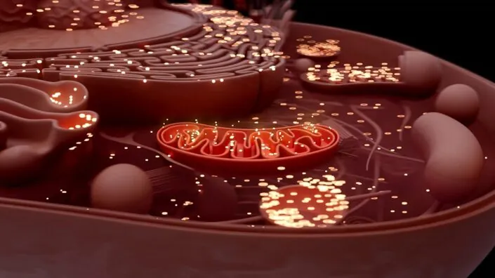
Mitochondria, the energy-producing organelles, have their own circular DNA (mtDNA) that resides inside each mitochondrion and codes for a number of essential proteins. Mutations in mtDNA cause several diseases and are also associated with aging [2]. Until very recently, there was no easy way to edit mtDNA since CRISPR-based tools are too large to enter mitochondria.

The situation began to change with the introduction of smaller editing tools, but more research is needed to test and refine them. In a new study published in PLOS Biology, scientists from the University Medical Center Utrecht in the Netherlands used the double-stranded DNA deaminase toxin A-derived cytosine base editor (DdCBE), paired with guiding proteins called TALE, “to develop in vitro disease models and assess therapeutic strategies for mitochondrial diseases in primary human cells.”

Creating a disease model

First, the team used DdCBE to introduce a loss-of-function mutation (m.15150G>A) in human primary adult liver stem cell-derived organoids. This particular mutation has not been associated yet with any known disease, but other mutations in the same gene (MT-CYB) have. The researchers report that their editing tool successfully introduced the mutation.

This is an important step in creating models of mitochondrial diseases so that they can be studied and cured. “Mitochondrial dysfunction and mtDNA alterations are implicated in several age-associated pathologies, however, our ability to understand the underlying mechanisms is limited by lack of appropriate models,” said Dr. Amutha Boominathan, a senior researcher at the Lifespan Research Institute, who was not involved in this study.

While a cell has only two copies of the nuclear DNA, one from each parent, there can be hundreds of thousands of mitochondria in each cell, each one with its own circular DNA. Therefore, an edit needs to be introduced to as many of those copies as possible. The presence of more than one type of mtDNA within a single cell is called heteroplasmy.

When the researchers introduced a pathogenic mutation into healthy liver organoids, they did not create cells that were 100% mutated. Instead, by isolating and growing single cells, they generated a collection of organoid lines with a wide range of heteroplasmy levels (from 0% to 80% mutated). This allowed them to study the effects of different levels on the severity of the disease, as naturally occurring DNA diseases also manifest themselves only past a certain heteroplasmy threshold.

Mutation fixed

The next step was to try fixing a known harmful mutation. In fibroblasts from a patient, the DdCBE system successfully corrected the pathogenic m.4291T>C mutation, which is linked to Gitelman-like syndrome, a group of inherited kidney disorders.

Heteroplasmy remained a challenge: when the researchers grew out colonies from single edited cells, they found a wide range of DNA correction levels. On the bright side, those levels remained stable over 50 days of follow-up and even slightly increased, showing that the corrected mitochondria were healthy and not at a selective disadvantage within the cell.

In cell lines with a high level of correction (76% and 81%), the mitochondrial membrane potential was successfully restored to the level of healthy control cells, suggesting functional rescue. In a line with low correction (35%), there was no improvement.

The results for overall energy production were more modest and inconsistent. While slight improvements were observed in some experiments, the effect was not as strong or reliable as the restoration of the membrane potential. The authors note that this warrants further study.

Initially, the team used a tried-and-true method of delivery: DNA carried by viral vectors. In later experiments, they showed that a better method was to deliver the editor as modified RNA (modRNA). The modifications included tweaking RNA nucleotides for greater stability and shielding the molecule from being detected by the immune system. Compared to DNA delivery, modRNA demonstrated much higher efficiency and less cytotoxicity.

The modified RNA molecules were delivered using lipid nanoparticles (LNPs). This is the same state-of-the-art technology used to deliver the mRNA in COVID-19, considered the most advanced non-viral system for in vivo delivery.

“Adapting precision DNA editing tools such as base editors to target the mitochondrial genome holds significant promise for both modeling and treating mitochondrial DNA (mtDNA) mutation-associated diseases,” said Dr. Boominathan. “However, this approach faces several challenges, including the high number of edits required per cell (due to the large mtDNA copy number), achieving homogeneous editing across cell populations, and minimizing off-target effects. In this study, the authors successfully generated a pathogenic mutation in liver organoids and corrected the m.4291T>C mutation in patient-derived fibroblasts. Nonetheless, limitations such as variability in editing efficiency – both in the extent and uniformity of edits – persist and warrant further optimization.”

We would like to ask you a small favor. We are a non-profit foundation, and unlike some other organizations, we have no shareholders and no products to sell you. All our news and educational content is free for everyone to read, but it does mean that we rely on the help of people like you. Every contribution, no matter if it’s big or small, supports independent journalism and sustains our future.

Literature

[1] Joore, I. P., Shehata, S., Muffels, I., Castro-Alpízar, J., Jiménez-Curiel, E., Nagyova, E., … & Koppens, M. A. (2025). Correction of pathogenic mitochondrial DNA in patient-derived disease models using mitochondrial base editors. PLoS biology, 23(6), e3003207.

[2] Sprason, C., Tucker, T., & Clancy, D. (2024). MtDNA deletions and aging. Frontiers in Aging, 5, 1359638.

Neural connections

Senolytics May Affect Inflammation-Related Cognitive Decline

Researchers have found that inflamed, senescent microglia prune too many synapses in the hippocampus and demonstrated that a senolytic compound can ameliorate this process in Aging Cell.

Some synapse pruning is normal

During brain development, the resident immune cells of the brain (microglia) prune unneeded synapses as a form of maintenance. This is normally a beneficial process both in young children [1] and mature adults [2], as the removal of unnecessary circuits facilitates the formation of more meaningful connections.

However, as with any of the body’s cleanup processes, disease states can send it into overdrive, causing damage. For example, during the inflammation brought on by blood sepsis, microglia tear apart functional synapses, leading to cognitive decline [3].

Like other cells, microglia can become senescent and unable to proliferate further. However, this state does not mean that they are turned off completely. While the two appear to be related, senescent microglia and disease-associated microglia are not quite the same [4].

Inflamed microglia express genes differently

This experiment began by exposing 8- to 10-week-old Black 6 mice to lipopolysaccharides (LPS) for one week in order to cause neuroinflammation. A gene expression analysis revealed that, of the 20 most upregulated genes, a full eight were related to debris clearing (phagocytosis), including genes related to Complement 1q, a compound related to synaptic pruning. Five more upregulated genes were related to senescence.

These findings were confirmed with an examination of lysosomal and activity markers. The microglia in the LPS-exposed mice were significantly more involved in phagocytosis and were also more senescent according to the p16 biomarker, which was significantly increased in the active microglia, and another examination showed that the senescent microglia in LPS-exposed mice had some morphological distinctions from the senescent microglia in the control group. Astrocyte activity was also increased by LPS. Interestingly, this phagocytosis appeared to be only limited to excitatory, rather than inhibitory, synapses, which were unaffected by this chemical.

Senolytics appear to be effective

As expected, the LPS treatment resulted in measurable levels of cognitive decline. The mice given LPS were less able to navigate a Y maze, less interested in novel objects, and less willing to navigate an open field. However, treatment with the senolytic compound ABT-737 reversed this decline, making most of their measurements indistinguishable from those of the control group.

This improvement was not due to benefits in neuroinflammation; multiple fundamental inflammatory biomarkers, including SASP biomarkers, were unaffected by ABT-737. Instead, it affected markers more directly related to senescence, such as p16 and p21. This reduction was accompanied by a decrease in the number of senescent microglia in the hippocampi of these mice.

Most importantly, ABT-737 treatment appeared to do what it set out to do. The phagocytosis of excitatory synapses was reduced in the treated mice, although, like with the behavioral analysis, not all markers were reduced to the levels of the control group. The number of dendritic spines, which decreases with LPS, was restored with ABT-737, and neuroplasticity, as measured by postsynaptic potential, also appeared to be partially restored.

These experiments used mice that were treated with an inflammatory compound, not aged mice. Further work will need to be done to determine if ABT-737 or any other senolytic is able to ameliorate the cognitive decline brought on by senescent microglia in the context of aging.

We would like to ask you a small favor. We are a non-profit foundation, and unlike some other organizations, we have no shareholders and no products to sell you. All our news and educational content is free for everyone to read, but it does mean that we rely on the help of people like you. Every contribution, no matter if it’s big or small, supports independent journalism and sustains our future.

Literature

[1] Bohlen, C. J., Friedman, B. A., Dejanovic, B., & Sheng, M. (2019). Microglia in brain development, homeostasis, and neurodegeneration. Annual Review of Genetics, 53(1), 263-288.

[2] Colonna, M., & Butovsky, O. (2017). Microglia function in the central nervous system during health and neurodegeneration. Annual review of immunology, 35(1), 441-468.

[3] Chung, H. Y., Wickel, J., Hahn, N., Mein, N., Schwarzbrunn, M., Koch, P., … & Geis, C. (2023). Microglia mediate neurocognitive deficits by eliminating C1q-tagged synapses in sepsis-associated encephalopathy. Science advances, 9(21), eabq7806.

[4] Rachmian, N., Medina, S., Cherqui, U., Akiva, H., Deitch, D., Edilbi, D., … & Schwartz, M. (2024). Identification of senescent, TREM2-expressing microglia in aging and Alzheimer’s disease model mouse brain. Nature neuroscience, 27(6), 1116-1124.

Gabriel Cian Interview

Gabriel Cian on Investment and the 2060 Longevity Forum

In this Lifespan interview, we speak with Gabriel Cian, founder of the 2060 Longevity Forum, about how his background in software shaped his views on healthspan innovation, the forum’s approach to scientific and investment credibility, and what he sees as the biggest bottlenecks and opportunities facing the field of longevity today.

Hi, Gabriel. Let’s start with your background. Can you introduce yourself and explain how your path led from software entrepreneurship into the longevity space?

When I sold my last startup in the tech space, I had just turned 40. According to Western health standards, I could expect to live in relatively good health until around 65 followed by 15 years of chronic illness.

That would mean I would die sometime around 80. That was it, just 25 more healthy years, with so much still to experience and achieve. I wanted more. That’s when I discovered longevity research.

I began to realize that the relentless pace of entrepreneurship had taken a serious toll on my health. I was prediabetic, under chronic stress, had a misaligned vertebra in my spine, and showed inflammatory markers comparable to those of a 72 year old.

It was a serious wakeup call for me!

Picturing myself dedicating the second half of my life to longevity, helping Humanity overcome aging felt fully aligned with myself.

This is how the 2060 Foundation, whose purpose is to help humanity defeat Aging, was born.

Would you say that your experience as a founder in tech has shaped how you think about healthspan, biotech, or systemic change more broadly?

When I started my first startup, everyone around me, friends, family, tried to convince me to give up. They said that I was crazy, that it would not work, but I chose to fight against this, and prove them wrong.

It took many years, even decades, but it eventually worked. Every argument against my startup and my project would make me even more motivated to make it work. Even though I had no money, no particular experience, and no precise idea on how I would succeed, I believed I could do it.

I’m roughly in the same position today, when it comes to longevity.

All of what I’ve done as a tech entrepreneur in the last 20 years feels now like training for what I’m doing now. I have to resist social pressure, and convince people around me that it is possible to slow down aging, and even reverse it.

But I have limited resources to make this happen; research in biotechnology is expensive! It may take decades until we know how to solve aging. Achieving this goal is all about grit, persistence, and audacity. Thinking and acting long term over the short term.

What motivated you to create the 2060 Longevity Forum, and how did you settle on the name and location?

I live in the south of France, one of the most beautiful, culturally rich, and longevity-friendly regions in the world.

My vision is to build a true Longevity Hub here: a place where longevity enthusiasts from all over the world can come and live with their families. They can then combine this with cutting-edge biotech research and medical infrastructure from advanced labs to preventative longevity clinics.

When it comes to the name of the conference, the year 2060 represents my commitment to invest the next 40 years of my life to humanity’s most audacious aspiration: solving aging. By that time, I’ll be 80!

With more longevity events emerging, how do you ensure this forum serves a unique purpose and doesn’t overlap with others?

My team and I have spoken with many attendees of longevity conferences and identified a clear gap: investment.

While numerous events rightfully focus on the academic and scientific side of longevity, very few put the spotlight on funding. There’s a real need for spaces where longevity startups can connect with investors, and where venture capitalists can engage with limited partners.

If we want to accelerate progress in this field, we need serious capital. At the end of the day, longevity isn’t just about science, it’s about funding.

The agenda spans well-supported interventions to more exploratory topics like cryonics and mind uploading. How do you decide what belongs on the program?

The core mission of the 2060 Longevity Forum is clear: channel as much capital as possible into the longevity ecosystem. More funding means more brilliant minds making more longevity breakthroughs.

Having said that, investors come in all shapes and sizes, so the projects and experts that come as keynote speakers need to cover the whole spectrum of possible interventions in longevity. With that in mind, I have invited longevity clinics, researchers, and startups working on whole body transplants.

How do you strike the balance between encouraging bold ideas and maintaining scientific credibility?

Having been a fundraising entrepreneur in the past and now an investor myself, I’ve come to realize that no two investors are the same. Each brings a unique mix of expertise, risk tolerance, and personal track record. Some are drawn to bold, visionary ideas; others prefer near-term, de-risked opportunities.

My mission is to engage as many of them as possible in the longevity space, which is why the 2060 Longevity Forum will showcase the full spectrum of projects from pragmatic, revenue-ready ventures to transformative moonshots.

I don’t have a specific algorithm to deal with this, it’s a case-by-case evaluation, and I’m also relying on experts that give me their opinion.

In short, I’m aiming for a balanced distribution between proven solutions and bold innovations across the whole spectrum of longevity startups.

You’ve called longevity the greatest investment opportunity of our time. What makes it so compelling to you personally?

I think AI is disrupting the field of biotechnology in an exponential and therefore unpredictable way, because we humans don’t know how to predict exponential curves, do we?

Rreally, the best way to predict the future is to create it yourself. So this feels like the right time to bet big time on major breakthroughs in the next years and decades.

Once I’ve said that, it becomes my mission to make that happen. Contributing to the field, and shortening the time before we discover something big, by channeling more funds into the field, makes it very compelling for me because I feel I can significantly contribute to that.

What do you see as the biggest bottlenecks to more investors getting involved in the space – and how are you trying to address them?

Investors could allocate significantly more capital to longevity, but several key obstacles stand in the way:

1. Lack of visibility and credibility

Many investors aren’t even aware that longevity is a serious investment opportunity. Without digging into the science, some still associate it with hype or snake oil, assuming aging is irreversible.

2. Lack of success stories

We need tangible wins both in terms of clinical breakthroughs and ROI to build trust. It’s a bit like AI before and after ChatGPT: investment was hesitant before, and exploded after. Longevity needs its own inflection point.

3. Lack of long-term thinking

Today’s investment models are geared toward 3x returns in 5-10 years, but longevity, with its R&D-driven nature, may offer 100x returns in 20 years. Imagine the market size for technologies that add even 10 extra years of healthspan. It’s massive. Investors can be educated to shift from short-term returns to exponential long-term impact.

4. Lack of meaning

ROI is important, but what about return on life? Most investors are trained to chase financial performance above all, but what if the most meaningful investment is one that helps extend life itself? Money is a tool, and using it to fund breakthroughs that allow us to live longer, healthier lives gives it its highest purpose.

This is why I’m taking a long-term approach to catalyze investment in longevity:

  • Through Ikare.Health, I help investors take care of their own health using the best longevity treatments available today. (Ikare is named after a small blue zone island in Greece.)
  • As they reach today’s limits, I challenge them to think bigger and invest in tomorrow’s breakthroughs through our 2060 Longevity Investment Club, a private community of investors backing longevity startups exclusively.
  • To unite the entire ecosystem, I’ve launched the 2060 Longevity Forum designed to increase visibility, spark connections, and create strong network effects. Virtual networks don’t work well until people meet in person.

I believe that with the right mix of education, community, and long-term vision, longevity investing is only just beginning. It may take decades to fully realize this ambition but it’s a mission well worth the journey.

This year’s event includes both startup pitch sessions and LP-GP networking. Why did you choose to spotlight both early-stage companies and fund managers?

The end game of what I’m trying to achieve revolves around one core mission: driving more investment into longevity. This includes supporting startups actively raising funds but also facilitating connections between GPs (fund managers) and LPs (capital providers) who believe in the long-term potential of the field.

Raising capital is never easy and it’s even more challenging in longevity, where timelines can be longer and the science more complex. That’s why I’m creating tailored opportunities for both types of investors:

  • For those who want to invest directly in startups, we host dedicated pitch sessions.
  • For those who prefer to back experienced fund managers, we facilitate direct connections with longevity-focused GPs.

I’ve seen strong enthusiasm for our LP-GP speed-networking format, and I’m fully committed to making it a standout success.

Some argue longevity may primarily benefit the wealthy. How do you think the field can evolve to better serve broader populations?

This is a crucial topic that deserves serious attention.

There’s a growing body of recent examples suggesting that once a life-extension technology proves effective, it’s likely to reach mass-market adoption from day one.

Take Large Language Models (LLMs) as an example. While not related to longevity, they represent one of the most disruptive technological leaps in human history. When ChatGPT launched in November 2022, no one expected it to become ubiquitous so quickly, yet just a few years later, it’s widely available, often for free, with multiple competitors offering similar tools at no cost. It’s used by millions, across all sectors of society.

One might argue that LLMs needed mass adoption to gather vast amounts of training data. But the same is true for longevity R&D we need large-scale datasets to uncover the right correlations between interventions and their impact on healthspan and lifespan.

We’re already seeing early signs of this mass-market trajectory in longevity-related drugs:

  • GLP-1 agonists (like semaglutide) are among the few longevity-linked treatments already on the market. While the extent of their healthspan benefits is still under review, these drugs are widely accessible and increasingly affordable.
  • Metformin, another life-extension candidate, is so inexpensive that it’s no longer of interest to big pharma. As a generic drug, it’s available to virtually everyone.
  • Rapamycin, also a promising compound in the aging field, is similarly low-cost and accessible to those who wish to explore its potential.

While concerns about life-extension technologies being reserved for a privileged elite are understandable, current trends suggest otherwise. The trajectory of innovation especially in tech and pharma is increasingly democratized. The evidence so far points to broad access, not exclusivity.

How geographically concentrated is longevity investment in your experience? Do you see notable differences – or advantages – emerging in particular regions?

From my experience, the US is leading by a short edge, but we’re very early on, and I strongly believe in a world where scientists from all the countries, organized in small and agile teams, can discover major things, taking advantage of their local scientific, regulatory, and cultural landscape.

For example, in some Eastern European countries, the cost of clinical trials is very low (thinking of The Cat Health Company), so they’re doing research in Romania.

In France, there are excellent, world-class scientists. Providing some funding and pairing them with more seasoned entrepreneurs could make terrific startups.

I was talking to another startup, Cyclarity; they’re doing their clinical trials in Australia, because their regulatory body is much more agile and comfortable to work with than the FDA.

The more I explore this space, the more I see original initiatives all over the world, each one of them having special advantages.

You’ve included policymakers in this year’s agenda. What role do you think governments should play in shaping the longevity field?

Essentially, private capital is invested in longevity projects right now. However, with an aging population and a lower birth rate, governments have no choice but follow the trend and invest massively in healthier living.

There’s no debate if they’ll do it, the only question is when and how we determine them to move faster in this direction. They’re the ones who will create the infrastructure of longevity, creating standards of care, and support long term R&D and massive deployment.

In my opinion, Governments are also the actors that will guarantee, at the end of the day, fair access to longevity treatment for everyone. This is what will make longevity not only aspiration, but also fair to everyone.

Are there any policy frameworks you’ve seen – locally or globally – that you think could serve as models to accelerate the field?

I see some early success stories, notably Singapore and South Korea, which have implemented effective longevity strategies, albeit for different motivations. These nations are among the most technologically advanced but also face some of the world’s lowest birth rates. Their demographic pressures left them with no choice but to act quickly and decisively.

The policy blueprint for life extension already exists. It includes:

  • Access to healthy nutrition
  • Promotion of physical activity
  • Reduction of environmental pollution
  • A shift from reactive to preventative healthcare
  • Significant investment in R&D for next-generation therapies

What’s missing now is broad public awareness. With greater public engagement, we can generate the political momentum needed to scale and implement these measures globally. Governments will do essentially what people want, and major corporations around the world will adapt to it to increase their revenue.

Zooming out, what would success look like for this year’s Forum, beyond attendance or press coverage?

If people just come here, spend some time relaxing and exploring longevity, and then feel like coming next year for the 2026 edition, I will be more than happy.

But having said that, inspiring participants to envision the south of France as a future home, a place to live with their families, work, and invest in longevity would be an even more powerful and meaningful goal to achieve.

Looking five years ahead: what are your aspirations for the Forum and for the longevity field more broadly?

My vision behind the 2060 Longevity Forum is to turn it into the “World Economic Forum of Longevity”, a global meeting point where investors of all types connect with the most promising longevity startups.

At a broader level, the mission of the 2060 Foundation is to help humanity reach a tipping point a “ChatGPT moment” for longevity where the potential for breakthrough innovations and exceptional returns becomes so clear that widespread investment becomes inevitable.

These are bold ambitions but with careful planning, persistence, and support of organizations like Lifespan.io, they are within reach.

If you could catalyze one major shift in the longevity space – scientific, financial, or cultural – what would it be and why?

The fundamental shift I’m working to promote not just in longevity, but for humanity as a whole is this: think and act long-term. This cultural mindset is the cornerstone of every meaningful decision we face. Longevity, at its core, demands long-term vision, planning, and action.

By the way, this is what I’m trying to teach to my kids, too: Longevity is thinking and acting long-term.

Finally, for those interested in supporting your work or getting involved with the Forum, what’s the best way to get in touch?

I’m reachable on Linkedin at https://www.linkedin.com/in/gabriel-cian-807b39156/ and by email at gabriel@2060.life.

We would like to ask you a small favor. We are a non-profit foundation, and unlike some other organizations, we have no shareholders and no products to sell you. All our news and educational content is free for everyone to read, but it does mean that we rely on the help of people like you. Every contribution, no matter if it’s big or small, supports independent journalism and sustains our future.
Cigarettes

Molecular Similarities Between Cigarette Smoking and Aging

Researchers have analyzed molecular patterns from different tissues obtained from over 700 people and learned that smoking acts as an aging accelerator and involves molecular changes in tissues beyond those directly exposed to cigarette smoke [1].

Millions of preventable deaths

Despite campaigns aimed at the reduction of tobacco smoking, it is still a very common practice and is considered the primary cause of preventable mortality globally, claiming 8 million lives annually [2]. Higher smoking-related mortality stems from an increased risk of respiratory, cardiovascular, metabolic, autoimmune, renal, and infectious diseases along with cancer [3, 4].

Beyond the lungs

Previous studies have addressed the effects of smoking by focusing mainly on airways and whole blood. In this study, the researchers expanded the investigation of the impact of cigarettes on multiple human tissues. They used the Genotype Tissue Expression (GTEx) project, which has data from 46 types of human tissues of 717 individuals, and compared the gene expression in different tissues between smokers and people who never smoked.

Smoking gene expression

The number of genes differentially expressed between smokers and non–smokers differed depending on the tissue, with most differences occurring in the lungs, pancreas, thyroid, and the cells lining the esophagus. Most of the changes were tissue-specific, and 86% of genes showing smoking-related changes in expression were altered in a single tissue, underscoring the importance of tissue-specific studies.

Only a few genes whose expression was upregulated by smoking were common across nine or more different tissues. A subset of those genes was previously reported to be upregulated by direct exposure to polycyclic aromatic hydrocarbons (PAHs) [5], chemicals formed during tobacco smoking. This connection suggests that toxic compounds from tobacco smoking also reach the tissues not directly exposed to the smoke. Another subset of genes altered by tobacco smoking in several tissues is linked to immune system functions and inflammation.

Along with epigenetics, gene expression can be affected by splicing changes. Genes consist of coding regions (exons) interspersed with non-coding regions (introns). When the DNA of a given gene is turned into RNA, coding regions are spliced together. However, this splicing doesn’t always happen in the same order, and sometimes, not all exons are spliced. This can affect the resulting proteins.

The researchers of this study observed alternative splicing events in 17 tissues from tobacco smokers, with the lung, thyroid, and heart being the most affected. About half of the alternative splicing led to the inclusion or exclusion of an exon, leading to changes in the protein. The other half of alternative splicing events led to the loss of properly coded functional proteins.

Further analysis was focused on the four tissues that showed the most smoking-related changes in gene expression: lung, thyroid, pancreas, and esophagus mucosa. An analysis of images from those tissues suggested structural changes, including at the cellular level. For example, in thyroid tissue, the researchers observed bigger colloid-containing follicles, the storage units of inactive thyroid hormones, which is consistent with the previously reported association between smoking and irregular growth of the thyroid gland [6]. Researchers suggest that the thiocyanate present in cigarette smoke might play a role here, as it inhibits iodine uptake by the thyroid gland, leading to problems with the production of thyroid hormones; however, this was not directly tested.

Inflammatory changes

Previous research observed similarities between gene expression changes in smoking and aging in the respiratory tract [7]. These researchers extended the analysis to different tissues. Eight tissues showed that the overlap between aging and smoking-related differentially expressed genes is higher than would be expected by chance. The changes in gene expression are in the same direction, with many genes associated with the immune system and inflammation.

Beyond these changes in gene expression, smoking also induced changes in methylation patterns. Comparing methylated sites to gene expression patterns revealed that, for the most part, smoking impacted DNA methylation and gene expression independently. However, there were also some shared patterns between the genes whose expression is smoking-associated and the smoking-related hypomethylation pattern. In both groups, the researchers noted enrichment in immune system-related functioning changes, suggesting immune system activation.

Beyond associations

Most observations described so far in this study were associations, not causal links. To establish causality, the researchers turned to the results of a previous study that identified specific methylation sites that have a causal effect on aging-related phenotypes [8]. Overlapping identified smoking-related methylation patterns with methylation sites that have a causal effect on aging-related phenotypes, and there was a substantial overlap in the lung tissues. Those results suggest a causal effect between cigarette smoking and accelerated tissue aging, which acts through DNA methylation of sites that have a causal impact on aging.

Further analysis of different methylation sites by a few epigenetic clocks suggested that age acceleration in the lung results from perturbations at protective methylation sites, that is, sites that contribute to healthy longevity.

Partial reversibility of smoking

Smokers are always advised to quit to improve their health outcomes; however, does quitting impact gene expression changes and DNA methylation patterns? These researchers used data from smokers and never-smokers and compared it to people who stopped smoking. This analysis suggested partial reversibility among most genes, splicing, and methylation events. However, the researchers observed expression changes to more reversible genes than non-reversible ones, making ex-smokers more similar to people who never smoked in terms of gene expression. In DNA methylation, there were fewer reversible sites than non-reversible ones, making ex-smokers more similar to smokers.

Analyzing the effects on gene expression and DNA methylation that are shared between smoking and aging, the researchers noted that in people who quit smoking, non-reversible DNA methylation sites in the lung were enriched in DNA methylation sites that are associated with aging signatures, but this wasn’t the case for reversible and partially reversible sites suggesting “that the smoking effects that affect DNA methylation in common to aging are more persistent in time.” This was not the case for gene expression changes.

Aging accelerator

Taken together, the results of this study support the hypothesis that smoking leads to accelerated aging, with dysregulation of the immune system and inflammation having a strong impact on both processes. While quitting can help reverse some of the smoking-related changes, there are molecular signatures that might persist for a long time.

We would like to ask you a small favor. We are a non-profit foundation, and unlike some other organizations, we have no shareholders and no products to sell you. All our news and educational content is free for everyone to read, but it does mean that we rely on the help of people like you. Every contribution, no matter if it’s big or small, supports independent journalism and sustains our future.

Literature

[1] Ramirez, J. M., Ribeiro, R., Soldatkina, O., Moraes, A., García-Pérez, R., Oliveros, W., Ferreira, P. G., & Melé, M. (2025). The molecular impact of cigarette smoking resembles aging across tissues. Genome medicine, 17(1), 66.

[2] GBD 2019 Tobacco Collaborators (2021). Spatial, temporal, and demographic patterns in prevalence of smoking tobacco use and attributable disease burden in 204 countries and territories, 1990-2019: a systematic analysis from the Global Burden of Disease Study 2019. Lancet (London, England), 397(10292), 2337–2360.

[3] Thun, M. J., Carter, B. D., Feskanich, D., Freedman, N. D., Prentice, R., Lopez, A. D., Hartge, P., & Gapstur, S. M. (2013). 50-year trends in smoking-related mortality in the United States. The New England journal of medicine, 368(4), 351–364.

[4] Carter, B. D., Abnet, C. C., Feskanich, D., Freedman, N. D., Hartge, P., Lewis, C. E., Ockene, J. K., Prentice, R. L., Speizer, F. E., Thun, M. J., & Jacobs, E. J. (2015). Smoking and mortality–beyond established causes. The New England journal of medicine, 372(7), 631–640.

[5] Stading, R., Gastelum, G., Chu, C., Jiang, W., & Moorthy, B. (2021). Molecular mechanisms of pulmonary carcinogenesis by polycyclic aromatic hydrocarbons (PAHs): Implications for human lung cancer. Seminars in cancer biology, 76, 3–16.

[6] Wiersinga W. M. (2013). Smoking and thyroid. Clinical endocrinology, 79(2), 145–151.

[7] Choukrallah, M. A., Hoeng, J., Peitsch, M. C., & Martin, F. (2020). Lung transcriptomic clock predicts premature aging in cigarette smoke-exposed mice. BMC genomics, 21(1), 291.

[8] Horvath S. (2013). DNA methylation age of human tissues and cell types. Genome biology, 14(10), R115.

Gero logo

Chugai and Gero Enter Into Research and License Agreement

Chugai Pharmaceutical Co., Ltd. (TOKYO: 4519, hereafter “Chugai”), and Gero PTE. LTD. (hereafter “Gero”), a Singapore-based biotechnology company, announced today that they have entered into a joint research and license agreement to develop novel therapies for age-related diseases.

In this collaboration, Chugai will create novel antibody drug candidates using its proprietary antibody engineering technologies for new drug targets discovered by Gero through analysis of human datasets using their unique AI target discovery platform. Under this agreement, Gero grants Chugai exclusive worldwide rights for the creation, research, development, manufacturing, and commercialization of antibodies for the identified targets. In addition to an upfront payment, Chugai will potentially pay up to approximately 250 million USD in total if predetermined development or sales milestones are achieved. If Chugai successfully launches a product, it will also pay royalties on sales to Gero.

“We believe that open innovation with external partners, including leading global players, is extremely important for achieving global first-class drug discovery outlined in our growth strategy toward 2030, TOP I 2030. By combining Gero’s target discovery technology with Chugai’s drug discovery technologies, we will accelerate the creation of innovation,” said Chugai’s President and CEO, Dr. Osamu Okuda.

“Our AI platform is built to identify therapeutic targets that drive multiple age-related diseases and potentially aging itself,” said Peter Fedichev, CEO of Gero. “In this collaboration, we aim to translate those insights into therapeutics that can help restore the lost function. This partnership with Chugai is an important step toward achieving Gero’s mission: to meaningfully target the biological processes of human aging.”

“We are excited to partner with Chugai, a leading pharmaceutical company, to unlock the synergy between human data-driven target discovery and cutting-edge therapeutic design technology platforms. Together, we aim to develop first-in-class therapeutics to address unmet needs of increasing number of patients suffering from age-related diseases,” said Alex Kadet, CBO of Gero.

Chugai Pharmaceutical Co., Ltd.

Chugai Pharmaceutical Co., Ltd., headquartered in Tokyo, is a research-based pharmaceutical company with world-class drug discovery capabilities, including proprietary antibody engineering technologies. Chugai is committed to creating innovative pharmaceutical products that may satisfy unmet medical needs. Chugai is listed on the Prime Market of the Tokyo Stock Exchange. While maintaining autonomy and management independence, Chugai is an important member of the Roche Group. Additional information is available at https://www.chugai-pharm.co.jp/english/

Gero PTE. LTD.

Gero PTE. LTD., headquartered in Singapore, is a preclinical-stage AI-driven biotechnology company creating therapeutics against age-related diseases with a mission to extend healthy human lifespan. Gero’s technology platform is grounded in physics-based machine learning and human data, enabling discovery of therapeutic targets and develop therapies that address age-related diseases and target the root causes of aging. Additional information is available at: https://www.gero.ai

Contact

Chugai Pharmaceutical Co., Ltd., Corporate Communications Dept.,

Media Relations Group Tel: +81-3-3273-0881 E-mail: pr@chugai-pharm.co.jp

Investor Relations Group Tel: +81-3-3273-0554 E-mail: ir@chugai-pharm.co.jp

Gero PTE. LTD.

Media Relations E-mail: media@gero.ai

Investor Relations E-mail: ir@gero.ai

We would like to ask you a small favor. We are a non-profit foundation, and unlike some other organizations, we have no shareholders and no products to sell you. All our news and educational content is free for everyone to read, but it does mean that we rely on the help of people like you. Every contribution, no matter if it’s big or small, supports independent journalism and sustains our future.

Orang Asli

Inflammaging Might Not Be Universal Across Populations

By comparing data from industrialized and non-industrialized societies, a new study calls into question some assumptions about the relationship between inflammation and aging [1].

Harmful protection

Inflammation accompanies us throughout our entire lives. Without it, we would not be able to fight off pathogens. Yet, inflammation also harms tissues and organs and, as such, is thought to be a major cause of aging [2]. In fact, this connection led to the appearance of the term “inflammaging”: the chronic low-grade inflammation that increases with age.

However, the relationship between inflammation and aging might not be as straightforward and universal as previously thought, according to a new study from Columbia University Mailman School of Public Health that was published in Nature Aging.

The researchers analyzed four populational datasets with extensive data on inflammatory molecules from four studies. The Italian InCHIANTI and the Singapore Longitudinal Aging Study (SLAS) originated from industrialized societies. The other two resulted from studying indigenous, non-industrialized populations: the Tsimane of the Bolivian Amazon and the Orang Asli of Peninsular Malaysia. The Tsismane are largely hunter-gatherers, while the Orang Asli live in a rural society with little development.

Diverse inflammation signatures

The team found that the first principal component, which explains the bulk of the variation in the data and can be used as a signature of inflammation, was largely similar between InCHIANTI and SLAS. The other two studies, however, had unique signatures that, importantly, did not show strong correlations with age.

In the Tsimane, acute inflammation, mostly from parasitic and helminth infections, was the major component of the inflammation score. This inflammatory signature did not correlate with chronic diseases and actually decreased rather than increased with age.

The Orang Asli presented an interesting intermediate case. This group’s inflammation score was linked to a high white blood cell count (leukocytosis), which is a general sign of infection or inflammation, but not to parasitic infections, a key feature of the hunter-gatherer group’s inflammation.

On the other hand, the Orang Asli did show a correlation, albeit a weaker one, between inflammation and aging, which puts them closer to the two industrialized groups. Their inflammatory drivers appear to be a mix of general infection and metabolic stress, distinct from both the chronic disease-driven pattern in industrial societies and the parasite-driven pattern in hunter-gatherers.

“In industrialized settings, we see clear links between inflammaging and diseases like chronic kidney disease,” said lead author Alan Cohen, PhD, associate professor of Environmental Health Sciences at Columbia Mailman School and faculty member of the Butler Columbia Aging Center. “But in populations with high infection rates, inflammation appears more reflective of infectious disease burden than of aging itself.”

The study’s findings suggest that inflammation can come in diverse shapes and show different relationships with aging, depending on the set of environmental exposures in a particular population. “If you move any species to a new environment that it has not evolved with or adapted to, it will develop inflammation as a natural response,” said Dr. David Furman, a prominent expert on inflammation, who was not involved in this study, in his recent interview with Lifespan. “If your body hasn’t seen something during its two-million-year evolution, you probably shouldn’t be exposed to it, because it will cause inflammation.”

This echoes Cohen’s words: “These results point to an evolutionary mismatch between our immune systems and the environments we now live in. Inflammaging may not be a direct product of aging, but rather a response to industrialized conditions.”

“These findings really call into question the idea that inflammation is bad per se,” Cohen added. “Rather, it appears that inflammation—and perhaps other aging mechanisms too—may be highly context dependent. On the one hand, that’s challenging because there won’t be universal answers to scientific questions. On the other hand, it’s promising, because it means we can intervene and change things.”

A crucial limitation

Demographic differences in the four datasets posed an important limitation on the study. InCHIANTI and SLAS were largely similar to each other, encompassing a wide age range with a mean age of 67.8 and 62.5, respectively. The Bolivian dataset (THLHP), on the other hand, only contained people aged 40 and older, hinting at a “survivor effect.” Essentially, in an environment with high infectious disease burden and limited medical care, people who survive into their 70s, 80s, and 90s are likely to be the most immunologically robust, similarly to centenarians in an industrialized society [3]. Inflammation decreasing with age in this group might not be telling us about a universal aging process but rather reflecting the fact that the surviving older population is enriched with exceptionally immunocompetent individuals.

Population inflammation

For the Orang Asli, the mean age of the dataset was 40.6: almost 20 or more years younger than in the other three. Consequently, the chronic, low-grade inflammation that defines “inflammaging” in older industrialized populations may simply not have had enough time to become the dominant signal in this younger group. Instead, inflammation driven by acute infections or other environmental challenges could still be a major factor, creating “noise” that makes Orang Asli’s overall inflammatory signature look different from that of the older groups.

We would like to ask you a small favor. We are a non-profit foundation, and unlike some other organizations, we have no shareholders and no products to sell you. All our news and educational content is free for everyone to read, but it does mean that we rely on the help of people like you. Every contribution, no matter if it’s big or small, supports independent journalism and sustains our future.

Literature

[1] Franck, M., Tanner, K., Tennyson, R., Daunizeau, C., Ferrucci, L., Bandinelli, S., Trumble, B., Kaplan, H., Aronoff, J., Stieglitz, J., Kraft, T., Lea, A., Venkataraman, V., Wallace, I., Lim, Y., Ng, K., Yeong, J., Ho, R., Lim, X., Mehrjerd, A., Charalambous, E., Aiello, A., Pawelec, G., Franceschi, C., Hertel, J., Fülöp, T., Lemoine, M., Gurven, M., & Cohen, A. (2025). Nonuniversality of inflammaging across human populations. Nature Aging, OnlineFirst, 1-10.

[2] Li, X., Li, C., Zhang, W., Wang, Y., Qian, P., & Huang, H. (2023). Inflammation and aging: signaling pathways and intervention therapies. Signal transduction and targeted therapy, 8(1), 239.

[3] Zhou, L., Ge, M., Zhang, Y., Wu, X., Leng, M., Gan, C., … & Dong, B. (2022). Centenarians alleviate inflammaging by changing the ratio and secretory phenotypes of T helper 17 and regulatory T cells. Frontiers in Pharmacology, 13, 877709.

DNA with cells

Five Hallmarks of Stem Cell Aging Proposed

In Cell Stem Cell, a trio of reviewers has proposed five hallmarks that are specific to the aging of stem cells.

Functional rather than molecular

This review begins with a note that its classifications focus on the physical features and overall behavior rather than what is going on biochemically. This is because the hallmarks of aging are largely universal across cells; things such as genomic instability, epigenetic alterations, mitochondrial dysfunction, and loss of proteostasis affect every cell in the body, not just stem cells. Additionally, these molecular markers vary greatly across cells; while these will surely have to be dealt with in future work targeting individual populations, these reviewers are endeavoring to deliver a broad understanding rather than a detailed analysis.

Therefore, this work focuses on what stem cells do in their roles and how they survive and proliferate. They propose five key hallmarks whose changes are fundamental to stem cell aging: quiescence, self-renewal propensity, cell fates, resilience, and heterogeneity.

Quiescence

The majority of stem cells are not actively dividing [1]. Instead, they remain quiescent, sitting idly by and waiting for some event to prompt their action. Some stem cell populations are exceptions; for example, the skin consistently renews itself [2].

Quiescence can be impaired in both directions. If the quiescence is too deep, the cells are slow to wake up; this has been found to impair muscle regeneration, as the stem cells responsible for replenishing muscle tissue (MuSCs) produce too few functional progeny [3], a problem that also happens in the brain [4] and the bone marrow [5]. On the other hand, shallow quiescence leads to a failure of stem cell populations to self-renew, thus leading to stem cell exhaustion [6].

Self-renewal

Changes to self-renewal are their own hallmark according to this framework. Like quiescence, this has problems in both directions. With age, some cells, such as hemapoietic stem cells (HSCs), replicate into more stem cells that fail to properly differentiate into somatic cells, leading to a buildup of useless cells [7].

On the other hand, cells that fail to properly replicate themselves and only differentiate into somatic cells will gradually become depleted. This occurs in multiple tissues, including the brain [4], and this is linked to the senescence-related tumor suppressor p16 [8].

The reviewers note here that the relationships between stem and somatic cells have not been fully explored and may vary greatly by tissue; some differentiated cells may, for example, revert back to a stem-like state, and this ability may be impaired by aging.

Altered cell fate

This paper highlights three ways in which differentiation can go wrong with aging. First, multipotent stem cells can produce too many of one cell type and not enough of another. This has been well-documented to occur in HSCs, with a variety of age-related disorders, such as thrombosis, being the result [9].

The second problem is when stem cells start dividing into cells that they should not have become. This occurs in muscle tissue; differentiated cells that were supposed to have become functional muscle cells become fibrotic instead [10]. This also occurs in HSCs, which are known to turn into fat tissue rather than functional bone marrow with age [11].

The third problem, of course, is cancer. The reviewers note that mutations that lead to other stem cell problems also lead to cancer.

Resilience

Resilience is the ability of cells to compensate for stresses, and this ability declines with aging. For example, the intestinal stem cells of older mice are much more likely to die by apoptosis when exposed to low doses of radiation [12]. This loss of resilience also leads to death in ordinary situations such as division, a phenomenon known as mitotic catastrophe [13].

Sometimes, undesirable cells gain resilience instead of losing it, compounding the self-renewal problems. Endlessly self-renewing HSCs, for example, have been found to have better mitochondrial energy generation than their functional counterparts [14].

Heterogeneity

The distinctiveness between individual stem cells changes with age. Due to the accumulation of mutations, this heterogeneity increases during adulthood, and the reviewers note that this may increase the heterogeneity of all the other hallmarks; some stem cells may be less or more resilient and willing to self-renew than others [15].

However, with truly advanced age, only a few clones survive, and heterogeneity dramatically decreases. The literature does not yet have a complete explanation for why this occurs. The reviewers suggest that this is due to certain mutations being able to outcompete others, particularly in an aged environment [16]. The extent to which mutations drive aging is also not yet fully understood.

Like the Hallmarks of Aging, these five broad hallmarks of stem cell aging are meant to serve as a guideline for understanding both aging and rejuvenation. The effectiveness of interventions that may reverse some aspects of stem cell aging, including basic interventions, such as dietary restriction and exercise, along with more advanced approaches, such as introducing factors that affect intercellular communication or replacing stem cells in their niche, can be judged by their impacts on these hallmarks.

We would like to ask you a small favor. We are a non-profit foundation, and unlike some other organizations, we have no shareholders and no products to sell you. All our news and educational content is free for everyone to read, but it does mean that we rely on the help of people like you. Every contribution, no matter if it’s big or small, supports independent journalism and sustains our future.

Literature

[1] Marescal, O., & Cheeseman, I. M. (2020). Cellular mechanisms and regulation of quiescence. Developmental cell, 55(3), 259-271.

[2] Giangreco, A., Qin, M., Pintar, J. E., & Watt, F. M. (2008). Epidermal stem cells are retained in vivo throughout skin aging. Aging cell, 7(2), 250-259.

[3] Benjamin, D. I., Brett, J. O., Both, P., Benjamin, J. S., Ishak, H. L., Kang, J., … & Rando, T. A. (2023). Multiomics reveals glutathione metabolism as a driver of bimodality during stem cell aging. Cell metabolism, 35(3), 472-486.

[4] Bast, L., Calzolari, F., Strasser, M. K., Hasenauer, J., Theis, F. J., Ninkovic, J., & Marr, C. (2018). Increasing neural stem cell division asymmetry and quiescence are predicted to contribute to the age-related decline in neurogenesis. Cell reports, 25(12), 3231-3240.

[5] Hammond, C. A., Wu, S. W., Wang, F., MacAldaz, M. E., & Eaves, C. J. (2023). Aging alters the cell cycle control and mitogenic signaling responses of human hematopoietic stem cells. Blood, 141(16), 1990-2002.

[6] Chakkalakal, J. V., Jones, K. M., Basson, M. A., & Brack, A. S. (2012). The aged niche disrupts muscle stem cell quiescence. Nature, 490(7420), 355-360.

[7] Sun, D., Luo, M., Jeong, M., Rodriguez, B., Xia, Z., Hannah, R., … & Goodell, M. A. (2014). Epigenomic profiling of young and aged HSCs reveals concerted changes during aging that reinforce self-renewal. Cell stem cell, 14(5), 673-688.

[8] Molofsky, A. V., Slutsky, S. G., Joseph, N. M., He, S., Pardal, R., Krishnamurthy, J., … & Morrison, S. J. (2006). Increasing p16 INK4a expression decreases forebrain progenitors and neurogenesis during ageing. Nature, 443(7110), 448-452.

[9] Poscablo, D. M., Worthington, A. K., Smith-Berdan, S., Rommel, M. G., Manso, B. A., Adili, R., … & Forsberg, E. C. (2024). An age-progressive platelet differentiation path from hematopoietic stem cells causes exacerbated thrombosis. Cell, 187(12), 3090-3107.

[10] Brack, A. S., Conboy, M. J., Roy, S., Lee, M., Kuo, C. J., Keller, C., & Rando, T. A. (2007). Increased Wnt signaling during aging alters muscle stem cell fate and increases fibrosis. Science, 317(5839), 807-810.

[11] Moerman, E. J., Teng, K., Lipschitz, D. A., & Lecka‐Czernik, B. (2004). Aging activates adipogenic and suppresses osteogenic programs in mesenchymal marrow stroma/stem cells: the role of PPAR‐γ2 transcription factor and TGF‐β/BMP signaling pathways. Aging cell, 3(6), 379-389.

[12] Martin, K., Potten, C. S., Roberts, S. A., & Kirkwood, T. B. L. (1998). Altered stem cell regeneration in irradiated intestinal crypts of senescent mice. Journal of cell science, 111(16), 2297-2303.

[13] Castedo, M., Perfettini, J. L., Roumier, T., Andreau, K., Medema, R., & Kroemer, G. (2004). Cell death by mitotic catastrophe: a molecular definition. Oncogene, 23(16), 2825-2837.

[14] Watanuki, S., Kobayashi, H., Sugiura, Y., Yamamoto, M., Karigane, D., Shiroshita, K., … & Takubo, K. (2024). SDHAF1 confers metabolic resilience to aging hematopoietic stem cells by promoting mitochondrial ATP production. Cell Stem Cell, 31(8), 1145-1161.

[15] Yang, D., & de Haan, G. (2021). Inflammation and aging of hematopoietic stem cells in their niche. Cells, 10(8), 1849.

[16] Mitchell, E., Spencer Chapman, M., Williams, N., Dawson, K. J., Mende, N., Calderbank, E. F., … & Campbell, P. J. (2022). Clonal dynamics of haematopoiesis across the human lifespan. Nature, 606(7913), 343-350.

David Furman Interview

Dr. David Furman on Inflammation and Aging

The longevity field hasn’t been very good at naming things, but one notable exception is “inflammaging”: the low-grade chronic inflammation that correlates with aging. Recognition has been growing that inflammation, the ubiquitous reaction of the immune system to various stressors, is a major driver of many age-related diseases and possibly one of the limiting factors for our species’ maximal lifespan.

Dr. David Furman, who has been studying inflammation for many years at Stanford and the Buck Institute for Research on Aging, might be the best authority to talk to about inflammation and aging. Recently, his team developed an inflammation aging clock that he aims to commercialize. David also leads with a personal example by minimizing environmental exposures that cause inflammation.

Tell me about your journey to where you currently are: a renowned geroscientist working at two top-tier institutions on inflammation in the context of aging.

I’m from Argentina. My journey started very early on when I realized I wanted to create a strong positive impact on humanity and decided to try and go for med school. Seeing how some can benefit from the best medicines and others simply have no access to clean water was shocking. I wanted to change that and help people live better and longer. Then, I had a conversation with my dad, who convinced me to study biology or biochemistry, and that way, I could have a much profound impact than being a physician.

I studied biology and then focused on immunology. This taught me how important inflammation is for fighting viruses and other pathogens. In the early 2000s, we first heard the idea that inflammation and the immune system also participated in age-related diseases. It sounded very surprising at the time. We know that inflammation and the immune system protect us from infectious diseases. We also know that if inflammation or an autoinflammatory condition goes awry, you can develop autoimmune diseases, but the idea that non-communicable diseases of aging, like cardiovascular disease or Alzheimer’s, had an immune root was very appealing to me.

That brought me to Stanford. I was recruited by Mark Davis, who I’m sure at some point will get the Nobel Prize for the discovery of T-cell receptors that recognize viruses and cancer cells. A few years after I joined the Stanford community in 2008 as a postdoc at Mark’s lab, he asked me to lead and be more involved with the Thousand Immunomes project, which was just starting at that time.

I became the director of that project, which looked at the immune system at large. We were doing multi-omics before it was even called multi-omics. We were analyzing a few hundred proteins, whereas today, we’re looking at over 10,000, but the premise is the same: by looking at many parameters in human cohorts, we can learn from humans and then apply those learnings directly to humans, skipping animal models entirely. We know they’re broken, right?

So, by analyzing this massive amount of data, I sort of became a data scientist by brute force. I learned from Rob Tibshirani, Trevor Hastie, and Daphne Koller, who essentially invented machine learning and AI at Stanford. Bridging computational sciences and immunology led to many findings and publications, and all of them had this aging component. It was striking that when looking at the immune system, the strongest signal by far was aging signatures.

That put me in a position to focus more on aging and longevity. In 2019, Eric Verdin recruited me as an Associate Professor and to lead the AI platform at the Buck Institute for Research on Aging.

Everything you’ve just said resonates with me strongly. I’ve also been fascinated with the role of inflammation in aging. I agree that its importance appears to be massive and has probably been overlooked. Can you tell me more about the connection between these two things?

Let me give you the historical perspective. We’ve been studying the immune system with decent technologies for about a hundred years, and we all understand that it protects us from infectious diseases, but the idea that aging is partly due to derangements in the immune system only started around the year 2000. It’s a very recent concept. Only 25 years ago, the first paper was published by Claudio Franceschi, who basically said that inflammation resulting from a number of environmental exposures will accelerate aging rates.

That was absolutely shocking to everybody. As a community, we started looking at the pathways, but at that time, we didn’t know what markers or cells were implicated in aging or age-related diseases. It turns out that inflammation not only affects the molecular hallmarks of aging, it can also drive particular diseases of aging.

Take cancer, for instance. I was shocked to see that if you take cancer cells and deprive the media of interleukin-6, they don’t grow, but if you put IL-6 in the media, they start proliferating like crazy. We now know that cancer – from the very early transformation of cells to metastasis and late stage four cancers – is largely dependent on inflammation.

Then we have the relationship between cardiovascular disease and inflammation. We’ve published several papers in major journals showing that inflammation in older adults is largely associated with a higher risk of heart attacks and other heart-related events, like arterial stiffening and ventricular remodeling.

Depending on the specific protein of the inflammatory process you’re looking at, you’re going to see a different effect. For example, eotaxin is a protein typically elevated in older adults that is associated with neurodegeneration. Who would have thought the immune system could drive Alzheimer’s disease? This is changing the paradigm in Alzheimer’s research, in cancer research, and in every single disease of aging as we understand more about the causal relationships between immune system cells and molecules and derangement at the organ level.

That naturally brings up the idea of intervening early. This seems to be an early type of accumulating damage, so theoretically, if we intervene soon enough, we could have a substantial impact on aging.

That’s very well put. I spent 17 years of my career on the idea of identifying early, preclinical signs of disease – molecular changes that are already happening in the body. Using these molecular and cellular changes, you can predict diseases and mortality rates in people who may not even show any symptoms yet.

If you can identify these changes, you can do more than just intervene; you can intercept these diseases. I like to call it the molecular interception of a disease that is developing very slowly and is not yet clinically observable. The whole premise of what we’re doing is not just predicting or understanding biomarkers. It’s about preventing disease and extending the healthspan of the population by intervening early in its course.

It sounds similar to epigenetic clocks, which work because we accumulate epigenetic damage from very early on. Epigenetic aging starts in the womb. Is that also the case with what we might call “inflammaging” or inflammatory aging? How early do the changes you track begin?

That’s an area of research that hasn’t flourished yet; it’s extremely early, so I can’t substantiate claims with strong science. The reason is a lack of sufficient data sets. The theory, however, is that inflammation may start two or three generations before an individual is born.

The concept of transgenerational epigenetic modifications also applies to inflammation. We’ve seen in some studies that the inflammatory state of new generations is impacted by what past generations have suffered from – things like psychological trauma or famine. These events can impact the epigenetic landscape and make individuals more susceptible to an increased inflammatory load later in life.

To give you an example, if an expectant mother has excessive inflammation – perhaps due to smoking or obesity – this can reflect on the growing fetus. Those children will have a higher risk of developing inflammation-related diseases like autism, early diabetes, cardiovascular disease, and even suicidal thoughts. In 2014, we published a paper in Molecular Psychiatry where we studied 500 individuals with major depression disorder and 500 controls. The inflammation levels were all over the place in those who suffered from the disorder. So yes, this starts very early on, possibly even before conception.

You used your 1000 Immunomes Project to build an aging clock that you believe is superior in some ways to existing clocks. If I understand correctly, it uses just a handful of proteins. Can you tell me more about it?

Yes. We leveraged the 1000 Immunomes dataset, which, at the time we built the clock, had data from over 12 years. We built a deep neural network to analyze the data. Protein networks have a lot of redundancy, and human data is typically very noisy. We addressed this using a specific type of neural network called a deep guided autoencoder, which is very different from what most people use for building clocks.

The beauty of autoencoders is that they can effectively deal with redundancy, non-linearity, and noise: the three main challenges in our data. So, it was the ideal tool. Using it, we predicted a person’s calendar age. What I really like about this clock is that it’s not perfectly accurate at predicting calendar age. That inaccuracy gives you room for biological interpretation as to why some people score so much higher or lower than the rest of the population.

So, this clock is trained on calendar age, not on intrinsic capacity?

You’re probably talking about a different aging clock we have that is trained on intrinsic capacity, from a collaboration with a group in France. The two clocks are interrelated, though. High intrinsic capacity, which is a very positive thing to have, correlates strongly with a low inflammatory age. We can explore that later, but for the inflammatory clock, which we call iAge, we predict chronological age. The model is “guided” because it’s trained on two target variables: the immune protein data you feed the algorithm with, and calendar age.

The output is the closest thing to an “immune age” out there. We then use the residual – the difference between a person’s calendar age and their predicted immune age – to see if the clock has clinical validity, and it does. We could associate an increase in the inflammatory clock with having multiple diseases at once (multimorbidity). We also saw a strong prediction of frailty; if I measure your inflammatory age today, I can predict with high accuracy whether you will become frail seven years from now. We then created a gene expression proxy for this protein clock and validated it in external datasets, like the Framingham Heart Study, where we were able to predict mortality in 2,500 people.

I remember you saying that your immune clock singles out centenarians as having a very different immune profile. I think this is amazing because it suggests that inflammation and immune system exhaustion might be what kill the oldest old and that centenarians are people who can somehow defend against this. What can you tell me about that?

Yes, that was one of our clinical validations. We looked at individuals with extreme longevity: centenarians and supercentenarians. We took a cohort of about 20 individuals from the Bologna area in Italy, all of them 100 years or older, and ran our iAge analysis on them. On average, their inflammatory age was 40 years younger than their calendar age.

There was one super-healthy 105-year-old male who had almost never seen a doctor. His inflammatory age was 25. That’s 80 years below his calendar age. He is an outlier, a really interesting person. What is it about his immune system that allows him such a level of control over inflammation? We don’t know yet. Other studies by Nir Barzilai, for example, show that the immune systems of centenarians are very different from their 80-year-old counterparts. They have peculiar CD4 T cells and a very different microbiome. Perhaps the explanation is that their microbiome and immune cell proportions are just shaped differently, but the fact remains: in supercentenarians and centenarians, their inflammatory age is dramatically lower than their calendar age.

Mimicking the immune system of centenarians can help us compress mortality. However, they also eventually die, and immune exhaustion is emerging as a central cause. By doing something about that, we might even be able to go one step further and extend maximum human lifespan.

That’s a hypothesis, right? We can think of multiple ways to try and push the healthspan of the population to, say, 120 years old, so people can be super healthy and then die quickly. But, I want to stress one thing about the difference between healthspan and lifespan. We all want to live healthier, for sure, but many people think of death as a very negative thing. Why? Because most people lack a humanitarian purpose and achievement in life, something that impacts more than themselves and their families. I think it is pretty scary to die and not leaving a legacy behind. Living is great, and dying shouldn’t be the worst thing that could ever happen to you.

Let’s pivot to something more down-to-earth. You said your inflammation clock gave you clues about what works in terms of diet, exercise, and other interventions.

I’m not sure if you’ve seen an article that is coming out in Business Insider about my experience reducing my own inflammation by modifying my environment and lifestyle. I was at a longevity investors meeting in Switzerland and told a reporter my story. She found it amazing and wanted to write a piece on it.

For 15 years, I’ve been studying the social and lifestyle determinants of inflammation, and I decided to start testing these principles on myself. It significantly changed my family’s life. We moved from the Palo Alto area to an off-the-grid cabin in a small valley called San Gregorio. There, we decided to apply the principles of evolutionary medicine to control inflammation.

The idea comes from a paper I published in Nature Medicine in 2019, which basically states that many environmental and lifestyle factors drive inflammation. I hate to call them “choices,” because someone in Fresno breathing polluted air has no choice. I dislike when people say, “lifestyle choices,” because for 90% of the population, there is no choice.

Anyway, I was guided by the following principle: if you move any species to a new environment that it has not evolved with or adapted to, it will develop inflammation as a natural response. The corollary is that the more distant a person’s life is from our species’ evolutionary experience, the more inflamed that person will be.

The immune system acts a sensory system of your environment, much like hearing capacity or vision; the only difference with classical sensory systems is that the output (inflammation) doesn’t reach your consciousness. You can measure this, and you can start thinking about how to implement this principle in your day-to-day life. It’s everywhere: the workplace, your household, your city. There are external and internal factors, some you can control and some you can’t. It applies to water quality, air quality, the food you eat, and the household products you use.

If your body hasn’t seen something during its two-million-year evolution, you probably shouldn’t be exposed to it, because it will cause inflammation. This applies to plastic containers with phthalates, and the microplastics and nanoplastics we are breathing that accumulate in our organs. Everything converges on inflammation and reactive oxygen species. When you read the literature, it becomes obvious that every one of these insults signals to your body through inflammation. That’s what causes issues in the brain, the heart, and the joints.

So, we made changes. We cut out wheat; humans haven’t been exposed to it for more than 8,000 years. Same with dairy products; we only started domesticating cows a few thousand years ago. Then there are hyper-processed foods. There’s a very long list of things you can start tweaking. And then you have to measure the effect. The problem is that the canonical markers of inflammation mostly work for acute inflammation.

You mean markers such as C-reactive protein (CRP)? It probably doesn’t tell you much about chronic inflammation.

Right, it doesn’t tell you anything. It’s worthless for this purpose. People look at CRP because there’s nothing else, but for predicting cardiovascular disease, its accuracy is about the same as flipping a coin. Paul Ridker built part of his career around CRP and now IL-1β, and he loves the idea of drugging these things.

High-sensitivity CRP is widely used, but a savvy cardiologist will tell you they don’t find it that useful for predicting risk. They use it now for suspected acute infections, of course. But it’s an acute-phase reactant; it goes up, but then it comes back down. IL-1β is similar, sometimes chronic, sometimes acute. Some proteins reflect chronic states, while others just change for a short period.

In terms of other interventions, simply not moving around will increase your inflammation. Your body will interpret a sedentary state as a sign that you’re sick, it’s actually called sickness behavior in psychology.

Basically, inflammation is a lifelong, adaptive reaction that can also be very destructive.

Exactly. It can be. Inflammation is built for repair and protection. Your skin and microbiome interact with inflammation all the time, but it becomes very detrimental if it’s sustained and doesn’t resolve. There’s remarkable work from Charles Serhan at Harvard on the biomarkers of inflammation resolution. That’s a whole other area of research that is super interesting and could be pivotal for finding solutions.

I want to ask you about one of your companies, Edifice Health. Is this how you’re commercializing the iAge clock?

Yes, exactly, and let me give you the high-level answer for why. The system for academic research is not ideal; it’s really broken. Think of the amount of money that goes from the government to academia. It’s incredibly inefficient. Why? Because in the academic setting, there is no incentive or training to start a commercial entity. If you don’t do that, the findings end up in a drawer, in the trash, or just as a publication. They don’t translate to the bedside, to households, to solving people’s problems in the marketplace. Federal money does not equal translation. The pathway is not from bench to bedside; it’s from bench to company to bedside.

It seems like the new agency ARPA-H is taking a different approach, and you have applied for one of their programs, correct?

Exactly, they are following this principle. A program we just applied for, called PROSPR, requires us to have FDA approval and a working commercial entity by year five. It’s very pro-startup; a commercialization strategy is a requirement for this ARPA-H funding. It’s a beautiful, dream program. It’s what everyone should be doing: putting their efforts into early diagnosis or interventions for aging but with a translational lens.

We would like to ask you a small favor. We are a non-profit foundation, and unlike some other organizations, we have no shareholders and no products to sell you. All our news and educational content is free for everyone to read, but it does mean that we rely on the help of people like you. Every contribution, no matter if it’s big or small, supports independent journalism and sustains our future.
TransVision Summit

Madrid Set to Become the Longevity Capital of Europe

We are thrilled to announce the second edition of the International Longevity Summit (www.TransVisionMadrid.com) in beautiful Madrid after the major success in 2024.

Organized by the Illustrious College of Medical Doctors of Madrid (ICOMEM) and the International Longevity Alliance (ILA), this year we expect over 500 participants with more international experts, including George Church (Professor, Harvard Medical School and MIT), Mehmood Khan (CEO, Hevolution Foundation), Steve Horvath (Principal Investigator, Altos Labs), Michael Ringel (COO, Life Biosciences), Aubrey de Grey (CEO, LEV Foundation: Longevity Escape Velocity), Joao Pedro de Magalhaes (Chair of Molecular Biology, University of Birmingham), Andrea B. Maier (Founding President, Healthy Longevity Medicine Society), Phil Newman (Founder, Longevity Technology), Liz Parrish (CEO, BioViva), David Wood (Executive Director, LEV Foundation: Longevity Escape Velocity), Natalie Coles (Director, Supercentenarians Project, R3 Bio), Ken Scott (Founder, Human Longevity and Rejuvenation Syndicate), Tina Woods (Executive Director, International Institute of Longevity), Juan Carlos Mendez (President, SOVEMAL), Sabinije von Gaffke (Director of Partnerships, Super Human Network), Roberto Grau (Cofounder, Juventas4Life), Sajidxa Mariño (Founder & CEO, Respira Libre), Jose Angel Olalde (Founder, Centro de Medicina Regenerativa), Jose Cordeiro (Director, International Longevity Alliance) and others to be announced soon, plus a select group of Spanish experts, from scientists and doctors all the way to the world of economics and politics, starting with the Mayor of Madrid: José Luis Martínez Almeida.

Madrid is the most longevous capital in the European Union, with a current life expectancy of 86.1 years at birth, and Spain is also the most longevous country in the region, according to Eurostat. Some of the oldest people in the world have been Spanish, like María Branyas Morera who was the oldest living person until she died last year at the age of 117 years and 168 days. In fact, life expectancy in Madrid is higher than in the so-called Blue Zones of Sardinia in Italy, Ikaria in Greece, Okinawa in Japan, Nicoya Peninsula in Costa Rica or Loma Linda in California. Therefore, Madrid can be considered as a Blue Zone 2.0, beating all the old Blue Zones, and with a much higher and pleasurable standard of living. Yes, it is the Mediterranean diet, and more, much more that makes Madrid the most longevous city in the EU!

Come to Madrid and discover the beauty of life and longevity in Spain, the second most longevous country in the world, among large nations, only after Japan. Thus, just before the International Longevity Summit (October 1-2), we will start that week organizing 2 days of pre-conference tours (September 29-30) to visit the UNESCO World Heritage sites around Madrid (Avila, Segovia, Aranjuez, Toledo, El Escorial and Alcala de Henares). You are more than welcome to join us for one or both tours so that you can really enjoy the beauty of Castilla La Mancha, the land of Cervantes and Don Quixote, the land of fiesta and siesta, the land of Picasso and Dali, the land of vino and sangria, the land of tapas and picoteo, the land of flamenco and fandango, the land of Plus Ultra as the Romans called Hispania over two millennia ago: https://youtu.be/X7cj1MptNwc

During October 1st, which corresponds to the International Longevity Day, we will announce the Madrid Longevity Declaration and will award the Madrid Longevity Prizes. Thus Madrid reinforces its position as the longevity capital of Europe, and Spain as the most longevous country in the EU. All these activities will be in the historic Great Amphitheater of the Illustrious College of Medical Doctors of Madrid (ICOMEM), the same place where Spanish Nobel laureate Santiago Ramon y Cajal gave his master lectures, and the most impressive Classical theater of Spain, rich in history. We will also premiere an award-winning longevity documentary and will walk the Madrid March for Longevity, between the world-renowned Puerta del Sol (Kilometer 0.0 of Madrid and Spain) and the famous Cibeles Fountain (where world champion Real Madrid celebrates its football victories). Come to Madrid, let´s all write history and create the future together, it will be an immortal experience, very literally!

For more information, contact: info@TransVisionMadrid.com
We would like to ask you a small favor. We are a non-profit foundation, and unlike some other organizations, we have no shareholders and no products to sell you. All our news and educational content is free for everyone to read, but it does mean that we rely on the help of people like you. Every contribution, no matter if it’s big or small, supports independent journalism and sustains our future.
Doctor holding brain

Fixing Sugar Metabolism Shows Promise Against Dementia

Scientists have shown that aberrant metabolism of glycogen in neurons is linked to the accumulation of harmful tau protein. Caloric restriction, genetic interventions, and small molecules might help [1].

Glycogen and the brain

Aberrant aggregation of microtubule-associated protein tau (MAPT), or simply tau protein, is a hallmark of several neurodegenerative diseases [2]. The most famous of them is Alzheimer’s disease, in which tau accumulation in the form of hyperphosphorylated neurofibrillary tangles (NFTs) damages neurons.

Another, less-known, characteristic of many of these diseases is abnormal glycogen metabolism and accumulation [3]. Glycogen is a stored form of glucose, used by the body as an energy source when nutrient levels are low. It is mostly found in the liver and muscle, but brain cells (predominantly astrocytes but also neurons) also contain small amounts of it.

Impaired glycogen metabolism in neurons hurts learning and memory, while dietary restriction (DR) is known to extend lifespan and delay neurodegeneration in animal models of neurodegenerative diseases. In this new study published in Nature Metabolism, scientists from the Buck Institute for Research on Aging tried to understand how these two facts might be connected.

Diet restriction rescues lifespan

The authors started with two Drosophila fly models. One exhibited accelerated accumulation of wild-type tau protein, while the other included a known mutation in MAPT (R406W), which, in humans, causes a severe familial disease called frontotemporal lobar degeneration with tau inclusions (FTLD-tau).

The flies were either freely fed or restricted in calories. DR significantly increased lifespan even in healthy controls. In the two disease models, the effect was even more dramatic. DR rescued lifespan in flies with aberrant accumulation of wild-type tau almost completely, and in mutation-carrying flies, the difference was highly significant. Accordingly, in DR flies, levels of neuronal death fell dramatically.

Tauopathies and dietary restriction

Proteomic analysis of the flies’ brains revealed that pathways related to fat and glycogen metabolism were among the most drastically changed by DR, and glycogen levels were indeed elevated in the brains of tauopathic flies.

Interestingly, however, DR did not seem to alter overall levels of glycogen, despite clearly having a strong beneficial impact. The researchers suspect that what might be important is the rate of glycogen turnover. The enzymes involved in this turnover, including glycogen phosphorylase (GlyP), were upregulated in mutant flies on DR. Overexpression of GlyP increased the lifespan of mutant flies by almost 70% and drastically reduced neuronal death.

More antioxidants!

The researchers used metabolomics and RNA sequencing to study the molecular effects of GlyP upregulation. Surprisingly, the pathways for energy production, namely glycolysis and the citric acid cycle, were actually downregulated. Instead, the glucose from the broken-down glycogen was being shunted into the pentose phosphate pathway (PPP). Its primary function is to generate antioxidants: molecules that combat oxidative stress. Reactive oxygen species (ROS) were indeed significantly reduced in the brains of the flies with enhanced glycogen breakdown.

According to the researchers, this might at least partially explain the benefits of DR and GlyP upregulation. In line with this hypothesis, blocking the PPP with a small molecule abolished the protective effects of glycogen breakdown. The team also successfully recreated the effects of genetic GlyP overexpression by using another small molecule, 8-Bromo-cAMP, to activate the GlyP-producing pathway.

A vicious cycle?

The team then ran experiments in vitro on human neurons derived from induced pluripotent stem cells (iPSCs) that were obtained from patients with FTLD-tau. Genetically corrected cells from the same donors were used as controls. The researchers demonstrated increased glycogen accumulation in FTLD-tau cells and also tested the rescue mechanism by overexpressing the human version of the glycogen breakdown enzyme (PYGB) in the diseased human neurons. This reduced abnormal glycogen accumulation and restored mitochondrial abundance, which declines with this disease.

Importantly, using these human neurons, the team showed that tau protein and glycogen co-localize within cells and physically interact, supporting the hypothesis that a direct interaction between the two might be part of the problem. The authors hypothesize that this may create a detrimental vicious cycle in which tau binding promotes glycogen accumulation, which, in turn, exacerbates tau pathology and oxidative stress.

“Our findings suggest that glycogen is more than just a metabolic reservoir – it may act as a sticky trap for tau, creating a dangerous feedback loop where tau promotes glycogen buildup, and glycogen in turn fuels tau aggregation,” said Dr. Pankaj Kapahi, the corresponding author of the study, to Lifespan.io. “Breaking this cycle could open a new therapeutic front in the fight against Alzheimer’s disease.”

We would like to ask you a small favor. We are a non-profit foundation, and unlike some other organizations, we have no shareholders and no products to sell you. All our news and educational content is free for everyone to read, but it does mean that we rely on the help of people like you. Every contribution, no matter if it’s big or small, supports independent journalism and sustains our future.

Literature

[1] Bar, S., Wilson, K. A., Hilsabeck, T. A., Alderfer, S., Dammer, E. B., Burton, J. B., … & Kapahi, P. (2025). Neuronal glycogen breakdown mitigates tauopathy via pentose-phosphate-pathway-mediated oxidative stress reduction. Nature Metabolism, 1-17.

[2] Goedert, M., Eisenberg, D. S., & Crowther, R. A. (2017). Propagation of tau aggregates and neurodegeneration. Annual review of neuroscience, 40(1), 189-210.

[3] Mann, D. M. A., Sumpter, P. Q., Davies, C. A., & Yates, P. O. (1987). Glycogen accumulations in the cerebral cortex in Alzheimer’s disease. Acta neuropathologica, 73, 181-184.

Healthy elderly

Researchers Connect Cellular Markers to Physical Well-Being

In Aging Cell, a team of researchers has described how the health of skin fibroblasts relates to physical and functional ability.

Frailty, capacity, and skin cells

This paper begins with an explanation of how frailty and capacity are defined. The World Health Organization defines it as an overall state of reduced strength, endurance, and physiogical functions that increase the risk of adverse health outcomes and lead to dependency [1]. Intrinsic capacity, while difficult to measure, refers to a collection of functional attributes that define overall health [2]. These metrics lead to a better understanding of how generally healthy someone is than the simple number of chronological age.

However, there is a gap between other clocks, such as epigenetic clocks, and biomarkers of functionality. The authors of this paper focus on a ‘gerophysical’ approach that links the two [3].

This study focuses on skin cells (fibroblasts) for several good reasons. Skin, of course, is easy to measure compared to other parts of the body, and fibroblasts preserve both the function and structure of multiple tissue types [4]. Previous work has found that fibroblasts play a significant role in immune responses [5] and metabolic regulation [6]. These cells have been heavily studied in the context of aging, including epigenetics and transcriptomics such as age-related mRNA strands [7]. However, these researchers note that no one had previously linked cellular aging biomarkers to intrinsic capacity metrics.

Some biomarkers are more telling than others

This study used skin samples from 133 volunteers of both sexes in the INSPIRE-T cohort. Their ages ranged from 20 to 96, encompassing healthy, pre-frail, and frail states.

The first part of this study cultured these fibroblasts in vitro, comparing chronological age to various biomarkers. As expected, the proliferation rate of the fibroblasts slowed with age, and markers of DNA damage increased along with the senescence marker p16 and the inflammatory factor IL-6. Interestingly, this study did not show any statistically significant correlation between chronological age and many other senescence markers, including SA-β-gal. However, older cells did express more SA-β-gal when exposed to the stressor doxorubicin.

In the next part of this study, the researchers focused on three key aspects of fibroblast function: tissue structure, immune responses, and metabolic regulation (SIM) along with senescence. This was a biomarker-based analysis, focusing on several key biomarkers in each of these domains: 31 in total.

SIM framework

The researchers then used a statistical measurement called Mahalanobis distance to quantify homeostatic dysregulation: how different a person’s biomarkers are from a normal baseline. As expected, this measurement was strongly correlated with chronological age, and the researchers suggest that it can be used as a biomarker of aging.

The researchers took a closer look at how the S, I, and M indices correlate with one another. They found that while many aspects of structure, such as MMP1, did not seem to be closely related with aging, others did, including Periostin and TIMP1. The researchers suggest that these metrics are closely tied to age-related changes in the extracellular matrix.

Cytokine production, as expected, is significantly upregulated with aging, including IL-6. Other well-known inflammatory compounds, such as TGF-β, were undetectable in most samples. Interestingly, aging increases the responsiveness of inflammation to viral infection, although this inflammation’s effect can be negative.

The examination of metabolism revealed a decrease in mitochondrial respiratory efficiency with aging, and antioxidant genes were found to be largely upregulated in response to increased oxidative stress. Once more, there were some interesting negative results: SIRT1 and the NRF genes were not found significantly correlated with aging in this study.

Putting it all together

Directly comparing each of these biomarkers to intrinsic capacity yielded crucial findings. Periostin was once more singled out as a significant contributor to functional aging, as were CD36 and mitochondrial respiration markers. The researchers believe that their overall SIM analysis allows for a holistic approach that provides a detailed analysis of multiple aspects of aging.

Periostin is known in the literature as being crucial to wound healing [8], but it has been little discussed in the context of aging. While the researchers do not directly suggest that it is a suitable target for future interventions, and they cannot demonstrate that it has a causal relationship in this paper, later work may investigate whether or not it is a valid target.

The researchers note this study’s limitations: there were a substantial number of analyses made from a limited number of skin cells, and the biopsies may have been poorly representative of the people from whom they were taken. External factors such as lifestyle and environment could not be accounted for. Larger cohorts would need to be utilized to further refine this SIM analysis.

We would like to ask you a small favor. We are a non-profit foundation, and unlike some other organizations, we have no shareholders and no products to sell you. All our news and educational content is free for everyone to read, but it does mean that we rely on the help of people like you. Every contribution, no matter if it’s big or small, supports independent journalism and sustains our future.

Literature

[1] Beard, J. R., Officer, A., De Carvalho, I. A., Sadana, R., Pot, A. M., Michel, J. P., … & Chatterji, S. (2016). The World report on ageing and health: a policy framework for healthy ageing. The lancet, 387(10033), 2145-2154.

[2] Gonzalez-Bautista, E., & Beard, J. R. (2023). The Challenge of Measuring Intrinsic Capacity. The journal of nutrition, health & aging, 27(10), 806-807.

[3] Kemoun, P. H., Ader, I., Planat-Benard, V., Dray, C., Fazilleau, N., Monsarrat, P., … & Casteilla, L. (2022). A gerophysiology perspective on healthy ageing. Ageing research reviews, 73, 101537.

[4] Plikus, M. V., Wang, X., Sinha, S., Forte, E., Thompson, S. M., Herzog, E. L., … & Horsley, V. (2021). Fibroblasts: Origins, definitions, and functions in health and disease. Cell, 184(15), 3852-3872.

[5] Haniffa, M. A., Wang, X. N., Holtick, U., Rae, M., Isaacs, J. D., Dickinson, A. M., … & Collin, M. P. (2007). Adult human fibroblasts are potent immunoregulatory cells and functionally equivalent to mesenchymal stem cells. The Journal of Immunology, 179(3), 1595-1604.

[6] Zhao, X., Psarianos, P., Ghoraie, L. S., Yip, K., Goldstein, D., Gilbert, R., … & Liu, F. F. (2019). Metabolic regulation of dermal fibroblasts contributes to skin extracellular matrix homeostasis and fibrosis. Nature metabolism, 1(1), 147-157.

[7] Tsitsipatis, D., Martindale, J. L., Mazan‐Mamczarz, K., Herman, A. B., Piao, Y., Banskota, N., … & Gorospe, M. (2023). Transcriptomes of human primary skin fibroblasts of healthy individuals reveal age‐associated mRNAs and long noncoding RNAs. Aging Cell, 22(11), e13915.

[8] Elliott, C. G., Wang, J., Guo, X., Xu, S. W., Eastwood, M., Guan, J., … & Hamilton, D. W. (2012). Periostin modulates myofibroblast differentiation during full-thickness cutaneous wound repair. Journal of cell science, 125(1), 121-132.

Ordinary lab mouse

Healthspan Effects of an Anti-Aging Vaccine on Mice

The researchers of a recent study published in Aging Cell described their novel CD38 peptide vaccine, which improved many measurements of physical health and prevented cognitive decline in aged mice [1].

A long-term anti-aging vaccine

Many anti-aging strategies, such as supplementation, require regular, daily intake. A vaccine, on the other hand, is a therapeutic approach that, once administered, can have lasting effects.

Anti-aging vaccination approaches have already been undertaken previously, and they were designed to target, among others, senescent T cells and the cells that line blood vessels (vascular endothelial cells) [2, 3]. These researchers chose a different target: the widely researched CD38 protein expressed in several types of immune cells and tissues, which has multifunctional enzymatic activities related to NAD metabolism.

The age-dependent increase in CD38 levels has been linked to NAD+ decline and mitochondrial dysfunction [4]. Previous research on targeting CD38 with small molecules for therapeutic purposes showed benefits in glucose intolerance, physical dysfunction, and neuroinflammation [5, 6], while CD38 antibody research showed benefits against age-related syndromes, such as fibrosis, NAD+ deficiency, and cardiotoxicity [7-9], making it a promising target for a vaccine.

First, the researchers needed to find which part of CD38 would induce the strongest response from the immune system, thus making it the best candidate for a vaccine. While the researchers encourage future studies to test different sequences, they limited themselves to three amino acid sequences with which to create three peptide vaccines. After a few weeks, they tested how the immune systems of mice responded to the vaccines and chose the one that spurred the strongest response.

Physically stronger and cognitively younger

The researchers immunized a group of 12-month-old mice with the chosen vaccine. At 15-18 months old, the researchers analyzed the mice’s healthspan, then immunized them again at 18 months and 3 weeks of age and euthanized them at 20 months to collect tissues for further assessment.

The mice showed a strong immune response to the vaccination, translating into physical and cognitive health improvements in aged mice. The researchers observed that vaccination prevented a decline in total walking distance, maximal walking speed, grip strength, and hanging endurance in both male and female animals. Similarly, measuring physical fitness by placing mice in an open arena and observing their behavior showed better measures of total movement distance and average exploring velocity. Aged vaccinated mice also scored better on frailty scores and generally appeared younger than controls.

Cognitive skills were tested using a classic Morris water maze experiment and a novel object recognition model. The results suggested that the CD38 vaccine prevented cognitive decline in mice.

Beyond physical and cognitive tests, the researchers noted that vaccination improves metabolic features, which tend to deteriorate with age. They reported improvements in glucose tolerance, insulin sensitivity, and different measures of body energy consumption, such as energy expenditure and oxygen consumption, in aged mice compared to controls.

Decreasing senescence

These results prompted the researchers to investigate the underlying molecular mechanisms. Therefore, they analyzed both gene expression and the full suite of liver proteins (the proteome). This focus on the liver stems from its essential role in metabolism, including glucose and NAD+ metabolism, and its role as a target for anti-aging therapeutics, such as metformin and NAD+ precursors [10, 11].

First, the researchers confirmed that the vaccine specifically targeted CD38. Both gene expression and proteome analysis confirmed reduced CD38 levels.

Among the aging-related processes impacted by CD38 was senescence, and CD38 vaccination led to decreased senescence in aged mice’s solid organs. The researchers observed a decrease of the senescence marker p21 but not p16 in the liver along with decreased senescence-associated secretory phenotype (SASP) factors. Similarly, the activity of another senescence marker, SA-β-gal, was reduced in the liver and spleen following vaccination.

In previous experiments, the researchers reported CD38 vaccination’s impact on metabolic processes. This was also reflected at the molecular level.

In the liver, there was decreased expression of some glucose metabolism-related genes, specifically key glycolytic enzyme genes, in vaccinated animals compared to controls, which was confirmed by the proteome analysis. The proteome analysis also identified upregulation of fatty acid metabolic processes, breakdown of complex molecules (catabolism), and peroxisome proliferator-activated receptor (PPAR) signaling pathways, which, among other roles, regulate metabolic processes and are closely associated with oxidative phosphorylation (OXPHOS), which generates most cellular energy.

Reversing the decline

Previous research linked a specific group of immune cells, CD38-expressing macrophages, to age-related NAD+ decline [12]. NAD+ metabolism is an important player in aging. Anti-aging approaches aimed at NAD+ repletion have shown some beneficial anti-aging and healthspan-improving results.

In this study, vaccination reduced the levels of CD38-expressing macrophages in the liver and increased the NAD+/NADH ratio in the aged mice’s liver and brain tissue, partly restoring the age-related NAD+ decline.

In total, these results suggest ‘that the CD38 peptide vaccine ameliorated abnormal metabolism-related proteome shifts’ and rejuvenated the protein profile.

Proof of concept

This proof-of-concept study showed the anti-aging potential of the CD38 vaccine. The observed promising effects are most likely caused by the elimination of CD38-positive cells; however, different yet unidentified mechanisms may exist.

Since this research was conducted on mice, further studies are necessary to confirm its effect in people. The researchers also speculate that future studies should address its potential in different age-associated diseases, such as Alzheimer’s disease.

Peptide vaccines generally have high specificity and safety. Based on these results, this vaccine was not an exception from the rule, as the researchers did not detect any side effecs in the vaccinated mice. However, since CD38 plays a role in infection response, it might lead to increased infection rates, so researchers should monitor for possible infections in future studies.

Additionally, the activation of inflammatory responses might cause side effects. Inflammation is linked to aging and is thought to drive senescence, and in the short term, activation of inflammation might accelerate aging processes. Indeed, the researchers observed increased IL-6, one of the SASP factors, following vaccination in middle-aged and older mice, but it decreased later. These changes in inflammatory response warrant further investigation into how this CD38 vaccine impacts the immune system.

We would like to ask you a small favor. We are a non-profit foundation, and unlike some other organizations, we have no shareholders and no products to sell you. All our news and educational content is free for everyone to read, but it does mean that we rely on the help of people like you. Every contribution, no matter if it’s big or small, supports independent journalism and sustains our future.

Literature

[1] Yu, S., Li, Z., Tang, Y., Chen, Y., Ma, Y., Du, K., Zong, Z., Feng, K., Wei, Y., Chen, L., & Deng, H. (2025). CD38-Targeting Peptide Vaccine Ameliorates Aging-Associated Phenotypes in Mice. Aging cell, e70147. Advance online publication.

[2] Yoshida, S., Nakagami, H., Hayashi, H., Ikeda, Y., Sun, J., Tenma, A., Tomioka, H., Kawano, T., Shimamura, M., Morishita, R., & Rakugi, H. (2020). The CD153 vaccine is a senotherapeutic option for preventing the accumulation of senescent T cells in mice. Nature communications, 11(1), 2482.

[3] Suda, M., Shimizu, I., Katsuumi, G., Yoshida, Y., Hayashi, Y., Ikegami, R., Matsumoto, N., Yoshida, Y., Mikawa, R., Katayama, A., Wada, J., Seki, M., Suzuki, Y., Iwama, A., Nakagami, H., Nagasawa, A., Morishita, R., Sugimoto, M., Okuda, S., Tsuchida, M., … Minamino, T. (2021). Senolytic vaccination improves normal and pathological age-related phenotypes and increases lifespan in progeroid mice. Nature aging, 1(12), 1117–1126.

[4] Camacho-Pereira, J., Tarragó, M. G., Chini, C. C. S., Nin, V., Escande, C., Warner, G. M., Puranik, A. S., Schoon, R. A., Reid, J. M., Galina, A., & Chini, E. N. (2016). CD38 Dictates Age-Related NAD Decline and Mitochondrial Dysfunction through an SIRT3-Dependent Mechanism. Cell metabolism, 23(6), 1127–1139.

[5] Roboon, J., Hattori, T., Ishii, H., Takarada-Iemata, M., Nguyen, D. T., Heer, C. D., O’Meally, D., Brenner, C., Yamamoto, Y., Okamoto, H., Higashida, H., & Hori, O. (2021). Inhibition of CD38 and supplementation of nicotinamide riboside ameliorate lipopolysaccharide-induced microglial and astrocytic neuroinflammation by increasing NAD. Journal of neurochemistry, 158(2), 311–327.

[6] Tarragó, M. G., Chini, C. C. S., Kanamori, K. S., Warner, G. M., Caride, A., de Oliveira, G. C., Rud, M., Samani, A., Hein, K. Z., Huang, R., Jurk, D., Cho, D. S., Boslett, J. J., Miller, J. D., Zweier, J. L., Passos, J. F., Doles, J. D., Becherer, D. J., & Chini, E. N. (2018). A Potent and Specific CD38 Inhibitor Ameliorates Age-Related Metabolic Dysfunction by Reversing Tissue NAD+ Decline. Cell metabolism, 27(5), 1081–1095.e10.

[7] Peclat, T. R., Agorrody, G., Colman, L., Kashyap, S., Zeidler, J. D., Chini, C. C. S., Warner, G. M., Thompson, K. L., Dalvi, P., Beckedorff, F., Ebtehaj, S., Herrmann, J., van Schooten, W., & Chini, E. N. (2024). Ecto-CD38-NADase inhibition modulates cardiac metabolism and protects mice against doxorubicin-induced cardiotoxicity. Cardiovascular research, 120(3), 286–300.

[8] Shi, B., Amin, A., Dalvi, P., Wang, W., Lukacs, N., Kai, L., Cheresh, P., Peclat, T. R., Chini, C. C., Chini, E. N., van Schooten, W., & Varga, J. (2023). Heavy-chain antibody targeting of CD38 NAD+ hydrolase ectoenzyme to prevent fibrosis in multiple organs. Scientific reports, 13(1), 22085.

[9] Ugamraj, H. S., Dang, K., Ouisse, L. H., Buelow, B., Chini, E. N., Castello, G., Allison, J., Clarke, S. C., Davison, L. M., Buelow, R., Deng, R., Iyer, S., Schellenberger, U., Manika, S. N., Bijpuria, S., Musnier, A., Poupon, A., Cuturi, M. C., van Schooten, W., & Dalvi, P. (2022). TNB-738, a biparatopic antibody, boosts intracellular NAD+ by inhibiting CD38 ecto-enzyme activity. mAbs, 14(1), 2095949.

[10] Radziuk, J., Bailey, C. J., Wiernsperger, N. F., & Yudkin, J. S. (2003). Metformin and its liver targets in the treatment of type 2 diabetes. Current drug targets. Immune, endocrine and metabolic disorders, 3(2), 151–169.

[11] Mitchell, S. J., Bernier, M., Aon, M. A., Cortassa, S., Kim, E. Y., Fang, E. F., Palacios, H. H., Ali, A., Navas-Enamorado, I., Di Francesco, A., Kaiser, T. A., Waltz, T. B., Zhang, N., Ellis, J. L., Elliott, P. J., Frederick, D. W., Bohr, V. A., Schmidt, M. S., Brenner, C., Sinclair, D. A., … de Cabo, R. (2018). Nicotinamide Improves Aspects of Healthspan, but Not Lifespan, in Mice. Cell metabolism, 27(3), 667–676.e4.

[12] Wu, S., & Zhang, R. (2020). CD38-expressing macrophages drive age-related NAD+ decline. Nature metabolism, 2(11), 1186–1187.

Rabbit ears

Study Discovers a Mammalian Mechanism of Tissue Regeneration

Scientists have analyzed the differences between mammalian species that can regrow ear tissue after injury and those that cannot. Their findings can pave the way for novel regenerative therapies [1].

The lost art of regeneration

Many animal species have amazing regenerative abilities. On the one side of the spectrum sit planarian worms: slice them up, and every slice will grow into a fully developed animal. Even more complex animals, such as amphibians, can fully regenerate limbs. Mammals, however, have largely lost this ability. If only we could “teach” human tissues how to regenerate, this would open completely new horizons for anti-aging therapies.

Interestingly, some mammals have retained certain regenerative potential. For instance, rabbits can fully regenerate damaged outer ear (“ear pinna”) tissue, while mice and rats cannot. In a new study published in Science, a team of Chinese researchers set out to discover what sets those species apart when it comes to regeneration.

One master regulator

First, they punched holes in the ears of three regenerating species (rabbits, goats, and African spiny mice), and two non-regenerating ones (laboratory mice and rats). As expected, in the first group, the injured ears fully regenerate, including the cartilage. In mice and rats, however, only the wound boundaries heal, leaving the hole.

However, the team made a crucial observation: the initial stages were surprisingly similar. Both species formed a blastema: a mass of formerly specialized cells, such as skin and muscle cells, that dedifferentiated into a more stem-like state to facilitate regeneration and initially showed robust cell proliferation. The difference was that in non-regenerative species, the process was weaker and soon petered out.

This showed that the failure in mice and rats wasn’t an inability to start the regenerative process but an inability to sustain it. Next, the researchers focused on finding the molecular causes for those differences in regenerative capacity.

Using state-of-the-art techniques, including single-cell RNA sequencing (scRNA-seq) and spatial transcriptomics, on the healing ear tissue, the team ran a deep comparison between rabbits and mice. The most significant differences in gene expression were found in wound-induced fibroblasts (WIFs), a subpopulation of fibroblasts that appeared only after tissue damage in both species.

The researchers ultimately zeroed in on nine regeneration-associated genes (RAGs) that were differentially expressed in the WIFs of regenerative and non-regenerative species and ran a series of experiments, overexpressing some of the top candidates in mice using viral delivery. One gene, Aldh1a2, was sufficient to fully restore ear pinna regeneration. The team then confirmed through RNA analysis that Aldh1a2 was strongly activated following injury in rabbits, goats, and African spiny mice but barely detectable in mice and rats.

The gene produces aldehyde dehydrogenase 1 family member A2, a rate-limiting enzyme in the synthesis of retinoic acid (RA), a known regeneration factor. Retinoic acid’s precursor is vitamin A (retinol).

Systemically treating mice with retinoic acid boosted ear regeneration. Retinol, on the other hand, did not have that effect, because in retinoic acid synthesis, it lies upstream of ALDH1A2. Conversely, blocking RA synthesis in rabbits impaired their natural ability to regenerate.Mammalian regeneration

The evolutionary trade-off

The investigation then shifted from a biological question to an evolutionary one: why did mice and rats lose the ability to activate Aldh1a2 during evolution? The researchers found that rabbits have several active enhancers that boost Aldh1a2 transcription after injury. While the DNA for these enhancers exists in mice and rats, it has accumulated mutations over time, rendering them non-functional.

Finally, the researchers created a line of transgenic mice by inserting a single functional rabbit enhancer into the mouse genome to control the mouse’s own Aldh1a2 gene. Reactivation of the RA pathway “transformed the nonregenerating response into a rabbit-like response and directed WIFs to form new tissues,” the paper states.

The authors propose an interesting evolutionary hypothesis as to why the ability to activate Aldh1a2 following injury was lost in some mammalian species. The retinoic acid pathway has multiple jobs. It is crucial both for regeneration and for the normal development and function of sensory systems, particularly hearing and vision.

The authors suggest that the need to build a highly specialized organ, like a high-performance ear, can create an evolutionary trade-off with the ability to regenerate it. In some lineages, like mice and rats, the genetic changes required to tightly control developmental pathways for building this complex ear resulted in the permanent disabling of those same pathways for regeneration after injury.

This might be a common theme in mammals. As the authors note, “recent evidence also suggests that the acquisition of endothermy and the metabolic shift from glycolysis to fatty acid oxidation contributed to cardiomyocyte cell-cycle arrest in adult mammals incapable of heart regeneration.” [2]

By identifying the dormant RA pathway as a master switch, this study provides a clear and actionable target for regenerative medicine. It suggests that reactivating these latent abilities in human tissues may one day be a practical therapeutic strategy.

We would like to ask you a small favor. We are a non-profit foundation, and unlike some other organizations, we have no shareholders and no products to sell you. All our news and educational content is free for everyone to read, but it does mean that we rely on the help of people like you. Every contribution, no matter if it’s big or small, supports independent journalism and sustains our future.

Literature

[1] Lin, W., Jia, X., Shi, X., He, Q., Zhang, P., Zhang, X., … & Wang, W. (2025). Reactivation of mammalian regeneration by turning on an evolutionarily disabled genetic switch. Science, 388(6754), eadp0176.

[2] Hirose, K., Payumo, A. Y., Cutie, S., Hoang, A., Zhang, H., Guyot, R., … & Huang, G. N. (2019). Evidence for hormonal control of heart regenerative capacity during endot1acquisition. Science, 364(6436), 184-188.

Rejuvenation Roundup June 2025

Rejuvenation Roundup June 2025

This month, in between reporting on new methods of affecting aging at its most basic levels, we caught up with AI developers and a longevity fund. Here’s what’s happened in June.

LEAF News

EditorialSpringtime for the Longevity Industry: If you are in the Northern Hemisphere, then spring is well underway and the weather is warming up. This is the season of renewal and growth. With that in mind, let’s take a look at what the Lifespan and LRI team has been up to.

Interviews

Rejuve.AI: Just Another App or a Longevity Research Network?: On its website, Rejuve.AI, a company co-founded by its dynamic CEO, Jasmine Smith, and a renowned AI researcher, Ben Goertzel, promises a lot of things: to “democratize longevity, globally,” to enable you to “take control of your data, and harness its earning potential,” and to “unite against aging.”

Boyang Wang InterviewBoyang Wang on Targeting Underfunded Longevity Projects: In this interview, Boyang Wang of Immortal Dragons discusses the kinds of projects he wants to fund, ways in which the industry can be encouraged to develop, relationships between the East and West in longevity research and development, and what got him involved in longevity.

Advocacy and Analysis

Longevity Policy, Advocacy in the Spotlight at Vitalist Bay: While the conference did not turn up huge crowds, it was an important first attempt to kick-start a discussion about how the longevity movement can take over the global agenda.

Research Roundup

Disappearing brainBlunting an Inflammatory Pathway Slows Alzheimer’s in Mice: Scientists have demonstrated that knocking out part of the cGAS-STING DNA-sensing pathway slows disease progression in a mouse model of Alzheimer’s, calming down microglia and protecting neurons.

Educated ‘Night Owls’ Might Have More Cognitive Decline Risk: A recent analysis of over 20,000 middle-aged and older adults showed an association between a later chronotype (‘night owls’) and cognitive decline among highly educated people.

NeuronsNew Insights Into How Neural Stem Cells Age: Researchers publishing in Aging Cell have used single-cell transcriptomics to discover new insights into how neural stem cells (NSCs) change with aging.

Younger Cohorts Show Less Dementia at the Same Age: While the overall prevalence of dementia might be rising due to population aging, a study has found that today’s older people seem to be less prone to dementia than in the past.

DNA Pasta ClockResearchers Find Age-Modulatory Perturbations at Scale: Scientists have developed a new open source transcriptomic aging clock and published their work as a pre-print. The newly identified rejuvenating drugs and gene perturbations could be applied in regenerative medicine and longevity therapies.

New Study Could Pave the Way for Better Cancer Vaccines: Scientists have found that only about 1% of presented tumor antigens come from oncogenic mutations. The remaining 99%, previously overlooked, may offer better treatment targets.

Gut bacteria close upHow Gut Microbiota Impact Endothelial Cell Senescence: In a recent study that included data from humans, mice, and cell culture experiments, researchers demonstrated that gut microbes and their metabolites can profoundly influence the senescence of endothelial cells.

Preventing CRISPR From Causing Senescence: Researchers publishing in Cell Reports Medicine have taken a look at what causes CRISPR/Cas9 gene-editing technology to drive cells senescent and investigated a potential method of preventing it.

Blood vessel cellsA Popular Sweetener Adversely Affects Blood Vessels: A new study has lent more support to previous epidemiological data that ties the popular sugar substitute erythritol to elevated cardiovascular risk.

A New Method of Modifying Stem Cells: Researchers have investigated a never-before-used method of transfecting senescent mesenchymal stem cells (MSCs) and published their results In the Cell journal Molecular Therapy Nucleic Acids.

Older people with financial problemsFinancial Behavior Might Help Diagnose Dementia Early: By analyzing a large UK dataset, scientists have identified various financial behaviors that might point to dementia years before it leads to loss of financial capacity.

Longer Reproductive Spans Linked to Younger Brains in Women: Analysis of over a thousand postmenopausal women suggests that women with longer reproductive spans, earlier first period (menarche), and later menopause experience slower brain aging.

Glial cellEngineering Microglia to Deliver an Anti-Alzheimer’s Drug: In Cell Stem Cell, researchers have described how genetically engineered microglia can be used to deliver therapeutic proteins to the brain.

Senolytics May Treat Some Long-Term Viral Lung Damage: In Aging Cell, researchers have published their findings that mice exposed to influenza experience long-term consequences that can be partially ameliorated with senolytics.

Elderly muscleLipid Metabolite Rejuvenates Muscle Stem Cells in Mice: A recent study investigated the effect of a single treatment of prostaglandin E2 on improving muscle strength and rejuvenating muscle stem cells in mice.

Targeting an Inflammatory Pathway Fights Alzheimer’s: Scientists have discovered that a rare mutation protects against Alzheimer’s disease by dampening a central inflammatory pathway. They recapitulated these results using a small molecule.

ScutellariaHow Part of the Krebs Cycle Affects Senescence: Researchers have discovered how and why α-ketoglutaric acid (AKG) affects cellular senescence and how a small molecule may be useful in affecting this process.

Subcutaneous and Visceral Fat React Differently to Obesity: Scientists have found that visceral fat and subcutaneous fat produce different responses to obesity in male mice and human patients and identified an important regulator of these processes.

AstrocyteResearchers Identify a New Dementia Target: Researchers have ascertained that excessive amounts of complement C3, an essential immune protein that increases with aging, are responsible for causing dementia in a mouse model.

The molecular impact of cigarette smoking resembles aging across tissues: This multi-tissue and multi-omic analysis of the effects of cigarette smoking provides an extensive characterization of the impact of tobacco smoke across tissues.

Long-term fasting and its influence on inflammatory biomarkers: A comprehensive scoping review: Results differ based on study design, fasting protocol, participant health status, and inflammatory baseline.

Enhancing active aging through exercise: a comparative study of high-intensity interval training and continuous aerobic training benefits: Future research should focus on longitudinal studies to assess the durability of these benefits and explore combining HIIT and CAT for optimal outcomes.

Coffee Consumption Is Associated With Later Age-at-Onset of Parkinson’s Disease: This relationship appears to be causal, although there is no evidence of an association with Parkinson’s risk or progression.

Green tea consumption and dementia risk in community-dwelling Japanese people aged 40–74 years: Higher consumption of green tea is independently associated with a lower risk of dementia.

Daily low-dose aspirin halves incident type 2 diabetes in elderly subjects with prediabetes: Daily treatment with 100 mg aspirin was associated with approximately a 50% reduction in the incidence of new-onset Type 2 diabetes, but also with an increased risk of gastrointestinal bleeding, in elderly individuals with prediabetes.

Nicotinamide Riboside Supplementation Benefits in Patients With Werner Syndrome: A Double-Blind Randomized Crossover Placebo-Controlled Trial: NR may be beneficial for preventing atherosclerosis, skin ulcers, and kidney dysfunction in patients with Werner syndrome.

Rapamycin, Not Metformin, Mirrors Dietary Restriction-Driven Lifespan Extension in Vertebrates: A Meta-Analysis: Overall, this study suggests that rapamycin and dietary restriction confer comparable lifespan extension across a broad range of vertebrates.

Senotherapy as a multitarget intervention in chronic obesity: In this study, sulforaphane was more broadly effective than the well-known combination of dasatinib and quercetin.

A non-genotoxic stem cell therapy boosts lymphopoiesis and averts age-related blood diseases in mice: These results suggest that non-genotoxic hematopoietic stem cell ransplantation could fundamentally change the clinical management of age-associated hematological disorders

Intrinsic health as a foundation for a science of health: These researchers provide a definition of intrinsic health as a quantifiable property of individuals that declines with age and interacts with context.

News Nuggets

Longevity Manhattan Project Launches: San Francisco, June 20: Viva.city and BerlinHouse have opened Viva Frontier Tower, a 6-week popup village in the newly purchased 16-floor Frontier Tower in downtown San Francisco.

Coming Up

6th TimePie Forum6th TimePie Longevity Forum: As China’s population ages rapidly, the development of the longevity industry has been prioritized to the level of national strategy since 2024. To drive its growth, government policies support a broad spectrum of longevity innovations, ranging from preventive wellness to nutritional supplements.

We would like to ask you a small favor. We are a non-profit foundation, and unlike some other organizations, we have no shareholders and no products to sell you. All our news and educational content is free for everyone to read, but it does mean that we rely on the help of people like you. Every contribution, no matter if it’s big or small, supports independent journalism and sustains our future.

Astrocyte

Researchers Identify a New Dementia Target

Researchers have ascertained that excessive amounts of complement C3, an essential immune protein that increases with aging, are responsible for causing dementia in a mouse model.

A necessary complement

The complement system is a key immune system component that is involved in removing misfolded proteins, infectious diseases, and various cellular fragments from the brain [1]. Deficiencies in C3 or its receptor lead to severe cognitive deficits in model mice [2]. However, another study found that C3-deficient mice, unlike their normal counterparts, do not suffer from age-related deterioriation of the hippocampus [3].

Just like in monkeys and mice, C3 increases with age in human beings [4]. In frontotemporal dementia patients, increased C3 is correlated with decreased volume of the frontal lobe [5]. Those studies led these researchers to inquire into the fundamental biochemistry of how and why a critical immune system protein might lead to damage.

Too much of a good thing

The researchers’ first experiment used genetically engineered mice that overexpress C3. After confirming that their modification worked, they performed behavioral tests on 16 C3-overexpresing mice and an equal number of wild-type controls. The C3-overexpressing mice were less likely to examine novel objects and showed a reduced ability to learn fear responses, and they performed worse on a rotarod test. These negative effects were only found in mature mice; 1-month-old mice did not seem affected by this overexpression.

These findings were confirmed with a more direct examination of brain synapses. PSD95, a biomarker of postsynaptic density, and NeuN, a biomarker of neuronal activity, were both decreased in the C3-overexpressing mice compared to the control group, although these effects appared to be limited to a specific area of the hippocampus. The number of astrocytes was also significantly increased in the C3-overexpressing mice, and dopamine release was decreased.

These negative effects were found to be related to impaired insulin signaling. Diabetes and obesity have been found to lead to cognitive problems [6], and a lack of functional insulin receptors has been found to make Alzheimer’s worse in model mice [7].

C3 is naturally cleaved into C3a and C3b. Introducing more C3a to astrocytes in cellular culture impaired insulin signaling, decreasing their survivability. It also had negative effects on the mitochondria: reactive oxygen species (ROS) were increased, while fundamental genes in the mitochondria became less strongly expressed. Matching the murine results, dopamine release was also significantly decreased.

A path to a potential treatment

The researchers then utilized SAMP8 mice, which age faster than normal mice and have accompanying cognitive decline. This was found to be accompanied by an increase in C3. Introucing C3 antibodies directly into the brains of these mice improved their performance on some memory tests, including the Y maze test, compared to a control group; however, it had no significant effects on the Barnes maze test, which examines spatial learning and memory.

This study did not examine wild-type mice, nor did it test a therapy that could be potentially used in human beings; the C3 antibody was introduced through a brain cannula. However, it did set the stage for further studies. If this immune system component is responsible for significant cognitive decline in older people, it may be possible to treat it directly or to determine and treat the root cause of its increase.

We would like to ask you a small favor. We are a non-profit foundation, and unlike some other organizations, we have no shareholders and no products to sell you. All our news and educational content is free for everyone to read, but it does mean that we rely on the help of people like you. Every contribution, no matter if it’s big or small, supports independent journalism and sustains our future.

Literature

[1] Stephan, A. H., Barres, B. A., & Stevens, B. (2012). The complement system: an unexpected role in synaptic pruning during development and disease. Annual review of neuroscience, 35(1), 369-389.

[2] Westacott, L. J., Haan, N., Evison, C., Marei, O., Hall, J., Hughes, T. R., … & Gray, W. P. (2021). Dissociable effects of complement C3 and C3aR on survival and morphology of adult born hippocampal neurons, pattern separation, and cognitive flexibility in male mice. Brain, Behavior, and Immunity, 98, 136-150.

[3] Shi, Q., Colodner, K. J., Matousek, S. B., Merry, K., Hong, S., Kenison, J. E., … & Lemere, C. A. (2015). Complement C3-deficient mice fail to display age-related hippocampal decline. Journal of Neuroscience, 35(38), 13029-13042.

[4] Cribbs, D. H., Berchtold, N. C., Perreau, V., Coleman, P. D., Rogers, J., Tenner, A. J., & Cotman, C. W. (2012). Extensive innate immune gene activation accompanies brain aging, increasing vulnerability to cognitive decline and neurodegeneration: a microarray study. Journal of neuroinflammation, 9, 1-18.

[5] van der Ende, E. L., Heller, C., Sogorb-Esteve, A., Swift, I. J., McFall, D., Peakman, G., … & Seelaar, H. (2022). Elevated CSF and plasma complement proteins in genetic frontotemporal dementia: results from the GENFI study. Journal of neuroinflammation, 19(1), 217.

[6] Kleinridders, A., Ferris, H. A., Cai, W., & Kahn, C. R. (2014). Insulin action in brain regulates systemic metabolism and brain function. Diabetes, 63(7), 2232-2243.

[7] Chen, W., Huang, Q., Lazdon, E. K., Gomes, A., Wong, M., Stephens, E., … & Kahn, C. R. (2023). Loss of insulin signaling in astrocytes exacerbates Alzheimer-like phenotypes in a 5xFAD mouse model. Proceedings of the National Academy of Sciences, 120(21), e2220684120.