×

The Blog

Building a Future Free of Age-Related Disease

Joe Betts-Lacroix Interview

Joe Betts-Lacroix on Retro Bio and Its Recent AI Advancement

Retro Biosciences, one of the hottest startups in the longevity field, was founded about five years ago by the tech entrepreneur Joe Betts-Lacroix with a $180 million investment from his friend Sam Altman, the CEO of OpenAI. Despite its hefty war chest, the company initially pursued an image of an agile, scrappy company headquartered in refurbished containers. It also boldly went after several big ideas at once.

Since then, things have settled down a bit. Now, Retro has four major programs in development, three of which are related to cellular reprogramming. This explains the big news that made quite a splash last month, highlighting the company’s OpenAI roots: using a dedicated AI model based on GPT architecture, Retro scientists were able to tweak some of the existing reprogramming factors (or create new ones, depending on how they are defined), greatly improving reprogramming efficiency. We talked to Joe about this big announcement and other exciting Retro-related topics.

Your personal journey to the longevity field is one of the most interesting I’ve ever seen. I wanted to ask how you feel about fighting aging and extending human healthspan and lifespan, and what led you down this path.

I think in some latent way, as a technologist, there was a general draw toward the idea that there should be some way of interacting with the biology of how and why bodies fall apart. But based on my prior experience, it stayed latent because it didn’t seem to me like there was any tractable approach. Biology just seemed unimaginably complicated, and it felt like there was nothing to be done.

Any time there was a discussion or proponents of some way of improving health, there would be an equal number of people arguing the opposite, with no way to resolve the controversies. People were saying, “You should eat low-fat,” and other people were like, “No, you should eat low-sugar or low-protein.” If you followed everybody’s rules, you’d basically eat nothing.

Then I read a book somewhat randomly by an actual legit aging biologist, Roy Walford, who unfortunately is no longer with us. That was around 2008 or 2009. He opened my eyes to the fact that there are solid ways of collecting evidence about the effects of interventions on complex organisms like mammals and humans. In humans, you can actually separate the signal from the noise – where noise also includes lots of people claiming things – by doing randomized controlled clinical trials with placebos and prospective hypotheses.

That got me thinking, “Huh, there’s real science in here. There are interventions in experimental animals, which can be compared to humans on all these different physiological parallels, where small changes to the structure of the animal can make big changes in their lifespan.”

Then I was working for a while with the guys who ran Halcyon Molecular, and a bunch of them were also interested in aging biology. It was one of their deeper motivations for wanting to do better DNA sequencing. Whether you believe the argument for that is potent with respect to making a big difference in age-related disease, it just created more energy among a bunch of people for the topic.

It got to the point where I decided, “Wait, we should really do something as a society about this”. A couple of years after I read Walford, I decided to start a nonprofit foundation.

And this was a few years after you sold your company and tried to retire?

Yeah, I kind of experimented with, “What if I just chill forever now?” And then I was like, well, forever isn’t very long. It’s just silly to sit here while people are dying of age-related diseases, and maybe I should at least make some difference.

The main tangible step I could take at that time was to create a nonprofit foundation called the Health Extension Foundation and just start elevating the conversation in Silicon Valley, which is where I was hanging out. At that point, the public conversation was essentially people trying to sell vitamin creams and random, markety stuff that makes a dollar but didn’t have good evidence to support it.

I wanted to educate people in Silicon Valley because that’s where a lot of change comes from in the world, and they’re my people. So, I started creating this lecture series where I brought in PIs from the aging biology academic community to come give talks to a hundred or two hundred interested folks at a time. That’s where the momentum started for me.

Then there was the Y Combinator connection with Sam Altman, right?

There were actually two Y Combinator connections, in a sense. The first was right around the time I was reading Walford. I was part of this private tech hacker community, and I ran into a guy who brought a self-driving unicycle. I had commuted from Harvard to MIT on a unicycle because I was living in Harvard Square and doing grad school at MIT, and it was very portable and fun. So, I was naturally interested in it. This guy, Trevor Blackwell, had built a self-balancing one – at least it took care of the front-back axis.

We just hung out a bit. He was working on robots, and then I started this health extension thing and mentioned it to him. He said, “Why don’t you use Y Combinator as the space for holding these public events?” which I did for roughly the first half a year of the foundation. Then the events got too big for the YC space, so I started finding other spaces around the peninsula and in San Francisco to run them in every month. Later, Jared Friedman invited me to be a part-time partner there, and that’s how I ran into Sam.

Do you think Sam is also passionate about life extension, or was it just an investment for him?

He was definitely interested in entrepreneurs doing meaningful things in biology. While I was at YC, he, Matt Krisiloff, and I created an experimental program called YC Bio. We attempted to adapt the YC model to biology, which is slower – you can’t have a total revolution in three months. So, it had a longer time period to demo, more money, it took more equity in exchange for the more money, and it also provided lab space. Clearly, he was interested at that point.

After we both left YC, we often discussed potential bio ventures. Then after the sale of my second company, he was like, “Great, come to dinner!” It took a few months for me to find exactly which next company I wanted to start. What became Retro didn’t really get started from an operational perspective until 2021.

Let’s move to some recent events. You have your first trial scheduled for later this year, for a molecule that’s supposed to reverse Alzheimer’s disease by improving autophagy. Can you tell me about it?

Yes, it is an orally bioavailable small molecule that also happens to cross the blood-brain barrier. It restores the autophagy process that tends to get stuck in older cells that get overloaded with internal waste products.

It operates on a fairly general mechanism, and we’ve shown that it improves the proxy molecular markers of multiple accumulation-related diseases. We decided that we would start with familial Alzheimer’s disease. Even though I know it is a really big swing, we are also a relatively well-funded and adventurous biotech that can do things like that. So, we’re going for it.

Like many companies, you’re planning trials in Australia. Why has it become such a popular place?

There are so many great things about Australia, other than it being a long plane flight. The Australian government is fairly inspired in its intention to make Australia have a vibrant R&D ecosystem. One of the ways they do that is by incentivizing companies that do R&D there with a tax rebate. That provides the financial incentive to go there, which is the spark to check it out.

But then, once you actually start talking to the people who do R&D there, you find they have a very adventurous, can-do spirit. The regulatory environment is very efficient, especially for running early-stage trials with small molecules. It’s very smooth, no-nonsense, and practical. It feels more startup-friendly for human clinical research than Silicon Valley, ironically.

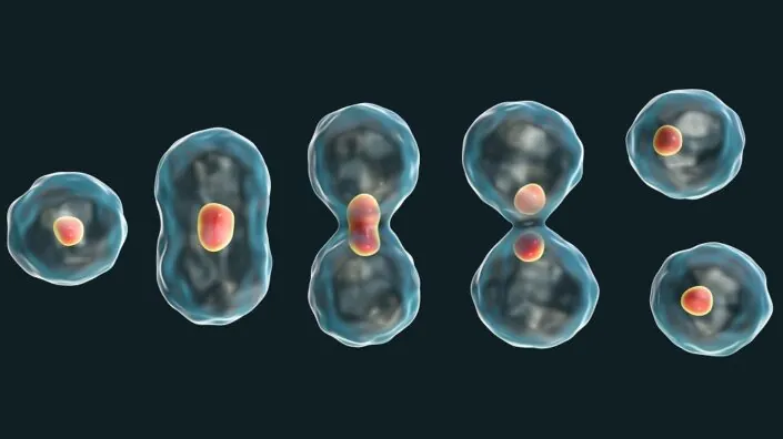
I believe three of your four candidates are based on cellular reprogramming. Has Retro effectively become a reprogramming company? Is this your biggest bet?

In one way or another, yes, we are for sure. We have a partial reprogramming research program that is not yet ready for clinical prime time, and then we have two clinical programs that use full reprogramming, which is exciting because it does essentially full rejuvenation, depending on the different ways of measuring the age of a cell. By most of those means, when you do full reprogramming all the way to an iPSC [induced pluripotent stem cell], you get essentially full rejuvenation.

What do you believe in more: the partial in vivo path or the full iPSC reprogramming in vitro path?

It’s hard to say what I’m most excited about. It’s like saying, “Which of my children do I love the most?” The problem with full reprogramming is that there are only a few cell types you can do it on. You need full control over the cell environment to differentiate it back to some adult cell type. So, a huge disadvantage is that you can’t do it in vivo. It pretty much only works for dissociated cells that are going to an individual cell type rather than a whole structured tissue. From an age-related disease intervention perspective, it can only work for cells that you can put back into the body as single cells.

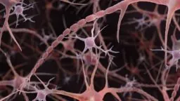
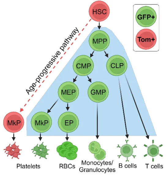
So that reduces the scope a lot. I am excited about it because I love the full rejuvenation and the ability to be reductionistic, controlling the entire environment step-by-step. But I wish there were more cell types that were amenable. Right now, we’re doing what we think are the two most viable and important ones, which are HSCs [hematopoietic stem cells] and microglia.

Fortunately, for HSCs, the healthcare community has gotten quite good at replacing the cells because of experience with leukemia, doing autologous bone marrow transplants for lymphoma or myeloma, or allogeneic transplants for various leukemias. It’s a well-developed path. But for the larger picture of the hundreds of cell types in other parts of the body, I think partial reprogramming is a huge opportunity. It’s just harder and more complicated.

Is that why you have that program on a back burner right now?

Yes, it’s operating outside of the clinical context. The moment we say, “Okay, time to go to the clinic,” that’s a very special mode for a program to go into. It requires a canonical indication, all these quality controls, preclinical safety, methods of measuring potency, setting up GMP manufacturing, regulatory work – all that stuff becomes a very different mindset and a huge, huge workstream.

Now, to the big news. You just announced a breakthrough with a ChatGPT-based model that was able to dramatically improve reprogramming efficiency. This is super exciting. Can you tell me more about it?

Yes, it’s been my thesis for quite a long time that biology is too complicated for humans to figure out alone, at least in the larger picture. Obviously, there’s a bunch of low-hanging fruit, especially things that involve single targets or a single pathway where you can disturb one protein-protein interaction and create a helpful effect for a particular disease. But I think there are only so many of those.

Age-related degeneration syndromes are often complex failures that are too hard for people to think about. A human can keep maybe a few different genes in their head at any given time, but then you add the sixth gene interaction with the third and the fifth, and downregulating the second and the fourth, and the network gets too big for humans to keep straight. They’re like, “Ah, can we go back to the single-gene, single-target therapeutics discussion, please?”.

I’ve thought – and hopefully, I get pushback from people smarter than me – that there is an evolutionary bias against single-target diseases. There’s this antagonistic pleiotropy concept where evolution is pretty good at trimming out diseases that pop up earlier in life and interfere with reproduction. Evolution is especially good at making a single mutation and seeing which replicator succeeds the best. It’s harder from a combinatorics perspective for an experiment to be randomly tried by evolution if it requires multiple changes on multiple genes all at the same time. So, I think evolution continually fixes these single-target diseases, and the ones you’re left with at the end of life are this messy, gross soup of multi-gene combinations of things that are starting to fall apart.

I couldn’t agree more on the potential of AI for biology. So, you basically retrained a large language model on all kinds of curated biological data, and the results were amazing.

The models that have been showing lots of traction lately were these LLMs, but they work on sequential data types, like text. A protein sequence is a sequential data type, essentially equivalent to a DNA sequence, but it’s a little closer to actual function, so there’s lower-hanging fruit operating at the protein level. There’s meaning based on proximity in similar ways to text, so how about if we train on that?

The thought was we don’t need to train it entirely from scratch because a lot of the information about proteins is encoded in English. If we co-train a language-based model with a protein sequence-based model, we should be able to capture information that’s in academic papers and annotation databases where humans have put great effort into noting protein functions and relationships. We did some iterations of that and also built a suite of evaluation functions that could give us quicker feedback as to whether the thing we’re training is starting to produce things that look like functional proteins that could actually fold. That was a guide for finding different ways of doing the training.

And your idea of trying of using it to make better reprogramming factors was pretty ingenious.

Well, we’re just already obsessed with those proteins anyway. The model itself was independent and wasn’t specific to transcription factors or Yamanaka factors; we’ve used it for multiple other things. But why not get started on these things? We’ve done a ton of reprogramming and have all kinds of metrics and assays.

Also, they tend to be types of proteins that are a bit hard to interact with using traditional tools because they have large disordered regions, so you can’t come at them from an entirely structured perspective. It just made sense to try these things out and see what happens. We weren’t necessarily expecting that they would be so functional so fast.

But they were. You kind of solved one of the biggest problems in reprogramming: low efficiency.

Yes, it’s so exciting to us that they were so functional. The function was so much better that they’re now in our latest FDA filing for one of the products that we’re taking to the clinic. We ran them through every metric we could think of in terms of whether they make legit, bona fide iPSCs. We wondered, “Is there something horribly, weirdly wrong with them?” It almost seemed too good to be true. So, we ran them through all the quality criteria that people use for iPSCs, and they seem perfect.

I also noticed the model basically made entirely new proteins. You call them enhanced factors, but the model changed 20-30% of the sequence. These could be novel antigens for all we know. On top of that, the model is a “black box,” meaning we don’t know how it did what it did. What are your thoughts on that? Do you see potential problems with safety and immunogenicity?

Actually, in some cases, it’s up to 80%. I think I had already come to terms with the notion that we’re not necessarily going to get to understand how AI does its thing. We’re going to become, at some point, spectators to science as it progresses. So, the fact that we don’t necessarily understand how it did everything – I’m willing to let go of that in exchange for new abilities to do incredibly humane things.

In terms of immunogenicity, it’s definitely a concern. For sure, the safest way is to use these initial proteins on cells outside the body, where they’re not exposed to a systemic immune system. The harder level will be if we want to use these to engineer therapeutic proteins that are circulating in the body. Unless and until we can apply an additional transform to the outer surfaces of the proteins to create a sort of “humanness” criterion – which we’re actively working on – it may be harder to employ them as random therapeutic proteins.

Do you have any thoughts on how we can speed up this process even more, such as with robotic labs for data generation and validation?

I think this is going to naturally happen. People have gotten extremely excited about robotic everything lately, and we’re in the elbow of an exponential curve on robotics. People seeing AI take off is a clear signal that we can build robots now that would not be dumb. In the past, it was demotivating; you could make this great robot, and it would just stand there and not know what to do. But now they’ll know what to do. We’re seeing billions of dollars invested in robotics that will be operated by AI.

In terms of pre-AI robotics, we’re doing a lot here at Retro. We’re supporting an open-source ecosystem called PyLabRobot that allows us to organize different laboratory instruments into clusters that can perform complex workflows for us, faster and more accurately than having humans do it. It’s better to use human brains for inventing the next experiment than having them sit there and tediously execute the last one.

We’ve noticed that ChatGPT is actually pretty good at writing code for PyLabRobot, which is cool. It’s an exciting vision. AI is going to be able to do more of the actions that create more data for the AI to get smarter, producing even more of the actions. We’re excited to be embedded right into one of those feedback loops.

Which brings me to my last question. You started Retro with a goal of extending human healthspan by 10 years. Have your ambitions and your ETA to target changed in the last five years, especially now that you’re using tools like LLMs that we didn’t think we’d have?

I hesitate to predict the future because such predictions are usually wrong. Us having to do clinical trials means we don’t understand biology. We can have an idea and think something is good, but then you must try it out in humans, and at least 80% of clinical trials fail. For me, this means that no one should be taken seriously if they are very confidently predicting the future.

But I could say that if everything goes well, our first drug should be out and prescribable by physicians by roughly the end of the 2020s. There’s a question of how much healthy lifespan extension we can get from making a significant dent in Alzheimer’s, but I think it’s a lot. Because of the regulatory environment we’re in, we have to think about everything in terms of prescribing for specific diseases.

In the meantime, we will operate within the organized constraints of the existing regulatory system, which exists for a purpose and is actually pretty reasonable in lots of ways. At the moment, it doesn’t slow us down hardly at all. The kinds of constraints and processes that are requested by a typical health authority right now don’t seem extreme to me.

I have all of my program leads apply this heuristic they call the “daughter test,” which is that if it were your daughter who had this disease and you’re making the Retro medicine for her, what tests would you want to run? Would you skip the six-month immunogenicity test for this particular therapy and just go straight to injecting it in her veins? Or would you want to run the test? When people roll their eyes about the FDA making us do a test, I’m like, “Okay, first I want to see the list from you of what you think actually matters.” And then I’m going to compare it to the FDA list and – oh, look at that. By and large, they’re pretty much the same.

We would like to ask you a small favor. We are a non-profit foundation, and unlike some other organizations, we have no shareholders and no products to sell you. All our news and educational content is free for everyone to read, but it does mean that we rely on the help of people like you. Every contribution, no matter if it’s big or small, supports independent journalism and sustains our future.
Dividing cells

Cell Cycle Stage Impacts the Efficacy of Senotherapeutics

A recent study reported that the effectiveness of the senotherapeutic drug ABT-263 depends on the cell’s DNA content, which is based on the cell cycle phase at which the senescent cell was arrested [1].

A personalized approach

Last week, we discussed clinical trials of senolytics and what we can learn from the data obtained so far. The authors of that paper discussed how it might be possible to personalize senolytic treatments to achieve better results.

This recent study published in Aging discusses similar ideas. The authors examine how cell population diversity impacts responses to senolytics and what differences in senescent cells drive those different responses.

Not all senescent cells are created equal

Even though senescent cells generally share many common characteristics, there is a significant variability among those cells. Such variability was reported in previous studies investigating the gene expression profiles of senescent cells; however, there is a scarcity of information about functional differences. Those researchers investigated those functional differences using high-content imaging, a technique that allows for measuring several protein markers at the single-cell level.

In the initial experiment, the researchers used a high-content image analysis to measure the expression of several senescence-associated markers following senescence induction through ionizing radiation (IR) in primary human endothelial cells and fibroblasts.

At the population level, their data confirmed IR-induced senescence. They noted that even in this high-level analysis, they identified differences in the levels of senescence markers between the two cell lines they used, suggesting cell-type-dependent differences in senescent cells.

Further analysis showed even more differences. In the following steps, the researchers investigated the senescent cell diversity at the single-cell level, and they noted two populations of cells that differ in senescence marker expression. They hypothesized that those populations might be associated with “the phase of the cell cycle at which senescent cells were growth-arrested.”

The tale of two cell phases

The cell cycle consists of two main phases: interphase and mitosis (cell division). Interphase is further divided into the G1 phase, where cell growth happens, the S phase, during which DNA is replicated so that it can be divided into two cells later, and the G2 phase, during which cells grow further and prepare for cell division.

The researchers analyzed the DNA content of the cells, as cells in the G1 and G2 phases have either low or high DNA content. G2-arrested cells expressed more senescent markers than G1-arrested cells, and the cells within each subgroup were roughly uniform in their expression of these markers. Identification of these two subgroups led to further testing of the differences between them.

In the next experiment, the researchers prepared cells so that each sample was enriched in either G1 or G2-phase arrested cells, irradiated them to induce senescence, and compared the secretion of IL-6, a SASP component that is associated with inflammation. IL-6 secretion was increased in the G2 group compared to the G1 group.

Most importantly, the researchers investigated the cells’ response to senolytics. Specifically, they tested ABT263, a senolytic that induces cell death (apoptosis) by inhibiting the anti-apoptotic proteins BCL-2 and BCL-xL. G2-arrested cells were more sensitive to ABT263 treatment than G1-arrested cells at the different concentrations tested.

The researchers noted that similar effects were obtained during cancer drug investigation; the cytotoxic effect of some drugs was impacted by DNA content and cell cycle phase, “with some drugs preferentially targeting cells in G1 and others in G2.” [2]

A piece of a bigger puzzle

This small study adds another piece of evidence to the idea that many cellular-level factors and interactions impact the efficacy of senotherapeutics. Those observations are essential in developing future clinical trials or therapies based on senolytics, as they will help to create personalized therapies that would be best tailored for particular patients.

The authors noted that much more needs to be explored in this topic. For example, this study was limited to only two cell lines and one mechanism of senescence induction; therefore, future investigation should expand to different cell types and senescence-inducing mechanisms. Furthermore, there is a need to investigate the diversity of senescent cells in living organisms and whether the effectiveness of senomorphics, which can reduce SASP factors and alleviate senescence-related tissue dysfunction instead of eliminating senescent cells, is similarly impacted.

Additionally, while this study reported on senolytics having different effects in various senescent subpopulations, it did not investigate the mechanism behind this observation. Similarly, these researchers examined only one senotherapeutic drug; it is highly possible that similar mechanisms can also be applied to different senotherapeutics, but this remains to be explored.

We would like to ask you a small favor. We are a non-profit foundation, and unlike some other organizations, we have no shareholders and no products to sell you. All our news and educational content is free for everyone to read, but it does mean that we rely on the help of people like you. Every contribution, no matter if it’s big or small, supports independent journalism and sustains our future.

Literature

[1] Neri, F., Zheng, S., Watson, M. A., Desprez, P. Y., Gerencser, A. A., Campisi, J., Wirtz, D., Wu, P. H., & Schilling, B. (2025). Senescent cell heterogeneity and responses to senolytic treatment are related to cell cycle status during senescence induction. Aging, 17(8), 2063–2078.

[2] Johnson, T. I., Minteer, C. J., Kottmann, D., Dunlop, C. R., Fernández, S. B. Q., Carnevalli, L. S., Wallez, Y., Lau, A., Richards, F. M., & Jodrell, D. I. (2021). Quantifying cell cycle-dependent drug sensitivities in cancer using a high throughput synchronisation and screening approach. EBioMedicine, 68, 103396.

Rejuvenation Roundup September 2025

Rejuvenation Roundup September 2025

Last month was full of news on both rejuvenation advocacy and rejuvenation advancements, including our new Public Longevity Group initiative along with our paper on Urolithin A being accepted into Aging Cell. Here’s what’s happened in September.

Team and activities

Public Longevity GroupLifespan Research Institute Launches Public Longevity Group: On September 17, we announced the launch of the Public Longevity Group (PLG), an initiative focused on bridging the cultural gap between scientific breakthroughs in aging and their public acceptance. To kickstart its work, PLG has opened a crowdfunding campaign to develop tools that measure and strengthen public trust in longevity science. It is developing the first data-driven cultural intelligence system for longevity—a platform designed to track real-time sentiment, test narratives, and identify which messages resonate and which backfire.

The PLG campaign is sponsored by the members of LRI’s Lifespan Alliance, a consortium of mission-aligned organizations that believe in the promise of extending healthy human lifespan. Newly-joined members include OpenCures, AgelessRx, and Lento Bio.

Looking Back at Summer, Looking Forward to Growth: For those of us in the Northern Hemisphere, autumn is underway. The fall is a time when the leaves that are green turn to brown, so let us see what the Lifespan team has been working on to help our field keep our own metaphorical leaves green and healthy.

Advocacy and Analysis

Ambulance backTwo People Almost Died at RAADfest. We Went to the Experts.: Two people nearly died, and several more sought treatment, after receiving peptide injections at the last RAADfest in Las Vegas. We might not know what happened until the ongoing investigations are concluded, but we asked several experts to share their thoughts on the broader context of unproven “rejuvenation therapies.”

Some Bioethicists Promote Lifespan Limitation: A paper published in Bioethics makes a startling case for people to die at 100 years old rather than live as long as they might choose.

Research Roundup

Mitigating Pro-Inflammatory SASP and DAMP With Urolithin A: A Novel Senomorphic Strategy: In Aging Cell, scientists from Lifespan Research Institute and the Buck Institute for Research on Aging have published their findings that Urolithin A, a molecule that has garnered a lot of attention in the longevity field, potently reduces senescence-related markers in human fibroblasts. We reported on this paper in its preprint stage, before it had been published in a journal.

Heart analysisA Non-Viral Gene Therapy Restores Mouse Hearts: A recent study investigated the roles of brown adipose tissue and a lipid-controlling hormone, 12,13-diHOME, in cardiac health. The researchers examined the molecular mechanisms behind 12,13-diHOME’s effects on the hearts of aged mice.

Cancer Cells Transfer Mitochondria to Fibroblasts: Scientists have discovered that cancer cells recruit fibroblasts to support tumor growth by transferring mitochondria into them. Blocking this process might be a new way to fight the deadly disease.

Protein aggregateA Mechanism Behind Protein Aggregation Discovered: Scientists have found a pathway that regulates protein aggregation, a cause of several age-related neurodegenerative diseases. For instance, amyotrophic lateral sclerosis (ALS), Huntington’s disease, and Alzheimer’s disease have all been linked to abnormal protein aggregation.

Study Boosts Brain Mitochondria, Rescues Memory in Mice: Scientists have found a way to directly stimulate the assembly of Complex I in mitochondria, rescuing memory deficits in mouse models of Alzheimer’s and frontotemporal dementia.

MicrogliaMicroglial Aging Is Determined by Their Environment: A new preprint study from Calico has found that the local brain environment is the primary driver of microglial aging. After being transplanted into old brains, young cells adopted aged characteristics, but their susceptibility to these signals could be turned off.

Regular Glucosamine Use Linked to Fewer Chronic Diseases: An analysis of UK Biobank data showed an association between regular glucosamine use and significantly lower risks of seven non-communicable chronic diseases.

Old timepieceEpigenetic Clocks Do Not Perfectly Capture Metabolic Health: In Aging Cell, researchers have published their surprising findings that epigenetic clocks are not significantly related to most measurements of metabolic health after weight loss interventions.

Microplastics Cause Cognitive Deficits in APOE4 Mice: Scientists have demonstrated that short-term exposure to microplastics causes Alzheimer’s-like effects in mice expressing human APOE4 versus APOE3. These effects were sex-dependent, mirroring the disease in humans.

MacrophageHow Macrophages Manage Obesity and Change With Age: In Nature Aging, researchers have identified and categorized several macrophage subtypes, including a subtype that appears with aging and another that manages nerve function.

A Short-Term High-Fat Diet Harms Memory in Mice: Scientists have demonstrated that even two days on a Western-like high-fat diet reduce hippocampal glucose availability, which activates a subset of inhibitory neurons and causes memory problems in mice.

DNA and cellsPartial Reprogramming Rejuvenates Aged Cells and Tissues: In this study, researchers investigated aging- and disease-associated changes in gene expression related to epithelial-mesenchymal transition. Inducing the Yamanaka factors in mice allowed them to rejuvenate cells and tissues and reverse some of these aging-associated changes.

Lipid Metabolism Is Causal in Some Alzheimer’s Cases: In Aging Cell, researchers have outlined the relationship between Alzheimer’s, increased pain sensitivity, and the enzyme LPCAT2.

Robot analysisAI Model Accurately Predicts Multiple Disease Risks: European scientists have created a GPT-based model that can predict the risk of more than a thousand diseases on par with single-disease tools and biomarker-based models.

Exercise Suppresses Appetite via a Brain Pathway: Scientists have discovered a pathway behind the known effect of exercise suppressing appetite: a lactate-related metabolite that acts directly on certain neurons.

Clinical documentationPersonalized Medicine Approach to Senolytics Clinical Trials: Recent commentary in Nature Aging summarized the results of clinical trials for senolytics and discussed recommendations for future clinical trials that use personalized medicine approaches.

A Potential Reason Why Clotting Increases With Age: In Aging Cell, researchers have described a method by which platelet-forming cells are rapidly generated from hematopoietic stem cells (HSCs), bypassing the intermediate cell types that are normally used to get there.

New Universal Therapy Effective in Multiple Tumor Types: Scientists have reported a breakthrough in treating solid tumor cancers using a Velcro-like tool that targets glycans, surface sugars especially abundant in cancer cells. This potentially off-the-shelf therapy does not need adjustment to individual cancer types or patients.

A Combination Greatly Extends Lifespan in Male Mice: The Conboy lab in Berkeley has discovered a treatment combination that greatly extends lifespan in old male mice and published its findings in Aging.

MitochondriaFaulty Mitochondrial DNA Copying Might Cause Inflammaging: Scientists have discovered a possible mechanism behind age-related inflammation. It involves wrong building blocks being incorporated into mitochondrial DNA during replication and can be countered by adding the correct ones.

Extension of lifespan by epicatechin, halofuginone and mitoglitazone in male but not female genetically heterogeneous mice: In addition to adding 3 new agents to the list of interventions identified by the ITP that extend lifespan, this report continues the strong male bias in the efficacy of life-extending drugs identified so far.

Short-term mTOR inhibition by rapamycin improves cardiac and endothelial function in older men: a proof-of concept pilot study: Cardiac and endothelial function improvements with RAPA were found and support future placebo-controlled studies in larger cohorts of healthy older persons as well as in patients with compromised diastolic and endothelial function.

A randomized, double blind, placebo-controlled, pilot study to fine tune an NT-proBNP-based method to assess the effect of anti-aging treatments: NT-proBNP levels increase exponentially with age and are associated with cardiovascular and all-cause mortality. From NT-proBNP concentration a surrogate for biological age (“proBNPage”) can be obtained.

Creatine and Cognition in Aging: A Systematic Review of Evidence in Older Adults: The current limited evidence suggests that creatine may be associated with benefits for cognition in generally healthy older adults. However, high-quality clinical trials are warranted to further validate this relationship.

Effect of henagliflozin on aging biomarkers in patients with type 2 diabetes: A multicenter, randomized, double-blind, placebo-controlled study: Metabolomic analysis shows that henagliflozin induces changes in various metabolites, including increased thiamine levels and enhanced thiamine metabolism. These findings suggest that henagliflozin may exert anti-aging effects through multiple pathways.

The multiomics blueprint of the individual with the most extreme lifespan: These findings provide a fresh look at human aging biology, suggesting biomarkers for healthy aging, and potential strategies to increase life expectancy.

Repeated Withdrawal of a GLPR Agonist Induces Hyperleptinemia and Deteriorates Metabolic Health in Obese Aging UM-HET3 Mice: These findings suggest that continuous GLP-1-based therapy is necessary to sustain metabolic benefits, while intermittent use may promote age-associated sarcopenia and metabolic decline.

Omega-3 Polyunsaturated Fatty Acids and Cognitive Decline in Adults with Non-Dementia or Mild Cognitive Impairment: These findings support n3-PUFA supplementation as a complementary approach to lifestyle-based strategies for cognitive health, including diet, physical activity, sleep optimization, and cognitive training.

Association of Eating Window With Mortality Among US Adults: Insights From a Nationally Representative Study: Moderate eating windows (~11–12 h/day) are linked to the lowest mortality risk, with deviations associated with higher risk. Differences across demographic groups highlight the need for personalized guidance.

Serum Vitamin C concentrations are inversely related to biological aging: a population-based cross-sectional study: Serum Vitamin C levels exhibit an inverse association with biological aging, particularly in older individuals and those with chronic conditions, highlighting the potential role of Vitamin C in healthy aging.

An Exercise Intervention May Counteract the Degradation of Nerve Conduction from Age-Related Disuse: The results of this study suggest that resistance training may be a viable method to counteract age-related nerve deterioration. These outcomes have the potential to improve quality of life and generate greater independence for our older populations.

SenolyticSynergy: An Attention-Based Network for Discovering Novel Senolytic Combinations via Human Aging Genomics: This framework paves the way for large-scale research into anti-aging drug combinations, advancing research capabilities in this field.

In Silico Assessment of Potential Geroprotectors: From Separate Endpoints to Complex Pharmacotherapeutic Effects: Validation using known geroprotectors (rapamycin, metformin, and resveratrol) demonstrated strong concordance between predicted activities and documented molecular mechanisms of action.

Partial Reprogramming in Senescent Schwann Cells Enhances Peripheral Nerve Regeneration via Restoration of Stress Granule Homeostasis: Dysregulated stress granule homeostasis drives the pathological accumulation of Runx2+ Schwann cells, representing a key mechanism underlying age-related axonal regeneration deficits in peripheral nerve repair.

Single-Short Partial Reprogramming of the Endothelial Cells Decreases Blood Pressure via Attenuation of EndMT in Hypertensive Mice: Overall, these data indicate that OSK treatment and EC reprogramming can decrease blood pressure and reverse hypertension–induced vascular damage.

News Nuggets

Longevity investorsCountdown to the Longevity Investors Conference 2025: From 22–25 September 2025, the sixth edition of this conference took place at the five-star hotel Le Grand Bellevue in Gstaad, Switzerland – a discreet alpine setting that combines world-class luxury with the privacy essential for meaningful dialogue.

LongX Hosts the Youth in Longevity Biotech Showcase: On September 18, 2025, Longevity Xplorer (LongX) hosted the first-ever “Youth in Longevity Biotech Showcase”, a virtual event featuring lightning talks from young professionals in longevity fellowships around the world.

YouthBioYouthBio Therapeutics Announces Positive FDA Feedback: YouthBio Therapeutics, a biotechnology company pioneering partial cellular reprogramming to treat diseases of aging, today announced a successful INTERACT meeting with the FDA for its lead Alzheimer’s candidate, YB002.

We would like to ask you a small favor. We are a non-profit foundation, and unlike some other organizations, we have no shareholders and no products to sell you. All our news and educational content is free for everyone to read, but it does mean that we rely on the help of people like you. Every contribution, no matter if it’s big or small, supports independent journalism and sustains our future.

Mitochondria

Faulty Mitochondrial DNA Copying Might Cause Inflammaging

Scientists have discovered a possible mechanism behind age-related inflammation. It involves wrong building blocks being incorporated into mitochondrial DNA during replication and can be countered by adding the correct ones [1].

Too similar to bacteria

Mitochondria, the cells’ energy-producing organelles, are considered to have developed from bacteria that once entered an ancient cell and stayed, enabling life as we know it [2]. Mitochondria’s microbial origins can still pose a problem: when mitochondrial DNA (mtDNA) gets into the cytoplasm, its resemblance to bacterial DNA might trigger an immune response [3].

Aging is accompanied by an increase in inflammaging, a form of inflammation that is unrelated to infections (sterile inflammation) and that harms cells and tissues [4]. The origins of inflammaging are not entirely understood, but mtDNA leakage has been proposed as a possible culprit. In this new study published in Nature, scientists from the Max Planck Institute for the Biology of Ageing describe a mechanism that might underlie this link.

Their central hypothesis, based on previous research, suggested that when deoxyribonucleoside triphosphates (dNTPs, the proper building blocks for DNA) are scarce relative to ribonucleoside triphosphates (rNTPs, the RNA building blocks), mitochondria mistakenly install rNTPs into mtDNA. These embedded rNTPs make the genome fragile during replication, creating fragments that spill into the cytosol and activate the well-studied inflammatory cGAS-STING pathway.

The wrong building blocks

The team started by using mice lacking MGME1, an enzyme needed for proper mtDNA replication, that naturally develop mtDNA leakage and inflammation. In the mice’s kidneys, they saw increased mtDNA fragment accumulation and innate immune activation, suggesting that the two are causally linked. These mice have been shown to develop kidney disease and die sooner. Knocking out the STING part of the cGAS-STING inflammatory pathway reduced inflammation and ameliorated kidney pathology.

The next question the researchers asked was whether this effect requires active mtDNA copying. When the researchers slowed or blocked mtDNA replication, the inflammatory response decreased, suggesting that the problem stems from breaks during copying rather than ambient damaged DNA. Deep mtDNA sequencing pointed at frequently aborted replication as the source of excessive mtDNA fragmentation.

The study then moved towards determining if the rNTP:dNTP ratio becomes imbalanced in cells where mtDNA copying isn’t working properly. Theoretically, numerous unsuccessful replication attempts should cause the limited dNTP pool in mitochondria to deplete, and this is what the researchers observed. Turning up the dNTP supply by knocking down SAMHD1, a dNTP-depleting enzyme, restored dNTPs and suppressed activation of the immune response.

In a complementary model lacking the mitochondrial protease YME1L, which also perturbs nucleotide metabolism, the researchers showed that raising the rNTP:dNTP ratio slows de novo mtDNA synthesis. They then measured rNMPs in mtDNA directly by two different methods and found that interfering with nucleotide metabolism caused an increase in rNMPs.

Confirmed in senescence and natural aging

The researchers then moved to senescent human fibroblasts, which are known to have decreased activity of RNR, the enzyme that converts rNTPs to dNTPs, and, consequently, a higher rNTP:dNTP ratio. Lowering it by adding back deoxyribonucleosides (dNs) reduced cytosolic mtDNA and caused the senescent cells to become less toxic without actually reversing senescence: a senomorphic effect. The treated fibroblasts produced less of the senescence-associated secretory phenotype (SASP), a mix of mostly pro-inflammatory molecules emitted by senescent cells.

Finally, the team confirmed that older healthy mouse tissues have a higher rNTP:dNTP ratio than younger ones do. This suggests that the mechanism is indeed characteristic of normal aging and is a promising target for future anti-aging interventions.

“Our findings explain on a molecular level how metabolic disturbances can lead to inflammation in senescent cells and in aged tissue and open up new strategies for possible interventions,” said Prof. Thomas Langer, who led the study.

“There is already a therapy for certain mitochondrial diseases that involves administering DNA building blocks. However, we do not yet know if it can also alleviate the inflammation that occurs more frequently with age. It would be interesting to test this,” noted Dusanka Milenkovic, one of the study’s lead authors.

On X, Harvard geroscientist Dr. David Sinclair called the study “an exciting new paper that could explain why inflammation rises as we age.” He added: “The paper shows that during cell stress & as mice age, their cells incorporate the wrong type of bases (ribonucleotides not deoxyribonucleotides) into replicating mDNA, causing the genome to eventually break and leak into the cytoplasm.”

We would like to ask you a small favor. We are a non-profit foundation, and unlike some other organizations, we have no shareholders and no products to sell you. All our news and educational content is free for everyone to read, but it does mean that we rely on the help of people like you. Every contribution, no matter if it’s big or small, supports independent journalism and sustains our future.

Literature

[1] Bahat, A., Milenkovic, D., Cors, E., Barnett, M., Niftullayev, S., Katsalifis, A., … & Langer, T. (2025). Ribonucleotide incorporation into mitochondrial DNA drives inflammation. Nature, 1-9.

[2] Andersson, G. E., Karlberg, O., Canbäck, B., & Kurland, C. G. (2003). On the origin of mitochondria: a genomics perspective. Philosophical Transactions of the Royal Society of London. Series B: Biological Sciences, 358(1429), 165-179.

[3] Hu, M. M., & Shu, H. B. (2023). Mitochondrial DNA-triggered innate immune response: mechanisms and diseases. Cellular & molecular immunology, 20(12), 1403-1412.

[4] Franceschi, C., Garagnani, P., Vitale, G., Capri, M., & Salvioli, S. (2017). Inflammaging and ‘Garb-aging’. Trends in Endocrinology & Metabolism, 28(3), 199-212.

YouthBio

YouthBio Therapeutics Announces Positive FDA Feedback

YouthBio Therapeutics, a biotechnology company pioneering partial cellular reprogramming to treat diseases of aging, today announced a successful INTERACT meeting with the FDA for its lead Alzheimer’s candidate, YB002. In its formal response, the FDA agreed that existing preclinical data support the bioactivity of YB002 and YouthBio’s proposed first-in-human trial. This feedback represents a major de-risking event for YouthBio, which will now focus on CMC activities and a pilot toxicology study to support a Pre-IND meeting and finalize designs for IND-enabling studies.

“We are thrilled with this INTERACT outcome,” said Yuri Deigin, CEO of YouthBio. “The FDA’s response confirms our capital-efficient development strategy for YB002 and provides a clear path to the clinic. This is a significant inflection point, shifting the conversation from scientific plausibility to execution, and positioning YouthBio to be the first to bring partial reprogramming to the human brain.”

This milestone continues YouthBio’s strong record of execution. It builds upon compelling scientific evidence, including a study with Dr. Alejandro Ocampo where YB002 ameliorated cognitive decline in mice. Additional Alzheimer’s models have demonstrated that partial reprogramming can reverse disease pathologies, counteract epigenetic aging, and rescue memory and learning.

“The FDA’s recognition of our proof-of-concept data is highly encouraging from a scientific perspective,” said Dr. João Pedro de Magalhães, CSO of YouthBio. “Their detailed feedback allows us to focus our resources effectively and build the most robust IND package possible.”

YB002 is a first-in-class gene therapy designed to safely and transiently express Yamanaka factors in the brain — a process known as partial reprogramming. Built on Nobel Prize-winning science, this approach aims to reverse epigenetic changes that accumulate with aging while preserving cell identity, thereby restoring youthful gene expression and improving cellular function.

“It is gratifying to see the field of partial reprogramming entering clinical translation,” said Dr. Alejandro Ocampo, whose pioneering 2016 paper launched the field of partial reprogramming. “In our joint study with YouthBio, YB002 ameliorated cognitive decline in old wild-type mice. This FDA feedback marks a critical milestone for both YouthBio and the entire field.”

Media contact: yuri@youthbiotx.com

About YouthBio Therapeutics, Inc.

YouthBio Therapeutics (www.youthbiotx.com) is developing a new class of medicines to combat age-related diseases by leveraging partial cellular reprogramming. The company’s lead program, YB002, is being advanced for the treatment of Alzheimer’s disease.

Black lab mouse

A Combination Greatly Extends Lifespan in Male Mice

The Conboy lab in Berkeley has discovered a treatment combination that greatly extends lifespan in old male mice and published its findings in Aging.

A combination with systemic effects

The researchers begin this paper with a discussion of well-known interventions and their drawbacks. For example, they note that while rapamycin is effective in extending the lifespans of mice [1], it is associated with cancer development [2]; while adding the anti-cancer drug trametinib mitigates this issue [3], this combination strongly inhibits mTOR, which is necessary for stem cell and brain function [4]. Similarly, knocking out the interleukin IL-11 extends lifespan in mice [5], but its levels only increase with severe disease [6] and it plays a major part in ovarian health [7].

Therefore, these researchers have built on their previous work involving the systemic environment. The Conboy lab is most famous for its work on heterochronic parabiosis and therapeutic plasma exchange, which remove harmful compounds from the bloodstream that accumulate with age [8]. However, such an approach involves repeatedly replacing a person’s blood, which comes with its own complications [9].

This experiment involves attempting to recapitulate the benefits of TPE by addressing its key molecular determinants. This team found two that it deemed likely to work in conjunction: oxytocin, a well-known compound involved in social bonding [10] and is crucial in healing and metabolism [11], and A5i, a compound that inhibits the age-related increase of TGF-β, a factor that promotes fibrosis and inflammation [12]. This lab’s previous work has found that this combination, OT+A5i, leads to tissue rejuvenation [13], and so it performed another experiment involving lifespan.

Sex-dependent effects

The researchers’ lifespan experiment involved four separate groups of males and females either receiving OT+A5i or serving as controls, each of which contained roughly a dozen animals. These mice, at 25 months of age, were already old and frail at the beginning of the study. For two weeks, they would be given this combination three times a week, then left for two weeks without the combination before they were physiologically assessed and the treatment cycle was repeated.

In male mice, the gains in lifespan were tremendous. While their lifespans were only increased by an average of 14% as measured from birth, they were increased by 74% as measured from the beginning of treatment. Six months after treatment, more than three quarters of the treated male mice were still alive, while only about a third of the male mice in the control group had survived this long.

Unfortunately, there were no benefits for lifespan in female mice. The survival curves of the controls and OT+A5i female mice looked similar, with a trend towards a decrease in lifespan instead.

OT+A5i survival curve

The researchers further assessed this combination’s effects on frailty. They found that OT+A5i improved healthspan as well as lifespan, increasing the time before the animals reached a certain threshold of assessed frailty according to multiple physical measurements, such as vision, gait, and physical challenges such as treadmill and limb hanging times. Additionally, the treated males were more likely to live longer even after becoming frail.

There were clear effects on the individual metrics used to assess frailty as well. The treated male mice were much more able to recognize novel objects, run for longer on gradually accelerating treadmills, and hang for longer from a wire ceiling. Similarly to the lifespan study, none of these beneficial effects on healthspan were found on old female mice.

Only short-term effects on females

These sex-dependent results appear to have been due to a lack of long-term efficacy in females. Immediately after administration of OT+A5i, the researchers discovered that both male and female mice began to synthesize a more youthful balance of proteins (the proteome). Proteomic noise was also, in the short term, decreased by OT+A5i in both sexes. However, these effects were attenuated in females but not males after four months of treatment.

The researchers note that oxytocin is already approved for clinical use by the FDA and that A5i drugs are already being investigated for the treatment of certain conditions, with no major adverse effects yet reported in clinical trials. Therefore, it would be logical to begin a clinical trial to investigate what, if any, effects that this combination may have on the lifespan and healthspan of older men.

We would like to ask you a small favor. We are a non-profit foundation, and unlike some other organizations, we have no shareholders and no products to sell you. All our news and educational content is free for everyone to read, but it does mean that we rely on the help of people like you. Every contribution, no matter if it’s big or small, supports independent journalism and sustains our future.

Literature

[1] Bjedov, I., & Rallis, C. (2020). The target of rapamycin signalling pathway in ageing and lifespan regulation. Genes, 11(9), 1043.

[2] Bitto, A., Ito, T. K., Pineda, V. V., LeTexier, N. J., Huang, H. Z., Sutlief, E., … & Kaeberlein, M. (2016). Transient rapamycin treatment can increase lifespan and healthspan in middle-aged mice. elife, 5, e16351.

[3] Gkioni, L., Nespital, T., Baghdadi, M., Monzó, C., Bali, J., Nassr, T., … & Partridge, L. (2025). The geroprotectors trametinib and rapamycin combine additively to extend mouse healthspan and lifespan. Nature Aging, 1-17.

[4] Garza-Lombó, C., Schroder, A., Reyes-Reyes, E. M., & Franco, R. (2018). mTOR/AMPK signaling in the brain: Cell metabolism, proteostasis and survival. Current opinion in toxicology, 8, 102-110.

[5] Widjaja, A. A., Lim, W. W., Viswanathan, S., Chothani, S., Corden, B., Dasan, C. M., … & Cook, S. A. (2024). Inhibition of IL-11 signalling extends mammalian healthspan and lifespan. Nature, 632(8023), 157-165.

[6] Ren, C., Chen, Y., Han, C., Fu, D., & Chen, H. (2014). Plasma interleukin-11 (IL-11) levels have diagnostic and prognostic roles in patients with pancreatic cancer. Tumor Biology, 35(11), 11467-11472.

[7] Cork, B. A., Li, T. C., Warren, M. A., & Laird, S. M. (2001). Interleukin-11 (IL-11) in human endometrium: expression throughout the menstrual cycle and the effects of cytokines on endometrial IL-11 production in vitro. Journal of reproductive immunology, 50(1), 3-17.

[8] Kim, D., Kiprov, D. D., Luellen, C., Lieb, M., Liu, C., Watanabe, E., … & Conboy, I. M. (2022). Old plasma dilution reduces human biological age: a clinical study. Geroscience, 44(6), 2701-2720.

[9] Mokrzycki, M. H., & Kaplan, A. A. (1994). Therapeutic plasma exchange: complications and management. American Journal of Kidney Diseases, 23(6), 817-827.

[10] Zeki, S. (2007). The neurobiology of love. FEBS letters, 581(14), 2575-2579.

[11] Breuil, V., Trojani, M. C., & Ez-Zoubir, A. (2021). Oxytocin and bone: review and perspectives. International journal of molecular sciences, 22(16), 8551.

[12] Tzavlaki, K., & Moustakas, A. (2020). TGF-β Signaling. Biomolecules, 10(3), 487.

[13] Mehdipour, M., Etienne, J., Chen, C. C., Gathwala, R., Rehman, M., Kato, C., … & Conboy, I. M. (2019). Rejuvenation of brain, liver and muscle by simultaneous pharmacological modulation of two signaling determinants, that change in opposite directions with age. Aging (Albany NY), 11(15), 5628.

New Universal Therapy Effective in Multiple Tumor Types

Scientists have reported a breakthrough in treating solid tumor cancers using a Velcro-like tool that targets glycans, surface sugars especially abundant in cancer cells. This potentially off-the-shelf therapy does not need adjustment to individual cancer types or patients.

No sugarcoating this

Antibody-based cancer immunotherapies, such as chimeric antigen receptor T (CAR-T) cells were once hailed as gamechangers in oncology. While their potential is indeed massive, several major hurdles remain. For instance, such tools need very high affinity to kill tumors, but that high affinity means they risk hitting the same target on normal tissues, where it can be present in low numbers (“on-target, off-cancer” toxicity) [2]. Moreover, targets tend to be specific to a cancer type or even a particular tumor, requiring expensive tailor-made approaches.

One of the most widespread adaptations that help cancer cells grow and propagate while evading the immune system is a remodeled glycocalyx, which is a cellular coating of glycans linked to proteins and fats (glycoproteins, glycolipids, proteoglycans) [3]. Tumors often thicken and re-pattern these sugars into tumor-associated carbohydrate antigens (TACAs), which can hide danger signals from immune cells, boost growth, and make it physically harder for immune cells to latch on.

However, this glycocalyx remodeling also differentiates cancer cells from the rest and provides an opportunity to target them. If we find a way to target TACAs safely, we can get a single approach that works across many tumors while sparing healthy cells. This idea is at the heart of a new study from the University of California Irvine, published in the journal Cell.

The two-armed bandit

The researchers used sugar-binding proteins called lectins in a way that causes many weak grips to add up (avidity), which the authors liken to Velcro, rather than the single high-affinity “key-lock” connection characteristic of antibodies. They created a bispecific protein that fuses a lectin carbohydrate-recognition domain (CRD) to a single-chain antibody.

Bispecificity means that one arm of the protein, containing four lectins, like four Velcro hooks, latches on to the glycans on the cancer cell’s surface, while the other arm attaches itself to a T cell, bringing it into proximity with the cancer cell. The researchers hypothesized that this construct, glycan-dependent T-cell recruiter (GlyTR, pronounced “glitter”), would recruit T cells to high-TACA-density cancer cells but not to normal cells, where glycan density is much lower.

“It’s the holy grail – one treatment to kill virtually all cancers,” said Michael Demetriou, MD, Ph.D., a professor of neurology, microbiology and molecular genetics at the UC Irvine School of Medicine and the paper’s corresponding author. “GlyTR’s velcro-like sugar-binding technology addresses the two major issues limiting current cancer immunotherapies: distinguishing cancer from normal tissue and cancer’s ability to suppress the immune system.”

The researchers confirmed that the GlyTR’s lectin arm binds to cancer cells but not to the T cells that GlyTR engages. They also tested GlyTRs on several types of healthy tissues and red blood cells (RBCs), confirming there was no strong interaction with non-cancer cells or concerning accumulation in normal organs in vivo.

Effective against multiple cancers

For cancer, however, the effect was devastating. In vitro, both GlyTR versions created by the researchers, GlyTR1 and GlyTR2, bound a wide variety of solid and liquid tumors, including breast, ovarian, prostate, pancreatic, colon, lung, AML, myeloma, and T-cell leukemia, much more than normal lymphocytes and drove T-cell-dependent killing even at very low concentrations. Notably, conventional immunotherapies mostly work for blood cancers, while solid tumors proved to be much harder to crack.

For their in vivo experiments, the team used immunodeficient mice given human T cells (humanized NSG mice) and implanted rejection-proof human tumors. When they injected fluorescent GlyTRs into the bloodstream, the proteins homed to lungs that harbored metastatic triple-negative breast cancer (TNBC) but not to lungs without tumors, providing evidence of tumor-seeking behavior in vivo.

Glycan treatment in cancer

In intraperitoneal models of TNBC, pancreatic, and ovarian cancers, GlyTRs produced dramatic response compared to just human CD8+ T cells, completely blocking tumor growth. The ovarian cancer model was independently replicated at the National Cancer Institute in a good sign for future clinical trials.

Clinical grade GlyTR1 protein manufacturing is already being developed at the NCI Experimental Therapeutics program labs in Maryland. Due to these phenomenal results, the team plans to bring the project to the clinic as early as possible, with human trials in about two years from now, according to the press release.

“This is the revolutionary approach to cancer treatment our patients have been waiting for,” said Farshid Dayyani, MD, Ph.D., medical director of the Stern Center for Clinical Trials and Research. “We are committing all available resources to bring this exciting new trial to UCI Health as fast as possible.”

We would like to ask you a small favor. We are a non-profit foundation, and unlike some other organizations, we have no shareholders and no products to sell you. All our news and educational content is free for everyone to read, but it does mean that we rely on the help of people like you. Every contribution, no matter if it’s big or small, supports independent journalism and sustains our future.

Literature

[1] Zhou, R. W., Purohit, P. K., Kim, J. H., Newton, B. L., Edwards, R. A., & Demetriou, M. (2025). Safe immunosuppression-resistant pan-cancer immunotherapeutics by velcro-like density-dependent targeting of tumor-associated carbohydrate antigens. Cell, 188, 1–17.

[2] Lamers, C. H., Sleijfer, S., Van Steenbergen, S., Van Elzakker, P., Van Krimpen, B., Groot, C., … & Gratama, J. W. (2013). Treatment of metastatic renal cell carcinoma with CAIX CAR-engineered T cells: clinical evaluation and management of on-target toxicity. Molecular therapy, 21(4), 904-912.

[3] Buffone Jr, A., & Weaver, V. M. (2019). Don’t sugarcoat it: How glycocalyx composition influences cancer progression. Journal of Cell Biology, 219(1), e201910070.

A Potential Reason Why Clotting Increases With Age

In Aging Cell, researchers have described a method by which platelet-forming cells are rapidly generated from hematopoietic stem cells (HSCs), bypassing the intermediate cell types that are normally used to get there.

Canonical and non-canonical formation

It is well known that blood clots, which form when platelets bind together, are a serious problem in older people [1]. Arterial clots directly cause both heart attacks and strokes, often being the final capstones on gradual cardiovascular aging, but changes in HSC behavior drive many other conditions as well [2].

In the canonical pathway, HSCs go through three intermediate cell types before finally becoming megakaryocyte progenitors (MkPs), which form the megakaryocytes that release platelets [3]. This team has previously discovered that a non-canonical pathway also exists and increases with age, turning HSCs directly into MkPs with no intermediate steps [4]. Worse, this previous work found that platelets created by non-canonical MkPs (ncMkPs) are hyperactive, heightening their potential contributions to clotting-related diseases.

Non-canonical platelet pathway

To illustrate these differences, the researchers had created a mouse model, FlkSwitch, that expresses green fluorescent protein (GFP) in MkPs that go through the canonical pathway and a different fluorescent protein, Tomato, in MkPs that go through the faster, non-canonical pathway. This paper is a closer examination into the differences between these MkPs, as the researchers wanted to be able to identify them without the need for specialized mice and to determine the functional differences between them.

Finding the right biomarkers

In their first experiment, the researchers combined their previous bulk-sequencing RNA results with single-cell sequencing. They were looking for membrane-bound proteins, which could potentially be used in future studies or even clinical practice to determine which MkPs are which, and they sought proteins for which commercially available antibodies already exist.

Initially, the researchers hit upon CD54 as their target protein, but further work found that CD48 was a better fit; MkPs expressing more CD48 are much more likely to be created through the canonical pathway, as determined by GFP and Tomato comparisons. Another protein, CD321, was found to be a strong predictor in the other direction; MkPs that express high amounts of this protein are much more likely to be created through the non-canonical pathway. This combination of CD48 and CD321 was highly useful in creating a firm basis for differentiating MkPs, as it yielded similar results to those gleaned from the FlkSwitch mice.

A further experiment found that ncMkPs are not entirely exclusive to aged animals. In 2- to 3-month-old male mice, approximately one in five MkPs is created through the non-canonical pathway. In aged male mice, however, this changes to an average of three in five, although there are large variations between animals. For female mice, there was a statistically significant increase with aging, but it was somewhat less noticeable and older female mice have fewer platelets overall than older male mice do, despite having similar numbers of MkPs.

Hardier survivors

One crucial and surprising finding was that older ncMkPs have a much greater ability to survive in vitro than all other MkPs. When derived from younger animals, ncMkPs proliferate poorly, less than canonical MkPs (cMkPs) of any age. Older ncMkPs, on the other hand, proliferate much more.and are much less likely to die by apoptosis.

This increase in activity persisted in vivo as well; older ncMkPs that were given to younger mice, which had their relevant cells previously destroyed by radiation, proliferated much more than younger ncMkPs did. Additionally, chemical stimulation determined that the platelets produced by older ncMkPs were, indeed, extremely reactive.

The environment plays a role

Taking HSCs from younger and older mice and observing their conversion into MkPs yielded entirely expected results; HSCs derived from the older animals were much more likely to produce ncMkPs. However, these findings changed when these cells were introduced into a younger irradiated animal; there, HSCs produced similar proportions of MkPs, regardless of whether they had come from a younger or older source. A closer examination to determine if any individual HSC is more or less likely to prouce ncMkPs yielded no useful results, suggesting that the aged environment is a strong factor in HSCs creating ncMkPs

As the researchers point out, however, humans and mice differ, and platelet generation is one of those differences; in humans, platelet count decreases with age [5]. Therefore, it is not clear if these murine findings could directly translate to human beings. However, with the identification of the proteins CD48 and CD321 as biomarkers, further work on human MkPs may reveal whether this change in cell fate is a significant factor in human clot-related diseases along with potential interventions to address it.

We would like to ask you a small favor. We are a non-profit foundation, and unlike some other organizations, we have no shareholders and no products to sell you. All our news and educational content is free for everyone to read, but it does mean that we rely on the help of people like you. Every contribution, no matter if it’s big or small, supports independent journalism and sustains our future.

Literature

[1] Le Blanc, J., & Lordkipanidzé, M. (2019). Platelet function in aging. Frontiers in cardiovascular medicine, 6, 109.

[2] Chung, S. S., & Park, C. Y. (2017). Aging, hematopoiesis, and the myelodysplastic syndromes. Blood advances, 1(26), 2572-2578.

[3] Manso, B. A., Rodriguez y Baena, A., & Forsberg, E. C. (2024). From hematopoietic stem cells to platelets: Unifying differentiation pathways identified by lineage tracing mouse models. Cells, 13(8), 704.

[4] Poscablo, D. M., Worthington, A. K., Smith-Berdan, S., Rommel, M. G., Manso, B. A., Adili, R., … & Forsberg, E. C. (2024). An age-progressive platelet differentiation path from hematopoietic stem cells causes exacerbated thrombosis. Cell, 187(12), 3090-3107.

[5] Jones, C. I. (2016). Platelet function and ageing. Mammalian genome, 27(7), 358-366.

Clinical documentation

Personalized Medicine Approach to Senolytics Clinical Trials

Recent commentary in Nature Aging summarized the results of clinical trials for senolytics and discussed recommendations for future clinical trials that use personalized medicine approaches [1].

From basic science to clinical trials

Destroying senescent cells using senolytics may mitigate some aging-associated symptoms or diseases. Preclinical studies on senolytics have determined that they can induce many beneficial effects, thus prompting researchers to test them against various conditions in human clinical trials. Those studies have determined that senolytics appear to be safe for use in people.

The authors of this commentary summarized the results of those clinical trials, dividing them into two groups. The first encompassed trials that used senolytics as systemic treatments, and the second encompassed trials in which senolytics were used locally due to possible toxicity. The discussion mainly focused on the systemic treatment trials.

Senolytics Trials 1

Of the systemic treatments, only the combination of dasatinib and quercetin has been tested; those trials included a small number of participants, with feasibility and safety being the primary focus. While some trials suggest efficacy, the authors caution against drawing firm conclusions since some studies didn’t have control groups to compare against.

Systemic senolytic treatment was also used in two randomized controlled trials. One of them was published by the same group that wrote this commentary, and it involved the most extensive study of this type with 60 postmenopausal women [2]. The researchers observed “a positive signal for an increase” in the bone formation marker procollagen type 1 N-propeptide (P1NP). They note that analyzing this study suggested some improvements that can be applied in the future design of senolytics trials. To explain this, they dived deeper into the results.

Assessing senescent cell burden

First, they describe that when analyzing the entire study population, they observed an increase in the bone formation marker P1NP, but it was modest (16% increase compared to controls). However, the study hypothesized that “the baseline senescent cell burden would determine the clinical response to the senolytic intervention.” To test that hypothesis, the researchers needed to assess senescence burden by measuring the gene expression of p16 in T cells. However, since such measurements are challenging, they also conducted additional tests and measured a panel of 36 SASP factors.

When women were divided into groups based on T cell expression of p16 mRNA, the women in the highest tertile (T3) had the highest P1NP levels. They also identified six SASP factors that showed increased levels at baseline in the T3 group compared to the T1 and T2 groups. They used those six factors to develop a SASP score that indicated senescent cell burden and used this score to predict responses to dasatinib and quercetin treatment: the SASP-score T3 group showed a similar increase in P1NP as the T cell p16 T3 group. Moreover, selecting participants in both the SASP-score T3 group and the T cell p16 T3 group (the people with the highest senescent cell burden) showed even higher levels of P1NP following dasatinib and quercetin treatment, supporting the original hypothesis.

Senolytics Trials 2

The authors note that while the levels of p16 in T cells appear to be helpful in grouping patients in clinical trials, there is a need to understand the underlying biology behind this connection and assess the variability of p16 levels over time and their correlation with the SASP.

The authors hypothesize that the relationship between senescent cell burden and senolytics might be even more complex. They elaborate that senescent cell burden increases as we age and during diseases. They suggest that the first-generation senolytics, such as dasatinib and quercetin, which have modest potency, are effective in people with high senescent cell burdens, and they hypothesize that second-generation, more potent senolytics (currently under development) should also be effective in people with lower senescent cell burdens. Their hypothesis should be tested once those compounds are developed.

Senolytics Trials 3

Developing resistance

The authors also point to another unresolved issue that their study identified. They observed the peak of increase in the bone formation marker serum P1NP at week four after starting the dasatinib plus quercetin treatment, but later those levels returned to baseline. One possible explanation can be an induction of compensatory mechanisms. However, the authors also suggest an alternative explanation. They suggest that after initial removal of a subset of senescent cells, remaining cells develop resistance to senolytics. Such a mechanism has never been reported in clinical trials; however, they believe it needs consideration while designing future senolytics clinical trials. The authors suggest two possible approaches that can be applied in future trials to address such an issue: either extending the time between dosing to prevent the development of resistance or incorporating combined treatment with another senolytic that targets different molecular pathways.

The personalized medicine of the future

The authors’ observations warrant further investigation into personalized approaches during senolytics treatment. Since multiple mechanisms drive aging, it is essential to identify the people for whom senescence is a major factor in aging, such as people with high senescent cell burdens, as they might benefit the most from senolytics treatment.

Further, the authors recommend future research that focuses on developing improved biomarkers of senescent cell burden. They believe that the currently used measurement of T cell p16 expression levels or the SASP score they developed, which should still be validated for outcomes unrelated to the skeleton, should be expanded. They propose using large-scale omics-based approaches to develop organ-specific biomarkers for senescent cell burden, which would aid in selecting clinical trial participants with a high senescent cell burden in any particular study’s relevant organ.

In summary, the authors recommend that future clinical trials of senolyics focus on better biomarkers of senescent cell burden and test whether individuals identified with those markers as having high senescent cell burdens indeed respond best to senotherapeutic therapies.

We would like to ask you a small favor. We are a non-profit foundation, and unlike some other organizations, we have no shareholders and no products to sell you. All our news and educational content is free for everyone to read, but it does mean that we rely on the help of people like you. Every contribution, no matter if it’s big or small, supports independent journalism and sustains our future.

Literature

[1] Khosla, S., Monroe, D. G., & Farr, J. N. (2025). Towards a personalized approach in senolytic trials. Nature aging, 10.1038/s43587-025-00964-5. Advance online publication.

[2] Farr, J. N., Atkinson, E. J., Achenbach, S. J., Volkman, T. L., Tweed, A. J., Vos, S. J., Ruan, M., Sfeir, J., Drake, M. T., Saul, D., Doolittle, M. L., Bancos, I., Yu, K., Tchkonia, T., LeBrasseur, N. K., Kirkland, J. L., Monroe, D. G., & Khosla, S. (2024). Effects of intermittent senolytic therapy on bone metabolism in postmenopausal women: a phase 2 randomized controlled trial. Nature medicine, 30(9), 2605–2612.

Running in autumn

Exercise Suppresses Appetite via a Brain Pathway

Scientists have discovered a pathway behind the known effect of exercise suppressing appetite: a lactate-related metabolite that acts directly on certain neurons.

Not just more calories burned

It has been long known that, somewhat counterintuitively, exercise transiently suppresses appetite. Scientists suspect that this contributes to exercise-related weight loss. However, the exact mechanisms behind this effect, which can possibly be used to help treat obesity and metabolic disorders, were not well understood. In this new study published in Nature Metabolism, researchers from Baylor College of Medicine, Jan and Dan Duncan Neurological Research Institute (Duncan NRI) at Texas Children’s Hospital, Stanford University School of Medicine, and collaborating institutions, reveal one such mechanism that works through the brain.

“Regular exercise is considered a powerful way to lose weight and to protect from obesity-associated diseases, such as diabetes or heart conditions,” said co-corresponding author Dr. Yang He, assistant professor of pediatrics – neurology at Baylor and investigator at the Duncan NRI. “Exercise helps lose weight by increasing the amount of energy the body uses; however, it is likely that other mechanisms are also involved.”

The two-stage neuronal pathway

Prior work demonstrated that intense exercise raises blood levels of N-lactoyl-phenylalanine (Lac-Phe, a small molecule made from lactate and the amino acid phenylalanine) and that dosing mice with Lac-Phe suppresses appetite without obvious side effects [2]. Another study found that Lac-Phe gets elevated by the anti-diabetes drug metformin [3]. The researchers of this new study set out to answer which brain cells Lac-Phe acts on to curb feeding.

First, they reconfirmed its basic effect by injecting Lac-Phe into the abdomen and brain ventricles of mice. This resulted in reduced food intake in both normal-diet and high-fat-diet mice, suggesting a brain-based mechanism of action rather than something related to gut distress. They also ran behavioral tests to show that the compound wasn’t just making mice feel sick.

The team then stained brains for c-Fos, a protein used as a marker of recently activated neurons, after Lac-Phe administration. Two regions showed increased activity: the nucleus tractus solitarius (NTS) and the paraventricular nucleus of the hypothalamus (PVH). This marked both as potential relay points.

The researchers then selectively silenced the exact NTS neurons or PVH neurons that Lac-Phe had activated. Silencing the NTS neurons did not change appetite suppression by Lac-Phe, while silencing the PVH neurons blunted it. This suggested that PVH activation is required for feeding suppression, while NTS activation is incidental.

However, PVH neurons turned out to be the downstream part of the pathway. Experiments showed that a large fraction of their input comes from Agouti-related peptide (AgRP) neurons in the arcuate nucleus (ARH). These neurons are classic “hunger” cells, well known for their role in appetite regulation through AgRP, neuropeptide-Y (NPY), and the neurotransmitter GABA. Indeed, applying AgRP or NPY directly inhibited PVH neurons.

Direct application of Lac-Phe to AgRP neurons dose-dependently reduced their firing, and a set of further experiments confirmed their role. This suggested that when Lac-Phe inhibits AgRP neurons, PVH neurons become disinhibited, causing animals to eat less. The researchers then showed that the post-exercise dip in feeding depended on the inhibition of AgRP neurons.

“Understanding how Lac-Phe works is important for developing it or similar compounds into treatments that may help people lose weight,” He said. “We looked into the brain as it regulates appetite and feeding behaviors.”

Fight or flight, but don’t eat

The researchers were also able to show that Lac-Phe directly quiets AgRG hunger neurons by opening KATP: energy-sensing potassium channels on cell membranes that inhibit neuronal activity when energy is plentiful.

“We found that Lac-Phe acts on a protein on AgRP neurons called the KATP channel, which helps regulate cell activity. When Lac-Phe activates these channels in AgRP neurons, the cells become less active,” Dr. He said. “When we blocked the KATP channels using drugs or genetic tools, Lac-Phe no longer suppressed appetite. This confirmed that the KATP channel is essential for Lac-Phe’s effects.”

These results suggest a specific ion-channel mechanism that can be targeted by future therapies to recapitulate the weight loss associated with exercise. “This finding is important because it helps explain how a naturally produced molecule can influence appetite by interacting with a key brain region that regulates hunger and body weight,” said co-corresponding author Dr. Jonathan Long at Stanford University School of Medicine.

Interestingly, according to the researchers, previous studies have shown that Lac-Phe is most elevated by sprinting, followed by resistance training and then endurance training. There might be a deep evolutionary reason here: if you have to run, it is possible that this is not an isolated event but rather a sign that you are in a dangerous environment that might require more running. Keeping appetite down for a while should make future sprinting easier.

We would like to ask you a small favor. We are a non-profit foundation, and unlike some other organizations, we have no shareholders and no products to sell you. All our news and educational content is free for everyone to read, but it does mean that we rely on the help of people like you. Every contribution, no matter if it’s big or small, supports independent journalism and sustains our future.

Literature

[1] Liu, H., Li, V. L., Liu, Q., Liu, Y., Su, C., Wong, H., … & Xu, Y. (2025). Lac-Phe induces hypophagia by inhibiting AgRP neurons in mice. Nature Metabolism, 1-14.

[2] Li, V. L., He, Y., Contrepois, K., Liu, H., Kim, J. T., Wiggenhorn, A. L., … & Long, J. Z. (2022). An exercise-inducible metabolite that suppresses feeding and obesity. Nature, 606(7915), 785-790.

[3] Xiao, S., Li, V. L., Lyu, X., Chen, X., Wei, W., Abbasi, F., … & Long, J. Z. (2024). Lac-Phe mediates the effects of metformin on food intake and body weight. Nature metabolism, 6(4), 659-669.

Robot analysis

AI Model Accurately Predicts Multiple Disease Risks

European scientists have created a GPT-based model that can predict the risk of more than a thousand diseases on par with single-disease tools and biomarker-based models [1].

Tell me the future!

For millennia, people have wanted to know what health events await them in the future. Models have been created that can forecast the onset of a single disease reasonably well. However, predicting several health outcomes simultaneously has been proven tricky. According to a new study published in Nature by an international group of researchers the immense power of AI can be harnessed to solve this problem.

The researchers created a model based on the GPT architecture, which powers chatbots such as ChatGPT. The model was fed data from UK Biobank, a huge repository of longitudinal health data on some half of a million British citizens.

The team notes that health events, biomarkers, and risk factors create an interconnected network that somewhat resembles language. Just like a large language model, such as GPT, predicts the next word, a model trained on health data can predict the next outcome. Age, sex, BMI, and risk factors such as tobacco use were also included in the model.

“Here we demonstrate,” the paper says, “that attention-based transformer models, similar to LLMs, can be extended to learn lifetime health trajectories and accurately predict future disease rates for more than 1,000 diseases simultaneously on the basis of previous health diagnoses, lifestyle factors and further informative data.”

Big results from big data

The model accepts the patient’s previous health history as a prompt. It then predicts the probability of the next health event in their life (for example, “pneumonia 18%, heart failure 9%, death 3%”, and so on) and the time to that event, with accuracy comparable to that of single-disease tools. Consequently, it can “generate entire future health trajectories,” study co-author Moritz Gerstung, a data scientist at the German Cancer Research Center in Heidelberg, said to Nature. “A health care professional would have to run dozens of them to deliver a comprehensive answer,” he added.

The model, called Delphi-2M (after the two million parameters it uses), also generally outperformed a multi-disease predictor trained on 67 UK Biobank biomarkers. However, its predictive power trailed some strong lab markers, such as HbA1c for diabetes, underscoring the value of biomarkers for some endpoints.

The model was validated and tested on UK Biobank data that was not used for training. Then, it was also tested on a separate Danish dataset of about 1.9 million health trajectories, where it showed only slightly reduced accuracy.

Delphi-2M can be used not only for flagging potential health concerns in individuals but also for populational modeling. For instance, it can model thousands of health trajectories for a given region and demographic mix, producing forward-looking estimates of incidence, hospitalizations, deaths, and years lived with disease. Because it preserves competing risks, it also enables ‘what-if’ analysis; for instance, it can estimate the gain in average life expectancy from eliminating cancer.

Delphi-2M, named after the legendary Greek oracle, cannot actually predict death with complete certainty. It can, by simulating many futures, draw a survival curve, showing how your risk of death changes with age. This is simply risk stratification and planning, with uncertainty bands and competing risks baked in, rather than fortune telling.

The data bottleneck

The researchers acknowledge several potential problems with their design. One is related to the data they used: UK Biobank recruits people aged 40-70. By this age, some people have already died, and the absence of their health trajectories from the data creates bias. The follow-up is limited in time, and hence, it misses people older than 80, meaning that very old-age dynamics are not represented.

Scarcity of data is a major bottleneck for most large AI models in biology. Health systems in various countries sit on mountains of health data spanning from birth to death. Figuring out ways to free this data, while addressing safety concerns and maintaining anonymity, can help accelerate progress in this area. Data from wearables, which are becoming increasingly popular, is another promising source.

Yet another way to augment data for large models is by using synthetic data: that is, data simulated by the model itself. Delphi-2M was asked to generate fake patient histories, and then a new model was trained only on those synthetic records without any real people’s data. Surprisingly, the model trained exclusively on synthetic data was almost as accurate in its predictions as the original model that used UK Biobank data. This approach can help solve the anonymity problem by minimizing the use of real patients’ data.

We would like to ask you a small favor. We are a non-profit foundation, and unlike some other organizations, we have no shareholders and no products to sell you. All our news and educational content is free for everyone to read, but it does mean that we rely on the help of people like you. Every contribution, no matter if it’s big or small, supports independent journalism and sustains our future.

Literature

[1] Shmatko, A., Jung, A. W., Gaurav, K., Brunak, S., Mortensen, L. H., Birney, E., … & Gerstung, M. (2025). Learning the natural history of human disease with generative transformers. Nature, 1-9.

Older man with Alzheimer's

Lipid Metabolism Is Causal in Some Alzheimer’s Cases

In Aging Cell, researchers have outlined the relationship between Alzheimer’s, increased pain sensitivity, and the enzyme LPCAT2.

Pain is among the earliest signs

The key characteristics of Alzheimer’s disease, such as cognitive decline and brain deterioration, are very well-known [1]. However, other symptoms, such as pain sensitivity, may precede these key manifestations, providing an early warning of its development [2].

Previous research has pinpointed lipid metabolism as a key aspect of the relationship between pain and Alzheimer’s [3]. Changes in these lipids have been found to be among the first signs of Alzheimer’s, and they contribute to damage and inflammation [4]. In particular, lysophosphatidylcholine (LPC) is strongly associated with pain [5] along with both neuroinflammation and the removal of protective myelin from axons (demyelination) [6].

However, there are confounding factors in this relationship. Previous work has found that the allele APOE4, which greatly increases sensitivity to Alzheimer’s, plays a role [7]. Alzheimer’s presentation and pain sensitivity also vary by sex [8].

The biological chain of causality between these facts, however, had not been examined. These researchers aimed to rectify that by taking a close look at LPC acyltransferase 2 (LPCAT2), a core part of lipid metabolism and brain inflammation.

Large databases help find a limited group

Because of the number of involved variables, the researchers needed to use multiple large databases: the Alzheimer’s Disease Neuroimaging Initiative, the ROSMAP database on memory and aging, a Mayo Clinic database on gene expression in the brain, and the Taiwan Biobank were all used as data sources along with human brain samples from the NIH.

The researchers discovered that pain sensitivity is increased with mild cognitive impairment in men that do not have APOE4. Women, and men with APOE4, did not have results that reached the level of statistical significance. In fact, women without APOE4 trended towards suffering less pain with cognitive decline as measured by the MMSE, a commonly used metric of cognitive ability.

A gene expression analysis found that this increase in pain sensitivity in non-APOE4 men was indeed related to LPCAT2, which was associated with both increased pain and with the onset of Alzheimer’s disease. Men who did not express increased levels of LPCAT2 were unlikely to experience dementia; men who did had a much greater risk.

These findings were corroborated with an analysis of brain tissue. Nearly every sample that was derived from a non-APOE4 man with Alzheimer’s disease contained elevated LPCAT2. This was even found to be true in mice; male mice that were modified to be susceptible to Alzheimer’s disease were considerably more likely to have elevated LPCAT2 in the hippocampus, although this finding did not extend to the cerebral cortex.

Genetic susceptibility

The researchers found that there is a genetic component. Eleven single-nucleotide polymorphisms (SNPs) were found to be associated with increased LPCAT2, pain sensitivity, and the risk of Alzheimer’s disease. Mendelian randomization, a statistical technique, was used to confirm that this relationship is causal; these mutations do indeed raise the risk of increased pain sensitivity and Alzheimer’s disease in non-APOE4 men.

The researchers offer some hypotheses as to why this might be the case. They note that APOE is directly related to circulating LPC levels [9] and microglial behavior [10] and that estrogen has been found to play a role in this area as well [11]. However, this study is observational and does not suggest any methods of modulating LPCAT2. Determining whether or not this is a druggable target will be the domain of future work.

We would like to ask you a small favor. We are a non-profit foundation, and unlike some other organizations, we have no shareholders and no products to sell you. All our news and educational content is free for everyone to read, but it does mean that we rely on the help of people like you. Every contribution, no matter if it’s big or small, supports independent journalism and sustains our future.

Literature

[1] Ballard, C., Gauthier, S., Corbett, A., Brayne, C., Aarsland, D., & Jones, E. (2011). Alzheimer’s disease. the Lancet, 377(9770), 1019-1031.

[2] Zhao, W., Zhao, L., Chang, X., Lu, X., & Tu, Y. (2023). Elevated dementia risk, cognitive decline, and hippocampal atrophy in multisite chronic pain. Proceedings of the National Academy of Sciences, 120(9), e2215192120.

[3] Yin, F. (2023). Lipid metabolism and Alzheimer’s disease: clinical evidence, mechanistic link and therapeutic promise. The FEBS journal, 290(6), 1420-1453.

[4] Bazan, N. G., Colangelo, V., & Lukiw, W. J. (2002). Prostaglandins and other lipid mediators in Alzheimer’s disease. Prostaglandins & other lipid mediators, 68, 197-210.

[5] Ren, J., Lin, J., Yu, L., & Yan, M. (2022). Lysophosphatidylcholine: Potential target for the treatment of chronic pain. International Journal of Molecular Sciences, 23(15), 8274.

[6] Freeman, L., Guo, H., David, C. N., Brickey, W. J., Jha, S., & Ting, J. P. Y. (2017). NLR members NLRC4 and NLRP3 mediate sterile inflammasome activation in microglia and astrocytes. Journal of Experimental Medicine, 214(5), 1351-1370.

[7] Romano, R. R., Carter, M. A., Dietrich, M. S., Cowan, R. L., Bruehl, S. P., & Monroe, T. B. (2021). Could altered evoked pain responsiveness be a phenotypic biomarker for Alzheimer’s disease risk? A cross-sectional analysis of cognitively healthy individuals. Journal of Alzheimer’s Disease, 79(3), 1227-1233.

[8] Aggarwal, N. T., & Mielke, M. M. (2023). Sex differences in Alzheimer’s disease. Neurologic clinics, 41(2), 343.

[9] Law, S. H., Chan, H. C., Ke, G. M., Kamatam, S., Marathe, G. K., Ponnusamy, V. K., & Ke, L. Y. (2023). Untargeted lipidomic profiling reveals lysophosphatidylcholine and ceramide as atherosclerotic risk factors in apolipoprotein E knockout mice. International Journal of Molecular Sciences, 24(8), 6956.

[10] Yamamoto, S., Hashidate-Yoshida, T., Yoshinari, Y., Shimizu, T., & Shindou, H. (2024). Macrophage/microglia-producing transient increase of platelet-activating factor is involved in neuropathic pain. Iscience, 27(4).

[11] Karpuzoglu-Sahin, E., Zhi-Jun, Y., Lengi, A., Sriranganathan, N., & Ahmed, S. A. (2001). Effects of long-term estrogen treatment on IFN-γ, IL-2 and IL-4 gene expression and protein synthesis in spleen and thymus of normal C57BL/6 mice. Cytokine, 14(4), 208-217.

Lifespan.io Editorial

Looking Back at Summer, Looking Forward to Growth

For those of us in the Northern Hemisphere, autumn is underway. The fall is a time when the leaves that are green turn to brown, so let us see what the Lifespan team has been working on to help our field keep our own metaphorical leaves green and healthy.

Top longevity news stories of Summer 2025

As always, we have been bringing you the best longevity and aging research news this summer; let’s take a look at some of the top stories.

Tragedy at RAADFest

Ambulance backTwo people nearly died, and several more sought treatment, after receiving peptide injections at the last RAADfest in Las Vegas. This highlighted some well-known problems and contradictions in the longevity field.

On one hand, there is a sense of urgency that pushes some people to offer and others to try therapies that have not been rigorously tested for safety and/or efficacy. Many people rightfully lament the increasingly lengthy and expensive regulatory process, which is not properly suited for rejuvenating therapies. There is a growing push to extend the “right to try” to all patients and all reasonably safe therapies, and to accelerate clinical trials by relaxing certain regulations and transitioning towards cost-effective methods, such as human organoids and in silico models.

On the other hand, a legitimate concern exists that the longevity movement might be overshadowed, compromised, and ultimately impeded by the murky wave of overblown claims, unfounded promises, unproven therapies, and flat-out “snake oil” cures. To make some sense of all this in the context of the RAADfest incident, we asked several prominent figures in the field about this incident and what led up to it.

A vertical longevity village in San Francisco

Viva Frontier TowerIn the heart of San Francisco, a tower in the downtown area is transforming into a center for longevity, artificial intelligence, cryptocurrency, and robotics.

In March, three young German business owners, Jakob Drzazga, Christian Nagel, and Christian Peters, acquired a 16-story building located in San Francisco’s Mid-Market district. They plan to convert the tower into a premier collaborative workspace for various innovative fields, including cryptocurrency, artificial intelligence, robotics, and longevity research.

Lifespan journalist Arkadi Mazin attended their inaugural longevity conference in June to cover these developments. Join us to find out what he learned at Viva Frontier Tower.

Rejuve.AI: A new app focused on longevity

Rejuve.ai InterviewRejuve.AI is a company founded by Jasmine Smith and well known AI researcher Ben Goertzel. Recently, they have launched a new longevity app, and Arkadi caught up with them to find out more.

Apparently, it is much more than simply yet another health and lifestyle app. The founders say it focuses on using real human data to study lifespan and potentially the efficacy of inventions that target aging.

In this interview we also discuss Ben and Jasmine’s predictions on the future for artificial intelligence, aging research, and how to create a world where medical data is controlled by patients.

But is this just another app or a longevity research network? Join us as we explore that question in this thought provoking interview.

Longevity in Ireland

2025 Longevity Summit DublinThe Longevity Summit Dublin has become quite the go-to event in the longevity community. The event first launched in 2022 and has grown in popularity since then.

Dublin is most famous for its Guinness brewery along with the pubs and restaurants of Temple Bar and its long history, but there is a significant buzz about longevity there too.

Hosted at Trinity College in the centre of Dublin, the event is very much in the heart of this vibrant city. The event saw a range of great speakers and workshops focused on aging research and advocacy. It really is great to see the popularity of this event growing and including a free public open day.

Lifespan Editor-in-Chief Steve Hill was at the Summit to bring you the highlights from the 2025 Longevity Summit Dublin. Join us as he shares his thoughts on some of the most interesting news from the conference.

Announcing the Lifespan Alliance

Lifespan Alliance

We have also launched the Lifespan Alliance, our corporate sponsorship program that brings together purpose-driven businesses and innovative organizations focused on promoting a longer, healthier human lifespan.

Sponsorship from Alliance members allows us to conduct innovative research, reach millions via news and other media, and link prominent figures in science, policy, and technology to speed up advancements.

If you are interested in supporting us as a company, including the benefits of doing so, please consider becoming a member of the Lifespan Alliance.

We would like to thank all of the organizations that have stepped up to join us so far. Your support means the world to us and allows us to continue our important research, advocacy, journalism, and educational activities.

Lifespan Alliance Members

Organizational leadership changes

KeithOliver

There have been some changes to leadership at LRI. Keith Comito and Dr. Oliver Medvedik have become the Chief Executive Officer and Chief Scientific Officer, respectively. They will spearhead LRI leadership and enhance the Institute’s outreach and scientific efforts.

These new roles demonstrate LRI’s dedication to merging innovative leadership with scientific precision and utilizing years of experience in ecosystem development to establish a network that can effectively identify and address key challenges in aging research.

A new lease on life for the Rejuvenation Roadmap

The Rejuvenation Roadmap is a database tracking the many interventions against aging currently being developed and tested. It holds information about each therapy’s progress, from initial drug discovery through to clinical trials. It is a one-stop shop for finding out where the field is in bringing aging under medical control.

Rejuvenation Roadmap

With that in mind, we are delighted to announce that Michael Rae is now curating the Rejuvenation Roadmap. Michael is the co-author of the well known rejuvenation biotechnology book Ending Aging, was a science writer for SENS Research Foundation, and is an active member of the longevity community.

We have also added two new aging damage types to the Roadmap: extracellular matrix damage and extracellular aggregates. We have added them to the Roadmap as they are important to our research and more accurately encompass our work.

Extracellular matrix damage involves the degradation and restructuring of its structural and functional elements that make up the extracellular matrix. This is the non-cellular part of tissues that offers structural support and a framework for cells.

Extracellular aggregates refer to a type of defective proteins that have lost their functionality. Instead, they have transformed into adhesive, misshapen forms that adhere to the surfaces of our cells and tissues, disrupting their proper functioning.

Michael has already been busy updating the Roadmap with new drugs and interventions. Keep your eyes out for more in the coming months as the database continues to grow.

A new look for our website

You may have noticed our website has changed a bit recently. Our regulars will recall that LEAF and SRF merged in October 2024 to create the Lifespan Research Foundation (LRI). It has taken a while, but we have now merged both org websites into one.

We like to think of our new site as a longevity switchboard, a place to connect with all of our initiatives. You can find the latest news about the field, learn about our exciting research projects at our Mountain View research centre, check out our educational programs, join the Longevity Investor Network, and much more!

Our goal is simple. Everything you need to know about rejuvenation biotechnology in one place. For those of you who visit us for the latest longevity news, please bookmark the news landing page.

Talking cellular senescence at Longevity Summit Dublin

At the 2025 Longevity Summit Dublin in June, Dr. Amit Sharma gave a talk about some of the work happening at our research center. Our Sharma Lab in Mountain View, California is conducting important research on senescent cells and therapeutic approaches to removing them.

Dr. Sharma in Dublin

Normally, cells becoming senescent is a helpful safety mechanism that helps us to avoid cancer. Damaged and worn-out cells pose a risk and need to be destroyed via a self-destruct process known as apoptosis.

The problem is that as we age, this safety system breaks down and cells that should be removed no longer are. They instead resist apoptosis and remain at large in the body, causing chronic inflammation. The accumulation of excessive numbers of senescent cells is one of the hallmarks of aging.

It has also been determined to affect the other reasons we age. This diagram shows how it affects other age-related damages. Senescent cells appear twice in the image because they can also encourage other nearby healthy cells to become senescent as well. This is caused by the inflammatory secretions known as the senescence-associated secretory phenotype (SASP).

Senescent cell effects

Senescent cell accumulation is linked to a wide variety of age-related diseases:

  • Cognitive decline
  • Frailty
  • Type 2 Diabetes
  • Obesity-induced dysfunction
  • Atherosclerosis
  • Non-alcoholic fatty liver disease (NAFLD)
  • Alzheimer’s disease
  • Parkinson’s disease
  • Idiopathic Pulmonary Fibrosis (IPF)
  • Chronic obstructive pulmonary disease (COPD)
  • Liver fibrosis
  • Osteoarthritis
  • Osteoporosis
  • Age-related muscle loss (sarcopenia)
  • Heart failure
  • Hypertension
  • Therapy-induced senescence (TIS)
  • Rheumatoid arthritis
  • Kidney disease (Glomerulosclerosis)
  • Macular degeneration
  • Chemotherapy-induced fatigue

The research we are doing at the Sharma Lab aims to tackle such diseases by targeting senescent cells. Potentially, this means that therapies based on our research might address many diseases of aging by attacking a core reason that they develop in the first place. Here’s some of the research we are doing at our Mountain View research center.

Iron dyshomeostasis and ferroptosis-based senolytics

Most studies on senescent cells have focused on primary senescent cells. These are the classic senescent cells that become this way due to DNA damage, replicative exhaustion, or mitochondrial dysfunction.

However, more recently, researchers have discovered what are known as paracrine senescent cells, which become senescent due to prolonged exposure to the SASP. This creates a vicious cycle where more and more cells become senescent due to SASP exposure.

Paracrine senescent cells play a key role in the buildup of senescent cells as we age. Previous attempts to define paracrine senescence have been inconsistent because they relied on analyzing mixed groups of both senescent and non-senescent cells.

Dr. Sharma and his team found that senescent cells accumulate iron but resist ferroptosis. This is a type of cell death that is accompanied by a large amount of iron accumulation and lipid peroxidation.

Senescent cells are not destroyed by accumulated iron like healthy cells are. They survive by repairing the plasma membrane and reducing the lipid peroxides induced by iron accumulation. These lipid peroxides create reactive oxygen species (ROS), which the senescent cells actively remove in order to survive.

Ferroptosis senescence

With this in mind, the team used dipeptidyl peptidase-4 (DPP4) as a surface maker to isolate senescent cells from regular cells. Our researchers found that ferroptosis dysregulation and ferrous iron accumulation are a common phenomenon in both primary and paracrine senescent cells.

Finally, our researchers identified drugs capable of inducing ferroptosis in target senescent cells. Essentially, the cells were already primed for ferroptosis cell death but were resisting it, and these drugs effectively tipped them over the edge. The approach works in primary and paracrine senescent cells.

These results have given us important insights into the nature of senescent cell populations and how we might approach their identification and clearance in a new way.

LAMP1 is a universal surface marker of senescence

Researchers at LRI have identified a cell surface marker of senescence that could make identifying senescent cell populations more reliable.

There have been many attempts to create accurate biomarkers in order to identify senescent cell populations. While there are some methods to do so, these have limitations and are less than adequate. It is therefore very important for reliable senescent cell biomarkers to be identified, otherwise it is impossible to confirm if an intervention is effective.

Senescent cells have increased lysosomal function. Lysosomes are bubble-shaped organelles that engulfs cellular waste and removes it from the cell. It does this by merging with the outer membrane to eject the waste through a process known as lysosomal exocytosis.

Researchers at LRI have used a database of membrane proteins to identify a protein called LAMP1. This protein is only present in healthy cells for a short time during lysosomal exocytosis, but in senescent cells, it persists.

Dr. Sharma and his team found a correlation between the expression of LAMP1 and senescence associated genes, including p21 and p16. They tested this association in human fibroblasts in a number of ways and found that LAMP1 expression increased significantly in senescent cells.

LAMP1 senescence

Untreated cells only expressed LAMP1 around 1% of the time, while cells that had induced senescence saw expression 20-60% of the time. How much depended on how senescence was induced in the cells.

We then investigated the possibility of utilizing LAMP1 as a therapeutic target. The team used an antibody-drug conjugate (ADC), which is comprised of an antibody aimed at a particular cell surface protein. The ADC was used to target LAMP1, which caused the senescent cells to die but didn’t affect healthy cells.

Ultimately, this opens up the possibility that LAMP1 could be a target for senolytic therapies that help clear harmful senescent cells. These are just two examples of how LRI is making important advances and driving progress in the aging research field.

Rejuvenation biotechnology has an advocacy problem

Advocacy is one of the most important things in our field, yet it is often the most overlooked thing. Often, the focus is understandably on the research side of things, but this comes at the cost of effectively engaging with policymakers, the medical world, and the public. While doing the actual research and achieving breakthroughs is critical, we also need to combine this with effective advocacy and communication strategies.

Group at conference

For years, we have watched the field develop, and one thing that has always struck us is how different groups in our community are often at odds with each other when it comes to advocacy. This has often led to completely opposing strategies, a disorganized community, and an unclear message. This is obviously not ideal.

Some groups suggest that public engagement isn’t important at all and that the focus should be on the science until there is something to show. Other groups believe that we should be engaging the public to pave the way for the emerging technologies that are to arrive. At face value, both of these seem like reasonable positions to take.

There may be a middle ground between both approaches, but how would we know, and how can we better understand public sentiment?

Developing a cultural intelligence platform for advocacy

This is why we are supporting the development of a cultural intelligence platform by the Public Longevity Group (PLG). Its goal is to create tools to gauge public sentiment, measure the impact of media coverage, study social media activity relating to the field, and develop optimal communication strategies to reach as-yet-untapped audiences.

In this video, Sho, the founder of PLG, highlights the importance of public trust in promoting rejuvenation biotechnology. He emphasizes that mere scientific advancements are insufficient; cultural acceptance and comprehension are vital. PLG employs data-driven techniques to assess public opinion, monitor discussions, and craft engaging narratives that appeal to various demographic groups.

By refining communication strategies and building partnerships with organizations like the Lifespan Research Institute, PLG aims to bridge the gap between science and society, ensuring that advancements are not only scientifically possible but are socially accepted.

Scientific narratives focusing on rejuvenation and longevity are gaining traction, while lifestyle-based messaging is plateauing, suggesting a shift in public interest towards more advanced interventions. Now is the perfect time to develop the tools to optimize our advocacy efforts.

Key points of the project:

  • The research is advancing, but for it to have a meaningful impact, gaining public trust, cultural acceptance, and legitimacy are essential. Without these, scientific breakthroughs may fail to reach the public or gain momentum.
  • Framing longevity in terms of long-term health and independence can significantly increase public support, shifting attitudes from opposition to acceptance.
  • The cultural intelligence platform will use strategic methods like tracking online discourse, conducting high-resolution surveys, and analyzing consumer behavior to understand public sentiment around longevity.
  • The cultural intelligence platform will also identify underserved regions and demographics, revealing new opportunities for targeted outreach.
  • Data-driven experiments will be used to test which narratives and communication strategies resonate across different demographics and political identities.
  • Finding new ways to communicate about the science using fresh metaphors, entry points, and language that engages the public without diluting the mission.
  • Recognizing the need for data-driven cultural insights to foster public engagement and collaboration within the rejuvenation and longevity field. This is a group effort, and we need to work together to make things happen.
  • PLG is already collaborating with key organizations like the Alliance for Longevity Initiatives and the University of Texas Medical Branch to turn cultural insights into actionable strategies for advancing longevity science.

The cultural intelligence platform campaign is live on the LRI website now; please help us to build the tools our movement needs to succeed! Thank you to everyone who has already stepped up to support this important initiative.

We would like to ask you a small favor. We are a non-profit foundation, and unlike some other organizations, we have no shareholders and no products to sell you. All our news and educational content is free for everyone to read, but it does mean that we rely on the help of people like you. Every contribution, no matter if it’s big or small, supports independent journalism and sustains our future.
DNA and cells

Partial Reprogramming Rejuvenates Aged Cells and Tissues

In a recent study, researchers investigated aging- and disease-associated changes in gene expression related to epithelial-mesenchymal transition. Inducing the Yamanaka factors in mice allowed them to rejuvenate cells and tissues and reverse some of these aging-associated changes [1].

Rejuvenating cellular identity

Aging-related epigenetic changes result in decreased capacity to maintain cellular identity [2], and some studies suggest aging-related changes in cellular states [3].

Such changes to the cell state can be a target for rejuvenation strategies. Probably the most famous rejuvenation strategy is the use of the Yamanaka factors, OCT4, SOX2, KLF4, and c-MYC (OSKM), which can rejuvenate fully differentiated aged cells into pluripotent stem cells; however, there is much to be understood regarding the mechanisms driving this rejuvenation.

The authors of this study focus specifically on the process of mesenchymal-epithelial transition (MET), which initiates the rejuvenation of fibroblasts into a pluripotent state by OSKM.

Drifting into old age

The authors analyzed gene expression in human tissue biopsies to identify aging-related trends. They noted some trends that are common among tissues and already known to be associated with aging, such as the aging-related increase in inflammation.

One specific observation, common across the tissues, captured the researchers’ attention: upregulation of pathways related to epithelial-mesenchymal transition (EMT). Given the common nature of this process across aging tissues, they named it ‘‘mesenchymal drift’’ (MD). They defined this as a process during which “cells undergo a partial or complete transition, leading to a compromised original cellular identity while acquiring new or intensifying existing mesenchymal traits, including altered extracellular matrix (ECM) production, cellular junction, tissue mechanical stiffening, and cytokine production.” They suggest that mesenchymal drift contributes to age-related organ dysfunction and might contribute to age-related diseases.

To investigate the latter, they analyzed available data from human biopsies obtained in various studies. They observed an increased expression, to a varying degree, of mesenchymal drift-associated genes in the disease-affected tissues taken from patients with lung diseases, liver diseases, chronic kidney diseases, cardiomyopathies, Alzheimer’s disease, osteoarthritis, and inflammatory bowel diseases as well as aged skin and endothelial cells from calcified atherosclerotic cores.

Prognostic marker

Because of the observed associations between multiple aging-associated diseases and mesenchymal drift, the researchers investigated its role as a predictor of clinical outcomes. They focused on idiopathic pulmonary fibrosis (IPF) in order to establish a proof of concept.

They divided IPF patients into low, middle, and high mesenchymal drift gene expression groups. The median survival of those groups was inversely correlated with the magnitude of mesenchymal drift. This suggests that mesenchymal drift genes can be used as prognostic markers and therapeutic targets.

Rejuvenating potential

Since the data obtained so far only establishes associations between the two observations, the researchers investigated the causality between mesenchymal drift and aging. They asked if biological age, as measured by epigenetic clocks, would be changed if they modulate the regulator of mesenchymal drift.

They re-analyzed publicly available data from breast cancer cell experiments that repressed ZEB1, a key EMT transcription factor that suppresses epithelial markers. Two epigenetic clocks (Horvath’s and PhenoAge) showed that repressing ZEB1 resulted in reduced biological age, suggesting that targeting mesenchymal drift has rejuvenating potential.

With this in mind, the researchers analyzed plasma proteins for potential drivers of mesenchymal drift across organs. They used UK Biobank data to identify mortality-associated plasma proteins. that have a stronger association with mortality were enriched in EMT pathways. They suggest that these findings show a significant association between mesenchymal drift regulators, aging, and increased mortality and that these plasma proteins are possible factors in driving mesenchymal drift across organs.

Rejuvenating without pluripotency

These researchers also aimed to mitigate mesenchymal drift using the Yamanaka factors, which are famous for their rejuvenating effects. Previous research has found that differentiated cells dedifferentiate by going through MET during the Yamanaka factor-driven rejuvenation process. [4] To learn more about the transition between the mesenchymal and epithelial states in aged cells, the researchers measured gene expression in human fibroblasts from a 96-year-old donor at different time points following OSKM induction.

Among different paths taken by cells, they identified a “partially reprogrammed” trajectory that shows some rejuvenation features. This trajectory was characterized by downregulation of mesenchymal genes in the first days following OSKM induction, while reprogramming-associated epithelial genes “were only beginning to be upregulated toward the pluripotency state.” Such a response suggests a critical window during early partial reprogramming that plays an essential role in the dampening of mesenchymal drift through partial MET.

Experiments in cell cultures with a non-functional OCT4 ene, which are unable to be reprogrammed to pluripotent stem cells, showed that those cells are still able to suppress the mesenchymal drift gene program following the expression of the three other Yamanaka factors. Those results suggest that mitigating mesenchymal drift is a potential mechanism behind rejuvenation, independent of inducting pluripotency.

Partial reprogramming’s impact on tissues and cells

Promising results obtained in the cell cultures led to further research in mice. First, in naturally aged mice, long-term (over 7 months) OSKM induction led to significantly decreased mesenchymal drift gene expression in the kidney and liver, and the trend toward decrease was also observed in most other organs. Short-term (around 1 month) OSKM induction at an advanced age significantly reduced mesenchymal drift in the spleen and showed a trend towards a decrease in the liver. Similar results (downregulation of mesenchymal drift in the bone marrow compartment and a similar trend in the skin and spleen) were seen in progeroid mice treated with a virus expressing OSK for two and a half months.

A cellular analysis followed the tissue-level analysis. The researchers used the available data from different studies regarding the mouse intestine and pancreas gene expression following a few days of OSKM induction. In the intestine, most cell types showed mesenchymal drift gene expression reduction, which the researchers linked to improved intestinal regenerative capacity. However, in the pancreas, they observed increased mesenchymal drift gene expression in most cell types; however, some subpopulations showed a decrease, suggesting cell-type-specific responses to OSKM induction that call for optimization of the Yamanaka factors induction duration and cell-type-specific targeting for optimal effects in further therapies.

The researchers concluded that their results show “that the partially reprogrammed state exhibited a notable decrease in the expression of mesenchymal drift genes through partial MET.” While those results are promising, this research was conducted on cell cultures and animal models and needs further validation in human trials.

We would like to ask you a small favor. We are a non-profit foundation, and unlike some other organizations, we have no shareholders and no products to sell you. All our news and educational content is free for everyone to read, but it does mean that we rely on the help of people like you. Every contribution, no matter if it’s big or small, supports independent journalism and sustains our future.

Literature

[1] Lu, J. Y., Tu, W. B., Li, R., Weng, M., Sanketi, B. D., Yuan, B., Reddy, P., Rodriguez Esteban, C., & Izpisua Belmonte, J. C. (2025). Prevalent mesenchymal drift in aging and disease is reversed by partial reprogramming. Cell, S0092-8674(25)00853-0. Advance online publication.

[2] Hernando-Herraez, I., Evano, B., Stubbs, T., Commere, P. H., Jan Bonder, M., Clark, S., Andrews, S., Tajbakhsh, S., & Reik, W. (2019). Ageing affects DNA methylation drift and transcriptional cell-to-cell variability in mouse muscle stem cells. Nature communications, 10(1), 4361.

[3] Izgi, H., Han, D., Isildak, U., Huang, S., Kocabiyik, E., Khaitovich, P., Somel, M., & Dönertaş, H. M. (2022). Inter-tissue convergence of gene expression during ageing suggests age-related loss of tissue and cellular identity. eLife, 11, e68048.

[4] Waryah, C., Cursons, J., Foroutan, M., Pflueger, C., Wang, E., Molania, R., Woodward, E., Sorolla, A., Wallis, C., Moses, C., Glas, I., Magalhães, L., Thompson, E. W., Fearnley, L. G., Chaffer, C. L., Davis, M., Papenfuss, A. T., Redfern, A., Lister, R., Esteller, M., … Blancafort, P. (2023). Synthetic Epigenetic Reprogramming of Mesenchymal to Epithelial States Using the CRISPR/dCas9 Platform in Triple Negative Breast Cancer. Advanced science (Weinheim, Baden-Wurttemberg, Germany), 10(22), e2301802.

Public Longevity Group

Lifespan Research Institute Launches Public Longevity Group

[Mountain View, September 17, 2025]Lifespan Research Institute (LRI) today announced the launch of the Public Longevity Group (PLG), a new initiative focused on bridging the cultural gap between scientific breakthroughs in aging and their public acceptance. To kickstart its work, PLG has opened a crowdfunding campaign to develop tools that measure and strengthen public trust in longevity science.

While the science of longevity biotechnology continues to advance, skepticism and cultural resistance limit progress, with some studies showing that more than half of Americans would reject a safe, proven therapy to extend life. This hesitation poses risks of raising costs, delaying health-promoting regulation, and slowing the delivery of treatments that could combat age-related diseases and extend healthy lifespan.

“The breakthrough that unlocks all other breakthroughs is public trust,” said Sho Joseph Ozaki Tan, Founder of PLG. “Without it, even the most promising therapies may never reach the people they’re meant to help. PLG exists to change that.”

“Persuasion is a science too,” said Keith Comito, CEO of Lifespan Research Institute. “To bring health-extending technologies to the public as quickly as possible, we must approach advocacy with the same rigor as our research. With PLG, we’ll be able to systematically measure and increase social receptivity, making the public’s appetite for credible longevity therapies unmistakable to policymakers, investors, and the public itself.”

PLG is developing the first data-driven cultural intelligence system for longevity—a platform designed to track real-time sentiment, test narratives, and identify which messages resonate and which backfire. Early tools include:

  • The Longevity Cultural Clock: a cultural barometer mapping readiness and resistance across demographics and regions.
  • Sentiment Dashboards: real-time monitoring of public, investor, and policymaker perceptions.
  • Narrative Testing Tools: data-driven analysis that will enable robust pathways to public support.

The crowdfunding campaign will provide the initial $100,000 needed to launch these tools, creating the cultural foundation required for healthier, longer lives.

With a lean, data-driven team, the group aims to provide open-access cultural insights for advocates and policymakers while offering advanced analytics to mission-aligned partners.

Campaign Timeline:

  • Campaign completion: November 2, 2025
  • Dashboard development: Dec 2025 – Feb 2026
  • First survey deployment: Feb – Apr 2026
  • Beta dashboard launch: May 2026
  • First public insight report: June 2026

Supporters can contribute directly at: https://www.lifespan.io/campaigns/public-longevity-group/

The PLG campaign is sponsored by the members of LRI’s Lifespan Alliance, a consortium of mission-aligned organizations that believe in the promise of extending healthy human lifespan. Newly-joined members include OpenCures, AgelessRx, and Lento Bio.

About Lifespan Research Institute

Lifespan Research Institute accelerates the science and systems needed for longer, healthier lives by uniting researchers, investors, and the public to drive lasting impact. LRI advances breakthrough science, builds high-impact ecosystems, and connects the global longevity community.

Media Contact:

Christie Sacco

Marketing Director

Lifespan Research Institute

christie.sacco@lifespan.io

(650) 336-1780

We would like to ask you a small favor. We are a non-profit foundation, and unlike some other organizations, we have no shareholders and no products to sell you. All our news and educational content is free for everyone to read, but it does mean that we rely on the help of people like you. Every contribution, no matter if it’s big or small, supports independent journalism and sustains our future.