×

The Blog

Building a Future Free of Age-Related Disease

Rejuve.ai logo

Rejuve.AI Launches App for Longevity Advice and Treatments

Longevity research network, Rejuve.AI, today launches Rejuve Longevity, an app that combines AI, cutting-edge research and blockchain technology to widen access to longevity treatments and advice. Rejuve Longevity analyzes users’ demographic, medical, and lifestyle data to give them a biological age estimate, alongside other personalized health insights. At its core, Rejuve Longevity is designed to help its users understand how to extend their healthy lifespans while putting power back into the hands of its users by ensuring they receive a fair share of proceeds gained from the use of their data.

Health Data

The data on which insight is built is gathered using demographic, medical, and lifestyle data from wearables or self-inputted by the user. Users then have the option to opt-in to studies and databases, being paid for participation using Rejuve.AI’s RJV crypto token, which can be used in-app to purchase products such as supplements and DNA tests. The app is designed to not only help users maximise their health, but also provide a deeper dive into data concerning lifespan. The app enables users to plunge into cutting-edge rejuvenation therapies as they come onboard, alongside a range of AI insights.

Rejuve Longevity calculates longevity recommendations using over 370 biomarkers, with over 300 present in the AI, one of the largest sets available in today’s market. Alongside this, the app’s systems are built on Bayes Expert, Rejuve.AI’s revolutionary approach to integrating diverse scientific studies into a holistic understanding of health risks and intervention. Rejuve.AI’s cutting-edge attitude to the intersection between technology and health makes today’s app launch an exciting development for the health technology community.

Jasmine Smith, Chief Executive Officer of Rejuve.AI, said: “The Longevity app is one-of-a-kind. While other wellbeing apps monitor one or a few aspects of a person’s health, Rejuve Longevity allows users to earn, discover trusted products and providers, and eventually combine various data types such as genetics, epigenetics and telomeres to get a truly holistic view of their health and longevity progress. It also, crucially, allows individuals to take back control of, and benefit from their personal data.

“The level and speed of progress being made in the longevity space is astounding. But that progress shouldn’t be gatekept. Anyone who wants to live a healthier, longer life should have the tools and insight to do so. This is the mission on which we founded Rejuve, and it’s so exciting to see this come to life two years after we penned the initial whitepaper.”

Dr Ben Goertzel, Chief AI Scientist at Rejuve.AI and CEO of SingularityNET, is just one of the AI leaders who has contributed to the app’s development, as well as Chief Technology Officer Dr Deborah Duong, an industry leader in AGI research. This expertise, paired with the app’s presence within the SingularityNET ecosystem (part of the ASI Alliance), means that the app is built on first-class technology.

Goertzel commented: “AI is advancing with remarkable speed, as is the accumulation of valuable biomedical data. There seems little doubt that the application of advanced AI to all this data has tremendous potential to move forward the science of longevity toward deeper understanding and toward powerful therapies for extending human healthspan. The pharmaceutical establishment is being distressingly slow at actualizing this potential. Their business models and ways of thinking are stuck in a previous era, focused on siloing data and insights in proprietary vaults and attacking one disease at a time in isolation and in a generic way, rather than approaching health in a holistic and personal fashion. Rejuve.AI has the modest mission of solving all this, by rolling out the world’s most advanced AI and systems biology modeling technology on decentralized networks leveraging crowdsourced biomedical data, and applying these tools to the reduction and eventual elimination of involuntary human death. It’s a complex and intensive job but someone’s got to do it, our lives are very literally at stake!”

Alongside developing Rejuve Longevity, Rejuve.AI has been building a network of best-in-class partners, spanning supplements, wearables, testing, DNA sequencing, and epigenetics. This includes companies like Garmin, Travala, TruDiagnostics, Glycanage, Lifelength and AVEA. This all happened while carrying out a 4,000-person beta test of the app to ensure the app was ready for launch.

The app is launching both on Android and iOS. The app is free to download, with premium services available by subscription, which is set to arrive later this year. Users can sign up here for Android and here for IOS devices.

About Rejuve.AI

Rejuve AI People

Rejuve.AI, the world’s first decentralized AI longevity research network, brings together blockchain, artificial intelligence, and cutting-edge longevity research. With a firm belief that an enhanced, healthy lifespan shouldn’t be an elite privilege, Rejuve.AI promotes equitable access to longevity benefits.

Users contribute health data via the Longevity app on iOS and Android, earning RJV tokens in return. These tokens unlock a wealth of wellness products and personalized longevity insights.

Central to Rejuve.AI is its unique tokenomic model, encompassing both the RJV utility token and innovative non-fungible tokens (NFTs) – the Data NFT (dNFT) and the Product NFT (pNFT). This structure guarantees a fair reward system for all contributors.

Beyond its platform, Rejuve.AI is carving out strategic partnerships across the longevity ecosystem, from supplement providers to biopharma companies, amplifying its impact.

In essence, Rejuve.AI isn’t just a platform—it’s a movement. Merging the technological promise of Web3 with the age-old quest for longevity, Rejuve.AI envisions a world where healthy aging is democratically accessible to all.

We would like to ask you a small favor. We are a non-profit foundation, and unlike some other organizations, we have no shareholders and no products to sell you. All our news and educational content is free for everyone to read, but it does mean that we rely on the help of people like you. Every contribution, no matter if it’s big or small, supports independent journalism and sustains our future.
Brain activity

Need for Cholesterol May Explain Alzheimer’s Brain Patterns

Some brain regions are more vulnerable to Alzheimer’s disease than others. A new study suggests that this might be due to how they uptake cholesterol [1].

Why do some parts of the brain succumb earlier?

Despite decades of research and vast amounts of funding, scientists still have limited understanding of how Alzheimer’s disease develops and progresses. The field urgently needs new approaches, and a recent study from the University of California, San Francisco, published in Alzheimer’s and Dementia, is an example of one.

The researchers capitalized on the fact that some brain regions appear to be much more vulnerable to Alzheimer’s than others. To understand why, the authors analyzed 22 post-mortem brain samples from patients at various stages of the disease, focusing on two regions: the locus coeruleus (LC) and the substantia nigra (SN). The former is among the first regions to exhibit Alzheimer’s-related damage, while the latter remains relatively resilient.

“These two regions are remarkably similar despite their markedly different vulnerabilities to Alzheimer’s disease,” said study first author Alexander Ehrenberg, Ph.D., an investigator at the UCSF Memory and Aging Center and translational health fellow at the Innovative Genomics Institute.

Both are anatomically and neurochemically similar, and both are also similarly vulnerable to other neurodegenerative diseases like Parkinson’s. Given this, we reasoned that the differences between the brain regions at the beginning of the study would offer clues into the baseline selective vulnerability of the LC to Alzheimer’s disease.

Someone’s hungry for cholesterol!

The researchers analyzed gene expression in these two regions and found that some genes and pathways were expressed markedly differently in the LC versus the SN. These included inflammation-related pathways, which is expected given the known connection between neuroinflammation and Alzheimer’s; estrogen pathways; and, notably, significant differences in cholesterol-related genes.

Cholesterol has previously been linked to Alzheimer’s disease [2]. Importantly, the gene most strongly associated with sporadic Alzheimer’s, APOE, is involved in cholesterol transport. The brain contains about one-fifth of the body’s total cholesterol, mostly within neuronal membranes and the myelin sheaths that cover axons, the long, slender projections of neurons transmitting electrical impulses. Brain cholesterol is primarily produced by glial cells, especially astrocytes, and delivered to neurons through specialized transport mechanisms.

“One key difference between the brain regions had to do with cholesterol metabolism and homeostasis,” said Ehrenberg. “The LC neurons exhibit signatures suggesting that they are super cholesterol-hungry—these neurons are doing both their best to produce their own cholesterol and take in as much as possible. The SN, on the other hand, doesn’t have the same level of demands.”

The paper makes an interesting suggestion as to why the LC has a higher cholesterol demand: “The LC projects widely throughout the neocortex to regions with high metabolic demand, while the SN, comparatively, projects less diffusely.” Essentially, longer and more extensive projections might increase cholesterol requirements for maintaining neuronal membranes, supporting synaptic function, and, where present, sustaining myelin sheaths, although this is currently hypothetical.

Same receptors take up amyloid beta

The researchers identified significantly increased expression of the LDLR gene in the LC compared to the SN. This gene codes for part of the transmembrane receptor complex Sigma-2, which facilitates the uptake of extracellular lipoproteins, including the notorious LDL (low-density lipoprotein, or “bad cholesterol”) and apoE, produced by its namesake gene. However, Sigma-2 also takes in soluble oligomers (small clumps) of amyloid beta, the misfolded peptide central to Alzheimer’s pathology.

While amyloid beta is commonly associated with extracellular plaques, research suggests that its soluble oligomers that enter cells might also be harmful. It should be noted that in 2022, fraud was found in several influential studies dealing with amyloid beta oligomers, but these primarily involved a specific oligomer type (Aβ*56). Nonetheless, substantial unrelated evidence supports a role for amyloid beta oligomers in Alzheimer’s [3].

The study was limited by its small sample size and exploratory nature. Further experimental research is required to confirm this hypothesis. However, the authors’ comparative approach provides valuable insights into some of the mechanisms underlying Alzheimer’s. While focusing on cholesterol metabolism, the study also highlights other gene expression differences between these two brain regions, which are also potentially relevant to Alzheimer’s mechanisms.

“The study highlights how cholesterol regulation not only explains differences between people’s vulnerability to Alzheimer’s but also differences in vulnerability between brain regions at early disease stages,” said senior author Lea Grinberg, MD, Ph.D., the John Douglas French Alzheimer’s Foundation Endowed Professor at the UCSF Memory and Aging Center.

A deeper understanding of the causal factors underlying LC degeneration—and the development of strategies to mitigate its vulnerability—could have a profound impact on the treatment of Alzheimer’s. LC dysregulation impairs critical functions such as sleep regulation and neuroinflammatory control, both of which are recognized as key risk factors that can accelerate Alzheimer’s disease progression.

We would like to ask you a small favor. We are a non-profit foundation, and unlike some other organizations, we have no shareholders and no products to sell you. All our news and educational content is free for everyone to read, but it does mean that we rely on the help of people like you. Every contribution, no matter if it’s big or small, supports independent journalism and sustains our future.

Literature

[1] Ehrenberg, A. J., Sant, C., Pereira, F. L., Li, S., Buxton, J., Langlois, S., Trinidad, M., Oh, I., Paraizo Leite, R. E., Diehl Rodriguez, R., Ribeiro Paes, V., Pasqualucci, C. A., Seeley, W. W., Spina, S., Suemoto, C. K., Temple, S., Kaufer, D., & Grinberg, L. T. (2025). Pathways underlying selective neuronal vulnerability in Alzheimer’s disease: Contrasting the vulnerable locus coeruleus to the resilient substantia nigra. Alzheimer’s & Dementia, 21, Article e70087.

[2] Feringa, F. M., & Van der Kant, R. (2021). Cholesterol and Alzheimer’s disease; from risk genes to pathological effects. Frontiers in Aging Neuroscience, 13, 690372.

[3] Jongbloed, W., Bruggink, K. A., Kester, M. I., Visser, P. J., Scheltens, P., Blankenstein, M. A., … & Veerhuis, R. (2015). Amyloid-β oligomers relate to cognitive decline in Alzheimer’s disease. Journal of Alzheimer’s disease, 45(1), 35-43.

Older man with back pain

Researchers Find New Target for Spinal Disc Degeneration

Researchers have found a new avenue for approaching spinal disc degeneration and published their work in Aging Cell.

A very common problem

Back problems in the elderly are widely known to be caused by a deteriorating spine. Some of these problems are due to the vertebrae themselves losing cohesion, such as in osteoporosis, but the soft discs between the vertebrae also wear out. This condition, intervertebral disc degeneration (IDD), makes discs less elastic, decreasing their ability to bear loads and hold the spine together [1].

Previous work has found that cellular senescence is a major part of disc degeneration, as the SASP has been linked to the degradation of the nucleus pulposus (NP) cells responsible for maintaining spinal discs [2]. Some recent work has directly contradicted other recent work in the mechanisms behind IDD; one paper concluded that the senescence-related STING pathway is responsible for it [3], while another concluded that it is not [4].

This paper, however, does not focus on STING. Instead, it focuses on BRD4, a compound that regulates transcription and gene expression. Previous work has found that BRD4 is directly related to the degradation of NP cells and thus IDD in diabetic patients [5], and these researchers have previously found that inhibiting BRD4 suppresses IDD in rats [6].

However, that prior study did not go into precisely why this is the case. Therefore, the researchers took a very close look at the biochemistry involved, discovering a mechanistic pathway that had not been previously described.

Confirming the target

In their first experiment, the researchers examined cells derived from IDD patients, including pathway analysis. They found that BRD4 indeed spurs senescence in NP cells, including upregulation of the well-known senescence inducer NF-κB. Senescence and severity are strongly correlated in IDD; patients with merely Grade II IDD had far less than patients with Grade V IDD, whose discs had become yellowed and solidified.

The researchers then turned to their rat population. Wild-type Sprague-Dawley rats naturally develop IDD over time in much the same way as humans; 2-month-old rats had no signs of it, while 9-month-old rats began to develop it and 20-month-old rats had notable degeneration. As expected, the SASP and other senescence biomarkers increased over time in these animals’ NP cells. Here, too, BRD4 was directly linked to this increase.

The researchers confirmed this link by cultivating NP cells and driving them senescent by either TNF-α or repeated replication. In both cases, the cells’ BRD4 increased alongside their SASP production. Overexpressing BRD4 caused these maintenance-focused cells to secrete compounds that break down the extracellular matrix (ECM); inhibiting BRD4 caused them to build the ECM instead.

Fundamental mechanisms

An examination of biochemical pathways found that the gene MAP2K7 is expressed alongside BRD4. Previous work has found this gene to be related to the well-known MAPK signaling pathway [7]. Knocking down BRD4 also knocked down MAP2K7, confirming that MAP2K7 is downstream of BRD4 and suggesting that it plays a role in cellular senescence.

Directly upregulating and downregulating MAP2K7 had the same effects as in BRD4, with overexpression leading to cellular senescence and ECM deterioration; similarly, inhibition led to a decrease in senescence and an increase in ECM construction.

Similar work on MAP2K7 and BRD4 found yet another downstream target, PGF. In cells derived from IDD patients, the expression of all three of these genes increased alongside disease severity and cellular senescence. Rat tissues were found to have similar results. Once more, direct upregulation and downregulation of PGF had the same effects as in BRD4 and in MAP2K7. These effects were confirmed in rats; in an IDD model induced by needle puncture, rats with BRD4 knocked down through a silencing lentivirus had reduced levels of both MAP2K7 and PGF, along with reduced senescence and better disc and ECM healing, compared to a control group.

Regulating these three components differently had interesting and sometimes contradictory effects. Downregulating BRD4 while upregulating MAP2K7 decreased senescence but also decreased ECM construction. Doing the reverse seemed to be beneficial, decreasing senescence and increasing ECM construction. Overexpressing MAP2K7 while inhibiting PGF also led to benefits in fighting senescence and improving ECM construction, while doing the reverse of this also reduced senesence but harmed construction.

Overall, the researchers concluded that BRD4, MAP2K7, and PGF form a signaling axis that modulates senescence and ECM maintenance in NP cells. They suggest that the components of this axis are potentially druggable targets, and focusing on this area may lead to effective therapies for age-related back pain and disc deterioration.

We would like to ask you a small favor. We are a non-profit foundation, and unlike some other organizations, we have no shareholders and no products to sell you. All our news and educational content is free for everyone to read, but it does mean that we rely on the help of people like you. Every contribution, no matter if it’s big or small, supports independent journalism and sustains our future.

Literature

[1] Azril, Huang, K. Y., Hobley, J., Rouhani, M., Liu, W. L., & Jeng, Y. R. (2023). Correlation of the degenerative stage of a disc with magnetic resonance imaging, chemical content, and biomechanical properties of the nucleus pulposus. Journal of Biomedical Materials Research Part A, 111(7), 1054-1066.

[2] Gao, J. W., Shi, H., Gao, F. P., Zhou, Z. M., Peng, X., Sun, R., … & Wu, X. T. (2025). Inhibition of OLR1 reduces SASP of nucleus pulposus cells by targeting autophagy-GATA4 axis. The Journals of Gerontology, Series A: Biological Sciences and Medical Sciences, 80(2), glae204.

[3] Wang, P., Zhang, S., Liu, W., Lv, X., Wang, B., Hu, B., & Shao, Z. (2024). Bardoxolone methyl breaks the vicious cycle between M1 macrophages and senescent nucleus pulposus cells through the Nrf2/STING/NF-κB pathway. International Immunopharmacology, 127, 111262.

[4] Ottone, O. K., Kim, C. J., Collins, J. A., & Risbud, M. V. (2022). The cGAS-STING pathway affects vertebral bone but does not promote intervertebral disc cell senescence or degeneration. Frontiers in immunology, 13, 882407.

[5] Wang, J., Hu, J., Chen, X., Huang, C., Lin, J., Shao, Z., … & Zhang, X. (2019). BRD4 inhibition regulates MAPK, NF‐κB signals, and autophagy to suppress MMP‐13 expression in diabetic intervertebral disc degeneration. The FASEB Journal, 33(10), 11555-11566.

[6] Zhang, G. Z., Chen, H. W., Deng, Y. J., Liu, M. Q., Wu, Z. L., Ma, Z. J., … & Kang, X. W. (2022). BRD4 inhibition suppresses senescence and apoptosis of nucleus pulposus cells by inducing autophagy during intervertebral disc degeneration: an in vitro and in vivo study. Oxidative Medicine and Cellular Longevity, 2022(1), 9181412.

[7] Zhang, H., Shao, Y., Yao, Z., Liu, L., Zhang, H., Yin, J., … & Bai, X. (2022). Mechanical overloading promotes chondrocyte senescence and osteoarthritis development through downregulating FBXW7. Annals of the rheumatic diseases, 81(5), 676-686.

Hot day

Heat May Speed Up Epigenetic Aging in Older Adults

A recent study reported significant associations between increased heat days and accelerated epigenetic aging [1].

Heat alters DNA

While warm summer days on the beach are pleasant, extreme heat is not as enjoyable, especially in daily life. It is also not healthy, since extreme heat has been linked to cardiovascular diseases and death [2, 3].

DNA methylation is a biological process that is known to respond to stressors, such as heat. By altering how DNA is methylated, organisms can react to stress by modifying their gene expression. These short-term, stress-related changes might have wider, long-term consequences that can eventually impact lifespan.

While there is ample evidence in different organisms, from worms [4] to mice [5], that heat impacts DNA methylation patterns, studies in humans are scarce. Therefore, these researchers used epigenetic clocks, which measure DNA methylation patterns, to assess whether outdoor heat has an effect on the speed of aging. They analyzed data from a representative sample of over 3,500 adults, aged 56 and up, in the United States.

Hot and humid

Those researchers calculated a daily heat index based on a National Weather Service (NWS) formula for each day between 2010 and 2016 in the contiguous United States. The heat index considers the daily maximum ambient temperature and the minimum relative humidity to approximate how the human body feels temperature. It can differ with the same ambient temperature but different humidity; with lower humidity, the apparent temperature can be lower than the air temperature.

“It’s really about the combination of heat and humidity, particularly for older adults, because older adults don’t sweat the same way. We start to lose our ability to have the skin-cooling effect that comes from that evaporation of sweat,” said Jennifer Ailshire, senior author of the study and professor of gerontology and sociology at the USC Leonard Davis School. “If you’re in a high-humidity place, you don’t get as much of that cooling effect. You have to look at your area’s temperature and your humidity to really understand what your risk might be.”

The heat index is an estimate of the stress that heat exerts on the human body and the potential for adverse health effects, and it is divided into categories that reflect it. Values between 80° to 90°F (26.7° to 32.2°C) and labeled “caution” and suggest increasing concern. Values between 90° and 103°F (32.2° and 39.4°C) are labeled “extreme caution” and indicate a moderate health risk, and values between 103° and 124°F (39.4° and 51.1°C) are labeled “danger”, indicating a high risk of adverse health effects. Finally, values above 124°F (51.1°C) are labeled “extreme danger”, as such temperatures are known to cause rapid heatstroke.

Heat epigenetics 1

The researchers examined different time windows in their analysis. By analyzing shorter time windows, they estimated the effects of immediate heat waves. Mid-length windows allowed them to observe delayed responses to heat exposure, and long-term windows show the consequences of prolonged exposure to heat, including its cumulative health impact and potential dose-response effect.

Heat epigenetics 2

However, the researchers were not able to take multiple methylation measurements at various time points throughout the study. This would have allowed them to track methylation changes in response to heat days.

Hotter days, quicker aging

Based on the available data, the researchers reported “significant associations between heat and accelerated epigenetic aging that differ across epigenetic clocks.”

A co-author of the study, Eunyoung Choi, USC Leonard Davis PhD in Gerontology alumna and postdoctoral scholar, commented on the results: “Participants living in areas where heat days, as defined as Extreme Caution or higher levels (≥90°F), occur half the year, such as Phoenix, Arizona, experienced up to 14 months of additional biological aging compared to those living in areas with fewer than 10 heat days per year,” Choi said. “Even after controlling for several factors, we found this association. Just because you live in an area with more heat days, you’re aging faster biologically.”

The longer, the worse

While the general conclusions were clear, there were differences in the more granular levels depending on the epigenetic clock used.

The PCPhenoAge epigenetic clock showed an association between the number of heat days and accelerated aging, at all levels of heat intensity and in all time windows. PCGrimAge and DunedinPACE also indicated the effect of heat on epigenetic age, but these results were only significant for longer time windows, not for the short and mid-length periods.

These differences might stem from variations in the methylation sites selected to build each clock, the various aspects of aging on which each clock focuses, and the different sensitivities to environmental stresses that each clock represents. If researchers want to understand the differences more deeply, they need to investigate the changes in methylation of specific sites and their short- and long-term impact on biological processes.

Heat epigenetics 3

Additionally, the differences in results regarding different time frames might reflect the differences in biological processes activated as a response to short-term compared to long-term heat stress. The effects observed on a short-term scale might be less robust and possibly transient. On the other hand, the long-term effects of heat exposure might accumulate over time. This is supported by previous studies that have also seen more pronounced effects of heat on methylation over longer periods than shorter ones [6, 7].

The authors also point to the possibility that extended periods of heat can change behavior, such as reducing physical activity and leading to increased stress and anxiety, which results from heat-related sleep disruptions and discomfort. Accumulated over time, these factors can result in health decline and accelerated aging.

Everyone is affected

When the researchers analyzed different sociodemographic subgroups based on age, gender, race/ethnicity, education, and wealth, they concluded that there was consistency among the subgroups, and their results did not suggest a higher vulnerability of any specific group. However, the researchers suggest that a lack of a subgroup-specific epigenetic clock might limit this part of the analysis.

This analysis is based on the weather conditions outdoors. However, as researchers admit, they cannot assess how much time the participants spend outdoors and whether they use air-conditioning. Therefore, those results should be interpreted “as reflecting the potential for heat exposure rather than direct, personal heat exposure.”

Risk mitigation

The authors stress that their results show the importance of including hot weather when discussing morbidity and mortality risk factors. Mitigating the risks of excessive heat should be considered when designing public policy and developing public health interventions.

“If everywhere is getting warmer and the population is aging, and these people are vulnerable, then we need to get really a lot smarter about these mitigation strategies,” Ailshire concluded.

We would like to ask you a small favor. We are a non-profit foundation, and unlike some other organizations, we have no shareholders and no products to sell you. All our news and educational content is free for everyone to read, but it does mean that we rely on the help of people like you. Every contribution, no matter if it’s big or small, supports independent journalism and sustains our future.

Literature

[1] Choi, E. Y., & Ailshire, J. A. (2025). Ambient outdoor heat and accelerated epigenetic aging among older adults in the US. Science advances, 11(9), eadr0616.

[2] Cleland, S. E., Steinhardt, W., Neas, L. M., Jason West, J., & Rappold, A. G. (2023). Urban heat island impacts on heat-related cardiovascular morbidity: A time series analysis of older adults in US metropolitan areas. Environment international, 178, 108005.

[3] Khatana, S. A. M., Werner, R. M., & Groeneveld, P. W. (2022). Association of Extreme Heat With All-Cause Mortality in the Contiguous US, 2008-2017. JAMA network open, 5(5), e2212957.

[4] Wan, Q. L., Meng, X., Dai, W., Luo, Z., Wang, C., Fu, X., Yang, J., Ye, Q., & Zhou, Q. (2021). N6-methyldeoxyadenine and histone methylation mediate transgenerational survival advantages induced by hormetic heat stress. Science advances, 7(1), eabc3026.

[5] Murray, K. O., Brant, J. O., Iwaniec, J. D., Sheikh, L. H., de Carvalho, L., Garcia, C. K., Robinson, G. P., Alzahrani, J. M., Riva, A., Laitano, O., Kladde, M. P., & Clanton, T. L. (2021). Exertional heat stroke leads to concurrent long-term epigenetic memory, immunosuppression and altered heat shock response in female mice. The Journal of physiology, 599(1), 119–141.

[6] Ni, W., Nikolaou, N., Ward-Caviness, C. K., Breitner, S., Wolf, K., Zhang, S., Wilson, R., Waldenberger, M., Peters, A., & Schneider, A. (2023). Associations between medium- and long-term exposure to air temperature and epigenetic age acceleration. Environment international, 178, 108109.

[7] Chiu, K. C., Hsieh, M. S., Huang, Y. T., & Liu, C. Y. (2024). Exposure to ambient temperature and heat index in relation to DNA methylation age: A population-based study in Taiwan. Environment international, 186, 108581.

Alzheimer's disease

Anti-Amyloid Drug Reduces Alzheimer’s Risk in Small Subgroup

According to an open-label study from Washington University in St. Louis, the anti-amyloid drug gantenerumab reduced the risk of developing familial Alzheimer’s disease in a subgroup of participants [1].

Is it about amyloid beta?

Despite billions of dollars invested in developing therapies against Alzheimer’s disease, the progress has been slow. The decades-old amyloid cascade hypothesis postulates that plaques of the insoluble peptide amyloid beta in the brain, first noticed by Alois Alzheimer himself more than a century ago, are the main culprit. The researchers learned to effectively clear those plaques, but this didn’t lead to a revolution in Alzheimer’s treatment. For instance, the recently approved state-of-the-art drug leqanemab (Leqembi) is only able to moderately slow the disease’s progression, despite being effective in removing the plaques.

As these successes are so modest, the amyloid hypothesis comes into question: after all, if amyloid plaques are the main cause, their removal should lead to the reversal of symptoms. The hypothesis’ advocates counter that interventions might occur too late, when the cascade of deterioration, which also includes the accumulation of tau-protein tangles inside brain cells, is already well underway.

Unfortunately, showing that anti-amyloid drugs can prevent Alzheimer’s rather than slow its advance is a massive multi-year undertaking. Treatment must start early in a big sample of healthy people and show that fewer of them eventually develop Alzheimer’s than those without the treatment. Such long-term trials have been largely unsuccessful – at least until now.

A tale of two studies

A paper published in Lancet Neurology presents data from a trial of the anti-amyloid drug gantenerumab. This trial focused on the dominantly inherited Alzheimer’s disease (DIAD), which is associated with certain genetic variants. People with DIAD have a very high risk of getting Alzheimer’s in their 30s to 50s. While the familial version of the disease is not similar to sporadic Alzheimer’s, working with the former simplifies things.

The study was an open-label extension (OLE) of a larger study, DIAN-TU-001, the world’s first Alzheimer’s prevention trial. That study ran between the years 2012 and 2019, and gantenerumab failed to impress. OLE is when participants in the original study, including the placebo arm, are invited to continue (or start) taking the drug, sometimes at different regimens.

OLEs are not blinded, as the participants know they’re taking the drug; they are not randomized, as participant selection is not random and is skewed towards those who didn’t drop out of the original study; and they are not controlled, as there is no control group. OLEs are conducted to gather additional safety or efficacy data about the drug, but their drawbacks make them less valuable than randomized controlled trials (RCTs).

In 2023, the drug’s sponsor, pharma giant Roche, gave up on gantenerumab. Since then, the researchers have been crunching OLE data and now released the results. The study goes on, but due to the discontinuation of gantenerumab, most participants are now receiving lecanemab. However, finishing this new part of the study requires more funds, and the grant is now under review by NIH.

Risk reduction in a small subgroup

Among the 73 participants in the open-label study, the researchers claim to have detected a statistically significant effect on the risk of developing Alzheimer’s in a subgroup of 22. Those were people who had little to no symptoms prior to their enrollment in the original study and took the drug for the longest period of time (eight years on average). According to the authors’ calculations, the risk of developing the disease in this subgroup was slashed by half by the treatment.

Since the study lacked a control group, scientists pitted the results against those from a comparable group of participants in the placebo arm of the original study and in a sister study, DIAN Observational. The researchers controlled for the expected age of onset derived from the participants’ familial history of Alzheimer’s.

“Everyone in this study was destined to develop Alzheimer’s disease, and some of them haven’t yet,” said senior author Randall J. Bateman, MD, the Charles F. and Joanne Knight Distinguished Professor of Neurology at WashU Medicine. “We don’t yet know how long they will remain symptom-free – maybe a few years or maybe decades. In order to give them the best opportunity to stay cognitively normal, we have continued treatment with another anti-amyloid antibody in hopes they will never develop symptoms at all. What we do know is that it’s possible at least to delay the onset of the symptoms of Alzheimer’s disease and give people more years of healthy life.”

Glass half empty

Not all researchers sounded as excited. Dr. Sebastian Walsh, NIHR Doctoral Fellow in Public Health Medicine, University of Cambridge, who was not involved in this study, said: “The results of this very small trial are actually ’null’ – meaning there is no strong evidence of a positive finding. A more accurate interpretation of the findings would be that this drug, like several other drugs before, demonstrated its effectiveness at removing the amyloid protein from the brain. But there was no convincing evidence in this trial that this led to any actual benefit for the participants in terms of the development or worsening of dementia symptoms. This is either because the effects were too small, or the study was too small, or a combination of both.”

Walsh cited several additional limitations of the study, including the fact that it was not blind and that familial Alzheimer’s differs from the sporadic form of the disease: “Evidence from population studies tells us that we cannot assume that findings from groups like this will translate to the majority of people who develop clinical Alzheimer’s disease – who often have other things going wrong in their brain, beyond the amyloid protein being targeted in this study, and typically also have other medical conditions affecting the rest of the body. These people are mostly older, frailer, and more complex.”

Bateman, however, is keeping his spirits up. “If late-onset Alzheimer’s prevention trials have similar results to the DIAN-TU trials, there soon could be Alzheimer’s preventions available for the general population,” he said. “I am highly optimistic now, as this could be the first clinical evidence of what will become preventions for people at risk for Alzheimer’s disease. One day soon, we may be delaying the onset of Alzheimer’s disease for millions.”

We would like to ask you a small favor. We are a non-profit foundation, and unlike some other organizations, we have no shareholders and no products to sell you. All our news and educational content is free for everyone to read, but it does mean that we rely on the help of people like you. Every contribution, no matter if it’s big or small, supports independent journalism and sustains our future.

Literature

[1] Bateman, R. J., Li, Y., McDade, E. M., Llibre-Guerra, J. J., Clifford, D. B., Atri, A., … & Schofield, P. R. (2025). Safety and efficacy of long-term gantenerumab treatment in dominantly inherited Alzheimer’s disease: an open-label extension of the phase 2/3 multicentre, randomised, double-blind, placebo-controlled platform DIAN-TU trial. The Lancet Neurology, 24(4), 316-330.

Joshua McClure Interview

Joshua “Scotch” McClure: “Infectious Disease Drives Aging”

Like many people, I’m both wary of and intrigued by people who make bombastic claims. Years in the longevity field have taught me caution but also that big claims are not necessarily outlandish, and few people make bigger claims than Joshua “Scotch” McClure, founder and CEO of Maxwell Biosciences. The company developed Claromers™, synthetic small molecules that mimic the natural immune system. According to McClure, his company’s synthetic version of the ubiquitous but naturally unstable anti-microbial peptide LL-37 can fend off almost any infection, potentially leading to a considerable increase in human healthspan and lifespan.

McClure is a fixture at geroscience conferences, where he pushes his company’s simple yet revolutionary idea with typical fervor, and he has some serious proof to back it up. He raises eyebrows, but he also raises money. Maxwell has entered numerous collaborations with the US military and countries like India. It also recently conducted successful non-human primate trials, a perfect prelude to the upcoming clinical trials. All of this meant one thing: it was time for us to have a chat.

People in our field who are not trained biologists usually have the most interesting stories of how they got here. What’s yours?

Ever since I was a kid, I’ve been very interested in genetics. I’ve been reading genetics books and scientific publications for a long time. I became very interested in longevity science when the Conboys started publishing about the benefits of heterochronic parabiosis. This convinced me that there was something in the plasma that would allow us to live longer.

I was working in commercial real estate AI at the time, making a lot of money. I thought this might be something I could fund as a lab or a side project, but then my dad and my daughter got really sick with antibiotic-resistant infections. One was a viral infection; the other was a bacterial infection.

At that point, I couldn’t look at healthspan as a side project anymore. This was a major emergency in my family. I had to take a leave of absence from my job to figure out what was going on with my daughter and my dad. Eventually, they both pulled through.

I understand that you’re a scientist, just not a biologist.

Yes, I’m a data scientist. Not a biologist, though I was always into physics and genetics. You could call me a biophysicist – understanding electromagnetic fields and electrostatic interactions of biology. That’s the kind of stuff I was studying on the side.

I looked at various business models, and the one I ultimately selected was mass-scale, hyper-affordable medicine. If you’re going to raise the average lifespan of humanity, the way to do it is by making just about everybody immune to whatever the biggest killer is. And the biggest killer is infectious disease, by far.

Pretty much everybody has chronic subacute infections, such as Epstein-Barr virus. Subacute means it hasn’t popped up above the radar. You’re not feeling sick, but you don’t know what your highest level of performance is. You wake up in the morning, you’re a bit groggy, so you take some coffee, and then you feel better. That could very well be Epstein-Barr virus, or herpes, or whatever.

You need a certain amount of energy to function. Underneath all the active operations are passive operations – your heart beating, your breathing, your thought processes – but also maintaining homeostasis, which draws core energy. This is like a spaceship’s life support system. If life support fails, the body dies, because it’s constantly being attacked by bacteria.

E. coli, Staphylococcus live inside us. They are our enemies; they just have their swords sheathed. They’re ready to eat us just the same way they’re ready to eat a hamburger coming through our gastrointestinal system, because we are essentially a hamburger.

The reason they’re not hurting us is because of our innate immune system peptides that reside inside the mucosal membranes of the gastrointestinal system, the nose, eyes, everywhere. They’re like pokey swords that are holding the bacteria and all the microbiome at sword point. If they go pathogenic, there’s an immediate attack within less than a second, so they don’t.

But as soon as you get weaker because of stress or old age, and you’re not able to have quite so many swords out there poking at them, they think, “He’s not really paying attention.” And then suddenly you get ulcerative colitis, gut permeability, and that’s the slow chronic disease decline that we’re trying to get rid of.

That may be one of the sources of inflammaging.

Correct. That’s the fundamental, multi-million-year competition between the tiny and the big, the ongoing battle that goes far beyond our species. If you look at sheer biomass on the planet, humans and all mammals don’t even show up on a graph. About 98 percent of the planet’s biomass is fungi and bacteria. It’s not a safe place for us to live.

The major thing killing us is infectious disease. It’s life eating life. And we are creating a synthetic immune system that is much better than your innate immune peptides and makes it impossible for you to be eaten when you get stressed. You still have all those blades out there keeping your E. coli in check, and you also completely get rid of the toxic microbiome. That’s where I think we’re headed – getting rid of infectious disease.

So, you have a technology that can mimic peptides and you’re specifically interested in the antimicrobial peptide LL-37. I understand it took you a long time to sift through a huge peptide database and zero in on LL-37 as a major regulator of the immune system.

Yes, and when we figured that out, we thought we had discovered something really amazing, but then we found that LL-37 is one of the most studied peptides in the human body. There are thousands of published articles about it.

So, I only discovered something for myself. Many bioengineers, chemists, and biologists knew that LL-37 is important, but people in our field also tend to think infectious disease doesn’t matter. There are sexier things like mapping the genome. People don’t care about some peptide that everybody has.

But the abundance of previous knowledge corroborated your science.

Yes, it totally corroborated it. Then I got involved with some of the top minds in what are called defensins, the antimicrobial peptides. I started talking with them, and they told me about this invention where the Department of Energy and DARPA got together to mimic peptides with a small molecule form factor.

How does this peptide actually work against pathogens?

It’s a positively charged linear peptide that looks like a corkscrew. It’s attracted to negatively charged pathogens, which includes almost all of them. Then, this positively charged corkscrew just drills into the pathogen and rips it apart. The only way a pathogen has a chance is to keep the peptide away or avoid detection.

LL-37 works against both Gram-positive and Gram-negative bacteria and enveloped viruses, and humans only have this one core peptide for their innate immune system. If it didn’t work against everything, you’d be dead. A lot of people don’t produce enough of it – babies, senior citizens, people not getting enough exercise, sunlight, good nutrition – so they are vulnerable.

I understand that LL-37 is as unstable as it is important, which was the main problem you had to overcome.

Yes, it’s like wet toilet paper – it falls apart very quickly.

So, you realized that to use it, you must stabilize it somehow, and you went for the peptoid architecture?

That’s right. We took the functional side chains that do all the work on the peptide and did a lot of extra math. We went through many very frustrating years of difficult work to do something that you’d think would be pretty easy: attaching the side chain to the nitrogen on the peptide’s backbone instead of the alpha-carbon.

The math of replicating the biological function of a peptide – that’s the thing. You wonder how that peptide works, and then you find out that even with thousands of published articles on it, people don’t really know that.
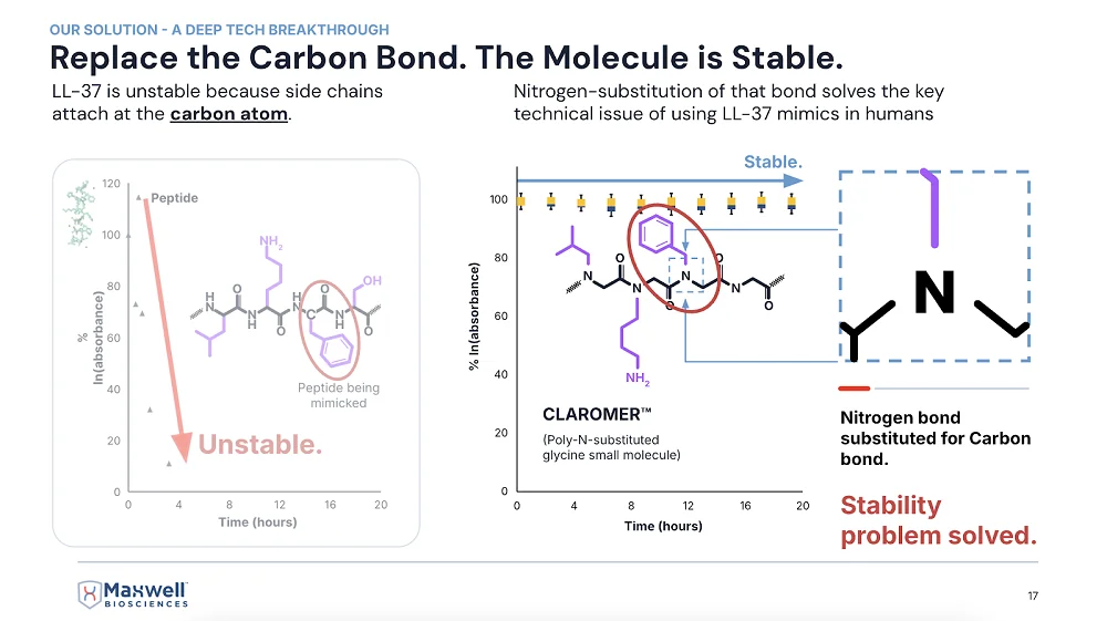
LL-37

A slide from McClure’s presentation at last year’s ARDD conference in Copenhagen

Are Claromers™ your proprietary version of peptoids?

Yes, the antimicrobial version of peptoids. We’ve got lots of new ones coming out that are way better than the ones we’re taking through the FDA right now, but even those are so incredible that the FDA loves them.

First, there is no toxic concentration that we have found yet. Second, they’re anti-inflammatory and extremely broad spectrum. We have one that works against bacteria and fungi, and another against viruses: all influenza, all coronaviruses, SARS-1, SARS-2, regular cold. It’s literally the cure for the common cold. It’s crazy but amazing.

What about long COVID?

It’s going to knock it out completely. I have no doubt about it. Of course, these are my opinions. I’m the CEO of the company, and I am paid to be the optimist. But these are not promissory statements. And I’m not asking for investment. We don’t need it.

The fact is that, in partnership with the government, we’ve passed non-human primate studies with a single molecule killing the worst type of bacteria and the worst fungus we could select – the worst multi-drug-resistant versions of both that would kill you because there’s no drug to treat it.

You barely need diagnostics anymore. Just use this all the time, and if it doesn’t work, then you can do some expensive diagnostics to figure out what wild thing is in there that isn’t being killed. It kills all the bad things and leaves the good stuff in the microbiome.

Because of how it mimics the natural thing?

Yes, it’s exactly the same natural mechanism of action, so it leaves your microbiome alone.

You’ve already touched on this, but let’s talk some more about how this relates to aging.

Infectious disease is the primary driver of aging. It is the primary disruptor of homeostasis. As soon as we start fighting infectious disease with our drug – the first time we’re approved by the FDA for any kind of infectious disease – it will start lengthening healthspan immediately.

I think it’s inevitable at this point because we passed the non-human primate study. The only thing that could stop us now would be lack of cash flow, but we don’t have a problem with that. We’re working on multiple big contracts right now with the US military, the governments of India, UAE – direct government contracts. We’re working on two big contracts with commercial companies.

This is going to be world-shaking. Just like AI – even though it’s not currently replacing your job, you can pretty much bet it’s going to replace most jobs. Same with this technology: it’s going to eradicate infectious disease and more.

The first thing that came to my mind when I heard about your technology is that infections are an insanely prevalent cause of death in the oldest old. Many think this is a major factor that limits our maximal lifespan.

It truly is. Many times, people say, “Great-grandma fell and broke her hip,” and everyone thinks that’s natural, that’s how very old people die, but why does breaking a leg or a hip make someone fall apart and die?

It’s because their body has been fending off infectious disease pretty well up to this point. Now it must redirect just a little bit of energy to healing the hip, and not enough is left to defend against infectious disease. You’re breathing millions of fungal spores every day that will eat you just like a rotten tree if you don’t produce enough of this peptide to keep them destroyed.

This big change – pretty much eliminating the ever-present attack of pathogens against us – is going to greatly expand our healthspan. I’m predicting 20 to 40 additional years for the whole population just from our drug, comparable to what advances against infectious diseases have already done for lifespan.

Once we solve this, once we clean everything up and you’re like, “Okay, bird flu is a big issue right now, but I don’t need to worry about it because I’ve got this drug; everything is clean – my food is clean, my water is clean, my air is clean,” – then you can start sort of moving up Maslow’s hierarchy of needs and getting into the sexy stuff – can I enhance my vision? Can I enhance other things?

How does a collaboration with a country like India work?

Countries have their own priorities and budgets established for those priorities. With India, it’s for rare, neglected tropical diseases. This budget has been around since 2017, but no one has gotten it because no one wanted to tackle the indications they want us to address.

With India, it’s more about antibiotic-resistant septic infections that happen out there in the villages. India has plenty of antibiotics – they’re the home of generic antibiotics. What they need is help with difficult situations where somebody is going to die before they can get diagnostics. There’s no diagnostics out there to tell you what type of bacteria it is, so you must have something very broad-spectrum at hand to cure whatever it is.

The whole pharma industry says, “No, that’s not how medicine works.” We come in and say, “This is exactly how medicine should work.” Our product is shelf-stable and thermal-stable. It can be hot enough to cook an egg – and our drug will still be effective.

It’s a $300 million deal over three years, and it’s helping us while we pursue bigger things like tuberculosis and long COVID. It’s non-dilutive funding, essentially.

You saw no toxicity in your preclinical studies?

Zero toxicity. Not only it’s not toxic to human cells, but it’s not toxic to the commensal microbiome. We specifically measured the commensal microbiome in rhesus macaque monkeys in the India project, where we went up against methicillin-resistant Staphylococcus aureus (MRSA).

In the microbiome test, we found that they have a huge amount of Staphylococcus aureus in their sinus cavity – it’s one of the predominant bacteria there. Our compound works great against Staph aureus, but all our data said it doesn’t affect the commensal microbiome.

So, we asked: does that mean it’s selective for commensal Staph aureus and only kills the pathogenic Staph aureus? That seemed crazy, but that’s what we found. The commensal Staph aureus was untouched and in fact helped to protect the primate against the infection.

We had to remove some of the commensal microbiome to even give our MRSA strain a seat on the bus, so to speak. It took us a long time to actually establish an infection. This tells me that many human infections have to do with microbiome disruption. We eat preservatives and other things that disrupt the protective microbiome, which is a major way to get infected. The good bacteria form a shield that prevents pathogenic bacteria from getting through.

There must be some mechanism that prevents the peptide or peptoid from attacking beneficial bacteria. Do you know what it is?

We do, and we’ve published on it. Like I said, LL-37 is extremely well studied. The mechanism of action of LL-37 and our compounds is that they target negatively charged phospholipids on the membrane of bacteria, fungi, or viruses. That negatively charged phospholipid in the membrane is required to merge with a human cell by connecting to TIM receptors (TIM-2, TIM-3, TIM-4). So, it’s a hallmark of aggressive pathogens.

The negatively charged phospholipids are present in the membrane of the commensal microbiome, too, but they are covered with a V-shaped or shield-shaped molecule called annexin-5 (and the whole annexin family). The annexin goes over the top of the negatively charged phospholipids, saying “I’m not going to attack you.”

If they take their annexin-5 off, they become pathogenic. That’s how Staphylococcus stays commensal as long as it’s getting fed inside your gut. But if you stop eating and stop producing antimicrobial peptides, it’s like “You’re what’s for dinner” – it comes after you.

Anything you can tell me about your collaboration with the military?

They want things like prophylactic wound care for combatants on the run; it’s pre-first aid, where you can squirt something into a puncture wound to stop bleeding and potential infection.

We have about seven Collaborative Research and Development Agreements (CRADAs) with the Pentagon. Some are for quite exotic things like anthrax and Ebola, things that could be weaponized. The military says, “We have to have something against that, and we’re willing to pay a billion dollars to stockpile it, just in case somebody uses a weaponized version against us.”

We’re also working on more common things, such as anti-diarrheal applications, which we could use for commercial purposes as well. How many battles have been won by dysentery rather than by the enemy army?

How far out are you in terms of human trials and approvals?

This year, we got the data back on the non-human primate studies, which is pretty much equivalent to a human Phase I. It was totally safe and very effective, so we have a very high probability for the human trials outcome. The value of the company has gone up significantly, and the certainty of the outcome has increased, which is why we’ve gotten all these contracts with governments and companies.

We would like to ask you a small favor. We are a non-profit foundation, and unlike some other organizations, we have no shareholders and no products to sell you. All our news and educational content is free for everyone to read, but it does mean that we rely on the help of people like you. Every contribution, no matter if it’s big or small, supports independent journalism and sustains our future.
RNA

Scientists Create Cytoskeleton-Like Structures From RNA

In a new study, researchers report producing self-assembling nanotubes and rings made from RNA molecules inside artificial cell-like lipid vesicles. In the future, this technology could facilitate the creation of synthetic cells for various research, diagnostic, and therapeutic applications [1].

Paperless origami

DNA and RNA molecules are central to life, carrying essential genetic information for protein production. However, their unique properties also make them excellent building materials. Nature discovered this eons ago; ribosomes, for instance, are partly built from RNA.

Scientists have experimented extensively with designing DNA and RNA sequences that cause molecules to self-assemble into predetermined shapes. The technique has become known under the poetic name “DNA (or RNA) origami” [2].

These engineered structures can be impressively intricate: for example, DNA “boxes” that carry drug molecules directly to target sites, then each open a molecular “door” to release their contents [3]. A new study from Heidelberg University, published in Nature Nanotechnology, takes RNA origami to the next level.

Tubes, rings, and networks

The researchers designed RNA molecules to self-assemble into structures resembling cellular cytoskeletons. The cytoskeleton, naturally composed of protein filaments and microtubules, is critical for maintaining cell shape and stability. “We designed RNA origami tiles that fold upon transcription and self-assemble into micrometer-long, three-dimensional RNA origami nanotubes,” the authors explain. Creating an artificial cytoskeleton is an important milestone on the path toward synthetic cells.

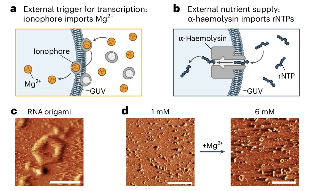
The team encapsulated DNA templates and RNA polymerase, the enzyme needed to transcribe DNA into RNA, into giant unilamellar lipid vesicles (GUVs), effectively creating proto-cells. To supply these synthetic cells with RNA building blocks (nucleotides), they used the bacterial transmembrane pore protein α-haemolysin. Magnesium ions (Mg2+) served as a trigger to prevent premature transcription, and the pore protein facilitated the removal of transcription byproducts.

When transcription was initiated, RNA strands immediately folded and assembled into nanotubes inside these synthetic cells. Remarkably, some nanotubes reached several micrometers in length, comparable to actual cellular cytoskeletal structures.

The researchers observed that subtle variations in the DNA template sequence significantly altered the RNA origami structures, demonstrating the method’s flexibility and ease of control. For instance, slight modifications switched the resulting structure from nanotubes to rings.

RNA cytoskeleton 1

To scale up their creations, the researchers included aptamers: RNA sequences capable of binding to specific molecular targets, including other RNA molecules. With aptamers incorporated, the nanotubes formed “cytoskeleton-like networks tens of micrometers across.”

RNA cytoskeleton 2

Aptamers also proved useful in creating cortex-like structures adhering to the lipid membrane, mirroring the cortices seen in real cells. Whether aptamers were used or not, a prolonged nucleotide supply led to structural networks becoming so extensive that they physically deformed the GUVs. Being able to maintain and alter the cell’s shape is another hallmark of a true cytoskeleton.

Possible applications

A major advantage of RNA origami is that these structures can be produced directly inside cells. Once DNA templates are introduced into cells, a single enzyme, T7 polymerase, can create numerous RNA products from these templates. In comparison, the full biological transcription-translation machinery requires over 150 genes.

“In contrast to DNA origami, RNA origami enables synthetic cells to manufacture their building blocks by themselves,” explained Dr. Kerstin Göpfrich, the study’s lead author, whose team, “Biophysical Engineering of Life,” conducts research at the Center for Molecular Biology of Heidelberg University (ZMBH). “This could open new perspectives on the directed evolution of such cells.”

This early-stage discovery has broad implications, including in aging research. It could help scientists better understand early cellular evolution, develop biomimetic systems, and engineer cells designed for specific tasks.

While creating fully functional synthetic eukaryotic cells remains distant due to their enormous complexity, the pathway toward viable, simplified prokaryotic “proto-cells” with limited but useful functions has just become shorter. Currently, bacteria are used extensively to produce biological molecules. However, minimal synthetic cells might simplify this process and even enable protein production directly within living organisms, circumventing bacterial immunogenicity issues.

Such proto-cells could, for example, produce essential proteins like collagen and elastin to maintain youthful extracellular matrix (ECM) function. Additionally, RNA origami structures could be introduced into existing cells to provide structural support and other functionalities.

The authors anticipate that future RNA origami structures will become more than passive scaffolds; they will actively perform complex cellular tasks by integrating ribozymes, RNA molecules capable of enzymatic activity. According to Göpfrich, the long-term research goal is the creation of fully functional molecular machinery for RNA-based synthetic cells.

We would like to ask you a small favor. We are a non-profit foundation, and unlike some other organizations, we have no shareholders and no products to sell you. All our news and educational content is free for everyone to read, but it does mean that we rely on the help of people like you. Every contribution, no matter if it’s big or small, supports independent journalism and sustains our future.

Literature

[1] Tran, M. P., Chakraborty, T., Poppleton, E., Monari, L., Illig, M., Giessler, F., & Göpfrich, K. (2025). Genetic encoding and expression of RNA origami cytoskeletons in synthetic cells. Nature Nanotechnology, 1 – 8.

[2] Dey, S., Fan, C., Gothelf, K. V., Li, J., Lin, C., Liu, L., … & Zhan, P. (2021). DNA origami. Nature Reviews Methods Primers, 1(1), 13.

[3] Udomprasert, A., & Kangsamaksin, T. (2017). DNA origami applications in cancer therapy. Cancer science, 108(8), 1535-1543.

DNA structure

A Core Senescence Biomarker Fights Inflammation

Researchers publishing in Nature Communications have found that p53, a biomarker and inducer of senescence, suppresses both inflammation and DNA damage in senescent cells.

Senescence against cancer

One of the main reasons why cells become senescent is to prevent cancer. The senescence-inducing compound p53, in particular, is known in the literature as a tumor suppressor, and its presence has been found to reduce, rather than exacerbate, the SASP [1]. The biochemical reasons behind this seemingly contradictory connection have not been previously explained.

However, these researchers have previously found that a biochemical pathway from the mitochondria to the nucleus is partially responsible for driving the SASP [2]. This pathway drives the expulsion of chromatin from the nucleus into the rest of the cell, which activates the cGAS/STING pathway and thus turns on NF-κB, the SASP’s master regulator [3]. Therefore, they sought to determine a link between p53 and this well-established pathway.

Preventing the SASP at its root

For their first experiment, the researchers created fibroblasts in which 53BP1, a suppressor of DNA damage that works with p53, was somewhat increased. These fibroblasts, after they were driven senescent through irradiation, had reduced amounts of chromatin in the nucleus and, thus, less SASP. Driving cells senescent by introducing p53 had similar effects, as did knocking down its target, MDM2.

Mutating 53BP1, on the other hand, had significant negative effects, spurring the release of chromatin into the nucleus after the cells were driven senescent with radiation. Silencing p53 had similar effects; four days after irradiation, cells with silenced p53 had significantly more chromatin in the nucleus. Further work found that mitochondria are required for this SASP upregulation; ablating away mitochondria prevented the chromatin from being expelled into the nucleus and thus prevented the upregulation of SASP elements.

Protecting cells from DNA damage

p53 was also found to be instrumental in DNA repair. γH2AX is a marker of DNA damage, and its levels in the nucleus were reduced in cells where MDM2 was downregulated and p53 was upregulated. The researchers found that this could be accomplished by introducing RG7388, a compound that promotes p53.

Similarly, silencing p53 increased the prevalence of this DNA damage marker. Interestingly, p21, another well-known biomarker of senescence, was found to be necessary for the effects of p53; without p21 being present, neither MDM2 nor p53 made any difference.

This was not just a change in a biomarker; p53 had real effects on the genome. In cells treated with RG7388 shortly after irradiation, the number of DNA amplifications and deletions was relatively small. In untreated cells, however, these signs of genetic damage were rampant. Most of this damage was located near the ends of the chromosomes, where the telomeres are. Unlike with chromatin ejection, however, mitochondria had nothing to do with these DNA damage effects.

Effects in female mice

The researchers then turned to in vivo experiments, introducing HDM201, a compound that suppresses MDM2, to a population of naturally aged mice for two weeks. In both males and females, HDM201 had no effects on body weight, blood cell counts, or liver pathological biomarkers. However, as the liver is where senescent cells normally accumulate in mice [4], the researchers focused their attentions there. Both p53 and p21 were significantly increased in these mice.

Interestingly, in gene expression, this treatment had substantially greater effects on female mice than on male mice. While it had no senolytic effects, it reversed many of the gene expression changes caused by aging. As expected, many of these genes were related to the SASP, and, likewise, the number of immune cells that infiltrated into these mice’s livers was decreased as well.

Both p53 and p21 have been investigated in the context of senescence, with previous researchers considering them as potential targets to be suppressed. This work, however, demonstrates that these compounds are both necessary and beneficial for preventing senescent cells from getting out of control. The researchers describe p53 as a senomorphic compound; while it clearly does not remove senescent cells, it significantly blunts their negative effects on the cells around them. They hold that “it may be possible to one day design a treatment that targets p53 to promote healthier aging.”

We would like to ask you a small favor. We are a non-profit foundation, and unlike some other organizations, we have no shareholders and no products to sell you. All our news and educational content is free for everyone to read, but it does mean that we rely on the help of people like you. Every contribution, no matter if it’s big or small, supports independent journalism and sustains our future.

Literature

[1] Coppé, J. P., Patil, C. K., Rodier, F., Sun, Y. U., Muñoz, D. P., Goldstein, J., … & Campisi, J. (2008). Senescence-associated secretory phenotypes reveal cell-nonautonomous functions of oncogenic RAS and the p53 tumor suppressor. PLoS biology, 6(12), e301.

[2] Vizioli, M. G., Liu, T., Miller, K. N., Robertson, N. A., Gilroy, K., Lagnado, A. B., … & Adams, P. D. (2020). Mitochondria-to-nucleus retrograde signaling drives formation of cytoplasmic chromatin and inflammation in senescence. Genes & development, 34(5-6), 428-445.

[3] Dou, Z., Ghosh, K., Vizioli, M. G., Zhu, J., Sen, P., Wangensteen, K. J., … & Berger, S. L. (2017). Cytoplasmic chromatin triggers inflammation in senescence and cancer. Nature, 550(7676), 402-406.

[4] Ogrodnik, M., Miwa, S., Tchkonia, T., Tiniakos, D., Wilson, C. L., Lahat, A., … & Jurk, D. (2017). Cellular senescence drives age-dependent hepatic steatosis. Nature communications, 8(1), 15691.

Hypothalamus

A Hypothalamus Neuropeptide Reduces Aging in a Mouse Model

Researchers have improved multiple health metrics in prematurely aged mice by re-establishing the production of neuropeptide Y in the hypothalamus [1].

An essential protein

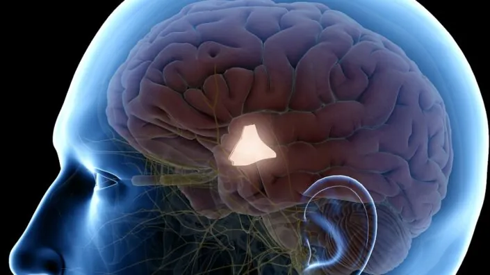
Age-related decline in the function of the hypothalamus, a core region of the brain, has been suggested to be a “key factor in the development of whole-body aging” [2]. Due to its essential role and implication in several aging-related processes, the hypothalamus may be a target for lifespan- and healthspan-extending therapeutic strategies.

The authors of this study focused specifically on hypothalamic neuropeptide Y (NPY). Several studies suggest that neuropeptide Y plays a role in lifespan through its involvement in lifespan-extending processes such as autophagy induction [3] and stress resistance [4], and it appears to play an essential role in caloric restriction-induced lifespan extension [5, 6]. Decreased levels have been connected to neurodegenerative diseases [7]. Therefore, the authors hypothesized whether re-establishing hypothalamic neuropeptide Y levels might slow down aging.

Aging too quickly

For this study, the researchers used animals that lack a gene encoding the protein Zmpste24 (Z24-KO), and this lack results in accelerated aging and premature death. Z24-KO mice have multiple defects in skin, bone, cardiovascular tissues, and skeletal muscles, similar to those observed in human accelerated aging processes. Therefore, the authors decided to test if their hypothalami exhibit the same changes as the hypothalami of naturally aged mice.

They tested an area of the hypothalamus called the arcuate nucleus (ARC), which contains neurons that release neuropeptide Y. They found lower levels of neuropeptide Y compared to age-matched wild-type mice and lower levels of a neuronal marker called NeuN, suggesting neuronal aging and fewer hypothalamic neurons.

Markers connected to neuroinflammation were also altered. The authors observed increased levels of a gliosis marker in Z24-KO mice. Gliosis is a process that occurs in glial cells (non-neuronal cells located in the central nervous system) in response to damage. Therefore, an increase in gliosis suggests neuroinflammation. On the other hand, a marker showing the activity of microglia, the immune cells of the brain, decreased in Z24-KO mice. This, according to the authors, suggests that immune response capacity is impaired.

Restoring youth

To re-establish decreased neuropeptide Y levels, the researchers used a genetically modified adeno-associated virus (AAV) that causes an increase in the expression of neuropeptide Y (AAV-NPY) and injected it into the mice. The virus increased neuropeptide Y in the ARC for at least four months, which is when the mice were sacrificed to analyze their organs.

The researchers tested the AAV-treated Z24-KO mice for the same biomarkers as the untreated Z24-KO mice. They observed an increase in NeuN and a reduction of the gliosis biomarker that is normally elevated in these mice. However, the biomarker of microglial immune capacity didn’t significantly change. Overall, AAV treatment made the brains of Z24-KO mice similar to those of age-matched wild-type mice, suggesting that neuropeptide Y plays a role in reducing neuroinflammation.

Further testing suggested positive changes in aging-related processes. The authors observed a reduction in NF-κB, a protein that increases with aging and is associated with neuroinflammation in the hypothalamus [8]. Other positive effects were a decrease in a marker of tau pathology, which is associated with neurodegenerative diseases [9], and an increase in autophagy, a process that plays an essential role in longevity.

Better looks and healthier minds

The authors also report positive changes in body weight, body composition, mobility, vitality, and fur among the virus-treated animals. The treatment also showed neuroprotective effects, as the spatial memory improvements accompanied physical health improvements, but there were no significant differences in locomotor activity.

One of the characteristics of the prematurely aged Z24-KO mice is lipodystrophy. This condition results in the organism losing fat from some parts of the body, including under the skin surface of different body parts, while gaining it in others, such as the liver.

The AAV-treated mice had fewer lipodystrophy symptoms. The treatment resulted in a thicker outermost skin layer (epidermis) and thicker layers of under-skin (subcutaneous) fat, probably resulting from increased proliferation of fat cells as suggested by increased levels of a cell proliferation marker. It also increases skin collagen, which decreases with age. Autophagy markers in the skin indicated increased autophagic activity, suggesting better cellular health.

Liver structure was also improved in the Z24-KO mice after treatment. The researchers observed increased cell proliferation, which might indicate improved liver protection and regeneration, a decrease in cellular death by apoptosis, and increased autophagic activity in the liver, suggesting better cellular health.

There were no differences between treated and untreated mice in different parameters of kidney health and heart structure, but heart cells showed improved cellular health.

Promising, but there are still many unknowns

Overall, this study’s results suggest that re-establishing neuropeptide Y has a positive effect on aging-associated hypothalamus-related symptoms. However, in most of the experiments performed, the researchers compared untreated, prematurely aged mice to prematurely aged mice treated with a virus expressing neuropeptide Y. It would be beneficial if a wild-type control was included, as this would allow for assessing whether the observed changes make the prematurely aged mice more similar to wild-type mice or if the changes are significant but modest.

Additionally, long-term studies are essential to determine the long-lasting effects and side effects of this approach, whether it induces the desired amount of neuropeptide Y expression, and how improvements in different biomarkers of cellular health translate to increases of healthspan and lifespan.

We would like to ask you a small favor. We are a non-profit foundation, and unlike some other organizations, we have no shareholders and no products to sell you. All our news and educational content is free for everyone to read, but it does mean that we rely on the help of people like you. Every contribution, no matter if it’s big or small, supports independent journalism and sustains our future.

Literature

[1] Ferreira-Marques, M., Carmo-Silva, S., Pereira, J., Botelho, M., Nóbrega, C., López-Otín, C., de Almeida, L. P., Aveleira, C. A., & Cavadas, C. (2025). Restoring neuropetide Y levels in the hypothalamus ameliorates premature aging phenotype in mice. GeroScience, 10.1007/s11357-025-01574-0. Advance online publication.

[2] Kim, K., & Choe, H. K. (2019). Role of hypothalamus in aging and its underlying cellular mechanisms. Mechanisms of ageing and development, 177, 74–79.

[3] Aveleira, C. A., Botelho, M., Carmo-Silva, S., Pascoal, J. F., Ferreira-Marques, M., Nóbrega, C., Cortes, L., Valero, J., Sousa-Ferreira, L., Álvaro, A. R., Santana, M., Kügler, S., Pereira de Almeida, L., & Cavadas, C. (2015). Neuropeptide Y stimulates autophagy in hypothalamic neurons. Proceedings of the National Academy of Sciences of the United States of America, 112(13), E1642–E1651.

[4] Michalkiewicz, M., Knestaut, K. M., Bytchkova, E. Y., & Michalkiewicz, T. (2003). Hypotension and reduced catecholamines in neuropeptide Y transgenic rats. Hypertension (Dallas, Tex. : 1979), 41(5), 1056–1062.

[5] Chiba, T., Tamashiro, Y., Park, D., Kusudo, T., Fujie, R., Komatsu, T., Kim, S. E., Park, S., Hayashi, H., Mori, R., Yamashita, H., Chung, H. Y., & Shimokawa, I. (2014). A key role for neuropeptide Y in lifespan extension and cancer suppression via dietary restriction. Scientific reports, 4, 4517.

[6] de Rijke, C. E., Hillebrand, J. J., Verhagen, L. A., Roeling, T. A., & Adan, R. A. (2005). Hypothalamic neuropeptide expression following chronic food restriction in sedentary and wheel-running rats. Journal of molecular endocrinology, 35(2), 381–390.

[7] Duarte-Neves, J., Pereira de Almeida, L., & Cavadas, C. (2016). Neuropeptide Y (NPY) as a therapeutic target for neurodegenerative diseases. Neurobiology of disease, 95, 210–224.

[8] Zhang, G., Li, J., Purkayastha, S., Tang, Y., Zhang, H., Yin, Y., Li, B., Liu, G., & Cai, D. (2013). Hypothalamic programming of systemic ageing involving IKK-β, NF-κB and GnRH. Nature, 497(7448), 211–216.

[9] Samudra, N., Lane-Donovan, C., VandeVrede, L., & Boxer, A. L. (2023). Tau pathology in neurodegenerative disease: disease mechanisms and therapeutic avenues. The Journal of clinical investigation, 133(12), e168553.

Unhealthy foods

Short-Term Overeating Alters Brain Insulin Sensitivity

A new study published in Nature Metabolism suggests that even a short period of eating loads of sweet and fatty snacks can cause brain changes reminiscent of those seen in obesity and type 2 diabetes [1].

What can go wrong in five days?

If you usually eat healthy and take care of your body, there’s no harm in letting your guard down for a while, right? Not so fast, says a new study by the University Hospital of Tübingen, the German Center for Diabetes Research (DZD), and Helmholtz Munich.

This controlled but not randomized study recruited 29 young, healthy men with a normal body mass index (BMI). At baseline, there was no significant difference between the study group and the control group in terms of calories and nutrients consumed. Participants in the study group were asked to increase their daily calorie intake by 1,500 calories for five days by consuming highly processed sweet and fatty snacks. The researchers measured various biomarkers at baseline, after five days of the experiment, and one more week later.

Altered insulin responses

They found that even though body mass did not significantly change in the study group compared to controls, the unhealthy diet caused a noticeable increase in liver fat contents. Probably more intriguing results related to how the participants’ brains reacted to this ‘food assault.’ While insulin directly stimulates glucose uptake in muscle and fat cells, its role in the brain is different: it helps regulate appetite, metabolism, and cognitive functions related to food choices. The “brain first” hypothesis postulates that those reactions play an important role in the development of insulin resistance and related metabolic disorders.

Immediately after the five days of the experiment, the study group showed increased insulin responses in reward-related brain regions (insula, midbrain). This pattern has been previously observed as an early response to overeating [2] – conceivably, to make food less attractive. However, if the system “overshoots,” the dampened reward response might cause the person to eat more to get to the same level of satisfaction. The changes in the reward and punishment responses were evident in a series of cognitive experiments that the researchers conducted.

Some researchers suggest that heightened insulin activity could eventually lead to insulin resistance if healthy eating habits are not restored, though this remains to be confirmed. Indeed, in this study, one week after resuming a normal diet, the brain insulin responsiveness of the hippocampus and fusiform gyrus was reduced in the study group. These two brain regions are important for memory and cognition, including food-related decision-making. Earlier studies have found comparable reductions in brain insulin responsiveness in people with obesity and type 2 diabetes [3].

“Our findings demonstrate for the first time that even a brief consumption of highly processed, unhealthy foods (such as chocolate bars and potato chips) causes a significant alteration in the brain of healthy individuals, which may be the initial cause of obesity and type 2 diabetes,” said Dr. Stephanie Kullmann, the study’s lead author. “Interestingly, in our healthy study participants, the brain shows a similar decrease in sensitivity to insulin after a short-term high calorie intake as in people with obesity. This effect can even be observed one week after returning to a balanced diet.”

Is it “brain first”?

While the participants in the study group did return to their normal eating habits, this might be because they knew they were temporarily overeating as part of an experiment, not as a personal choice. This awareness likely made it easier for them to consciously give up overeating. People who start overeating in less controlled settings, such as in emotional distress, might not be so lucky.

Importantly, the researchers did not find signs of impaired peripheral insulin signaling, suggesting that brain insulin dysfunction may occur before systemic changes. This finding is in line with the “brain first” hypothesis, which suggests that brain insulin resistance may precede whole-body metabolic dysfunction. However, more rigorous studies are needed to confirm or disprove this.

The study had several important limitations, such as the small sample size and short follow-up duration. The scientists chose to limit participation to males because previous research has shown differences in insulin dynamics in males and females. However, this also affected the study’s generalizability and interpretability.

Dr. Andreas Birkenfeld, the study’s co-author, concluded, “We assume that the brain’s insulin response adapts to short-term changes in diet before any weight gain occurs and thus promotes the development of obesity and other secondary diseases.”

We would like to ask you a small favor. We are a non-profit foundation, and unlike some other organizations, we have no shareholders and no products to sell you. All our news and educational content is free for everyone to read, but it does mean that we rely on the help of people like you. Every contribution, no matter if it’s big or small, supports independent journalism and sustains our future.

Literature

[1] Kullmann, S., Wagner, L., Hauffe, R., Kühnel, A., Sandforth, L., Veit, R., … & Birkenfeld, A. L. (2025). A short-term, high-caloric diet has prolonged effects on brain insulin action in men. Nature Metabolism, 1-9.

[2] Wingrove, J. O., O’Daly, O., Forbes, B., Swedrowska, M., Amiel, S. A., & Zelaya, F. O. (2021). Intranasal insulin administration decreases cerebral blood flow in cortico‐limbic regions: A neuropharmacological imaging study in normal and overweight males. Diabetes, Obesity and Metabolism, 23(1), 175-185.

[3] Arnold, S. E., Arvanitakis, Z., Macauley-Rambach, S. L., Koenig, A. M., Wang, H. Y., Ahima, R. S., … & Nathan, D. M. (2018). Brain insulin resistance in type 2 diabetes and Alzheimer disease: concepts and conundrums. Nature Reviews Neurology, 14(3), 168-181.

Older woman lifting weights

Elamipretide, a Potential New Drug, Reduces Frailty in Mice

In Aging Cell, Dr. Vadim Gladyshev and a team of researchers have described how elamipretide beneficially affects mitochondrial pathways and reverses frailty in mice.

A drug on its way to the clinic

As a mitochondria-affecting antioxidant peptide, elamipretide has been, and continues to be, the subject of multiple investigations. As far back as 2004, researchers were testing whether its effects against reactive oxygen species (ROS) had benefits against ischemia-reperfusion injury [1], which occurs when blood is restored to tissue after it has been cut off, such as when a heart attack is treated. Other work has used it to treat heart failure in dogs [2]. Right now, Stealth Biotherapeutics is waiting for the FDA to approve it as a new drug for treating Barth syndrome, a rare mitochondrial disease.

Despite its clinical benefits, it has been little investigated in the context of aging biomarkers. Theoretically, a drug that has significant benefits for people suffering mitochondrial disorders and age-related diseases would similarly improve known biomarkers, such as epigenetic clock measurements and, relatedly, gene expression. These researchers, therefore, attempted to investigate if that is the case.

What elamipretide affects

The first experiment described in this paper tested function in 5-month-old and 24-month-old Black 6 mice, both males and females. Osmotic pumps under the mice’s skin delivered elamipretide for 8 weeks, after which the mice were tested and examined again.

The frailty index, a measurement of flaws in overall physical function that is often used to diagnose age-related weakness, was decreased in older mice of both sexes that had received elamipretide, despite being two months older at the end of the study. Heart function, as measured by ejection fraction and pumping force, was significantly improved. There were also benefits against fatigue, particularly in females.

Multiple molecular pathways received significant benefits. As expected of a drug that treats the mitochondria, many of the affected pathways were related to mitochondria organization and transport along with ATP production and oxygen processing. Immunological pathways that are upregulated with aging were downregulated with elamipretide, suggesting some benefits against inflammaging.

What elamipretide doesn’t affect

The heart enlarges with age, and elamipretide did nothing to ameliorate this. The mass of the gastrocnemius, a crucial muscle in the leg, was unaffected. Despite its effects on frailty, it did not appear to have consistently significant effects on overall skeletal muscle force; it appeared to increase maximum force in older female mice while decreasing maximum force in older male mice.

Most interestingly, despite the effects on pathways, there were no effects on differentially expressed genes related to aging. Aging clocks, including transcriptomic clocks and the Horvath epigenetic clock, were used on multiple tissues, and none of them revealed any difference; the old treated mice, despite being significantly less frail, did not become biologically younger. While there appeared to be trends towards gene expression that are similar to the effects of rapamycin, these findings were not statistically significant.

Similarly, although there were a couple of findings involving effects on cap-independent translation targets, which are alternate metrics of longevity, the researchers hold that these unclear results were largely negligible and that elamipretide does not, overall, have significant effects on molecular longevity predictors.

This study sheds light on the difference between functional benefits and biomarker metrics, potentially revealing the limitations of clocks. Elamipretide may have no benefits against epigenetic aging, but it has clear benefits for the mitochondria, and it is poised to reach the clinic. Further clinical determinations will reveal if it is effective against frailty in human beings.

We would like to ask you a small favor. We are a non-profit foundation, and unlike some other organizations, we have no shareholders and no products to sell you. All our news and educational content is free for everyone to read, but it does mean that we rely on the help of people like you. Every contribution, no matter if it’s big or small, supports independent journalism and sustains our future.

Literature

[1] Zhao, K., Zhao, G. M., Wu, D., Soong, Y., Birk, A. V., Schiller, P. W., & Szeto, H. H. (2004). Cell-permeable peptide antioxidants targeted to inner mitochondrial membrane inhibit mitochondrial swelling, oxidative cell death, and reperfusion injury. Journal of Biological Chemistry, 279(33), 34682-34690.

[2] Sabbah, H. N., Gupta, R. C., Kohli, S., Wang, M., Hachem, S., & Zhang, K. (2016). Chronic therapy with elamipretide (MTP-131), a novel mitochondria-targeting peptide, improves left ventricular and mitochondrial function in dogs with advanced heart failure. Circulation: Heart Failure, 9(2), e002206.

LongX Logo

LongX Launches 2nd Edition of the Xplore Program

LongX, an initiative dedicated to providing avenues into the longevity space, announced a call for applications for the 2025 Xplore Program. With its inaugural cohort launching last year, the Xplore Program is a remote fellowship open to early career professionals and students looking for their first steps in longevity biotechnology. The 2nd edition will take place from June 2025 to August 2025, and features 1 month of aging biology and biotechnology courses followed by the opportunity to gain industry experience with partnered biotech companies for 2 months.

Xplore Program fellows will receive guidance from working professionals in longevity while also having the opportunity to network with peers and experts. The fellowship is accepting applications until March 31, 2025.

To learn more, visit LongX or apply directly to the Xplore Program.

About LongX

Longevity Xplorer (LongX) was formed in 2023 to lower the barrier of entry for emerging early career professionals in longevity. Our goal is to drastically increase the number and capacity of people who meaningfully contribute to the longevity industry on a global scale. We encourage exploration beyond traditional roles and aim to equip future experts with the skills to drive progress in the field. The LongX Substack is home to various articles and interviews showcasing developments within the longevity space, resources, opportunities, experiences, and advice.

Contact

Marvin Yan, Co-Founder

team@longx.bio

We would like to ask you a small favor. We are a non-profit foundation, and unlike some other organizations, we have no shareholders and no products to sell you. All our news and educational content is free for everyone to read, but it does mean that we rely on the help of people like you. Every contribution, no matter if it’s big or small, supports independent journalism and sustains our future.
Lung fibroblasts

Using a Surface Biomarker to Target Senescent Cells

Scientists have identified a senescence-associated surface protein that can be targeted using antibodies [1]. This discovery might help distinguish between beneficial and harmful senescent cells and could speed up the development of senolytic therapies. This work was done by the Lifespan Research Institute, which was formed last year by the merger of SENS Research Foundation and Lifespan.io.

Shedding light on senescence

Most geroscientists agree on two things: cellular senescence is an important driver of aging, and senescent cells are notoriously difficult to study and target because of their heterogeneity. Senescent cells are often described as damaged or exhausted cells that stop dividing but remain alive, releasing pro-inflammatory factors that can induce senescence in neighboring cells. While cellular senescence plays a beneficial role in some processes related to development, wound healing, and anti-cancer surveillance, the overall senescence burden grows with age, tipping the scale towards harm.

The answer is on the surface

Senescent cells are known to exhibit increased lysosomal activity [2]. Lysosomes are bubble-like organelles that envelop intracellular junk and transport it outside the cell by fusing with the cell’s outer membrane in a process called lysosomal exocytosis. The underlying causes of excessive intracellular waste in senescent cells are not entirely clear, but this process may contribute to senescence-related inflammation.

Working with a publicly available database of membrane proteins, the researchers zeroed in on the protein LAMP1, which is abundant in lysosomal membranes. In healthy cells, “LAMP1 is only briefly found at the cell surface due to the fusion of lysosomes with the plasma membrane, and thus, mostly undetectable,” the paper says. However, LAMP1 is known to linger in membranes of some cancer cells, probably due to their increased lysosomal exocytosis.

The dataset revealed a strong correlation between LAMP1 expression and other known senescence-related genes, such as p21 and p16, but the researchers had to confirm this in actual cells. After inducing senescence in human fetal lung fibroblasts in three different ways, they observed a significant increase in the proportion of LAMP1-positive cells. While only about 1% of untreated cells expressed LAMP1 on their membranes, 20% to 60% of treated cells expressed it, depending on how senescence was induced.

The researchers then isolated both LAMP1-positive and LAMP1-negative cell populations from the liver and lungs of middle-aged mice and found that the former expressed additional senescence markers.

Telling good from evil

As their in vivo model, the researchers chose mice treated with bleomycin, which induces a condition similar to idiopathic pulmonary fibrosis (IPF) – a deadly and currently uncurable age-related lung disease. IPF has long been suspected to be driven by increased cellular senescence.

Experiments detected a 1.5- to 3-fold increase in the number of LAMP1-positive cells after the bleomycin treatment relative to controls. Interestingly, in healthy mice, LAMP1 was expressed mostly by a fraction of one cell type: endothelial cells. In contrast, bleomycin-treated mice showed LAMP1 expression across multiple cell types.

This difference might be important for distinguishing between beneficial and harmful senescent cells. “Another interesting observation is that in healthy mice, most senescent cells in the lung were endothelial cells,” said Dr. Amit Sharma, who led the study. “In contrast, in bleomycin-treated mice that showed increased inflammation, the majority of senescent cells were of myeloid origin. It is possible that another layer of complexity in the heterogeneity of senescence is the type of cells contributing to pathology vs tissue repair.”

He further explained, “There has been a discussion of good vs bad senescent cells, so is it possible that these myeloid senescent cells are the bad ones? There is some evidence by others that this might be true, at least in liver fibrosis [3], where eliminating p16-expressing fibroblasts slows down tissue repair, while p16-expressing macrophages can be beneficial. Of course, this must be further tested. If LAMP1 is reliably seen as a senescence biomarker, we can develop drugs specifically targeting bad senescent cells, which would be safer.”

Targeting LAMP1

Lastly, the researchers made the first steps towards confirming LAMP1 as a possible target for intervention. Working on cells in culture, they used an antibody-drug conjugate (ADC), a construct consisting of an antibody that targets a specific surface protein, in this case LAMP1, and a drug that kills the targeted cell (technically, the ADC targeted another antibody, which targeted LAMP1). The treatment caused substantial cytotoxicity in senescent cells and virtually none in non-senescent cells.

LAMP1

According to Sharma, identifying LAMP1 as a surface biomarker of senescence is significant. Unlike cytosolic markers that remain inside the cell, surface biomarkers might allow easier detection, study, and targeting of senescent cells.

“Senescent cell biomarker discovery is the holy grail of senescence research,” Sharma said. “This will be useful for understanding how senescent cells contribute to pathology and validating the efficacy of interventions. If what we found in the IPF mouse model and in middle-aged mice is true for other senescence models, a surface marker offers a huge possibility for diagnostic tool development.”

We would like to ask you a small favor. We are a non-profit foundation, and unlike some other organizations, we have no shareholders and no products to sell you. All our news and educational content is free for everyone to read, but it does mean that we rely on the help of people like you. Every contribution, no matter if it’s big or small, supports independent journalism and sustains our future.

Literature

[1] Meca-Laguna, G., Qiu, M., Hou, Y., Barkovskaya, A., Shankar, A., Dixit, B., Rae, M. J., Boominathan, A., & Sharma, A. (2025). Cell-Surface LAMP1 is a Senescence Marker in Aging and Idiopathic Pulmonary Fibrosis. Aging cell, e70141. Advance online publication.

[2] Rovira, M., Sereda, R., Pladevall‐Morera, D., Ramponi, V., Marin, I., Maus, M., … & Serrano, M. (2022). The lysosomal proteome of senescent cells contributes to the senescence secretome. Aging Cell, 21(10), e13707.

[3] Zhao, H., Liu, Z., Chen, H., Han, M., Zhang, M., Liu, K., … & Zhou, B. (2024). Identifying specific functional roles for senescence across cell types. Cell, 187(25), 7314-7334.

Lab mice

Cellular Senescence Prevents Unlearning in Some Male Mice

In Aging Cell, researchers have established a link between cellular senescence and cognitive decline in unmodified male mice.

Resilience versus decline

The authors begin their paper by noting that cognitive decline in older people varies greatly. Some people suffer serious cognitive defects; other people are scarcely impacted at all [1]. The researchers have developed an automated tool called PhenoTyper to measure this in unmodified male Black 6 mice, establishing a benchmark set at 5 to 7 months old and using it to classify older mice as either intact or impaired [2]. They previously used this benchmark to ascertain that some mice remain fully functioning throughout their lives while others suffer serious cognitive decline [3].

This decline has nothing to do with Alzheimer’s, which wild-type mice cannot get. Instead, the researchers point to reactive gliosis, which, under normal circumstances, is the brain’s response to injury [4]. Sustained, chronic inflammation in aging is known as inflammaging, and sustained reactive gliosis is this process in the brain. The same compounds that are responsible for systemic inflammaging, such as the senescence-associated secretory phenotype (SASP), occur in the brain as well, and previous work has found that removing these cells leads to cognitive benefits [5].

However, these researchers hold that such previous work did not adequately distinguish between cognitively impaired and intact mice. Therefore, they performed an experiment of their own, attempting to more thoroughly document the relationship between brain senescence and cognitive decline.

Teaching old mice new tricks

For their first experiment, the researchers used their PhenoTyper system to assess the capabilities of their mice. In this experiment, mice were taught for 50 hours that they must enter the leftmost hole of a three-hole setup in order to receive food pellets; for another 40 hours, however, they had to use the rightmost hole instead (“reversal learning”).

In learning the initial hole, young (6 months) mice and old (22 to 24 months) mice performed similarly well. However, in the reversal learning task, there was an immense amount of difference between the two groups; the older mice’s performance was bimodal, with some older mice completely failing to unlearn what they had learned.

Failure to unlearn

These dramatic differences within the aged group were not related to the total distance moved by the mice, nor were they related to changes in circadian rhythms (this test was performed during the dark hours in which mice are most active). They also only applied to male mice; female mice did not have a similarly sharp stratification.

The researchers then examined the differences between these stratified groups. They found substantial and stark differences in both morphology and in biochemistry. Microglial activity was greatly increased in the impaired group, while the intact group was indistinguishable in this area from younger mice. Some biomarkers of reactive gliosis were moderately increased in the intact group, but all of them were far more elevated in the impaired group. These researchers, therefore, hold that they have found a distinct phenotype of neurological impairment.

This was linked to biomarkers of cellular senescence. Interestingly, the p16 senescence biomarker, which was significantly more elevated in intact older mice compared to young mice, was only slightly more elevated in the impaired group. p21 was nearly the same in both older groups and elevated compared to younger mice. However, other biomarkers were significantly different. The interleukin IL-6 was notably upregulated only in the impaired group, as was the key senescence marker SA-β-gal.

Senolytics appear to help

The researchers then administered the well-known senolytic combination of dasatinib and quercetin (D+Q) to 22-month-old mice and performed cognitive tests at 24 months. Nearly all the older animals given the senolytic were considered cognitively intact mice, with very few failing the reversal learning task. Their senescent cell biomarkers were similarly reduced to those of intact mice, with IL-6 reaching approximately the level of young mice; similar beneifits were found in microglial morphology and biochemistry. Once again, these findings only applied to males.

The researchers surmise that such sex-related differences may also apply to human beings. Additionally, this work applies specifically to ‘normal’ cognitive decline that may not be directly related to proteostasis diseases such as Alzheimer’s. However, if a relevant human population can be identified, a senolytic or senomorphic regimen may allow them to retain their cognitive abilities.

We would like to ask you a small favor. We are a non-profit foundation, and unlike some other organizations, we have no shareholders and no products to sell you. All our news and educational content is free for everyone to read, but it does mean that we rely on the help of people like you. Every contribution, no matter if it’s big or small, supports independent journalism and sustains our future.

Literature

[1] Marron, M. M., Wojczynski, M. K., Minster, R. L., Boudreau, R. M., Sebastiani, P., Cosentino, S., … & Long Life Family Study. (2019). Heterogeneity of healthy aging: comparing long-lived families across five healthy aging phenotypes of blood pressure, memory, pulmonary function, grip strength, and metabolism. Geroscience, 41, 383-393.

[2] Baier, M. P., Nagaraja, R. Y., Yarbrough, H. P., Owen, D. B., Masingale, A. M., Ranjit, R., … & Logan, S. (2022). Selective ablation of Sod2 in astrocytes induces sex-specific effects on cognitive function, d-serine availability, and astrogliosis. Journal of Neuroscience, 42(31), 5992-6006.

[3] Logan, S., Baier, M. P., Owen, D. B., Peasari, J., Jones, K. L., Ranjit, R., … & Sonntag, W. E. (2023). Cognitive heterogeneity reveals molecular signatures of age-related impairment. PNAS nexus, 2(4), pgad101.

[4] Sochocka, M., Diniz, B. S., & Leszek, J. (2017). Inflammatory response in the CNS: friend or foe?. Molecular neurobiology, 54, 8071-8089.

[5] Ogrodnik, M., Evans, S. A., Fielder, E., Victorelli, S., Kruger, P., Salmonowicz, H., … & Jurk, D. (2021). Whole‐body senescent cell clearance alleviates age‐related brain inflammation and cognitive impairment in mice. Aging cell, 20(2), e13296.

Environmental exposure

Human Exposome Project Explores Environmental Disease Causes

Recent research confirms the relatively minor role that genetics plays in our health, with the ‘exposome’, defined as the totality of exposures individuals experience over their lives affecting their health, responsible for 10 times more variation in mortality risk than genetic predisposition [1].

A paper published in Nature Medicine today, ‘Cities, communities and clinics can be testbeds for human exposome and aging research’ [2], sets out ways to start measuring how humans are affected by the exposome and furnish long-overdue evidence to design environments that enhance healthy life expectancy while reducing health and wellbeing inequalities.

The publication of the paper coincides with a tipping point for an international movement behind the ‘Human Exposome Project’, a generation on from the Human Genome Project, to understand how external exposures (including social, behavioural and geo-physical factors) and their interaction with internal factors (such as genetics and physiology), affect an individual’s health and overall resilience.

The Exposome Moonshot Forum is meeting for the first time in Washington, DC, 12-15 May 2025, to launch an unprecedented international scientific endeavour to map the combined impact of environmental factors that impact human health from conception to death.

Specific environmental factors can activate pathological pathways that contribute to disease and accelerate aging. The ability to capture, analyse and link individual data outside the medical record can show how external exposures affect a person’s health across their lifetime. These interactions can now be much better understood at an individual level and traced with unprecedented precision using artificial intelligence, representing a significant leap forward in determining the impact of the exposome at an aggregated, population health level.

This work is crucial to define new ways to address the chronic disease epidemic and ageing demographic now creating an economic drag in many nations around the world. The evidence will shape more effective public health interventions urgently needed to shift investment and policy away from an unsustainable healthcare model to one more rooted in prevention.

Tina Woods, steering committee member, Exposome Moonshot Forum; CEO, Collider Health; executive director of the International Institute of Longevity, and corresponding author says: ‘The time for the Human Exposome Project has come and I am excited to be participate in the Exposome Moonshot Forum to move it from concept to reality. We need to measure the exposome to demonstrate the return on investing in health and incentivising prevention.’

Professor David Furman, Buck Institute for Research on Aging, director of the Stanford 1000 Immunomes Project, steering committee member, Exposome Moonshot Forum, and corresponding author says: ‘At a time of increasing environmental threats to human health such as air pollution and microplastics, we have the technologies like applied artificial intelligence to help us to unravel the complex interactions between environment, immunity and health at an individual level that can be aggregated up to get a true picture of the relative impact drivers of population health’.

Professor Nic Palmarini, director of the National Innovation Centre for Ageing, and author, says: ‘We have the technologies and tools to understand the human exposome with clinics, communities and cities acting as ideal real-world testbeds to understand what solutions will promote healthier behaviours and ultimately, outcomes.’

Buck Institute

The mission of the Buck Institute is to end the threat of age-related disease for this and future generations. It is the first biomedical research institution devoted solely to research on ageing revolving around our commitment to helping people live better longer.

Media contact:

Kris Rebillot, Senior Director of Communications

415-209-2080

krebillot@buckinstitute.org

National Innovation Centre for Ageing

The UK’s National Innovation Centre for Ageing is a world-leading organisation to help co-develop and bring to market products and services which create a world in which we people live better, for longer.

Media contact:

Lynne Corner

+44 (0) 7713 245780

lynne.corner@ncl.ac.uk

International Institute for Longevity

The International Institute of Longevity (IIOL) is focused on driving global excellence, industry standards and best practice in the real-world application of longevity science into ‘longevity clinics’ as well as scientific and medical innovation to extend human healthspan, resilience and flourishing in the wider context of corporate and urban health and wellness.

Media contact:

Tina Woods

+44 (0) 7808 402032

t.woods@l-institute.com

Exposome Moonshot Forum

The Exposome Moonshot Forum on 12-15 May in Washington DC is intended to define and accelerate the future of the Human Exposome Project (HEP). The central aim of the Forum is to identify the resources, policies, and collaboration necessary to drive the successful implementation of the HEP, ensuring longevity and impact. Outcomes will centre around:

  • Defining clear, actionable steps toward the scalable implementation of the HEP.
  • Building consensus on the essential policy changes needed to support and expand research.
  • Establishing long-term collaborative partnerships that will bring together diverse sectors, including academia, industry, government agencies, and non-profit organizations, and divert critical funds toward this project and toward successful integration of active working groups

Media contact:

Eliza Cole, Communications Specialist

ecole28@jh.edu

We would like to ask you a small favor. We are a non-profit foundation, and unlike some other organizations, we have no shareholders and no products to sell you. All our news and educational content is free for everyone to read, but it does mean that we rely on the help of people like you. Every contribution, no matter if it’s big or small, supports independent journalism and sustains our future.

Literature

[1] Argentieri, M.A., Amin, N., Nevado-Holgado, A.J. et al. Integrating the environmental and genetic architectures of aging and mortality. Nat Med (2025). [2] Woods, T., Furman D., Palmarini N. et al. Cities, communities and clinics can be testbeds for human exposome and aging research. Nat Med (2025).