The Blog

Building a Future Free of Age-Related Disease

Public Longevity Group

Lifespan Research Institute Launches Public Longevity Group

[Mountain View, September 17, 2025]Lifespan Research Institute (LRI) today announced the launch of the Public Longevity Group (PLG), a new initiative focused on bridging the cultural gap between scientific breakthroughs in aging and their public acceptance. To kickstart its work, PLG has opened a crowdfunding campaign to develop tools that measure and strengthen public trust in longevity science.

While the science of longevity biotechnology continues to advance, skepticism and cultural resistance limit progress, with some studies showing that more than half of Americans would reject a safe, proven therapy to extend life. This hesitation poses risks of raising costs, delaying health-promoting regulation, and slowing the delivery of treatments that could combat age-related diseases and extend healthy lifespan.

“The breakthrough that unlocks all other breakthroughs is public trust,” said Sho Joseph Ozaki Tan, Founder of PLG. “Without it, even the most promising therapies may never reach the people they’re meant to help. PLG exists to change that.”

“Persuasion is a science too,” said Keith Comito, CEO of Lifespan Research Institute. “To bring health-extending technologies to the public as quickly as possible, we must approach advocacy with the same rigor as our research. With PLG, we’ll be able to systematically measure and increase social receptivity, making the public’s appetite for credible longevity therapies unmistakable to policymakers, investors, and the public itself.”

PLG is developing the first data-driven cultural intelligence system for longevity—a platform designed to track real-time sentiment, test narratives, and identify which messages resonate and which backfire. Early tools include:

  • The Longevity Cultural Clock: a cultural barometer mapping readiness and resistance across demographics and regions.
  • Sentiment Dashboards: real-time monitoring of public, investor, and policymaker perceptions.
  • Narrative Testing Tools: data-driven analysis that will enable robust pathways to public support.

The crowdfunding campaign will provide the initial $100,000 needed to launch these tools, creating the cultural foundation required for healthier, longer lives.

With a lean, data-driven team, the group aims to provide open-access cultural insights for advocates and policymakers while offering advanced analytics to mission-aligned partners.

Campaign Timeline:

  • Campaign completion: November 2, 2025
  • Dashboard development: Dec 2025 – Feb 2026
  • First survey deployment: Feb – Apr 2026
  • Beta dashboard launch: May 2026
  • First public insight report: June 2026

Supporters can contribute directly at: https://lifespan.io/campaigns/public-longevity-group/

The PLG campaign is sponsored by the members of LRI’s Lifespan Alliance, a consortium of mission-aligned organizations that believe in the promise of extending healthy human lifespan. Newly-joined members include OpenCures, AgelessRx, and Lento Bio.

About Lifespan Research Institute

Lifespan Research Institute accelerates the science and systems needed for longer, healthier lives by uniting researchers, investors, and the public to drive lasting impact. LRI advances breakthrough science, builds high-impact ecosystems, and connects the global longevity community.

Media Contact:

Christie Sacco

Marketing Director

Lifespan Research Institute

christie.sacco@lifespan.io

(650) 336-1780

We would like to ask you a small favor. We are a non-profit foundation, and unlike some other organizations, we have no shareholders and no products to sell you. All our news and educational content is free for everyone to read, but it does mean that we rely on the help of people like you. Every contribution, no matter if it’s big or small, supports independent journalism and sustains our future.
Brain and peripherals

Peripheral Inflammation May Drive Parkinson’s

A new study suggests that aging or Parkinson’s-triggering mutations create inflammation in peripheral tissues, and then circulating extracellular vesicles spread it to the brain, which might contribute to the disease [1].

Brain on fire

Nature made the brain remarkably well-protected, including from the elements by the skull and from pathogens by the blood-brain barrier (BBB). The brain was also long considered to be immune-privileged – that is, limiting local immune responses to reduce swelling and damage from inflammation; another example is the eye.

However, more recent studies have shown that some brain cells, particularly microglia, can act as immune cells, and brain inflammation exists and probably drives brain aging [2]. What has been largely unknown is whether systemic inflammation, such as inflammaging, affects brain inflammation, and if yes, what pathways are involved?

One well-characterized molecular engine of sterile inflammation is the cGAS-STING pathway, which senses DNA in the cell’s cytosol. Normally, it detects foreign DNA, such as from viruses and bacteria. The problem is, in aging and senescence, a cell’s own DNA – from a damaged nucleus or leaky mitochondria – can spill into the cytosol. cGAS can’t distinguish self from non-self and fires anyway, producing a smoldering, chronic interferon type I (IFN-I) response [3].

Exporting inflammation

A new study by an international collective of scientists, published in Cell Reports, asks whether cGAS-STING-driven systemic inflammation can be a factor in Parkinson’s disease, and what mechanisms might be involved. Leucine-rich repeat kinase 2 (LRRK2 in humans, Lrrk2 in mice) regulates the cell’s degradation and recycling machinery (endolysosomal system). The most common genetic cause of Parkinson’s is the G2019S mutation in the associated gene, which increases the enzyme’s activity. The authors refer to this gain-of-function mutation as LRRK2GoF.

The authors’ central hypothesis is that LRRK2GoF accelerates aging by degrading endolysosomal function, which causes self-DNA to accumulate in the cytosol, instead of being promptly recycled, and to be exported in DNA-carrying extracellular vesicles (EVs). Those EVs activate cGAS-STING not just in the original cell but also in distant cells – including, eventually, the brain.

First, the researchers took plasma and cerebrospinal fluid (CSF) samples from young healthy donors, aged healthy donors, and Parkinson’s patients. The latter showed elevated systemic IFN-I activity but not NF-κB activity, which belongs to a more general inflammation pathway.

At the cellular level, blood monocytes from Parkinson’s patients showed elevated IFNB1 transcripts, but an LRRK2 inhibitor normalized that IFNB1 elevation back to healthy levels, pointing to LRRK2’s causal involvement. However, the sample sizes in this and several other experiments were small (n=3-4).

To test causality, the team moved to a G2019S knock-in mouse model. Compared to controls, these Lrrk2GoF mice had a markedly elevated, age-dependent IFN-I signature across plasma, monocytes, bone marrow, and spleen. RNA sequencing of spleen showed elevated inflammatory and senescence-associated gene expression.

The researchers then asked whether the brain also becomes inflamed and whether behavior is affected. In aged Lrrk2GoF mice, neurons and microglia showed increased IFN-I and interferon-stimulated genes (ISGs) as well as a pronounced age-dependent motor decline. Importantly, the mutant mice also had increased BBB permeability and smaller brains. A leaky BBB might explain how peripheral inflammatory signals reach the brain.

Using RNA sequencing at different time points, the team found that the peripheral IFN-I signature was already present at 3 months, whereas the brain IFN-I signature and locomotor decline did not appear until 12 months. The author’s interpretation, crucial for the entire paper, is that inflammation starts peripherally and reaches the brain later.

The tiny Trojan horses

Mechanistically, IFN-I has triggers other than cGAS/STING. However, the researchers determined that eliminating those other triggers had no effect, while deleting STING completely reversed the elevated IFN-I response to wild-type levels in both splenocytes and microglia, reflecting changes in both a peripheral tissue and the brain. STING deletion also reduced microglial inflammation markers and protected the mice from the motor decline.

Lrrk2GoF mice lost 51% of dopaminergic neurons with age, a major hallmark of Parkinson’s, versus about 30% in wild-type mice, and STING deletion prevented that loss. Changes in other neuronal populations were inconsistent.

Moving back in vitro to hunt for additional mechanistic insights, the authors showed that Lrrk2GoF mouse fibroblasts reached senescence earlier than their wild-type counterparts and had elevated IFN-I activity, which could be normalized by inhibiting Lrrk2. Intriguingly, in transwell co-culturing (which blocks direct cell contact but allows diffusible/vesicular signals), senescent fibroblasts evoked a STING-dependent IFN-I response in physically separated macrophages.

Defective endolysosomal clearance is known to cause increased EV secretion, which is exactly what the researchers found in both Lrrk2GoF and naturally aged fibroblasts. Again, this effect was abrogated by blocking Lrrk2. EVs taken from Lrrk2GoF fibroblasts contained more genomic and mitochondrial DNA and induced STING-dependent IFN-I response in recipient macrophages.

The team then confirmed the EV mechanism in vivo and in humans. In Lrrk2GoF mice, EV accumulation in plasma appeared early, but in the cerebrospinal fluid (CSF), it started much later, consistent with the “peripheral inflammation slowly causes brain inflammation” hypothesis. In humans, Parkinson’s patient macrophages showed reduced endolysosomal degradation. Plasma and CSF from Parkinson’s patients had more DNA-containing EVs, and EVs derived from these patients induced STING-dependent IFN-I, demonstrating a clean mouse-to-human bridge.

We would like to ask you a small favor. We are a non-profit foundation, and unlike some other organizations, we have no shareholders and no products to sell you. All our news and educational content is free for everyone to read, but it does mean that we rely on the help of people like you. Every contribution, no matter if it’s big or small, supports independent journalism and sustains our future.

Literature

[1] Öberg, M., Myers, C., Saffarzadeh, N., Maric, I., Murillo-León, M., Strömberg, A., … & Härtlova, A. (2026). STING-dependent peripheral inflammaging drives neurodegeneration via extracellular vesicles. Cell Reports, 45(7).

[2] Yin, F., Sancheti, H., Patil, I., & Cadenas, E. (2016). Energy metabolism and inflammation in brain aging and Alzheimer’s disease. Free Radical Biology and Medicine, 100, 108-122.

[3] Gulen, M. F., Samson, N., Keller, A., Schwabenland, M., Liu, C., Glück, S., … & Ablasser, A. (2023). cGAS–STING drives ageing-related inflammation and neurodegeneration. Nature, 620(7973), 374-380.

Gabriel Cian Interview

Gabriel Cian on Building the 2060 Longevity Ecosystem

In this follow-up interview, we speak with Gabriel Cian about how the 2060 ecosystem has evolved since last year’s Forum, what he learned from the first edition, and how he is thinking about investment, scientific credibility, health optimization, policy, and the future of longevity in Europe.

Last year, we spoke about your path from software entrepreneurship into longevity and your goal of building a broader 2060 ecosystem. One year later, what has changed most in how you think about the mission?

What is the shortest path between two points? In mathematics, we would say it’s a straight line, right?

Yet a river never flows in a straight line from its source to the sea. Why is that?

Let me explain how this applies to the 2060 Foundation—and, more broadly, to the longevity field.

The mission of the 2060 Foundation is to defeat aging before the year 2060. At first glance, the obvious way to achieve this is to invest massively in R&D until we gain a precise understanding of the biology of aging and discover how to slow it down, stop it, or even reverse it. There’s no time to lose. 2060 is only 34 years away—it’s tomorrow. Understanding aging is incredibly difficult, so we should start funding large-scale research immediately, right?

Well, not so fast.

People are not ready for this. Governments are not ready for this. Much of the scientific community is not ready for this. Humanity, as a whole, is not ready for this. Trying to push society directly toward radical life extension meets fierce resistance at every level.

At the same time, however, the world is embracing preventive medicine at an exponential pace. Here, there’s no disruption, no futuristic biotech, no radical treatments—just healthier lifestyles, personalized diagnostics, and targeted supplementation. Nothing too controversial.

People resonate with this message. Governments support it because it can help contain healthcare costs. Investors are pouring massive amounts of capital into the space. Suddenly, longevity medicine seems like common sense.

I think you can see where I’m going with this.

The straight line from today to defeating aging would be to invest massively in R&D. But the resistance is simply too strong. That direct path doesn’t work.

By contrast, helping humanity move through an intermediate phase – embracing preventive medicine before becoming comfortable with the idea of radical life extension – is the path of least resistance. And it works.

Once this global movement toward preventive medicine is fully underway, even a small fraction of the enormous financial resources flowing into it can be redirected toward biotechnology and aging research. Sometimes, 1% of a huge pie is worth far more than 100% of a small one.

The biggest lesson I’ve learned over the past year is this:

In the real world, the shortest path is often not a straight line. It’s the path of least resistance.

What did you learn from the first 2060 Longevity Forum that shaped how you are approaching the 2026 edition?

The biggest insight from last year’s edition of the 2060 Forum was that financial resources are still so scarce in the longevity field that almost every participant had a strong financial incentive to attend.

VC funds were looking for new LPs. Longevity startups were raising capital. Investors were searching for promising investment opportunities. Sponsors were showcasing their products to generate revenue—and some of them were fundraising as well.

In a nutshell, everything revolved around capital.

This realization allowed us to rethink our communication strategy for this year’s edition. By aligning our messaging with the financial objectives of our audience, we were able to make our communication significantly more effective.

Looking back at the 2025 Forum, what were the most concrete outcomes? Were there partnerships, investments, or projects that came directly from people meeting there?

We’ve already received confirmation that several startups secured funding after pitching at last year’s edition of the 2060 Forum. We were thrilled to have played a part in making that happen.

Participants also loved the venue, and many are planning to return this year—this time with their partners and children. Southern France, a beautiful setting, outstanding sports facilities, and a unique blend of keynote speeches, meaningful conversations, swimming, and yoga sessions create an experience that is difficult to replicate elsewhere.

Another major outcome—although one that is much harder to quantify—has been planting the idea in investors’ minds that longevity may become one of the greatest investment opportunities of our time. Those ideas don’t transform into investments overnight. They take root, mature over time, and eventually reach a tipping point where investors begin allocating significant capital to longevity startups.

But perhaps the most significant outcome of the 2060 Forum has been the creation of the 3060 Fund of Funds. That is where the real epiphany happened.

But more on that later in this interview!

Were there any parts of last year’s event that did not work as well as you hoped, or that you deliberately changed this time?

Well, the most common piece of feedback we received from participants was about the food. It turns out that protein is just as important as vegetables – and all the other longevity-friendly nutrients.

The good news is that this is an easy fix, and we’re taking care of it this year!

The 2026 Forum appears to be larger and more ambitious than the first edition. What is substantively different this year beyond having more speakers or attendees?

For the 2026 edition, we’re aiming to go even further by bringing more investors to the event. We believe we have a responsibility to educate the investment community about the extraordinary opportunities in longevity, so this year we’re placing an even stronger focus on investors.

This should be great news for fundraising startups, as well as for investment funds looking to attract new LPs.

What did you learn from trying to bring scientists, startups, investors, clinicians, and policy advocates into the same room, and how has that shaped the 2026 program?

The biggest lesson we’ve learned is that longevity is not a vertical – it’s a mega-trend growing at an exponential pace, one that is reshaping multiple industries simultaneously: hospitality, healthcare, real estate, longevity clinics, fitness, insurance, tourism, and many others.

Conversations with participants from these different sectors made it clear just how broad and far-reaching the implications of longevity really are.

Another important insight from last year’s edition is that there is still no consensus on which industries, companies, or business models will emerge as the biggest winners. Will it be longevity therapeutics? Longevity clinics? Consumer digital health platforms? Advanced diagnostics? The truth is, nobody knows – and that’s precisely what makes the future so exciting.

This realization has strongly influenced the design of the 2026 program. We’ve placed an even greater emphasis on the diversity of our keynote speakers and on the range of longevity startups we’ll be showcasing on stage. We want investors to discover the broadest possible spectrum of opportunities across the longevity ecosystem.

Longevity conferences can sometimes become echo chambers for people who already agree with each other. How are you trying to include disagreement, skepticism, or more critical perspectives?

I don’t think the 2060 Forum is likely to become an echo chamber, because our mission is to educate investors about longevity as a new investment opportunity—not to promote a single narrative.

We want investors to understand both the opportunities and the risks, as well as the different timelines involved. That’s why we deliberately bring together experts with very different perspectives, who often disagree with one another.

For example, we’ll have:

  • Medical doctors who practice longevity medicine and place greater confidence in interventions that are already available than in long-term biotechnology projects.
  • Scientists who appreciate the extraordinary complexity of biology and believe that truly disruptive technologies may take many years to reach the market.
  • Pragmatic investors who focus on preventive medicine businesses—companies that may not revolutionize healthcare but are more likely to become profitable in the near term while delivering meaningful health benefits.
  • Long-term investors who back ambitious biotech startups, pursuing high-risk, high-reward opportunities.
  • Futurists who believe that artificial general intelligence is only a few years away and could dramatically accelerate the cure of many, if not all, diseases.
  • Startup founders developing breakthrough biotechnology who are advancing their therapies toward the clinic and the market, and who are optimistic about the results of the validation process in the coming years.

As you can imagine, these groups don’t always agree – and that’s exactly the point.

Our role is not to tell people what to think. It’s to expose them to the strongest arguments from different perspectives so they can form their own views.

Ultimately, our goal is not to provide all the answers, but to help our participants ask the right questions.

After speaking with investors through the 2025 Forum and the 2060 Investment Club, what do you now think investors most misunderstand about longevity?

Rather than saying investors misunderstand longevity, I would say that most of them simply don’t understand it yet – and for good reason. That’s exactly why the 2060 Forum exists.

On one side, there is biotechnology. The biology of aging is extraordinarily complex and largely inaccessible to most investors. Yet, this is where the truly disruptive breakthroughs are likely to emerge. This is where the next trillion-dollar company could be created. Because so few investors understand the field today, it remains a blue ocean with relatively little competition.

On the other side, there is preventive medicine: consumer health apps, advanced diagnostics, epigenetic clocks, and many other solutions that are already experiencing exponential growth. These businesses are easy to understand, highly relatable, and therefore already attract significant amounts of capital. But, because the barriers to entry are often relatively low, competition is intense. Investors can certainly generate attractive returns in this segment, but identifying the long-term winners is far from straightforward.

In my view, a sound longevity investment strategy combines both approaches. Yet many investors remain reluctant to invest in biotechnology and allocate most of their capital to preventive medicine instead.

I believe that’s a mistake.

Since last year, the 2060 ecosystem seems to have expanded beyond the Forum itself, including the Investment Club, Ikare.Health, and newer investment initiatives. How do these pieces fit together?

From day one, the vision of the 2060 Foundation has been to build a constellation of profitable, mission-driven ventures, each led by a dedicated and highly competent team, and all deeply interconnected in a mutually reinforcing ecosystem. That vision is becoming more tangible every day.

  • The 2060 Club makes longevity investing accessible to a broader audience by lowering the minimum investment ticket while carefully selecting high-potential startups.
  • The 3060 Fund of Funds enables qualified investors to gain diversified exposure to the entire longevity sector without the complexity of selecting dozens of startups or actively managing a private equity portfolio. In many ways, it is the closest thing to an S&P 500 for longevity—except that it invests in private companies rather than publicly traded ones.
  • Ikare helps people stay healthy for as long as possible by giving them access to the latest advances in preventive medicine. After all, investors are people too. Why invest in the longevity technologies of tomorrow if we don’t even benefit from the longevity technologies that already exist today?
  • Last but certainly not least, the global longevity community remains relatively small and geographically dispersed. And as social creatures, we eventually need to meet face to face.

That’s where the 2060 Forum comes in.

  • We want investors to shake hands with the founders they’ve backed. We want longevity physicians to meet their patients. We want Ikare patients to become investors, and investors to become Ikare patients.
  • In other words, we want every part of the longevity ecosystem to strengthen every other part.

Can you explain the 2060 Longevity Investment Club in practical terms? Who is it for, what role does it play, and how does it decide which startups or opportunities to present?

The 2060 Club is a community of investors who co-invest alongside me in carefully selected longevity startups.

Our goal is to invest in five to ten companies each year. Individual investments typically range from $5,000 to $20,000 per startup, allowing us to aggregate approximately $500,000 per financing round. For each investment, we create a dedicated Special Purpose Vehicle (SPV) that pools capital from all participating investors. I personally invest in every startup alongside our members.

The 2060 Club follows a straightforward and well-established venture capital model. It has two revenue streams: an annual membership fee and a 10% carried interest on successful exits.

The Club manages each investment throughout the life of the company until a liquidity event occurs, at which point the proceeds are distributed to investors.

Overall, the 2060 Club is an ideal solution for investors who want exposure to the longevity sector but lack the expertise to identify the best opportunities or the network and scale required to negotiate attractive investment terms.

When your team evaluates a longevity startup, what does the diligence process actually look like? Who assesses the science, the market, and the investment risk?

We often co-invest alongside longevity-focused venture capital funds such as Apollo Health Ventures and LongGame. Whenever possible, we leverage their due diligence as an additional layer of validation.

The broader 2060 Foundation ecosystem is also a significant advantage. Over the years, we’ve built strong relationships with universities, research institutions, leading scientists, and industry experts. In many areas of biotechnology, only a handful of people in the world have the expertise to fully understand what a company is developing. Being able to consult those experts is an invaluable part of our evaluation process.

My partner, Martial Trigeaud, brings decades of relevant experience to this effort. Before joining the 2060 Foundation, he was a serial entrepreneur in the medtech sector and later became the co-founder and General Partner of B21 Ventures, a venture capital fund focused on longevity.

Martial leads our due diligence process. His experience, network, and deep understanding of the field make him exceptionally well qualified for this role.

Has your threshold for what counts as investable longevity science changed over the past year?

I’m very bullish on disruptive biotechnology, and we will continue investing directly in these companies through the 2060 Club.

At the same time, we also invest in preventive medicine companies because they represent the path of least resistance for investors (as I explained earlier). They are easier to understand, easier to adopt, and attract significantly more capital today.

So yes, in a sense, our investment thesis has broadened. While we remain convinced that disruptive biotechnology will generate the largest long-term returns, we now also actively invest in preventive medicine companies as an essential part of the longevity ecosystem.

You have described longevity as a major investment opportunity, but biotech timelines are long and failure rates are high. How do you communicate the upside without encouraging unrealistic expectations?

Most of the time, we invest in biotechnology companies at very specific stages of their development. For example, we often invest when a company is close to completing Phase I or Phase II clinical trials, and is potentially just two or three years away from being acquired by a major pharmaceutical company.

Of course, there is never any certainty that such an outcome will materialize. But this illustrates an important point: investing in biotechnology is not necessarily riskier—or more long-term—than investing in many other sectors. When you invest at the right stage, the risk-reward profile can be remarkably attractive.

You have also discussed newer investment concepts around broader exposure to longevity, including 3060.vc. What are you trying to build there, and what problem does it solve for investors?

Let me tell you the story behind the 3060 Fund of Funds.

During last year’s edition of the 2060 Forum, a friend of mine, Martin Beaujouan—an exceptionally successful real estate investor—made an observation that completely changed the way I thought about longevity investing.

He pointed out that longevity is not a single industry. It’s an ecosystem that spans biotechnology, medtech, diagnostics, preventive medicine, nutrition, fitness, hospitality, real estate, insurance, and many other sectors. The field is simply too broad and too specialized for any individual investor to cover effectively.

To build a truly diversified longevity portfolio, you would need to invest in hundreds of startups, review thousands of investment opportunities every year, and possess deep expertise across multiple disciplines.

In short: mission impossible.

What we needed was the equivalent of the S&P 500 or the NASDAQ but for longevity: a single investment vehicle providing broad exposure to the entire longevity ecosystem.

Because there are still very few publicly traded longevity companies, an index fund isn’t a viable solution today. The closest equivalent is a fund of funds.

Our idea is simple. Each year, we select what we believe are the five best longevity-focused venture capital funds, based on factors such as track record, team quality, credibility, and historical performance. If each of those funds holds a portfolio of around 50 companies, our investors immediately gain exposure to roughly 250 startups. By carefully selecting funds with complementary investment strategies, we can provide broad exposure across the entire longevity landscape.

Today, there are roughly 50 venture capital funds dedicated primarily to longevity worldwide. As the industry matures, that number is likely to grow significantly. Our role is to continuously screen the market, evaluate these funds, rank them, and invest in the most compelling ones. The objective is to give our investors exposure to the longevity economy in much the same way that the S&P 500 provides exposure to the U.S. economy or the NASDAQ provides exposure to the technology sector.

Initially, we built this strategy for ourselves and a small group of close friends, but we quickly encountered another challenge. Many of the world’s leading venture capital funds require minimum commitments of around $500,000. Building a diversified portfolio across five funds therefore requires an investment of approximately $2.5 million.

The obvious solution was to bring together a community of like-minded investors so that we could invest alongside the world’s leading longevity funds.

That’s how the 3060 Fund of Funds was born.

To the best of my knowledge, it is the only investment vehicle specifically designed to give qualified investors broad, diversified exposure to the entire longevity sector through a single product.

Ikare.Health appears to focus on helping people act on today’s available longevity and preventive health tools. How do you distinguish responsible health optimization from overpromising or biohacking hype?

At Ikare.Health, we’re obsessed with the 80/20 principle. We constantly challenge our longevity physicians and health coaches with one simple question: “What are the 20% of interventions that will generate 80% of the health benefits for this particular patient?”

Ikare is the exact opposite of a service that overwhelms people with long lists of generic recommendations. Instead, we identify the one or two interventions that are likely to have the greatest impact for each individual.

For one person, the priority may be improving sleep. For another, it may be building muscle mass. For someone else, it could be reversing prediabetes or optimizing metabolic health. Whatever the priority, we translate it into clear, actionable steps that can be integrated into the patient’s daily routine.

This 80/20 approach naturally helps patients focus on what matters most, ignore unnecessary complexity, and consistently execute on the changes that will have the greatest impact on their long-term health.

What evidence threshold do you personally use before you are comfortable recommending or building around a health intervention?

I’m a man of extremes: very low-tech when it comes to preventive medicine, and very high-tech when it comes to biotechnology. 🙂

When it comes to my own health, I follow a set of simple, evidence-based principles:

  • Managing stress
  • Prioritizing both the quantity and quality of sleep
  • Eating an appropriate diet
  • Maintaining good mental health through meaningful relationships and a strong sense of purpose
  • Engaging in regular, varied physical activity
  • Using personalized diagnostics, such as genetic testing, blood biomarkers, VO₂ max assessments, and other objective measurements

The challenge isn’t knowing what to do – it’s doing it consistently.

These principles may sound like common sense, but following them requires persistence and the ability to stay on your own path instead of constantly being pulled back toward the habits of your surrounding environment. It becomes even more challenging when you recognize that every individual is different and that the optimal balance between these interventions varies from person to person.

Personally, I believe that once these fundamentals are in place, the returns from pursuing increasingly sophisticated optimization strategies diminish rapidly. Rather than spending more time and money chasing marginal gains, I think those resources are often better invested in advancing the longevity field itself—by supporting research and development, advocacy, or other initiatives that accelerate scientific progress.

Since last year’s Forum, have you seen any concrete movement from policymakers, or is longevity still mostly being driven by private capital?

Government officials are beginning to take an interest in the longevity field, but concrete initiatives remain modest.

Influencing public policy is both surprisingly easy and surprisingly difficult. On the one hand, it doesn’t necessarily require enormous financial resources – meaningful advocacy efforts can begin with budgets of around $50,000 per year. On the other hand, changing laws is a long-term endeavor that often requires 10 to 15 years of patient, persistent work by experienced advocacy professionals.

For now, our priority is to build sustainable revenue streams that will allow us to support these efforts over the long term. Once that financial foundation is in place, we intend to allocate part of those resources to advocacy and public policy initiatives designed to accelerate the development of the longevity field.

A recurring criticism of longevity is that it could become a field for wealthy early adopters. Given the high cost of conferences, clinics, and early-stage investment access, what does democratization mean in practice?

The biggest obstacle to the widespread adoption of longevity isn’t financial – it’s cultural. It’s a matter of mindset.

Many of the most powerful interventions are not expensive. In fact, they often cost less than the alternatives:

  • Practicing intermittent fasting often costs less than eating more frequently.
  • Walking, running, or exercising costs less than spending hours in front of Netflix.
  • Preventing disease is generally less expensive than treating it.
  • Getting eight hours of sleep costs less than regularly staying out late.
  • Building meaningful relationships and living with purpose can be far more valuable – and often less costly – than dealing with the consequences of chronic stress, loneliness, or poor mental health.
  • Quitting smoking and reducing alcohol consumption cost less than maintaining those habits.

Of course, changing behavior is much harder than making these comparisons. The challenge is rarely financial – it is psychological, social, and cultural.

The mission of the 2060 Foundation is to help make these ideas mainstream – to make healthy longevity aspirational, accessible, and, yes, even “sexy.”

It’s an ambitious mission, but we’re convinced the world is moving in that direction.

You have talked about building the South of France into a longevity hub. What parts of that are already real today, and what remains aspirational?

Today, most of our initiatives are virtual. Our teams and collaborators are spread across the world, working remotely, and that model has served us well. The projects are moving forward.

For now, we only come together in person for two days each year during the 2060 Forum. That’s valuable – but it’s not enough. We need a permanent home for the longevity ecosystem.

I imagine a high-end longevity campus in Southern France: a place where families who aspire to live according to the principles of healthy longevity can also work, build companies, conduct research, and collaborate every day. A place that truly embodies the ideal of a healthy mind in a healthy body.

It would be much more than a real estate project. It would be a living community and a catalyst for innovation.

That vision hasn’t become reality yet – but we still have 34 years until 2060. 🙂

If we speak again next year, what would you want to be able to say 2060 accomplished in 2026?

Our goal for 2026 is to establish the 2060 Foundation as the leading organization promoting longevity in Europe.

We’re building that visibility on multiple fronts: appearing on television, participating in podcasts and conferences, helping channel more investment into the longevity sector, and bringing people who have only recently discovered longevity into direct contact with world-class scientists, physicians, entrepreneurs, and investors.

We’re making steady progress—and we believe we’re on the right path to achieve that vision.

Cells versus cells

Combining Senolytics and Stem Cells Shows Promise in Mice

A new study associated with Immorta Bio suggests that combining a senolytic vaccine with mesenchymal stem cells might create a synergistic impact. However, the findings rest on acute, artificially induced injury models rather than natural aging [1].

Clearing out senescent cells to help stem cells work

Mesenchymal stem cell (MSC) therapies have largely underperformed in the clinic. MSCs are connective-tissue stem cells that help mostly not by becoming new tissue but by secreting repair-promoting factors. Despite strong preclinical promise, clinical MSC trials in fibrosis, inflammation, and organ failure have shown only modest benefits [2].

One of the reasons may be an unwelcoming environment full of senescent cells, which secrete a mix of inflammatory and tissue-degrading molecules called the senescence-associated secretory phenotype (SASP). Prior work suggests that SASP factors actively suppress stem cell proliferation, differentiation, and survival [3]. In a new study published in the Journal of Translational Medicine and associated with the biotech startup Immorta Bio, the authors suggest a solution: combining MSCs generated from pluripotent stem cells with the company’s proprietary senolytic agent SenoVax.

As evident from its name, SenoVax is a “senolytic vaccine” that primes the immune system against the body’s own senescent cells. Notably, Immorta describes SenoVax in two different ways. In its patent, IND, and press materials, SenoVax is presented as an autologous, personalized cellular immunotherapy: the patient’s own cells are taken via biopsy and driven into accelerated senescence, then used as an antigen source to pulse the patient’s dendritic cells generated ex vivo. The dendritic cells are then reinfused and prime T cells. This is a personalized, work-intensive, and expensive procedure. In the study, however, SenoVax is described as a simpler peptide-based vaccine: peptides derived from senescence-associated proteins and injected subcutaneously along with an immune-triggering adjuvant in the hope that resident dendritic cells will “learn the lesson” in vivo.

The combination wins every time

The researchers tested the combination in two mouse models of senescence-driven damage, asking whether the combination beats either therapy alone on inflammation, regeneration, organ function, physical performance, and survival. One model involved injecting carbon tetrachloride (CCl₄), a liver toxin, to emulate chronic liver injury and senescence-associated inflammation. The other one was based on injecting low-dose doxorubicin, a chemotherapy drug that drives cells into senescence. In each model, induced mice were split into four arms: untreated control, SenoVax alone, MSCs alone, or the combination.

The team then measured four inflammatory/SASP markers – IL-11, IL-23, IL-6, and YKL-40 – in the liver injury model. All four fell below the injured baseline in every treated arm, and the combination lowered each one the most, suggesting that both agents dampen SASP signaling and that combining them produces the largest effect. Importantly, these factors are less senescence-specific than p16, p21, or SA-β-gal, so the senolytic mechanism is rather inferred than shown. Conversely, the regeneration markers Klotho, FGF-2, VEGF, and GDF-11 rose above the injured baseline, while the liver-damage enzymes AST and ALT fell; both of these shifts pointed toward improvement. In each case, the combination moved furthest, supporting the idea that clearing SASP takes the brakes off regeneration.

To show the pattern isn’t specific to chemical liver injury, the researchers then repeated the SASP and the regenerative markers panels in the doxorubicin-induced “accelerated aging” model. The results were similar: most positive with the combination.

To test physical function in the “accelerated aging” model, the team used the “T-climbing” test, which times how long a mouse takes to climb down a vertical pole – a standard motor-coordination and strength assay. The combination improved climbing performance by roughly 65%. However, this claim, made in the Discussion session, is not supported by the correspondent figure, which only contains bars for single interventions, not for the combination. Numbers for monotherapies do not appear in the paper.

Senovax 1

Large lifespan effects – but short lifespans

In the capstone experiment, which tested survival, mice received doxorubicin until death or a humane endpoint. The combination again gave the best results: about 50% of the animals were alive at Day 35 and 20% at Day 40, versus complete mortality by Day 30 in untreated doxorubicin controls. Monotherapies extended median survival only modestly, to about Day 35.

Senovax 2

While the accompanying press release touts a 73% increase in mean survival and ~84% extension of median lifespan compared with untreated controls in validated murine aging models, the extremely short lifespan puts it more into the “acute toxin-related damage” territory, as opposed to accelerated aging, much less natural aging.

Despite the several drawbacks and quirks, the study lends certain support to the intriguing concept behind Immorta Bio: using senolytics to create an auspicious niche for MSCs to work their magic. Hopefully, the company will keep developing this concept further.

We would like to ask you a small favor. We are a non-profit foundation, and unlike some other organizations, we have no shareholders and no products to sell you. All our news and educational content is free for everyone to read, but it does mean that we rely on the help of people like you. Every contribution, no matter if it’s big or small, supports independent journalism and sustains our future.
Literature

[1] Ichim, T. E., Markov, N., Lopes, G., Pascual, K. A., Evans, A., Reznik, R., … & Reznik, B. N. (2026). Synergistic senolytic–regenerative therapy significantly extends healthspan and lifespan Journal of Translational Medicine, 24(1), 745.

[2] Levy, O., Kuai, R., Siren, E. M., Bhere, D., Milton, Y., Nissar, N., … & Karp, J. M. (2020). Shattering barriers toward clinically meaningful MSC therapies. Science advances, 6(30), eaba6884.

[3] Moiseeva, V., Cisneros, A., Sica, V., Deryagin, O., Lai, Y., Jung, S., … & Muñoz-Cánoves, P. (2023). Senescence atlas reveals an aged-like inflamed niche that blunts muscle regeneration. Nature, 613(7942), 169-178.

Underground laboratory

An Experimental Proposal for Blocking Ambient Radiation

A perspective published in Aging and Disease has recommended the use of underground laboratory space in order to remove the effects of surface radiation on biological clocks.

A question of entropy

The cascading failure of bodily systems, leading to the loss of organ function, sits downstream of fundamental damage to genomics and epigenomics. However, how much of this damage is due to random, outside sources, such as unavoidable radiation, has been repeatedly questioned. Depending on the clock used, the stochastic portion of epigenomic damage ranges anywhere from two-thirds to nine-tenths [1], with the rest being attributed to deterministic processes.

The most well-known and well-established source of this sort of damage is radiation, which is impossible to completely avoid under ordinary conditions. Even if a sample or organism is kept safe from such well-known damage sources as ultraviolet radiation, fundamental particles known as muons, which originate from cosmic ray collisions with the atmosphere, will inevitably strike.

Where the muons aren’t

To prevent these particles from disrupting sensitive experiments, physics researchers have made use of deep underground laboratories (DULs), which take advantage of the fact that far fewer muons get through a substantial barrier of solid rock. This perspective paper holds that the same technique can and should be used for fundamental aging research in order to determine what happens when tissues are grown in an environment insulated from muon radiation.

Some work has already been done in this area, and the results were surprising. A population of Drosophila fruit flies grown in a DUL was found to have its natural repair mechanisms severely impaired without regular use [2].

This paper proposes an experiment with a different aim: to determine how much age-related epigenetic damage is caused by muon radiation. Specifically, the authors wish to use the Laboratorio Subterráneo de Canfranc (LSC) in Spain, which is the second-largest in Europe and one of only 14 DULs that exist around the world.

In such an experiment, the LSC would be used with one set of cells, while an above-ground lab would be used with the same type of cells grown in otherwise identical conditions to serve as a control group. Of course, as the authors note, this cannot possibly eliminate all random sources of damage; internal enzymes and oxidative stress would still exist, along with other chemistry-related issues and internal radiation, such as from imperfectly stable atoms of carbon and potassium. However, the purpose is simply to remove one source in order to determine its effects on the clock. While they do not expect its contribution to be large, they note that “this framework enables us to quantitatively test the muon-depletion hypothesis instead of presuming its mechanism.”

Many variables to measure

The authors also note that epigenetic clocks measure more than just genomic damage and that muon exposure may be having effects on both the epigenome and the genome, which must both be measured to gauge the effects of muon depletion. They intend to measure a great many factors of epigenetic aging, including repair signaling, senescence, inflammation, and cellular division. Similarly, they intend to measure radiation in all its forms, including radiation derived from objects around the samples, in order to have a background value to compare the effects of muon flux against.

Two competing hypotheses are entertained in this paper. The first is that epigenetic clocks will have more stability outside the effects of muon radiation and that variance will be decreased. However, the second is that, as the fruit fly experiments suggested, a certain level of background radiation is required for maintenance. Additionally, under this hypothesis, deviant, metastable cell lineages that would normally be obliterated by background muon radiation would proliferate. The results of an epigenetic clock under these conditions would be “increasingly governed by residual internal biases and long-lived states rather than by a simple narrowing of diffusion around an unchanged programmed drift”.

This is a perspective paper that recommends a direction of research, so these experiments have yet to be carried out. If they are, they could teach the research community valuable information about the relationship between background muon radiation and epigenetic aging. It would also be particularly valuable for astronauts, who are constantly beset by cosmic radiation, and anyone seriously considering long-term occupation of the Moon or Mars.

We would like to ask you a small favor. We are a non-profit foundation, and unlike some other organizations, we have no shareholders and no products to sell you. All our news and educational content is free for everyone to read, but it does mean that we rely on the help of people like you. Every contribution, no matter if it’s big or small, supports independent journalism and sustains our future.

Literature

[1] Tong, H., Dwaraka, V. B., Chen, Q., Luo, Q., Lasky-Su, J. A., Smith, R., & Teschendorff, A. E. (2024). Quantifying the stochastic component of epigenetic aging. Nature Aging, 4(6), 886-901.

[2] Morciano, P., Iorio, R., Iovino, D., Cipressa, F., Esposito, G., Porrazzo, A., … & Cenci, G. (2018). Effects of reduced natural background radiation on Drosophila melanogaster growth and development as revealed by the FLYINGLOW program. Journal of cellular physiology, 233(1), 23-29.

Protein-Rich Diet

Optimizing Guidelines Toward Optimal Health Outcomes

In a recently published perspective paper, the author argues that the UK’s official health guidelines on physical activity and protein intake should be revised to recommend levels necessary to achieve optimal health, rather than the bare minimum currently recommended [1].

From bare minimum to optimizing for healthspan

Current government guidelines on nutrition and physical activity are designed to prevent nutrient deficiencies; however, preventing deficiencies is only the bare minimum in regard to health outcomes. In a recently published paper, the author makes a clear distinction between these ‘bare minimum’ guidelines and what the recent scientific literature teaches us about the protein intake and physical activity thresholds required to maintain good health.

“Public health advice often focuses on the minimum people need to avoid problems,” said the paper’s author, Chris Macdonald, a lab director at Lucy Cavendish College, University of Cambridge. “But many people want to know what they should do to remain strong, independent, and mentally sharp throughout life.”

The more the merrier

Physical activity is a well-known lifestyle factor necessary for maintaining good health, but there is no perfect amount to achieve benefits. Researchers have observed that as little as 15 minutes of physical activity per day is associated with reduced all-cause mortality [2]. That is not to say this is enough for optimal health, but even a small amount can make a difference. When this number was increased up to multiple hours per day, the researchers observed that mortality steadily declined [2, 3], suggesting that, to a certain point, more movement is associated with better health.

The kind and intensity of activity also matter. Both muscle-strengthening and aerobic activities, such as walking, cycling, and running, are important, and including both in an exercise routine appears essential. Muscle strengthening is important because muscle loss is one of the most common conditions in older populations, increasing the risks of falls and fractures, frailty, disability, and loss of independence and quality of life.

One of the most striking statistics highlighting the importance of improving muscle strength was the comparison between the least- and most-active study participants. In this case, low muscular strength was associated with a roughly 200% increase in all-cause mortality risk compared with the high-strength group [4]. Similarly, another study showed that having “very low cardiorespiratory fitness is associated with a ~ 400% higher mortality risk compared with high cardiorespiratory fitness” [5]. In comparison, smoking is associated with a 50% increase in mortality risk [6]. Additionally, more vigorous activity yields a greater return on time invested in exercise than less vigorous activity does.

The author summarized that the evidence is clear: to achieve optimal health, “more is better: more variation, more time, and more intensity.” This is not reflected in the current government recommendations, which, the author believes, also lack sufficient explanation of the health benefits of exercise. Additionally, public messaging often links exercise to something that should be done by those who struggle with maintaining a healthy weight. Such framing overlooks the greater importance of exercise as an intervention whose primary goal is to improve health and help people stay more independent as they age. Health-focused framing presents exercise as a lifestyle choice that benefits everyone, regardless of their weight and age.

The protein gap

The author also believes that the UK’s protein intake recommendation is insufficient. It currently sits at 0.34 grams of protein per pound of body weight per day (g/lb/day), originally calculated as a minimum maintenance level for a sedentary person.

Higher protein intake is often associated with people engaging in physical exercise, especially resistance training, which makes sense as those people need it in higher amounts. Some research suggests that 1 g/lb/day of protein is associated with optimal health outcomes for people engaged in resistance training [7]. Of course, if someone followed this paper’s recommendation of more physical activity, that person would also need a higher protein intake for that reason.

However, the author also mentions other groups who have been found to benefit from higher protein intake, regardless of physical activity: the elderly and pregnant women. The elderly, due to their age, are prone to sarcopenia (loss of strength and function). One way to address it is to increase protein intake. Research has found improved muscle mass composition and function in elderly people who consume twice the UK’s recommended amount [8-10]. Doubling the UK’s recommended protein amount is also beneficial for fetal growth and is associated with better pregnancy outcomes [11-13]. Beyond those two groups, there are also benefits for a wider population, especially people struggling to maintain a healthy weight, as research suggests that high-protein diets might facilitate fat loss by increasing satiety.

Following the science

Given all this evidence, the author recommends that the government commission a review of exercise and protein intake guidelines and update them to reflect the most recent data for optimal health outcomes. However, he also recognized that there is a scarcity of reliable data in this field, and the available studies have many limitations, especially on nutrition, which often relies on self-reported data. Therefore, he calls for future studies that would include larger sample size and broader variation in doses.

Those changes should be followed by communication efforts to raise public awareness. Those messages should be communicated in a way that is easy for an average person to understand and implement, such as by using the recommended grams of protein per meal or by creating an easy-to-use calculator that aids meal composition to hit the target protein amount per meal.

Macdonald summarized the benefits behind the changes as follows: “High-intensity exercise and high-protein diets also empower the general population to extend their lifespan and healthspan. Therefore, it is less about having ‘abs’ and a ‘beach body’ and more about being able to lift up, play with, and even remember your grandchildren thanks to a strong and resilient body and mind.”

“When we see a stereotypical image of a hunched-over, slow, fragile person in ill health in their later years, it seems like an inevitable consequence of ‘Father Time.’ However, I propose that in most cases, it is evidence of a non-evidence-based lifestyle. In short, we should not be quick to normalize and accept the consequences of a largely sedentary lifestyle; we should proactively empower people to reclaim their health and their independence. The reduction in unnecessary suffering would be profound.”

We would like to ask you a small favor. We are a non-profit foundation, and unlike some other organizations, we have no shareholders and no products to sell you. All our news and educational content is free for everyone to read, but it does mean that we rely on the help of people like you. Every contribution, no matter if it’s big or small, supports independent journalism and sustains our future.
Literature

[1] Macdonald C. (2026). Beyond the bare minimum: the case for revised physical activity guidelines and protein intake recommendations that maximise healthspan. Frontiers in nutrition, 13, 1853124.

[2] Wang, F., Wang, Y., Wang, K., Wu, S., Chen, Y., Li, Y., Chen, S., & Gao, X. (2025). Dose‒response relationship between physical activity and all-cause mortality in Chinese adults. Scientific reports, 15(1), 43359.

[3] Samitz, G., Egger, M., & Zwahlen, M. (2011). Domains of physical activity and all-cause mortality: systematic review and dose-response meta-analysis of cohort studies. International journal of epidemiology, 40(5), 1382–1400.

[4] García-Hermoso, A., Cavero-Redondo, I., Ramírez-Vélez, R., Ruiz, J. R., Ortega, F. B., Lee, D. C., & Martínez-Vizcaíno, V. (2018). Muscular Strength as a Predictor of All-Cause Mortality in an Apparently Healthy Population: A Systematic Review and Meta-Analysis of Data From Approximately 2 Million Men and Women. Archives of physical medicine and rehabilitation, 99(10), 2100–2113.e5.

[5] Mandsager, K., Harb, S., Cremer, P., Phelan, D., Nissen, S. E., & Jaber, W. (2018). Association of Cardiorespiratory Fitness With Long-term Mortality Among Adults Undergoing Exercise Treadmill Testing. JAMA network open, 1(6), e183605.

[6] Aune, D., Schlesinger, S., Norat, T., & Riboli, E. (2018). Tobacco smoking and the risk of sudden cardiac death: a systematic review and meta-analysis of prospective studies. European journal of epidemiology, 33(6), 509–521.

[7] Bosse, J. D., & Dixon, B. M. (2012). Dietary protein to maximize resistance training: a review and examination of protein spread and change theories. Journal of the International Society of Sports Nutrition, 9(1), 42.

[8] Ishaq, I., Noreen, S., Maduabuchi Aja, P., & Atoki, A. V. (2025). Role of protein intake in maintaining muscle mass composition among elderly females suffering from sarcopenia. Frontiers in nutrition, 12, 1547325.

[9] Wu, W., Chen, F., Ma, H., Lu, J., Zhang, Y., Zhou, H., Yang, Y., Nie, S., Wang, R., Yue, W., Li, M., & Yang, X. (2025). Dietary protein requirements of older adults with sarcopenia determined by the indicator amino acid oxidation technology. Frontiers in nutrition, 12, 1486482.

[10] Yuan, W., Ao, P., Ma, Y., Ma, Y., Song, J., Wei, S., & Yuan, L. (2025). Association between dietary intake of protein and amino acids and sarcopenia: a cross-sectional study. PloS one, 20(11), e0337095.

[11] Stephens, T. V., Payne, M., Ball, R. O., Pencharz, P. B., & Elango, R. (2015). Protein requirements of healthy pregnant women during early and late gestation are higher than current recommendations. The Journal of nutrition, 145(1), 73–78.

[12] Yang, J., Chang, Q., Tian, X., Zhang, B., Zeng, L., Yan, H., Dang, S., & Li, Y. H. (2022). Dietary protein intake during pregnancy and birth weight among Chinese pregnant women with low intake of protein. Nutrition & metabolism, 19(1), 43.

[13] Kramer, M. S., & Kakuma, R. (2003). Energy and protein intake in pregnancy. The Cochrane database of systematic reviews, (4), CD000032.

Rescuing Calcium Ion Homeostasis Extends Mouse Lifespan

Scientists have linked disrupted Ca²⁺ homeostasis to aging in both progeroid and naturally aging mice. Rescuing it with a well-known antidepressant significantly increased the animals’ median and maximum lifespan [1].

Calcium ions in progeria and natural aging

Cells use calcium ions (Ca²⁺) for signaling, and a rise or fall in cytoplasmic Ca²⁺ switches many pathways on or off. Ca²⁺ homeostasis is known to break down in many age-related diseases, including heart failure, hypertension, sarcopenia, and Alzheimer’s [2]. What has been missing is a concrete molecular chain that connects this phenomenon to cellular aging. A new study from Chinese scientists, published in Nature Communications, aims to bridge that gap.

Prior research had found that Ca²⁺ is elevated in cells taken from patients with Hutchinson-Gilford progeria syndrome (HGPS), a rare genetic disease characterized by accelerated aging [3]. However, nobody had previously shown that this Ca²⁺ disruption causes the aging phenotype or explained how.

The inflammation connection

The researchers started by applying unbiased proteomics on lung tissue from 12-week-old normal and progeroid mice. The Ca²⁺ pathway was among the most upregulated in the latter, and so was one of the proteins involved in it, S100A6. The team confirmed S100A6 elevation across heart, lung, skin, and muscle of progeroid mice and in fibroblasts from HGPS patients. Knocking down S100A6 in HGPS patient cells lowered the DNA damage marker γH2AX and the senescence markers p16 and p21, and it restored the cells’ ability to divide.

The authors then asked where the excess Ca²⁺ was coming from and found cytoplasmic Ca²⁺ elevated across multiple tissues and in patient cells, with a matching drop in the endoplasmic reticulum (ER), the cell’s main protein-producing organelle. Since mitochondrial Ca²⁺ stayed stable, it became obvious that the ER was the leak’s source. The leak channel turned out to be IP3R, the main Ca²⁺ release valve on the ER, which was overexpressed in patient cells. Blocking IP3R lowered both Ca²⁺ and cytoplasmic S100A6.

To find S100A6’s partners, the researchers used mass spectrometry and identified PARP1, a central DNA-repair enzyme, as a binding partner. PARP1 is known to be downregulated in HGPS. Knocking down S100A6 raised the PARP1 protein level without changing its messenger RNA (mRNA) source, meaning the regulation happens at the protein level: the same amount is produced, but less is degraded, as was confirmed by further experiments.

How does PARP1 loss accelerate aging? Cells overexpressing progerin, the main culprit in HGPS, showed increased signs of DNA breaks and more cytoplasmic chromatin fragments (CCF) – bits of DNA that escape the nucleus and trigger inflammation via the cGAS-STING pathway. Inflammation and DNA break markers were reduced by S100A6 knockdown, while inhibiting PARP1 and forcing CCF formation reversed the effect.

Mianserin improves healthspan and lifespan

Inhibiting IP3R improved body weight and locomotion, and it extended median survival by 14.15% in progeroid mice, but the drug used (2-APB) also caused tremors, which is a dealbreaker for long-term therapy. This pushed the researchers to look for a more tolerable option.

What they found was mianserin, a long-known anti-depression medication. It antagonizes serotonin receptors that normally activate IP3R, so blocking them should shut the Ca²⁺ leak from ER. Mianserin indeed reversed senescence markers, restored proliferation, and reduced γH2AX and CCF. Artificially raising intracellular Ca²⁺ abolished these anti-aging effects.

Treating progeroid mice with mianserin from 4 weeks of age extended median survival by about 30% and improved appearance, weight, cardiac, pulmonary, and muscle function. It also lowered S100A6 and inflammatory factors while raising PARP1 – closing the loop back to the proposed mechanism.

Confirming that S100A6 is relevant to normal aging, the researchers found that it was elevated in senescent fibroblasts, in skin fibroblasts from very old people, and in aged rat tissues. Interestingly, S100A6 was actually reduced in models where senescence was caused by acute stress, suggesting that high cytoplasmic S100A6 is a signature specifically of chronic/physiological senescence, not senescence in general.

Finally, the team treated naturally aged mice with mianserin or saline every other day for four months. Mianserin-treated mice had larger body size, glossier fur, less spinal curvature (kyphosis), less leg osteoporosis, slowed weight loss, and better locomotion. The treatment also extended median survival by 17.5%.

Calcium Treatment Survival

This result is quite impressive, especially considering how late in life the treatment began, but it comes with several important caveats. First, the size of this healthspan/lifespan cohort was small, with 7 or 8 mice in each group. Second, the controls’ lifespan was low for Black 6 mice (823 days median and 869 days maximum). This problem plagues many longevity studies [4], but here, healthspan experiments provide some reassurance by pointing in the same direction and suggesting that “real” slowing of aging is happening. Finally, the cohort was entirely male, which makes it harder to generalize results.

We would like to ask you a small favor. We are a non-profit foundation, and unlike some other organizations, we have no shareholders and no products to sell you. All our news and educational content is free for everyone to read, but it does mean that we rely on the help of people like you. Every contribution, no matter if it’s big or small, supports independent journalism and sustains our future.
Literature

[1] Xiang, W., Hu, Q., Sun, P., Wu, X., Jiang, H., Qu, M., … & Zhang, Y. (2026). Ameliorating calcium homeostasis improves longevity and healthspan in progeroid and naturally aged mice. Nature Communications.

[2] Berridge, M. J. (2012). Calcium signalling remodelling and disease. Biochemical Society Transactions, 40(2), 297-309.

[3] Fafián-Labora, J. A., Morente-López, M., de Toro, F. J., & Arufe, M. C. (2021). High-throughput screen detects calcium signaling dysfunction in hutchinson-gilford progeria syndrome. International journal of molecular sciences, 22(14), 7327.

[4] Pabis, K., Barardo, D., Gruber, J., Sirbu, O., Malavolta, M., Selvarajoo, K., … & Kennedy, B. K. (2024). The impact of short-lived controls on the interpretation of lifespan experiments and progress in geroscience–Through the lens of the “900-day rule”. Ageing Research Reviews, 101, 102512.

Man with dumbbell

How Muscle Loss and Bone Loss Are Related

Researchers have elucidated some of the links between the age-related loss of muscle (sarcopenia) and the age-related loss of bone (osteoporosis).

Related in multiple ways

Substantial previous research has described the relationship between muscle and bone health. While misloading can be dangerous, ordinary mechanical loading, which necessarily involves muscle use, maintains the density of bone minerals [1]. Unsurprisingly, reduced bone mineral density is associated with an increased risk of sarcopenia [2].

The combination of sarcopenia and osteoporosis is known as osteosarcopenia [3], and people with this combination have an even greater risk of injury from falls and similar mishaps than people with only one of these conditions [4]. Considering that skeletal muscle and bone are closely related and share a similar developmental origin [5], these researchers sought to carefuly examine this relationship and its commonalities.

Bidirectional risk

Using UK Biobank data as their source and confirming previous research [6], the researchers’ first finding was that sarcopenia and osteoporosis are each associated with an increased risk of the other. According to this analysis, people with decreased hand grip strength or reduced walking speeds are likelier than average to have osteoporosis; similarly, people with reduced bone mineral density in the heel are likelier than average to have sarcopenia. This association was found to be particularly strong in men and in younger people, and people with multiple symptoms of sarcopenia were even more likely to have osteoporosis than people with only one symptom.

The researchers also discovered a U-shaped relationship between muscle mass and osteoporosis: people with very little muscle mass were likely to have bone deterioration, but people with excessive muscle mass were also likely to have the condition. The researchers ascribe this to people who train their muscles too hard or in the wrong way, overstressing their bones and weakening them in the long run.

Genes, proteins, and metabolites

Multiple biomarkers were listed as being potentially related to this relationship. The researchers singled out the ratios of omega-3 fatty acids and polyunsaturated fatty acids to total fatty acids, along with inflammatory biomarkers that were largely localized in inflammation-regulating cells outside the immune system. They surmise that the decline of muscle tissue decreases the prevalence of myokines, which causes an inflammatory reaction that leads to osteoporosis [7].

A proteomic analysis found that of all the proteins associated with the likelihood of either sarcopenia or osteoporosis, nearly a third were related to both diseases, and nearly all of them were found to have the same direction of impact. Unsurprisingly, many of them were related to inflammatory pathways, such as the NF-κB signaling pathway. A metabolic analysis yielded similar results, with many of the metabolites being related to inflammation and immune function.

There were a dozen genetic regions that were found to impact the likelihood of both disorders. In two of them, there was a negative correlation, but in the other ten, the correlation was positive: genes that were more likely to lead to sarcopenia were also more likely to lead to osteoporosis, and vice versa. Some of the key genes included TFAM, which helps mitochondria maintain their internal DNA; COMMD7, which is related to NF-κB; and MGP, a protein that relies on Vitamin K to prevent soft tissues from accumulating too much calcium. Other related genes were, as expected, linked to immune function and inflammation, and still others were linked to diabetes.

Lifestyle and mediators

Another unsurprising finding was that lifestyle was strongly related to both disorders. Sedentary people are much more likely to have sarcopenia and/or osteoporosis than active people. Lifestyle factors were also found to be closely related to the metabolic factors that are related to both sarcopenia and osteoporosis; a lack of physical activity was related to poor lipid metabolism, which links both diseases. Smoking and short sleep duration were also found to be strongly related to osteosarcopenia, and poor gut health was found to be a mediator in both of these relationships.

This is an exploratory study seeking to find mutual relationships between age-related diseases, and by the nature of the UK Biobank data that served as its basis, there were several potential confounders and limitations that could not be fully accounted for or worked around. The researchers used heel thickness to gauge osteporosis rather than more established metrics, and sarcopenia was based on grip strength rather than a more in-depth analysis. Causal and temporal relationships could also not be established; for example, the researchers had no way of knowing whether or not sarcopenia occurred before or after the prevalence of sarcopenia-related inflammatory metabolites.

Despite these limitations, however, this study serves as evidence that both sarcopenia and osteoporosis have strong links to inflammatory and other systemic issues. It is probable that treatments for either or both disorders will require such problems to be directly dealt with in order for lasting clinical benefits to be achieved.

We would like to ask you a small favor. We are a non-profit foundation, and unlike some other organizations, we have no shareholders and no products to sell you. All our news and educational content is free for everyone to read, but it does mean that we rely on the help of people like you. Every contribution, no matter if it’s big or small, supports independent journalism and sustains our future.

Literature

[1] Novotny, S. A., Warren, G. L., & Hamrick, M. W. (2015). Aging and the muscle-bone relationship. Physiology, 30(1), 8-16.

[2] Cheng, L., & Wang, S. (2023). Correlation between bone mineral density and sarcopenia in US adults: a population-based study. Journal of Orthopaedic Surgery and Research, 18(1), 588.

[3] Teng, Z., Zhu, Y., Teng, Y., Long, Q., Hao, Q., Yu, X., … & Lu, S. (2021). The analysis of osteosarcopenia as a risk factor for fractures, mortality, and falls. Osteoporosis international, 32(11), 2173-2183.

[4] Chen, S., Xu, X., Gong, H., Chen, R., Guan, L., Yan, X., … & Huang, P. (2024). Global epidemiological features and impact of osteosarcopenia: A comprehensive meta‐analysis and systematic review. Journal of cachexia, sarcopenia and muscle, 15(1), 8-20.

[5] DiGirolamo, D. J., Kiel, D. P., & Esser, K. A. (2013). Bone and skeletal muscle: neighbors with close ties. Journal of bone and mineral research, 28(7), 1509-1518.

[6] Yu, X., Sun, S., Zhang, S., Hao, Q., Zhu, B., Teng, Y., … & Teng, Z. (2022). A pooled analysis of the association between sarcopenia and osteoporosis. Medicine, 101(46), e31692.

[7] Nelke, C., Dziewas, R., Minnerup, J., Meuth, S. G., & Ruck, T. (2019). Skeletal muscle as potential central link between sarcopenia and immune senescence. EBioMedicine, 49, 381-388.

Hungry mouse

Intermittent Fasting Increases Lifespan in Male Mice

Restricting food access to an 8-hour window increased median lifespan in male mice by 12%. However, that might be due to voluntary caloric restriction induced by the regimen [1].

You are when you eat

Time-restricted eating (TRE), also known as intermittent fasting, has become a popular health practice due to a slew of animal studies showing its benefits for healthspan and lifespan. Its record in humans, however, has been uneven [2]. One of the central questions that remain in both animal and human studies is whether TRE, known as time-restricted feeding (TRF) in animal research, provides benefits beyond simply inducing mild caloric restriction (CR), which is one of the most potent extenders of lifespan in animal models.

A new study from the University of Texas, published in Nature Aging, addresses this and several other questions, including whether TRF acts differently in male and female mice. While many previous studies have focused on obese mice (where almost anything that curbs overeating looks beneficial), this one involved lean, healthy animals on a normal chow. This is the long-term follow-up to the same group’s 2022 Science study, which showed that circadian-aligned CR extends lifespan in male mice [3].

Is it just CR?

The researchers individually housed 264 male and 264 female mice in cages with running wheels and automated feeders, starting at two months of age. Feeders dispensed purified-diet pellets and logged the timing and quantity of every pellet taken.

After an eight-week baseline on ad libitum (AL) feeding, at four months, each sex was split into three lifelong groups: 12-hour TRF, which has food available during a 12-hour nighttime window; 8-hour TRF, which uses a shorter nighttime feeding window; and AL controls. Mice are active at night, so nighttime feeding fits their circadian rhythms and corresponds to daytime eating in humans. Hoarding of food was quantified and subtracted and did not substantially affect the results. Crucially, the daily food allotment always exceeded what any group ate, so no CR was imposed by design; any caloric reduction had to be voluntary.

TRF indeed caused mild CR in every group except in 12-h females. 12-h males had mild, transient CR (8-14% over time, mid-life only), and both 8-h TRF groups had deeper and sustained CR (10-22% in females, 9-23% in males). This makes it hard to decouple the effects of TRF and CR, except in 12-h females, who did show significant improvements in some metrics. For instance, a 12-h window was enough to improve body weight and composition in both sexes.

In this respect, 8-h TRF gave females no extra benefit over 12-h despite their added CR, whereas males clearly benefitted more from the 8-hour regimen (up to 16% less weight gain, larger fat/lean mass improvement). Apparently, females max out the body-composition benefit in the milder window, while males keep gaining as restriction tightens.

TRF slowed the rise in frailty, with effects depending on dose and sex. Using a 31-item mouse frailty index that combines coat, eyes, hearing, musculoskeletal and other deficits, the researchers found that both TRF windows reduced frailty at specific ages, with 8-h TRF producing the largest and longest-lasting reductions in both sexes. 8-h TRF delayed the median onset of health problems in males, and no other group showed this effect. Additionally, only 8-h males showed elevated activity from mid-life onward.

Interestingly, systemic metabolic, inflammatory, and blood markers were largely unchanged by the intervention. Fasting glucose and glucose tolerance showed only modest, mostly early improvements, and mostly in males. A panel of blood markers, including leptin, BDNF, and cytokines such as TNFα, IL-1β, IL-6, IL-10, and MCP-1, showed no sustained effects of TRF. The authors hypothesize that TRF’s health benefits, however obvious, do not depend on large shifts in systemic endocrine or inflammatory signaling.

Lifespan gains for males only

12-h TRF did not affect lifespan, while 8-h TRF extended, in males only, median lifespan by 12% and maximal lifespan by 3%. However, a composite healthspan index (frailty plus activity, feeding, body composition, etc.) showed benefits in both sexes and in both regimens, proportionally greater in females and on 8-h TRF. This was not uniform; the males fared better on some individual metrics such as physical activity.

The study had several noteworthy limitations. For instance, only early-onset, lifelong TRF was tested, while late-life initiation, a more clinically relevant scenario, was not. More importantly, females had shorter lifespans than males, which is the opposite of what you usually see in mice. The authors attribute this partly to cold stress in singly-housed females without nesting material. If the female cohort was chronically thermally stressed, that could mask a lifespan benefit from TRF that might emerge under better housing. Another major issue with mouse TRF studies is that due to their higher metabolic rate, an 8-hour feeding window actually represents a harsher TRF regimen for them than for humans, which limits translation.

We would like to ask you a small favor. We are a non-profit foundation, and unlike some other organizations, we have no shareholders and no products to sell you. All our news and educational content is free for everyone to read, but it does mean that we rely on the help of people like you. Every contribution, no matter if it’s big or small, supports independent journalism and sustains our future.
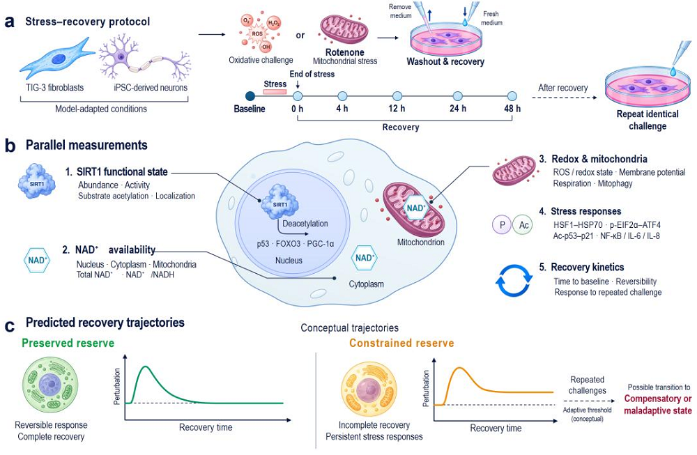
Literature

[1] Iiams, S. E., Skinner, N. J., Wight-Carter, M., Acosta-Rodríguez, V. A., Green, C. B., & Takahashi, J. S. (2026). Time-restricted feeding extends healthspan in both sexes and lifespan in male C57BL/6 J mice. Nature Aging, 1-17.

[2] Lowe, D. A., Wu, N., Rohdin-Bibby, L., Moore, A. H., Kelly, N., Liu, Y. E., … & Weiss, E. J. (2020). Effects of time-restricted eating on weight loss and other metabolic parameters in women and men with overweight and obesity: the TREAT randomized clinical trial. JAMA internal medicine, 180(11), 1491-1499.

[3] Acosta-Rodríguez, V., Rijo-Ferreira, F., Izumo, M., Xu, P., Wight-Carter, M., Green, C. B., & Takahashi, J. S. (2022). Circadian alignment of early onset caloric restriction promotes longevity in male C57BL/6J mice. Science, 376(6598), 1192-1202.

Rejuvenation Roundup June 2026

Rejuvenation Roundup June 2026

Doing something about aging seems to require action at the most basic genomic level. Here’s what’s been done there and in other areas in June.

Interviews

Todd White InterviewThe Thalion Initiative: A New Non-Profit With Big Ambitions: The longevity field remains small and starved for resources, especially the subfield devoted to the fundamental biology of aging, despite near-universal agreement that solving aging requires understanding it first.

Research Roundup

Preventing Load-Induced Arthritis at the Cellular Level: Researchers have discovered that the osteoarthritis-inducing effects of excessive mechanical stress can be mitigated by increasing miR-330, a key regulator in cartilage and bone cells.

Inside a cellNeurons’ Protein Disposal Trick Offers Alzheimer’s Insights: According to a new study, a special protein disposal system, currently found only in neurons, is linked to central hallmarks of Alzheimer’s disease.

Fighting Parkinson’s by Restoring Protein Degradation: Researchers have explained how a protein found in both yeast and humans facilitates the destruction of the core protein responsible for Parkinson’s disease.

Lungs affectedHow Gut Bacteria Affect Lung Fibrosis: In Aging Cell, researchers have described how a strain of Lactobacillus gut bacteria sends chemical signals that enter the bloodstream and decrease fibrosis in the lungs.

The Immune System Maintains the Microbiome: Researchers have proposed that the immune system and immune surveillance play a central role in maintaining microbial composition throughout life by suppressing microbial proliferation and that aging weakens these processes.

Firing neuronsInducing NREM-Like Neuronal Patterns Mimics Sleep Benefits: Scientists have “faked” sleep in mice by artificially creating the on/off neuronal firing pattern similar to that seen in non-REM sleep. This produced sleep-like effects, including improved learning memory.

How Senescent Cells Grow the Homes of Cancerous Tumors: A team of reviewers has taken a look at the relationship between cancer, cellular senescence, and vascular overgrowth and published this information in Aging Cell.

Lab mouse facing viewerLate-Life Gene Therapy Boosts Lifespan in Mice by 20%: In a new study, muscle-targeted viral-vector-based delivery of the protein FGF21 significantly increased median lifespan in male mice and improved many healthspan markers.

How Antioxidants Can Selectively Remove Some Senescent Cells: In Aging Cell, researchers have described the way that antioxidants work against senescence in muscle cells by altering mTOR signaling.

Blood vesselEpigenetic Drug Targets Fat, Improving Blood Vessel Health: Scientists have targeted the thin fat layer around blood vessels with a transcription inhibitor, reducing symptoms of cardiometabolic disease.

A Transcriptional Failure Leads to Systemic Inflammation: Researchers have found that bound pieces of RNA and DNA in the cytoplasm of senescent cells encourage these cells to secrete inflammatory factors.

Young man as old manBiological Aging May Be Driving Increased Early-Onset Cancer: A new study links accelerated aging to early-onset solid cancers, while showing that this gap is becoming wider with each new generation.

Glycosylation’s Role in Alzheimer’s Disease: A recent study suggests that hyperglycosylation in brain tissue can be a hallmark of Alzheimer’s disease, which was observed in human brains and in two mouse models.

Knee painUpregulating a Key Cartilage Factor Leads to Osteoarthritis: Researchers have found that sustained expression of excess hypoxia-inducible factor (HIF)-1α leads to unwanted formation of blood vessels (angiogenesis) that destroys cartilage and causes osteoarthritis.

Cell Type-Specific Aging Predicts Disease Onset: A new study has used aging trajectories of various cell types to predict diseases such as Alzheimer’s and lung cancer. This expands on previous research into organ-specific aging.

Monkeys and peopleHow a Primate-Specific RNA Strand Worsens Senescence: Researchers have discovered a primate-specific piece of non-coding RNA that is linked to aging and makes senescence worse.

Study Maps Existing Drugs to the Hallmarks of Aging: A new study suggests a way to predict whether existing drugs can extend human lifespan. This method uses a network approach that detects longevity signals in protein interactions.

Time-restricted feeding extends healthspan in both sexes and lifespan in male C57BL/6J mice: The benefits were more prolonged in female mice relative to their total lifespan. Median lifespan was significantly extended in male mice under 8-h TRF by 12%, whereas female mice showed no significant lifespan extension.

Rapamycin Attenuates Age-Related Changes in Marmoset Submandibular Gland: A Non-Human Primate Model of Human Oral Aging: The marmoset represents a valuable NHP model for studying SG aging biology and testing therapeutic strategies aimed at attenuating age-related structural degeneration associated with SG dysfunction.

A combination of ketones and NAD+ precursor preserves white matter integrity in mild cognitive impairment: Improved myelin density may help explain the positive association between increased WM ketone uptake and improved processing speed in MCI after a ketone salt and NAD+ precursor supplementation.

Plasma proteomic signatures of cellular aging predict human disease: These findings establish a framework for quantifying human physiology at cellular resolution, revealing heterogeneous aging trajectories and their impact on disease susceptibility and resilience.

Functional rejuvenation of endothelial cell aging by transient reprogramming: While the compounds used are individually approved for other indications, their combined use in this context highlights a conceptual translational potential.

Synergistic senolytic–regenerative therapy significantly extends healthspan and lifespan: Targeted senolytic immunotherapy enhances the efficacy of regenerative interventions and represents a promising combinatorial strategy for chronic disease management and potentially for modifying biological aging itself.

Corylin promotes healthy aging via RAGA–mTOR suppression and sex-dependent activation of SIRT3: Integrated multi-omics analyses across multiple tissues reveal coordinated age-associated molecular changes modulated by Corylin.

Ameliorating calcium homeostasis improves longevity and healthspan in progeroid and naturally aged mice: Together, these findings uncover the mechanism of Ca2+ homeostasis disruption during premature and natural aging, and suggest MIA as a potential therapeutic strategy to extend healthy lifespan by augmenting Ca2+ homeostasis.

Long-term selection for extended lifespan reshapes host physiology and gut microbiome structure in an insect model: Long-term selection can be associated with the emergence of strain-specific gut microbiome configurations.

From metaphor to metric: the entropic framework as a unifying theory of aging: This framework transforms entropy from a rhetorical analogy into an operational concept in aging biology.

Why we age – Integrating error, program, and selective pressure: This review argues that aging should not be interpreted exclusively as the result of random molecular damage or the output of a specific genetic program, but rather as a regulated modulation of how organisms acquire defects and epigenetic drift over time.

Integrating evolutionary theory into a framework for the mechanistic evaluation of candidate anti-aging interventions: In this framework, persistent activity of growth and developmental programmes, alongside insufficient somatic maintenance, define two broad biological axes.

Epigenetic Clocks in the Cosmic Silence of a Deep Underground Laboratory: Implications for Aging and Space Exploration: This reductionist framework could clarify the physical constraints on epigenetic timekeeping and inform how aging clocks function in shielded terrestrial, lunar, and Martian habitats during aging, disease, and future space habitation.

News Nuggets

Forever Healthy FoundationForever Healthy Foundation Launches Evipedia.ai: The Forever Healthy Foundation publicly launched evipedia.ai, an open online encyclopedia of in-depth evidence reviews covering more than 500 health and longevity interventions.

Coming Up

TimePie Longevity Forum Spotlights Evidence-Based Medicine: On September 12–13, 2026, the 7th TimePie Longevity Forum will convene nearly 2,000 researchers, physicians, healthcare operators, technology companies and investors in Shanghai to examine how aging science can be translated into evidence-based, real-world medicine.

Younger 2027NeuroAge Therapeutics Launches Younger 2027: NeuroAge Therapeutics announced Younger 2027, a six-month biological aging contest in which competitors are measured on a clinical-grade aging panel and retested six months later. Baseline testing kits begin shipping September 1, 2026, with at-home baseline testing open through February 1, 2027.

We would like to ask you a small favor. We are a non-profit foundation, and unlike some other organizations, we have no shareholders and no products to sell you. All our news and educational content is free for everyone to read, but it does mean that we rely on the help of people like you. Every contribution, no matter if it’s big or small, supports independent journalism and sustains our future.
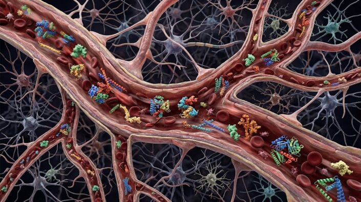
Drug compartments

Study Maps Existing Drugs to the Hallmarks of Aging

A new study suggests a way to predict whether existing drugs can extend human lifespan. This method uses a network approach that detects longevity signals in protein interactions [1].

Slowing aging with existing drugs?

Finding drugs that can slow aging, a multifactorial process involving thousands of genes, is hard. Directly measuring the effect of a prospective drug on human aging would take decades, so scientists need proxy markers, such as epigenetic clocks. Another hurdle is regulation, as the system is geared towards single-disease indications. On the other hand, thousands of drugs for these diseases have already been approved. What if some of them also affect the rate of aging, and how can we unlock this potential?

A new study from Northeastern University and Harvard, published in Nature Aging, offers an interesting way forward. “As someone whose hair turned gray years ago, I share the universal wish that there might one day be a pill that slows aspects of aging,” said Albert-László Barabási, Distinguished University Professor of Physics at Northeastern who oversaw the study. “The challenge is figuring out which drugs are worth testing.”

Hitting the hallmarks

The study is based on network medicine, a framework that the Barabási lab has developed over fifteen years. Its core idea is that proteins do not act in isolation but form a giant graph (the interactome) that shows which proteins interact physically or functionally. Genes underlying a given disease tend to clump together into a “disease module.” Once such a module is outlined, scientists can ask which drugs have targets in the vicinity of that “neighborhood” and are thus candidates for perturbing it. This approach has been used before for asthma, heart disease, and COVID-19 [2].

The Hallmarks of Aging has been a defining paradigm in geroscience for many years. It includes crucial biological features and processes that get disrupted with age, such as DNA stability and intercellular communication. This study is built on the premise that each hallmark of aging behaves not unlike a disease module and that the same machinery that network medicine applies to diseases can therefore be used with these hallmarks.

“You have genes related to aging by some definition or by some reasoning, but it feels like you just have a very big pile of genes related to aging,” said Bnaya Gross, a postdoctoral researcher in Barabási’s lab at Northeastern and lead author of the study. “Networks allow us to organize them, saying, OK, it’s not just a pile of genes. They are connected to each other. They form some sort of organization. It’s not a random process.”

The researchers started from the OpenGenes database, a manually curated resource linking 2,358 genes to aging/longevity. Each gene is tagged with a confidence level from 1 (changing the gene’s activity actually extends mammalian lifespan) to 5 (lowest, weak association). Notably, only 26 genes sit at confidence level 1. 1,250 of the genes could be assigned to at least one hallmark of aging; the remaining 1,108 are still aging-linked but couldn’t be pinned to a specific hallmark. 860 genes belong to a single hallmark and 390 to multiple hallmarks, with TP53 spanning the most (seven).

The fact that many genes are related to several hallmarks shows their interconnectedness via shared molecular machinery. The network approach is quite good at catching this complex web of interactions, while also showing that the hallmarks occupy distinct nodes in it. The team then validated that their 1,250-gene set is largely related to aging and mapped the genes onto the human interactome: a network of more than 500 thousand experimentally supported interactions among proteins.

The team then took 6,442 compounds from DrugBank and, for each hallmark module, measured each drug’s network proximity – the average shortest-path distance from the drug’s protein targets to the nearest hallmark genes. Drugs whose targets sit significantly closer than chance are predicted to perturb that hallmark. However, they found that some of the hits acted in the opposite direction – for instance, induced rather than lowered cellular senescence. So, proximity alone correctly identifies drugs that act on a hallmark but not whether they help or harm.

Turning the pAGE

The researchers devised a metric that accounts for directionality and called it pAGE. They outlined a Systematic Hallmark-based Aging Repurposing Pipeline (SHARP) based on proximity and pAGE, and they validated it against drugs tested in mammalian longevity studies. For instance, out of the eight compounds that increased mouse lifespan in the Intervention Testing Program (ITP) trials and also had interactomic data, all had a positive pAGE for at least one hallmark. Of the drugs that failed in the ITP, less than half did. Interestingly, some pro-longevity drugs were beneficial for some hallmarks but harmful for others, highlighting possible trade-offs.

Another test came from drugs currently in human longevity trials, such as metformin and rapamycin. Of the 17 compounds, 11 had significant proximity. Interestingly, aspirin was mapped to six hallmarks and dasatinib to five, whereas rapamycin hit only one: intercellular communication. Finally, the team successfully tested their method on 10 compounds from a parallel study whose results appeared after their predictions [3] – the closest thing to a prospective test.

The researchers then ran SHARP across all hallmarks to generate candidates. They identified 370 drugs that are proximal to at least one hallmark, and 83 of them are “network drugs” that do not directly target any aging gene but affect a hallmark module through the network topology. These would be invisible to any method that looks solely at direct drug-target relationships, and they are the strongest argument for the network approach. The team showed their predictions are mechanistically interpretable by analyzing how exactly oxymetazoline – a nasal/topical decongestant and rosacea drug – affects longevity-related genes.

We would like to ask you a small favor. We are a non-profit foundation, and unlike some other organizations, we have no shareholders and no products to sell you. All our news and educational content is free for everyone to read, but it does mean that we rely on the help of people like you. Every contribution, no matter if it’s big or small, supports independent journalism and sustains our future.

Literature

[1] Gross, B., Ehlert, J., Gladyshev, V. N., Loscalzo, J., & Barabási, A. L. (2026). Network-driven discovery of repurposable drugs targeting hallmarks of aging. Nature Aging, 1-16.

[2] Morselli Gysi, D., Do Valle, Í., Zitnik, M., Ameli, A., Gan, X., Varol, O., … & Barabási, A. L. (2021). Network medicine framework for identifying drug-repurposing opportunities for COVID-19. Proceedings of the National Academy of Sciences, 118(19), e2025581118.

[3] Shindyapina, A. V., Tyshkovskiy, A., Bozaykut, P., Castro, J. P., Gerashchenko, M. V., Trapp, A., … & Gladyshev, V. N. (2025). Molecular signatures of longevity identify compounds that extend mouse lifespan and healthspan. bioRxiv.

Monkeys and people

How a Primate-Specific RNA Strand Worsens Senescence

Researchers have discovered a primate-specific piece of non-coding RNA that is linked to aging and makes senescence worse.

The packing matters

While non-coding RNA strands (ncRNAs) do not themselves produce proteins, they serve crucial regulatory functions. Short ncRNAs, such as microRNAs, are involved in the fine-tuning of transcriptional pathways, modulating the proteins produced by coding RNAs [1]. Long ncRNAs (lncRNAs), on the other hand, serve significant transcriptional and regulatory purposes; some of them even encode micropeptides despite being noncoding as a whole [2]. Unsurprisingly, dysfunction of lncRNAs is associated with aging [3].

While protein-coding sequences are strongly conserved across mammalian evolution, lncRNAs are not. The total length of lncRNAs is associated with lifespan in mammalian species, and the length of lncRNAs in humans is nearly three times that of mice [4]. Some of these sequences have already been found to be associated with youth; for example, PCAT14, which exists in other primates but not in mice, is active in young human cells but declines with age, and this decline is associated with blood vessel degeneration [5]. LINC00507, which is specific to the primate cortex, is also altered with age [6].

Conserved across primates

In their initial examination, the researchers pulled data from the NONCODE database in order to find commonalities between species. While there were substantially fewer lncRNAs in other primate species than in people, the researchers note that this was most likely due to discrepancies in data collection; humans are, of course, more thoroughly studied than other primates.

However, even with this limited data, it was clear that many lncRNAs are conserved across primates. A subset of these sequences was found to be correlated with chronological age, and some of these expression changes were tissue-specific. While they found several promising candidates, the researchers settled on LINC01021 as the strongest and most representative lncRNA for their further experiments, as its expression significantly changes with aging in seven distinct tissues.

Another examination found that in an RNA sequencing database of senescent cells, LINC01021 is strongly upregulated in four distinct types of fibroblasts that had been driven senescent through two separate means. The researchers used a population of their own human embryonic lung fibroblasts (HELFs) to confirm this data, driving them senescent through radiation, doxorubicin toxicity, and replication; in all three groups, LINC01021 was indeed upregulated.

In order to determine if this lncRNA was a contributor to or a defense against senescence, the researchers then created a population of HELFs in which LINC01021 is overexpressed. These cells were more likely to become senescent than their unmodified counterparts, as measured by decreased proliferation and an increase in the crucial biomarker SA-β-gal. Other key senescence-related genes and their downstream proteins were similarly upregulated by increased LINC01021. Knocking down LINC01021, as expected, produced the opposite effect; HELFs without this lncRNA were significantly less likely to become senescent.

The precise mechanisms

Further work found that this is due to LINC01021‘s suppression of RBMX, which does encode a protein. Knocking down RBMX was found to cause significant upregulation of senescence-associated genes. This was found to be strongly linked to the tumor suppressor P53; knocking down RBMX drastically increased the levels of P53, while silencing P53 through RNA significantly diminished the effects of LINC01021. The researchers noted that this is distinct from the effects of this lncRNA in the context of cancer, as it has been found to promote tumor growth [7].

The researchers went into detail, discovering the mechanistic relationship between LINC01021 and RBMX. While there were weak links between the proteasome and this lncRNA, the strongest result was that overexpressing LINC01021 was found to deplete the levels of DAZAP1, a protein responsible for RNA stability. This lack of DAZAP1 was directly responsible for the rapid depletion of RBMX protein levels.

Premature aging in a mouse model

The researchers then created a humanized mouse with an LINC01021 knock-in. Compared to wild-type mice, mice with this alteration became frail earlier, as measured by multiple physical tests; while not all of these tests’ results reached the level of statistical significance, the altered mice took longer to cross a beam, and they had reduced grip strength. There were signs of increased inflammation, and they had significantly upregulated biomarkers of senescence.

This study serves as a sobering reminder that mice are not people, and some of the changes that occur with aging affect the very things that make us human. It also illustrates the serious difficulties involved in studying such changes, as there are no naturally occurring short-lived species that can be used to study them. Similarly, if an mRNA-based or gene therapy can be used to directly affect such apparently detrimental lncRNAs, we will be fortunate if it can possibly be tested on other primates first.

We would like to ask you a small favor. We are a non-profit foundation, and unlike some other organizations, we have no shareholders and no products to sell you. All our news and educational content is free for everyone to read, but it does mean that we rely on the help of people like you. Every contribution, no matter if it’s big or small, supports independent journalism and sustains our future.

Literature

[1] Turko, R., Hajja, A., Magableh, A. M., Omer, M. H., Shafqat, A., Khan, M. I., & Yaqinuddin, A. (2025). The emerging role of miRNAs in biological aging and age-related diseases. Non-coding RNA research, 13, 131-152.

[2] Yao, R. W., Wang, Y., & Chen, L. L. (2019). Cellular functions of long noncoding RNAs. Nature cell biology, 21(5), 542-551.

[3] Marttila, S., Chatsirisupachai, K., Palmer, D., & de Magalhães, J. P. (2020). Ageing-associated changes in the expression of lncRNAs in human tissues reflect a transcriptional modulation in ageing pathways. Mechanisms of Ageing and Development, 185, 111177.

[4] Wang, A. (2025). Noncoding RNAs evolutionarily extend animal lifespan. Global Medical Genetics, 12(2), 100034.

[5] Drekolia, M. K., Talyan, S., Cordellini Emídio, R., Boon, R. A., Guenther, S., Looso, M., … & Bibli, S. I. (2022). Unravelling the impact of aging on the human endothelial lncRNA transcriptome. Frontiers in genetics, 13, 1035380.

[6] Mills, J. D., Ward, M., Chen, B. J., Iyer, A. M., Aronica, E., & Janitz, M. (2016). LINC00507 is specifically expressed in the primate cortex and has age-dependent expression patterns. Journal of molecular neuroscience, 59(4), 431-439.

[7] Kaller, M., Forné, I., Imhof, A., & Hermeking, H. (2024). LINC01021 Attenuates Expression and Affects Alternative Splicing of a Subset of p53-Regulated Genes. Cancers, 16(9), 1639.

Organs

Cell Type-Specific Aging Predicts Disease Onset

A new study has used aging trajectories of various cell types to predict diseases such as Alzheimer’s and lung cancer [1]. This expands on previous research into organ-specific aging.

Age is more than one number

Gone are the days when aging was assumed to be happening uniformly across every tissue at once. Today, we know that certain organs and systems in the body often exhibit either accelerated aging or, conversely, unusual resilience, and that can influence morbidity and mortality. This idea is associated in particular with the group led by Tony Wyss-Coray of Stanford University and their 2023 seminal paper on organ-specific aging [2]. The diagnostic and therapeutic potential of this area of research is hard to overstate.

In a new study published in Nature Medicine, Wyss-Coray and colleagues took their idea further, making a conceptual jump from organs to cell types. The researchers “developed machine learning models to estimate the biological age of over 40 cell types,” per the abstract, and the results are informative and, at times, surprising.

Clocks and predictions

Using single-cell RNA sequencing from the Human Protein Atlas, the authors classified a gene as “cell-type-enriched” if it was expressed much more strongly in one cell type than in any other and then linked those genes to their plasma protein products. For each cell type, the researchers trained a machine-learning model to estimate a person’s chronological age from how those associated proteins rise, fall, or hold steady across the lifespan. In total, data from about 60,000 people across three cohorts was analyzed to test whether these cell-type aging signatures track disease and death.

Notably, the clocks’ predictive power varied widely; for instance, predictions from liver cells (hepatocytes) were more robust than predictions from excitatory neurons. These cell type-specific clocks revealed that 35% of people had no extreme age gaps in any cell type, 24% had extreme aging in exactly one cell type, and 1.5% exhibited extreme aging across ten or more cell types. The rest showed extreme aging in anywhere from two to nine cell types.

Apparently, at least some of these clocks can actually predict diseases. For instance, extreme aging of skeletal muscle cells (myocytes) strongly predicted incident amyotrophic lateral sclerosis (ALS), a devastating neurodegenerative disease, even for cases diagnosed more than three years after the blood draw. Extreme aging of neuron-supporting brain cells (astrocytes) predicted incident Alzheimer’s disease. Other diseases the researchers were able to predict, although less robustly, included lung cancer, lymphoma, type 2 diabetes, COPD, and stroke.

“Twenty years ago, we started to explore the idea that immune and other signaling proteins in the circulation could provide insights into Alzheimer’s disease,” said Wyss-Coray to Lifespan News. “Taking advantage of technical innovations that allow us to quantify thousands of proteins in a drop of blood, we pushed this concept further to show that circulating proteins derived from specific cell types in the brain and other tissues allow us to assess the physiological state and the risk to develop disease at an individual level. We were most surprised about strong links between accelerated aging of astrocytes and muscle cells with Alzheimer’s disease and ALS, respectively.”

Interactions with genetic risk factors

Cellular aging interacted with the APOE genotype, the strongest genetic predictor of Alzheimer’s, in a peculiar way. People homozygous for APOE4 – the most Alzheimer’s-prone genotype – were almost three times as likely to develop the disease if they also had rapidly aging astrocytes. This result may eventually lead to earlier and more robust prediction of Alzheimer’s risk and new therapeutical options aimed at keeping astrocytes young.

Interestingly, APOE4 carriers showed older astrocytes, but younger macrophages, and the carriers of the protective allele APOE2 showed the opposite. The authors suggest this might be a case of antagonistic pleiotropy – when the same trait is beneficial in one respect but costly in another [3]. APOE4-enhanced immune vigilance may have been an asset in our pathogen-laden past, so it was favored by selection despite an accompanying cost of faster brain aging. The latter only became consequential once more people began surviving into the advanced age where Alzheimer’s strikes.

Another famous risk factor, smoking, was also mitigated by younger cells: in this case, alveolar type 2 cells and respiratory epithelial cells. Smokers whose cells stayed young were much less susceptible to lung cancer. These cellular clocks could also predict mortality. For all-cause death, skeletal myocyte aging carried the strongest signal, followed by neurons, fibroblasts, alveolar type 2, and myeloid cells.

We would like to ask you a small favor. We are a non-profit foundation, and unlike some other organizations, we have no shareholders and no products to sell you. All our news and educational content is free for everyone to read, but it does mean that we rely on the help of people like you. Every contribution, no matter if it’s big or small, supports independent journalism and sustains our future.

Literature

[1] Ding, D. Y., Bot, V. A., Chen, K. L., Groves, J. W., Pálovics, R., Masuda, D., … & Wyss-Coray, T. (2026). Plasma proteomic signatures of cellular aging predict human disease. Nature Medicine, 1-13.

[2] Oh, H. S. H., Rutledge, J., Nachun, D., Pálovics, R., Abiose, O., Moran-Losada, P., … & Wyss-Coray, T. (2023). Organ aging signatures in the plasma proteome track health and disease. Nature, 624(7990), 164-172.

[3] Austad, S. N., & Hoffman, J. M. (2018). Is antagonistic pleiotropy ubiquitous in aging biology? Evolution, medicine, and public health, 2018(1), 287-294.

Knee pain

Upregulating a Key Cartilage Factor Leads to Osteoarthritis

Researchers have found that sustained expression of excess hypoxia-inducible factor (HIF)-1α leads to unwanted formation of blood vessels (angiogenesis) that destroys cartilage and causes osteoarthritis.

A necessary but destructive factor

Being required for extracellular matrix (ECM) construction and energy in the absence of oxygen, HIF-1α is necessary for cartilage-building cells (chondrocytes), which normally live in a low-oxygen environment, to function properly [1]. However, excessive HIF-1α is commonly found in osteoarthritic joints [2], and some previous work has linked it to angiogenesis in the cartilage, placing blood vessels where none should exist [3].

Still, given its beneficial nature under normal circumstances, researchers had struggled to determine whether upregulated HIF-1α was a protective reaction to damage or a source of damage itself. We have reported on previous work finding that HIF-1α has benefits in the spine by inhibiting iron-related cellular death (ferroptosis). That study, however, focused on nucleus pulposus cells. This research focuses on chondrocytes and their compartments, and what it has to say about excessive HIF-1α is nothing good.

Results in mice and people

In this paper’s first study, the researchers examined cartilage and synovium derived from osteoarthritis patients. Samples harvested from severely damaged areas were considerably richer in HIF-1α than samples from undamaged areas. Vascular endothelial growth factor (VEGF), which promotes angiogenesis, was also significantly upregulated in the damaged areas.

Comparing joint samples from 6-month-old and 24-month-old mice found similar results, with both compounds being upregulated in the older mice. Artificially inducing osteoarthritis in 3-month-old mice through surgery caused similar upregulations, with the cartilage taking longer to exhibit long-term upregulation than the synovium, where it occurred immediately.

Mice that express more HIF-1α withstand more pain

The researchers then used a population of mice that were genetically engineered to express excessive HIF-1α, and induced arthritis into one of their knees. Compared to wild-type mice, these altered mice endured more pain and had greater joint deterioration. Interestingly, the altered mice expressed both more building-related (anabolic) and destruction-related (catabolic) factors compared to the wild-type mice. According to the researchers, “this indicates that sustained HIF-1α activation drives a ‘metabolic paradox’ where compensatory synthetic efforts are overwhelmed by parallel catabolic signaling.”

Even without induced arthritis, the mice modified to express more HIF-1α began to develop it at 9 months of age anyway, with abnormal bone remodeling and a loss of cartilage. By 12 months of age, these mice had developed severe osteoarthritis, including complete erosion of cartilage. Therefore, excessive HIF-1α is, by itself, sufficient to drive the disease.

Progression is determined by tissue expression

Osteoarthritis progression in these animals was found to be driven by a loss of the hypoxic environment where chondrocytes normally live. At 9 months of age, this loss of hypoxia coincided with significant increases in cellular senescence as well, and markers of senescence were found throughout the tissues at 12 months of age. This was preceded by the ‘metabolic paradox’ that the researchers had discovered in the previous experiment; at 6 months of age, these mice exhibited increases in both anabolic and catabolic factors, “irreversibly tipping the cartilage from a state of frustrated repair into overwhelming, chronic degeneration.” Similar results were found when a population of wild-type mice was injected with mRNA causing excessive HIF-1α to be produced in the cartilage.

The authors turned to a different type of genetically engineered mouse, which expresses excessive HIF-1α only in the superficial tissues around the joint. These mice took longer to show disease symptoms: they experienced inflammation in the joints at 12 months of age, and at 15 months of age, they had developed full-blown osteoarthritis. There was significant angiogenesis in their joints, along with significant upregulation of inflammatory factors. Their osteoarthritis occurred alongside the suppression of anabolic factors and large upregulations of catabolic ones.

The researchers are clear that there is a substantial difference between transiently upregulated and normally fluctuating HIF-1α and the sustained increases in HIF-1α that lead to, and exacerbate, osteoarthritis. They describe it as a long-term stressor that ages the joints over time, and they note that instead of attempting to bolster it in tissues as any kind of treatment, it may make sense to attempt to remove it from the joints of older patients instead.

We would like to ask you a small favor. We are a non-profit foundation, and unlike some other organizations, we have no shareholders and no products to sell you. All our news and educational content is free for everyone to read, but it does mean that we rely on the help of people like you. Every contribution, no matter if it’s big or small, supports independent journalism and sustains our future.
Literature

[1] Sophia Fox, A. J., Bedi, A., & Rodeo, S. A. (2009). The basic science of articular cartilage: structure, composition, and function. Sports health, 1(6), 461-468.

[2] Qing, L., Lei, P., Liu, H., Xie, J., Wang, L., Wen, T., & Hu, Y. (2017). Expression of hypoxia-inducible factor-1α in synovial fluid and articular cartilage is associated with disease severity in knee osteoarthritis. Experimental and therapeutic medicine, 13(1), 63-68.

[3] Caliogna, L., Berni, M., Torriani, C., Mancuso, M. E., Di Minno, M. N. D., Brancato, A. M., … & Pasta, G. (2024). Pathogenesis of osteoarthritis, rheumatoid arthritis, and hemophilic arthropathy: The role of angiogenesis. Haemophilia, 30(6), 1256-1264.

Younger 2027

NeuroAge Therapeutics Launches Younger 2027

NeuroAge Therapeutics announced Younger 2027, a six-month biological aging contest in which competitors are measured on a clinical-grade aging panel and retested six months later. Baseline testing kits begin shipping September 1, 2026, with at-home baseline testing open through February 1, 2027. A live kickoff conference open to competitors and the broader longevity community takes place January 11-12, 2027 at the Gateway Pavilion at Fort Mason in San Francisco. The contest concludes with a finale in November 2027.

Participants complete a clinical-grade baseline across brain, body, and face, run a six-month intervention window, and then are retested on the same panel.

The contest is hosted by NeuroAge Therapeutics in collaboration with TruDiagnostic, Harvard FaceAge, and Vero Bioscience, with science contributed by researchers from MIT, Harvard, Yale, and Stanford.

The Panel

Every Competitor receives baseline and retest measurements across brain, body, and face. The standard panel includes TruDiagnostic’s TruAge platform (epigenetic clocks, SymphonyAge across 11 organ systems, and TruHealth clinical biomarkers), NeuroAge’s NeuroGames cognitive assessment, Harvard’s FaceAge model, and functional measurements (VO2 max, grip strength, sit-to-stand). Results from each partner are combined into a single composite score that tracks change between baseline and retest.

Ultra Competitors receive an extended panel that adds two brain MRIs, additional pre-release biological clocks including proteomic and methylation aging clocks, two sets of 100+ clinical biomarker panels, and two DEXA body composition scans. Several of these clocks are pre-release, giving Ultra participants early access ahead of public availability.

Kickoff at Fort Mason

The Fort Mason event is a two-day longevity conference open to Younger 2027 competitors and the wider longevity community, featuring keynote talks, a curated longevity expo, and hands-on workshops. Confirmed speakers include Dr. Christin Glorioso (NeuroAge Therapeutics), Ryan Smith (TruDiagnostic), Dr. Ray Mak (FaceAge / Harvard Medical School), Dr. Raghav Sehgal (Yale), Dr. Ronjon Nag (Stanford Medicine / R42 Group), Dr. Steve Horvath (UCLA), Dr. Amy Killen (Humanaut Health), Paul Coletta (Vero Bioscience), and Dr. Momo Vuyisich (Viome). Participating clinic teams include California Center for Functional Medicine, VitOS, Humanaut Health, and Prime Health Associates.

Prizes and Awards

Younger 2027 award categories include:

  • The Younger Award (most years of biological age turned back)
  • The Forever Young Award (youngest biological age relative to chronological age)
  • The Ultra Younger Award (most years turned back on the Ultra panel)
  • The Ultra Forever Young Award (youngest biological age on the Ultra panel)
  • Organ-system category winners

Sponsoring partners contribute a $50,000 equivalent prize pool in cash, products, and services.

Registration

Registration is open at neuroagetx.com/events/younger. Early-bird pricing closes September 1, 2026.

About NeuroAge Therapeutics

NeuroAge Therapeutics combines cognitive testing, AI, and advanced diagnostics to help people measure and track brain health. Its multi-modal platform combines MRI, genetics, blood biomarkers, and cognitive testing into a single NeuroAge score tracked over time. NeuroAge was founded by Dr. Christin Glorioso, MD, PhD, a scientist trained at MIT and Harvard and the author of 30+ peer-reviewed publications. The company convenes Younger 2027. Learn more at neuroagetx.com.

Media Contact

Marah Doria

NeuroAge Therapeutics

Email: marah@neuroagetx.com

Web: neuroagetx.com/events/younger

We would like to ask you a small favor. We are a non-profit foundation, and unlike some other organizations, we have no shareholders and no products to sell you. All our news and educational content is free for everyone to read, but it does mean that we rely on the help of people like you. Every contribution, no matter if it’s big or small, supports independent journalism and sustains our future.
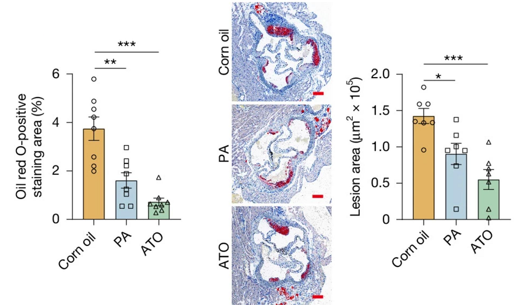
Alzheimer's MRI

Glycosylation’s Role in Alzheimer’s Disease

A recent study suggests that hyperglycosylation in brain tissue can be a hallmark of Alzheimer’s disease [1].

Lesser-known Alzheimer’s molecular features

Pathological deposits of amyloid-β plaques and tau tangles in the brain are probably the most well-known features of Alzheimer’s disease. However, Alzheimer’s also involves various metabolism-related pathologies, such as alterations in glucose metabolism [2], mitochondrial function [3], lipid homeostasis [4], and N-linked glycosylation, a reaction in which a complex carbohydrate (a glycan) is attached to a protein.

N-linked glycans have been found to contribute to protein stability, intercellular communication, and immune signaling [5] and to regulate the blood–brain barrier [6]. Previous studies have reported a link between alterations in glycosylation patterns and neurological disorders [7] and suggested changes in glycosylation in Alzheimer’s disease [8]. Glycosylation has also been found to impact tau post-translational modifications and aggregation [9] and might alter amyloid-β plaque composition and clearance [10].

The authors of this study delved deeper into this topic and leveraged advances in -omics technologies to elucidate the role of glycosylation in Alzheimer’s disease.

Hyperglycosylation appears in Alzheimer’s

Advancements in laboratory techniques enabled researchers to conduct spatial metabolomics, lipidomics, and glycomics (techniques that allow comprehensive examination of metabolites, lipids, and glycans) on frontal cortex samples from deceased patients with Alzheimer’s disease and from healthy donors. “This technology allows us to examine thousands and thousands of molecules created when the body breaks down food or drugs and to uncover intricate pathways that otherwise would stay hidden,” said senior author Ramon Sun, Ph.D., director of the Center for Advanced Spatial Biomolecule Research and associate director for innovation of UF’s McKnight Brain Institute.

While they observed Alzheimer’s disease-specific changes in metabolomics and lipidomics analyses, the differences that caught their attention most were in glycan abundance, which was significantly increased across both white and grey matter regions in samples from patients with Alzheimer’s disease.

The hyperglycosylation observed in human brains was also observed in two mouse models of Alzheimer’s disease. These experiments discovered that those modifications were brain region-specific and preferentially affected brain regions associated with memory, cognitive processing, and neuroinflammation.

Subsequent experiments in human and mouse models found that the increase in glycosylation was the result of increased glycan biosynthesis rather than reduced degradation and recycling, and that the hyperglycosylation in Alzheimer’s disease “predominantly involves an increase in glycosylation modifications on pre-existing glycoproteins rather than the appearance of novel glycosylated proteins.”

Additionally, the researchers identified neurons as the predominant cell population affected by increased glycosylation, supporting the role of glycosylation in Alzheimer’s disease pathogenesis.

A cause or a consequence?

The observed changes in glycosylation can be either a driver of neurodegeneration or a consequence of it. To determine this relationship, the authors experimentally decreased or increased N-glycan levels in the brain.

First, they used two approaches to block glycosylation: genetic engineering tools to reduce the levels of a key enzyme involved in the production of glycans, and a small molecule to block transfer of glycans to proteins. Mouse models of Alzheimer’s disease treated this way showed improvement on a social memory test (which measures “social interaction time across repeated exposures”) compared to controls. Results from those experiments suggest that reducing excessive N-glycosylation results in behavioral benefits.

Increasing glycosylation in mice with Alzheimer’s had the opposite effect. The authors used glucosamine, a molecule that can be incorporated into brain glycans, and supplemented the mice with Alzheimer’s disease with it. Glucosamine supplementation increased brain glycosylation and further exacerbated social memory deficits in those mice. Supplementing glucosamine to healthy mice didn’t result in hyperglycosylation in the brain nor impaired social recognition memory, suggesting that normal brains have mechanisms that can protect them against glucosamine supplementation-induced perturbation.

Together, these results show a causal role for hyperglycosylation in Alzheimer’s disease-associated cognitive dysfunction and suggest that glycosylation can be used as a therapeutic target for Alzheimer’s disease-related neurocognitive deficits. This finding is independent of other Alzheimer’s features such as neuroinflammation or amyloid deposits, which were not affected when glycosylation was experimentally blocked. The authors suggest future studies into small-molecule inhibitors with the ability to selectively decrease pathological glycosylation levels in Alzheimer’s patients’ brains.

“Our results suggest that altered metabolism is a significant contributor to Alzheimer’s progression and, in addition, addressing the metabolic defect could be an important complement to approaches focused on Alzheimer’s plaques and tangles,” Sun said.

Glucosamine supplementation drawbacks

While the results of these experiments can be used for future therapy development, the authors found that the knowledge they gathered here has immediate implications. They demonstrated this by examining glucosamine use in Alzheimer’s disease patients. Glucosamine, which they used in mouse supplementation experiments, is an over-the-counter supplement used for joint health.

From the University of Florida Health system health records, the authors identified 24,481 patients with Alzheimer’s disease-related dementias; among them, 1,896 patients had at least 1 year of documented glucosamine usage following a dementia diagnosis. 41,884 people with mild cognitive impairment were used as controls; 2,750 of them received glucosamine. The analysis indicated that “glucosamine usage was associated with a 25% increase in mortality risk” among patients with Alzheimer’s disease-related dementias, but no significant increase was observed among people with mild cognitive impairment.

When the rate of mild cognitive impairment-to-Alzheimer’s disease-related dementias conversion rate (indicating disease progression) was quantified, the conversion rate was increased by 25% among people who took glucosamine, suggesting that it might accelerate disease progression.

“The electronic health record data are very provocative,” said Matt Gentry, Ph.D., chair of UF’s Department of Biochemistry and Molecular Biology and a study co-author. “While it’s an association and not proof of causality, it does raise an important clinical question that now deserves much more attention.”

“In the United States, there are about seven million people living with Alzheimer’s and millions more with related dementias such as Lewy body or frontotemporal dementia,” Sun adds. “A lot of these people actively take an over-the-counter supplement that could be making their disease progression worse.”

The authors emphasize the need for a well-designed clinical trial to evaluate glucosamine’s impact on cognitive health in people with dementia and to identify which groups are at risk for worsening their dementia symptoms.

We would like to ask you a small favor. We are a non-profit foundation, and unlike some other organizations, we have no shareholders and no products to sell you. All our news and educational content is free for everyone to read, but it does mean that we rely on the help of people like you. Every contribution, no matter if it’s big or small, supports independent journalism and sustains our future.
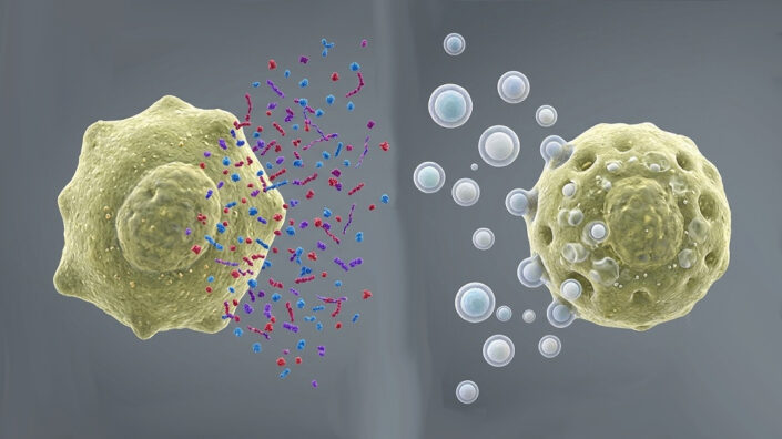
Literature

[1] Hawkinson, T. R., Liu, Z., Ribas, R. A., Medina, T., Nielsen, R. S., Clarke, H. A., Ma, X., Mueller, A. C., Plasencia, A. F., Sheer, A. L., Simpson, S. T., Soto, C. M., Sudderth, J., Cai, F., Cantrell, A. R., Colpaert, M. G., Shedlock, C. J., Wu, L., Young, L. E. A., Kooser, D. D., … Sun, R. C. (2026). Hyperglycosylation is a metabolic driver of Alzheimer’s disease. Nature metabolism, 10.1038/s42255-026-01538-4. Advance online publication.

[2] Butterfield, D. A., & Halliwell, B. (2019). Oxidative stress, dysfunctional glucose metabolism and Alzheimer disease. Nature reviews. Neuroscience, 20(3), 148–160.

[3] Swerdlow R. H. (2018). Mitochondria and Mitochondrial Cascades in Alzheimer’s Disease. Journal of Alzheimer’s disease : JAD, 62(3), 1403–1416.

[4] Di Paolo, G., & Kim, T. W. (2011). Linking lipids to Alzheimer’s disease: cholesterol and beyond. Nature reviews. Neuroscience, 12(5), 284–296.

[5] Conroy, L. R., Hawkinson, T. R., Young, L. E. A., Gentry, M. S., & Sun, R. C. (2021). Emerging roles of N-linked glycosylation in brain physiology and disorders. Trends in endocrinology and metabolism: TEM, 32(12), 980–993.

[6] Nesselbush, M. C., Luca, B. A., Jeon, Y. J., Jabara, I., Meador, C. B., Garofalo, A., Binkley, M. S., Hui, A. B., van ‘t Erve, I., Xu, N., Shi, W. Y., Liu, K. J., Sugio, T., Kastelowitz, N., Hamilton, E. G., Liu, C. L., Olsen, M., Bonilla, R. F., Wang, Y. P., Jiang, A., … Diehn, M. (2025). An ultrasensitive method for detection of cell-free RNA. Nature, 641(8063), 759–768.

[7] Freeze, H. H., Eklund, E. A., Ng, B. G., & Patterson, M. C. (2015). Neurological aspects of human glycosylation disorders. Annual review of neuroscience, 38, 105–125.

[8] Hawkinson, T. R., Clarke, H. A., Young, L. E. A., Conroy, L. R., Markussen, K. H., Kerch, K. M., Johnson, L. A., Nelson, P. T., Wang, C., Allison, D. B., Gentry, M. S., & Sun, R. C. (2022). In situ spatial glycomic imaging of mouse and human Alzheimer’s disease brains. Alzheimer’s & dementia : the journal of the Alzheimer’s Association, 18(10), 1721–1735.

[9] Losev, Y., Frenkel-Pinter, M., Abu-Hussien, M., Viswanathan, G. K., Elyashiv-Revivo, D., Geries, R., Khalaila, I., Gazit, E., & Segal, D. (2021). Differential effects of putative N-glycosylation sites in human Tau on Alzheimer’s disease-related neurodegeneration. Cellular and molecular life sciences : CMLS, 78(5), 2231–2245.

[10] Perdivara, I., Petrovich, R., Allinquant, B., Deterding, L. J., Tomer, K. B., & Przybylski, M. (2009). Elucidation of O-glycosylation structures of the beta-amyloid precursor protein by liquid chromatography-mass spectrometry using electron transfer dissociation and collision induced dissociation. Journal of proteome research, 8(2), 631–642.