In a preprint paper, scientists have reported that senescent cells transiently adhere to neighboring cells, and, upon departure, leave in place large membrane-enclosed fragments of themselves [1].
Mysterious fragments
Cellular senescence is central to aging, but there are still a lot of unknowns around this complex and heterogeneous phenomenon. For instance, we know that senescent cells are highly “communicative”: they can influence their environment, inducing senescence in neighboring cells [2] and eliciting immune response [3]. They do this via the senescence-associated secretory phenotype (SASP), a diverse mix of molecules such as pro-inflammatory cytokines and chemokines. However, this might not be the only signaling mechanism at their disposal.

The researchers started by studying human primary fibroblasts rendered senescent by irradiation. To observe the progression of senescence in single cells, they created a model in which cells marked by the fluorescent protein GFP were mixed at a ratio of 1:1000 with other, non-fluorescent cells. In one group, the fluorescent cells were senescent, but others were healthy and proliferating. In the second group, it was the other way around. In the third group, all cells were senescent, and, finally, the control group contained only healthy cells.
To the scientists’ amazement, after a few days, in addition to GFP-positive cells, smaller GFP-positive spots began to appear. When the researchers imaged them using higher-resolution equipment, those turned out to be not cellular debris but cellular fragments attached to the membranes of adjacent cells. This did not happen in the control group, where both GFP-positive and GFP-negative cells were non-senescent, which ruled out the possibility that the strange effect was caused by GFP.
Origin: senescent cells
To determine the origin of the fragments, the researchers turned to time-lapse imaging, which showed that both senescent and non-senescent cells moved around and routinely made contact with their neighbors.
Contacts between non-senescent cells were brief and ended without damaging either cell. However, when at least one of the cells was senescent, the contacts were sometimes more persistent and resulted in the cells being glued together. As the cells finally moved apart, their bodies stretched until the senescent cell was torn, leaving a part of itself attached to the other cell. Interestingly, the membranes on both the cells and the fragments they left behind quickly sealed, showing effective membrane repair.
Those fragments began appearing in the culture at around day 3 after the induction of senescence, and by day 4, an average of 10-12 fragments per each senescent cell were observed. The fragments were quite large, 7-8 micrometers across, and contained many types of organelles, including mitochondria, ribosomes, and autophagosomes. However, no nuclear matter was found in any of the fragments. In some instances, the researchers also observed adherens junctions: large protein complexes that tie cells together.
When isolated, those senescent cell adhesion fragments (SCAFs) exhibited “spinning, projecting and retracting arms, or crawling-like behavior”. Most of them disintegrated by day 3, creating extracellular debris, although a small part of SCAF contents ended up inside the surrounding cells.
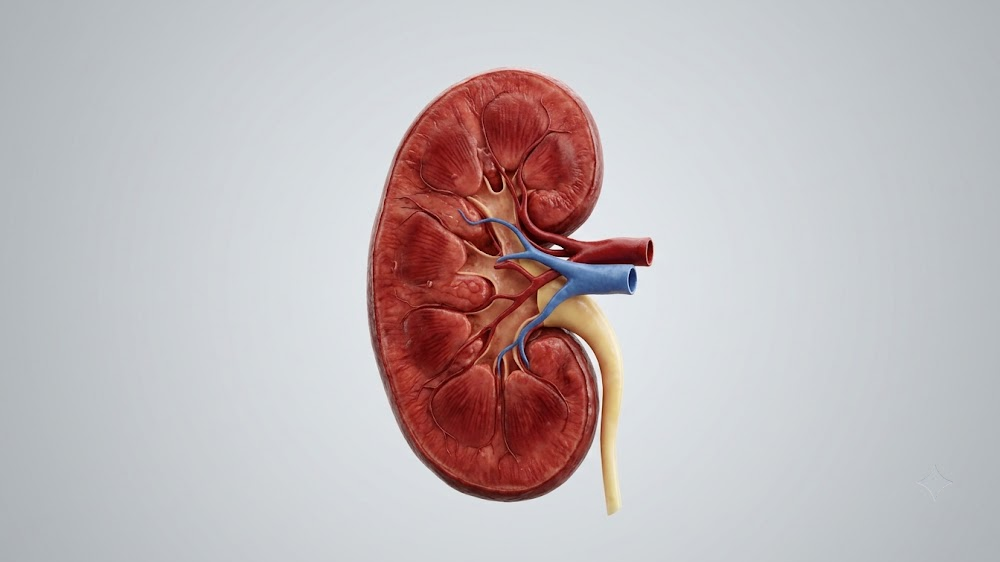
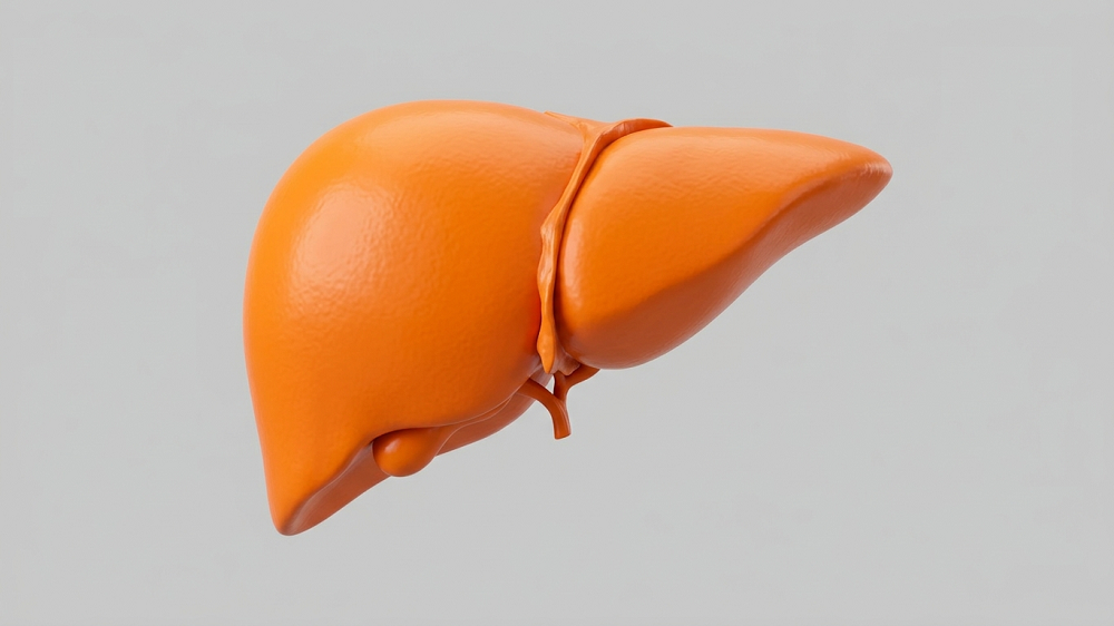
To make sure that SCAFs are not exclusive to irradiated senescent fibroblasts, the researchers experimented with several other combinations of senescence triggers and cell types, such as oncogene-induced senescence in primary fibroblasts and chemotherapy-induced senescence in human liver cancer cells. They also showed that SCAF formation occurs in vivo in mice.
Associations with inflammation and cancer
The researchers analyzed the protein content of SCAFs and discovered associations with damage signaling, immune cell recruitment and activation, inflammation, and neurodegenerative disease.
The researchers also wanted to see what effects SCAFs have on the cells that they end up piled upon. DEG (differentially expressed genes) analysis of those cells produced many correlations with SCAFs, pointing at pathways associated with cell proliferation, migration, and invasion as well as wound healing, cell adhesion, and cancer.
When exposed to SCAFs isolated from senescent cells, healthy fibroblasts showed a significant increase in proliferation and migration velocity. Due to a strong association with cancer signaling, the researchers also investigated the effects of SCAFs on cancer cells. Adding SCAFs to liver cancer cells increased their proliferation. In 3D culture, SCAF-treated cancer cells became more active, invading the gel and forming branches.
This supports the theory that cellular senescence is a double-edged sword that facilitates organismal development, wound healing, and probably cancer resistance, but later in life, it turns from transient to permanent, driving inflammation and, somewhat paradoxically, cancer [4].
Conclusion
This study points to a previously unknown quality of senescent cells that might contribute to our understanding of cellular senescence and its complex relationship with cancer. Given that several different types of senescent cells had exhibited this phenomenon, the researchers also suggest that it might be used as a senescence marker.
Literature
[1] Durik, M., Goncalves, D. S., Spiegelhalter, C., Messaddeq, N., & Keyes, B. (2023). Senescent cells deposit intracellular contents through adhesion-dependent fragmentation. bioRxiv.
[2] Nelson, G., Wordsworth, J., Wang, C., Jurk, D., Lawless, C., Martin‐Ruiz, C., & von Zglinicki, T. (2012). A senescent cell bystander effect: Senescence‐induced senescence. Aging cell, 11(2), 345-349.
[3] Marin, I., Boix, O., Garcia-Garijo, A., Sirois, I., Caballe, A., Zarzuela, E., … & Serrano, M. (2022). Cellular senescence is immunogenic and promotes anti-tumor immunity. Cancer Discovery, CD-22.
[4] Giaimo, S., & d’Adda di Fagagna, F. (2012). Is cellular senescence an example of antagonistic pleiotropy?. Aging cell, 11(3), 378-383.