Researchers publishing in Aging Cell have found that the efficiency of autophagy, a cellular maintenance process, increases rather than decreases in some T cells derived from healthy older people.
Keeping cells healthy
The researchers introduce their paper by discussing autophagy and its decline in aging. They focus specifically on its effects on T cells, noting that a decline in autophagy causes mitochondrial dysfunction in CD4+ T cells due to old mitochondria not being cleared [1] and is associated with DNA damage in CD8+ T cells [2].
This paper focuses specifically on CD4+ T cells, building upon previous work demonstrating that an age-related deficiency of a glycolytic enzyme prevents these cells from using autophagy as an energy source [3] and that better autophagy in these cells is hereditary and associated with longer lifespans [4].
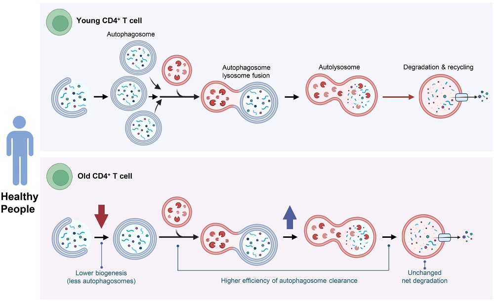
This study began by measuring the base rate of autophagy in CD4+ T cells derived from one group of 28- to 35-year-olds and another group of 67- to 93-year-olds. The first analysis was of compartments (puncta) that tested positive for microtubule-associated protein 1A/1B-light chain 3 (LC3), a marker of autophagosomes.
No autophagic decline in cells from healthy people
Interestingly, while the number of LC3+ puncta trended towards being lower in the older group, this did not reach the level of statistical significance. The number of protein-destroying lysosomes was also similar, as was the number of autolysosomes, which are combinations of autophagosomes and lysosomes that actually destroy unwanted mitochondria and organelles.
Experimentation with an inhibitor of autophagic degradation found that there is an increase in autophagic flux with age. This led to the researchers’ surprising conclusion: in CD4+ T cells, a reduction in the number of lysosomes is compensated for by an increase in efficiency, allowing these cells to maintain themselves through autophagy. The researchers suggest that “when autophagy is stimulated, the greater increase in older adults may be the result of a much larger amount of damaged material that needs to be processed.”
There was also some evidence that the autolysosomes of older CD4+ T cells are more heterogenous and possibly less stable, and introducing a molecule that affects autophagy through stress revealed that these cells may have a reduced ability to respond to this stress.
Unexpected results
Overall, these findings ran counter to the researchers’ assumptions, who noted that previous work has found a decline in autophagy with age in various models, including human cells, and that autophagy has been repeatedly found to play a role in aging [5]. However, the researchers also suggest that animal models may not translate well to humans in this respect, as it is plausible that increased autophagic maintenance is one of the reasons we live longer. Furthermore, some previous work has found that certain conditions, such as Type 2 diabetes, have been found to increase rather than decrease autophagic flux [6].
These experiments were performed only on a certain subset of T cells, and these findings may not be universal across cell types. Additionally, this study included only cells derived from healthy donors, and the researchers note that this may have played a large role in its negative results: “A burning question is whether the preservation of effective autophagy in these healthy individuals free of chronic diseases is one of the reasons for the maintenance of their healthy status.” They recommend further studies that compare health status to autophagic maintenance.
Literature
[1] Bektas, A., Schurman, S. H., Gonzalez-Freire, M., Dunn, C. A., Singh, A. K., Macian, F., … & Ferrucci, L. (2019). Age-associated changes in human CD4+ T cells point to mitochondrial dysfunction consequent to impaired autophagy. Aging (Albany NY), 11(21), 9234.
[2] Phadwal, K., Alegre-Abarrategui, J., Watson, A. S., Pike, L., Anbalagan, S., Hammond, E. M., … & Simon, A. K. (2012). A novel method for autophagy detection in primary cells: impaired levels of macroautophagy in immunosenescent T cells. autophagy, 8(4), 677-689.
[3] Yang, Z., Fujii, H., Mohan, S. V., Goronzy, J. J., & Weyand, C. M. (2013). Phosphofructokinase deficiency impairs ATP generation, autophagy, and redox balance in rheumatoid arthritis T cells. Journal of Experimental Medicine, 210(10), 2119-2134.
[4] Raz, Y., Guerrero-Ros, I., Maier, A., Slagboom, P. E., Atzmon, G., Barzilai, N., & Macian, F. (2017). Activation-induced autophagy is preserved in CD4+ T-cells in familial longevity. Journals of Gerontology Series A: Biomedical Sciences and Medical Sciences, 72(9), 1201-1206.
[5] Aman, Y., Schmauck-Medina, T., Hansen, M., Morimoto, R. I., Simon, A. K., Bjedov, I., … & Fang, E. F. (2021). Autophagy in healthy aging and disease. Nature aging, 1(8), 634-650.
[6] Bensalem, J., Teong, X. T., Hattersley, K. J., Hein, L. K., Fourrier, C., Liu, K., … & Sargeant, T. J. (2023). Basal autophagic flux measured in blood correlates positively with age in adults at increased risk of type 2 diabetes. Geroscience, 45(6), 3549-3560.