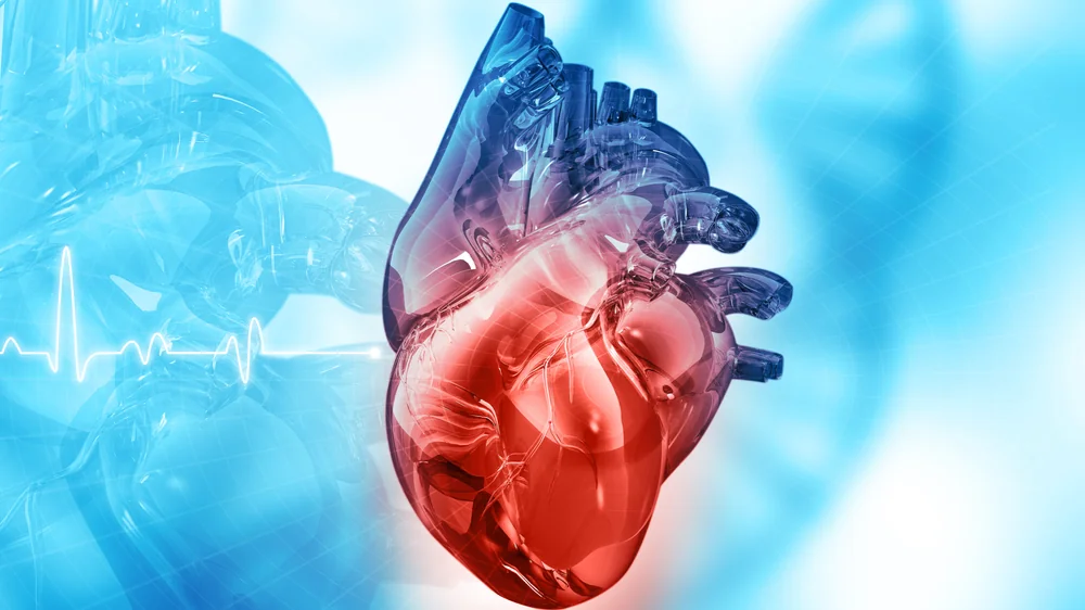
Researchers publishing in Nature Biotechnology have generated epicardioids, which are pluripotent, self-organizing stem cells that allow for better understanding, research, and potentially prevention and treatment of heart disease.
A background of heart development
The epicardium, a layer of cells surrounding the heart, plays a major role in human embryonic development. Some of these cells migrate towards the heart, developing into vascular and connective tissue, making them a prime target for potential therapies [1]. Whether or not they develop into muscle cells is still under debate [2].
Organoids are created from human cells for the purposes of research, testing, and potential therapeutic use. Previous work has created human organoids from stem cells that mimic some features of the heart [3], but none of this work has focused on the epicardium and its development. Here, the researchers present organoids that organize themselves into seemingly functional cardiac muscle and epicardial tissue: epicardioids.
From pluripotency to specificity
This study begins with human pluripotent stem cells (hPSCs) that the researchers drove towards an epicardial phenotype through the introduction of a host of signaling factors. This research found retinoic acid to be a critical factor in the differentiation and formation of organoids into epicardial tissue and muscle cells, as organoids grown without this compound were loosely organized and poorly differentiated. Supplementing the nascent epicardioids with vascular endothelial growth factor (VEGF) was sufficient for the development of blood vessels within them. This procedure was effective in four separate hPSC lines.
Gene expression analysis showed that these organoids developed into cells necessary for heart function, including pacemaker cells. These cells had similar gene expression profiles as natural cardiac cells in fetal tissue, although the progression of the epicardioids was much faster. The layering and cellular patterns found in these epicardioids was found to be similar to that of human beings, and they responded similarly to signals, such as the growth and regeneration factor NRP2.
The researchers also found evidence for the first heart field (FHF) and juxtacardiac field (JCF) [4], which are transitional stages between hPSCs and epicardial progenitors. These stages had been previously found in animals, but this research reported that they exist in humans as well.
Potential for research and treatment
The researchers were able to replicate medical problems that occur in human beings. Introducing endothelin-1 into their environment recapitulated signs of ventricular hypertrophy, including fibrosis. Using cells from a patient with Noonan syndrome, a genetic defect that leads to fibrosis, led to that same fibrosis in the resulting epicardioids. This gives researchers a potential workbench for treating such diseases.
Conclusion
At the end of their discussion, the researchers note that this research lends itself to the development of therapies that replace cardiac muscle cells lost to heart attacks, including further research on the regenerative factor NRP2. If epicardioids behave similarly to human tissues, particularly if they are more similar to hearts than mouse models, this may speed research and allow for more rapid development of treatments. However, it is unclear if epicardioids can replicate the myriad problems found in aging. More work will be done to determine if stem cells that form heart tissue can be used to develop therapies or be used as an effective therapy themselves.
Literature
[1] Quijada, P., Trembley, M. A., & Small, E. M. (2020). The role of the epicardium during heart development and repair. Circulation research, 126(3), 377-394.
[2] Christoffels, V. M., Grieskamp, T., Norden, J., Mommersteeg, M. T., Rudat, C., & Kispert, A. (2009). Tbx18 and the fate of epicardial progenitors. Nature, 458(7240), E8-E9.
[3] Hofbauer, P., Jahnel, S. M., Papai, N., Giesshammer, M., Deyett, A., Schmidt, C., … & Mendjan, S. (2021). Cardioids reveal self-organizing principles of human cardiogenesis. Cell, 184(12), 3299-3317.
[4] Tyser, R. C., Ibarra-Soria, X., McDole, K., Arcot Jayaram, S., Godwin, J., van den Brand, T. A., … & Srinivas, S. (2021). Characterization of a common progenitor pool of the epicardium and myocardium. Science, 371(6533), eabb2986.


